inhibitory effect of berberine on human skin squamous … effect of berberine on human skin squamous...
TRANSCRIPT

©FUNPEC-RP www.funpecrp.com.brGenetics and Molecular Research 14 (3): 10553-10568 (2015)
Inhibitory effect of berberine on human skin squamous cell carcinoma A431 cells
D.X. Li1*, J. Zhang1*, Y. Zhang2, P.W. Zhao3 and L.M. Yang3
1Department of Dermatology, The Affiliated Hospital of Inner Mongolia Medical University, Hohhot, Inner Mongolia, China2Medical College of Shandong University, Jinan, Shandong, China3Pathogenic Organisms and Immunology Lab, Inner Mongolia Medical University, Hohhot, Inner Mongolia, China
*These authors contributed equally to this study.Corresponding author: D.X. Li E-mail: [email protected]
Genet. Mol. Res. 14 (3): 10553-10568 (2015)Received February 19, 2015Accepted July 10, 2015Published September 8, 2015DOI http://dx.doi.org/10.4238/2015.September.8.17
ABSTRACT. Berberine (BBR) is a natural alkaloid with significant anti-tumor activity against many types of cancer cells. In this study, we investigated the molecular mechanisms employed by BBR to repress the proliferation and growth of skin squamous cell carcinoma A431 cells. Berberine was reported to inhibit the proliferation of A431 cells in a dose- and time-dependent manner and was observed to induce a series of biochemical events, including the loss of mitochondrial membrane potential, release of cytochrome-c to cytosol, induction of proteins of the Bcl-2 family and caspases, and the cleavage of poly(ADP)-ribose polymerase. This suggested its ability to induce apoptosis. The results of a wound healing test revealed that berberine inhibited the migration of A431 cells. Ezrin was transfected into A431 cells by RNA interference. The level of expression of Ezrin in the transfected A431 cells was observed to decrease with berberine treatment, which suggested that berberine might inhibit the invasion of A431 cells through Ezrin. The results of this study

10554D.X. Li et al.
©FUNPEC-RP www.funpecrp.com.brGenetics and Molecular Research 14 (3): 10553-10568 (2015)
demonstrated that berberine could potentially inhibit proliferation, induce apoptosis, and inhibit the invasion of A431 cells.
Key words: Berberine; Squamous cell carcinoma; A431 cells; Bcl-2; Proliferation; Apoptosis
INTRODUCTION
Skin is the outermost organ of the human body; it mirrors body health and functions as a barrier, in addition to aiding in absorption, secretion, and immunity. The increase in environmental pollution and ultraviolet radiation intensity has resulted in a rapid increase in the morbidity and mortality rate of malignant skin tumor over the past few years. Squamous cell carcinoma (SCC) is a common non-melanocytic skin tumor seen in China. This tumor can be easily detected at an early stage; however, a majority of the population lacks awareness regarding skin cancer, leading to delays in diagnosis and timely and efficient treatment. This leads to deep tumor invasion or metastasis, causing a serious impact on patient health and life. Berberine is an isoquinoline alkaloid extracted from several plants, including Hydrastis canadensis (goldenseal), Berberis aquifolium (Oregon grape), and Berberis vulgaris (barberry). Berberine has strong antimicrobial activities, and has been used to treat bacterial diarrhea, intestinal parasitic infections, and ocular trachoma infections for several decades (Imanshahidi and Hosseinzadeh, 2008; Sun et al., 2009).
Recent studies have revealed that berberine suppresses the growth of some tumor cell lines (Letasiova et al., 2006; Jantova et al., 2007; Liang et al., 2008; Tang et al., 2009). Several studies have attempted to elucidate the mechanism of antitumor activity of berberine (Iizuka et al., 2000; Lin et al., 2004; Lee et al., 2006; Tan et al., 2006). Berberine has been reported to suppress cancer cell growth and proliferation by inducing cell cycle arrest, stimulate cancer cell caspase-dependent apoptosis, reduce the levels of Bcl-2 and Bcl-xL and increase the levels of Bax and Bak, and inhibit metastasis by downregulating the expression of matrix metalloproteinases (Jantova et al., 2003; Lin et al., 2006). These findings indicate the multiple mechanisms involved in the anti-cancer effects of berberine on different tumor cell types. However, the effect of berberine on the growth characteristics of SCC remains to be elucidated. This study aimed to investigate the proliferation-promoting, apoptosis-inducing, and metastasis-inducing effect of berberine on the skin SCC cell line A431 in vitro.
MATERIAL AND METHODS
Chemicals and reagents
Berberine (>98% pure) and molecular biology-grade dimethyl sulfoxide (DMSO) were purchased from Sigma-Aldrich (St. Louis, MO, USA). Dulbecco’s modified Eagle’s medium (DMEM) and all chemicals used for cell culture were purchased from HyClone (Logan, UT, USA). Penicillin, streptomycin, trypsin, and ethylene diamine tetraacetic acid (EDTA) were also purchased from Sigma-Aldrich. Commonly used chemical reagents in this study were mostly procured from China. Rabbit anti-human Bax, Bcl-2, caspase-3, caspase-9, cyto-C, poly ADP ribose polymerase (PARP), and Ezrin were procured from Cell Signal Technology (Danvers, MA, USA). Rabbit anti-human β-actin antibody and horseradish peroxidase (HRP)-conjugated goat anti-rabbit secondary antibodies used in this study were purchased from KPL (Gaithersburg, MD, USA). The reagents

10555Berberine inhibits human skin SCC A431 cells
©FUNPEC-RP www.funpecrp.com.brGenetics and Molecular Research 14 (3): 10553-10568 (2015)
used for polymerase chain reaction (PCR) and western blot analysis were procured from Tiangen Biotech (Beijing, China) and Beyotime (Jiangsu, China), respectively. Unless otherwise mentioned, control treatment involved the treatment of cells with an equal volume of complete medium.
Cell culture and maintenance
SCC (A431) cells were procured from the Shanghai Cell Bank of the Chinese Science Institute. A431 cells were cultured in DMEM supplemented with 10% fetal bovine serum (FBS). The cell types were maintained in a CO2 incubator at 37°C and 85 ± 5% relative humidity. At 70% confluence, the cells displaying normal morphology and multiplication patterns were obtained for use in further experiments.
Cell proliferation assay
A431 cells (1 x 10⁶ cells/mL) were treated with different concentrations (12.5, 25, 50, 100 µg/mL) of berberine. The treated cells were incubated for 12, 24, 48, and 72 h. Cell viability was measured spectrophotometrically using the 3-(4,5-dimethylthiazol-2-yl)-2,5-diphenyltetrazolium bromide (MTT) assay; DMSO was used to dissolve formazan.
Trypan blue dry exclusion assay
The cytotoxic effects of berberine were analyzed using the trypan blue dye exclusion assay. Briefly, A431 cells (1 x 10⁶ cells/mL) were seeded onto six-well culture plates, and incubated under standard culture conditions overnight (in an incubator). The cells were then treated with berberine at final concentrations of 0, 25, 50, and 100 µg/mL for 24 h. The cells were harvested by trypsinization at the stipulated time point; the cells that appeared blue (had taken up the dye; dead cells) were observed under a microscope.
Wound healing assay
Cell motility and migration characteristics were analyzed by a wound healing assay, using a previously described method (with minor modifications). Briefly, the cells were grown to a confluent monolayer on a six-well tissue culture plate. The cell monolayers were “wounded” using a P200 micropipette tip. The wounded monolayers were washed twice with Dulbecco’s phosphate buffered saline (Boster, Wuhan, China) to remove the cell debris, and incubated in culture medium supplemented with 0.1% FBS in the absence or presence of BBR for 0 or 24 h. The migrating cells in the denuded zone were monitored using an inverted microscope equipped with a camera (Leica, Wetzlar, Germany).
Detection of Bax, Bcl-2, Ezrin mRNA expression by quantitative reverse transcriptase PCR (qRT-PCR)
The cultured A431 cells were treated with different concentrations of berberine (0, 25, 50, and 100 µg/mL), and incubated for an additional 4, 8, or 12 h. Total RNA was subsequently extract-ed from the cells using TRIzol reagent (Tiangen Biotech, Beijing, China). The RNA was reverse-transcribed to cDNA. The cDNA samples were subjected to real-time PCR, using a Real Master

10556D.X. Li et al.
©FUNPEC-RP www.funpecrp.com.brGenetics and Molecular Research 14 (3): 10553-10568 (2015)
Mix (SYBR Green) Kit (Tiangen Biotech). The following primers were used for PCR amplification: Bax, forward 5'-GGATGATTGCCGCCGT-3', reverse 5'-CCCAGTTGAAGTTGCCGT-3' (melting temperature, Tm = 55°C); Bcl-2, forward 5'-TGCACCTGACGCCCTTCAC-3', reverse 5'-AGACAGCCAGGAGAAATCAAACAG-3' (Tm = 55°C); Ezrin, forward 5'-ACGTCTGAGAATCAACAAGC-3', reverse 5'-TTCTCCTCATAGTCCTGCAG-3' (Tm = 53°C); and GAPDH, forward 5'-GGTGAAGGTCGGAGTCAACGGA-3', reverse 5'-GAGGGATCTCGCTCCTGGAAGA-3' (Tm = 61°C). The PCR conditions were set as follows: 1 cycle at 95°C for 10 min, 40 cycles at 95°C for 15 s and Tm for 1 min, and 72°C for 7 min. The amplification reaction, PCR product detection, and data analy-ses were performed using the ABI Prism7900 Sequence Detection System (Applied Biosystems, Waltham, MA, USA).
Detection of Bax and Bcl-2 expression by immunocytofluorescence
A431 cells treated with berberine (0, 50 µg/mL) for 24 h were fixed in 4% paraformaldehyde (in PBS) for 15 min. The cells were then washed with PBS, permeabilized with 0.5% Triton X-100 in PBS for 15 min (wash again), and blocked with 5% FBS in PBS for 30 min. The cells were then incubated overnight at 4°C with the polyclonal anti-Bax antibody (1:2000 in PBS; Santa Cruz Biotechnology, Dallas, TX, USA) and the polyclonal anti-Bcl-2 antibody (1:1000 in PBS; Santa Cruz Biotechnology). Subsequently, the cells were washed and incubated with secondary fluorescein isothiocyanate (FITC)-conjugated anti-rabbit IgG (Sigma-Aldrich) for 1 h in the dark at room temperature. Finally, the samples were washed, counterstained with 1 µg/mL 4',6-diamidine-2'-phenylindole (DAPI, Sigma-Aldrich), and mounted in an anti-fade solution (PBS, Boster, Wuhan, China) in the dark. The cells were examined using a Leica epifluorescence microscope (Leica). FITC and DAPI images were serially captured for samples subjected to each of the treatment conditions.
Determination of related protein expression by western blotting
The cells were treated with berberine for 48 h. The whole protein extract was extracted by washing the cells twice, scraping the washed cells in 1 mL PBS, and incubating the same on ice for 30 min in cell lysis buffer (Beyotime) supplemented with Halt protease and phosphatase inhibitor cocktail (Life Technologies, Carlsbad, CA, USA). The cell lysates were centrifuged at 12,000 rpm for 5 min at 4°C. The protein content in the supernatant was measured using the Bio-Rad protein assay kit (Bio-Rad, Hercules, CA, USA). The samples were directly subjected to sodium dodecyl sulfate polyacrylamide gel electrophoresis (SDS-PAGE), and blotted onto the nitrocellulose (NC) membrane. The membranes were blocked with 5% non-fat milk in Tris-buffered saline, supplemented with Tween-20 (TBST) for 1 h at room temperature. The blocked NC membranes were incubated with primary antibodies at 4°C overnight, and subsequently incubated with a HRP-conjugated secondary antibody for 1 h at room temperature. The reactive signals were visualized using the enhanced chemiluminescence (ECL) kit (Tiangen Biotech, Beijing, China). The resulting images were scanned and quantified using the Image J software.
Transient transfection of siRNA against ezrin
Plasmid vector expressing ezrin-siRNA Gene name: Homo sapiens ezrin (EZR) RNAi sequence: S1 GGGTCCTACGCTGTGCAGGCC; S2 AACAGCTGGAAACAGAGAAGA .

10557Berberine inhibits human skin SCC A431 cells
©FUNPEC-RP www.funpecrp.com.brGenetics and Molecular Research 14 (3): 10553-10568 (2015)
Isolation of plasmid
Preparation of the kanamycin (Kana) stock (100 mg/mL) solution: 100 mg Kana was dissolved in 1 mL deionized water, filtered through a 0.22 μm membrane, and stored in a 1 mL Eppendorf tube at -20°C.
Preparation of the Luria-Bertani/Kana (LB/Kana) liquid medium: 10 g Tryptone, 5 g yeast extract, and 10 g NaCl were dissolved in deionized water, and the solution diluted to 1 L. This solution (50 mL) was autoclaved and cooled to 60°C; subsequently, 25 μL Kana stock solution was added to the autoclaved LB medium, and the solution was mixed evenly.
LB/Kana liquid medium (3 mL) was added to a 4-mL Eppendorf tube; this was inoculated with 2 μL bacterial glycerol stock and incubated on a shaker for 16 h at 37°C.
Preparation of the LB/Kana solid medium: 10 g Tryptone, 5 g yeast extract, 10 g NaCl, and 15 g agar were dissolved in 1 L deionized water. This solution (100 mL) was autoclaved and cooled to 60°C; Kana stock solution (50 μL) was added to the agar solution. This mixture was transferred to an autoclaved cell-culture dish (20 mL/dish), sealed, inverted, and stored at 4°C.
Inoculation of bacterial glycerol stock: 200 μL bacteria was inoculated to the center of a cell-culture dish in a “Z”-shape using an inoculation loop. The plates were incubated at 37°C for 24 h; the colonies were punctiform, and displayed good growth.
The amplified bacteria (3 mL) were used for plasmid isolation; the isolation was performed using a Tiangen small plasmid extraction kit (Tiangen Biotech), according to the manufacturer protocols; the extracted plasmid was then stored at -20°C.
Preparation of 1% agarose gel: 0.2 g agarose was dissolved in 20 mL tris-borate-EDTA (TBE); this mixture was heated to boiling in a microwave (three times) until the solution became translucent. The solution was cooled to 50°C, mixed evenly with 2 μL nucleic acid dye, poured into a mold, and allowed to solidify (approximately 10 min); the gel was then transferred to an electrophoresis tank.
The comb was pulled out from the gel apparatus. The isolated plasmid sample (5 μL) was added to the well. The gel was covered with TBE buffer. Electrophoresis was performed at a 110 V for 25 min. The gel was observed using an imaging system (Bio-life, USA) in order to determine the results of plasmid isolation.
Preparation of A431 cells
A431 cells in the logarithmic phase of growth were digested with trypsin. The cells were then cultured in antibiotic-free DMEM at a final cell density of 2.5 x 10⁵ cells/mL. The cells were seeded onto six-well plates at a concentration of 2 mL/well, and the plates incubated in a cell incubator at 37°C, under conditions of 5% CO2 and saturated humidity.
Plasmid transfection [adapted from the protocol provided in the Invitrogen (Life Technologies) plasmid reference manual]
A431 cells at 90% confluence were transfected with plasmid ezrin-siRNA (isolated from the bacterial glycerol stock) or the empty vector plasmid HK.
The plasmid (4 μg) was dissolved in 250 μL serum-free medium, and the solution mixed gently and evenly. Liposome (10 μL) was diluted to 250 μL using serum-free medium; this mixture was allowed to stand at room temperature for 5 min. The plasmid and liposome solutions were

10558D.X. Li et al.
©FUNPEC-RP www.funpecrp.com.brGenetics and Molecular Research 14 (3): 10553-10568 (2015)
mixed together (total volume = 500 μL), and the mixture allowed to stand at room temperature for 20 min. Meanwhile, A431 cells cultured on six-well culture plates were washed twice with serum-free medium. Subsequently, 2 mL serum-free medium was added to each of the wells. The plasmid-liposome mixture was pipetted into the wells of the six-well culture plate (and the contents mixed evenly). The plate was incubated at 37°C and 5% CO2 conditions. After 4-6 h of incubation, the medium-liposome-plasmid mixture was removed, and the cells were incubated in normal culture medium. The results of transfection were observed under a fluorescence microscope (Leica) after 24 h. The total protein was subsequently extracted.
Analysis of ezrin protein expression (post-plasmid ezrin-siRNA transfection) by western blotting
A431 cells were transfected with plasmid ezrin-siRNA or plasmid HK for 72 h. The cells were treated with berberine (0, 25, 50, and 100 μg/mL) for 48 h, and subsequently lysed to extract total proteins. The extracted total proteins were separated by SDS-PAGE and blotted onto NC membranes. The NC membrane was then blocked with non-fat milk, and incubated with rabbit anti-human Ezrin monoclonal antibody (1:1000) or β-actin monoclonal antibody (1:1000) (primary antibodies). Finally, the cells were incubated with HRP-conjugated goat anti-rabbit secondary antibodies (1:2000). The reactive signals were visualized by ECL (image analysis).
Statistical analysis
All results are reported as means ± standard deviations. Statistical significance was assessed by one-way ANOVA, following appropriate transformation to normalized data and equalization of variance, where necessary. All statistical analyses were performed using the SPSS v.17.0 software platform (IBM, Armonk, NY, USA). P values less than 0.05 were considered to be statistically significant for all tests.
RESULTS
Cell viability
Treatment with berberine was observed to have an anti-tumor effect compared to the untreated group (blank control group) and the samples treated with DDP (cisplatin 250 µg/mL). The increase in dosage of berberine and duration of action resulted in a corresponding decrease in OD490 (P < 0.05) (Table 1). This suggested a significant decrease in cell viability with the increase in time and drug concentration (Figure 1), which in turn indicated a gradual dose- and time-dependent increase in the inhibitory effect of berberine on A431 cells.
Effect of berberine on A431 cell morphology
Phase-contrast microscopy (Figure 2) revealed that the A431 cells were epithelioid adherent cells. Normal cells of the control group (Figure 2A) showed good, homogenous growth, were flat and polygonal in shape, and showed strong refraction and intercellular mutual convergence properties. These cells were tightly packed and integrated into the film. When treated with low concentrations of berberine for 24 h, the cells showed decreased refraction and were shrunk to

10559Berberine inhibits human skin SCC A431 cells
©FUNPEC-RP www.funpecrp.com.brGenetics and Molecular Research 14 (3): 10553-10568 (2015)
a round shape; in addition, we observed a reduced cell volume, widened gaps between cells, and scattering of cells in suspension. Cells treated with higher concentrations of berberine were increasingly round, scattered, and suspended in the medium (Figure 2C and D).
Table1. Results of the MTT assay of berberine on A431 cells (means ± SD).
Group (µg/mL) OD at 490nm
12 h 24 h 48 h 72 h
Control 0.842 ± 0.499 0.764 ± 0.076 0.707 ± 0.019 0.980 ± 0.04512.5 0.689 ± 0.18* 0.383 ± 0.013* 0.113 ± 0.002* 0.106 ± 0.005*25 0.646 ± 0.136* 0.347 ± 0.007* 0.108 ± 0.004* 0.097 ± 0.003*50 0.543 ± 0.001*# 0.336 ± 0.015* 0.100 ± 0.002* 0.094 ± 0.003*100 0.387 ± 0.007*# 0.327 ± 0.007* 0.098 ± 0.003* 0.089 ± 0.003*DDP 0.599 ± 0.018* 0.187 ± 0.006* 0.127 ± 0.013* 0.095 ± 0.005*
Significant differences: *P < 0.05 compared to the control group; #P < 0.05 compared to the DDP-treated group (cisplatin 250 µg/mL).
Figure 1. Effect of berberine on A431 cell viability. Significant differences were indicated by *P < 0.05 (compared to the control group) and #P < 0.05 [compared to the DDP (cisplatin 250 µg/mL)-treated group].
Figure 2. Morphological changes in A431 cells among the control samples (A), and those treated with 25 µg/mL (B), 50 µg/mL (C), and 100 µg/mL (D) of berberine, as observed under a phase contrast microscope (100X magnification).

10560D.X. Li et al.
©FUNPEC-RP www.funpecrp.com.brGenetics and Molecular Research 14 (3): 10553-10568 (2015)
Results of the trypan blue dye exclusion assay
A431 cells treated with berberine were stained with trypan blue (Figure 3); the dead cells showed a clear blue staining, while exclusion stained colorless and transparent cells were live cells. As seen in Figure 3, the cells of the control group were round, colorless, and transparent, with a regular shape, showing intercellular adhesion, with very few blue-stained dead cells (Figure 3A). The cells treated with low concentrations of berberine were severely affected; a few mutual adherent colorless cells were observed, while a number of cells were stained blue, and were scattered (Figure 3B). The increase in treated berberine concentration led to a corresponding increase in the number of blue-stained cells; these cells were shrunken, displayed a number of different forms, and were scattered (Figure 3C and D).
Figure 3. The results of trypan blue staining of control (A) (untreated) and berberine-treated [25 µg/mL (B), 50 µg/mL (C), and 100 µg/mL (D)] A431 cells (at 100X magnification).
Results of the wound healing assay
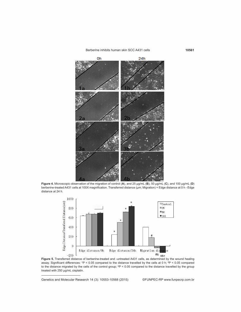
The A431 cells showed good growth and cell characteristics; the scratch width was basically uniform. S images were taken of the scratch surface at different time points (with no focal slips), as shown in Figure 4 (Figures 4-1a, 4-2a, 4-3a, and 4-4a). The scratch made to the control plate showed gradual healing (Figure 4-1b) compared to those made to the plates treated with different concentrations of berberine after 24 h; the migration distance of the cells in the low dosage group was much smaller than that shown by the cells of the control group (Figure4-2b). The edge distance was significantly higher in the high dosage group after 24 h, compared to that observed at 0 h (Figures 4-3b, 4-4b, and 5).
10561Berberine inhibits human skin SCC A431 cells
©FUNPEC-RP www.funpecrp.com.brGenetics and Molecular Research 14 (3): 10553-10568 (2015)
Figure 4. Microscopic observation of the migration of control (A), and 25 µg/mL (B), 50 µg/mL (C), and 100 µg/mL (D) berberine-treated A431 cells at 100X magnification. Transferred distance (µm; Migration) = Edge distance at 0 h - Edge distance at 24 h.
Figure 5. Transferred distance of berberine-treated and -untreated A431 cells, as determined by the wound healing assay. Significant differences: *P < 0.05 compared to the distance travelled by the cells at 0 h; #P < 0.05 compared to the distance migrated by the cells of the control group; &P < 0.05 compared to the distance travelled by the group treated with 250 µg/mL cisplatin.

10562D.X. Li et al.
©FUNPEC-RP www.funpecrp.com.brGenetics and Molecular Research 14 (3): 10553-10568 (2015)
Results of the qRT-PCR amplification
PCR was performed using GAPDH as the internal control; the results revealed a low level of Bax mRNA expression in cells treated with low concentrations of berberine. The increase in berberine dosage led to a corresponding increase in Bax mRNA expression. On the other hand, Bcl-2 mRNA was significantly expressed in cells treated with low concentrations of berberine, with the mRNA level decreasing with the increase in berberine concentration. That is, the increase in berberine drug dose led to a corresponding increase and decrease in Bax and Bcl-2 mRNA levels. The levels of expression of Bax/Bcl-2 were relatively low in the low berberine concentration groups (25 and 50 µg/mL), and significantly higher in cells treated with high concentrations (100 µg/mL) of berberine. We also observed a gradual increase in the relative expression levels of Bax/Bcl-2 with the increase in treatment time (Figure 6).
Analysis of the Ezrin mRNA expression by PCR revealed a gradual decrease in mRNA expression levels with the increase in treatment time and berberine dosage (Figure 7).
Figure 6. Relative expression of Bax/Bcl-2 mRNA in A431 cells treated with different concentrations of berberine. Significant differences: *P < 0.05 compared to the group treated with 25 µg/mL of berberine; #P < 0.05 compared to thegroup treated with 50 µg/mL of berberine.
Figure 7. Relative expression of Ezrin mRNA in A431 cells treated with different concentrations of berberine. Significant differences: *P < 0.05 compared to the cells treated with 25 µg/mL berberine; #P < 0.05 compared to the cells treated with 50 µg/mL berberine.
10563Berberine inhibits human skin SCC A431 cells
©FUNPEC-RP www.funpecrp.com.brGenetics and Molecular Research 14 (3): 10553-10568 (2015)
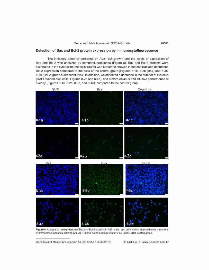
Detection of Bax and Bcl-2 protein expression by immunocytofluorescence
The inhibitory effect of berberine on A431 cell growth and the levels of expression of Bax and Bcl-2 was analyzed by immunofluorescence (Figure 8). Bax and Bcl-2 proteins were distributed in the cytoplasm; the cells treated with berberine showed increased Bax and decreased Bcl-2 expression compared to the cells of the control group [Figures 8-1b, 8-2b (Bax) and 8-3b, 8-4b (Bcl-2; green fluorescent dye)]. In addition, we observed a decrease in the number of live cells (DAPI stained blue cells; Figures 8-2a and 8-4a), and a more obvious and intuitive performance of overlay (Figures 8-1c, 8-2c, 8-3c, and 8-4c), compared to the control group.
Figure 8. Analysis of theexpression of Bax and Bcl-2 proteins in A431 cells, and cell viability, after berberine treatment, by immunofluorescence staining (200X). 1 and 3: Control group; 2 and 4: 50 µg/mL BBR-treated group.

10564D.X. Li et al.
©FUNPEC-RP www.funpecrp.com.brGenetics and Molecular Research 14 (3): 10553-10568 (2015)
Determination of protein expression by western blot analysis
Western blotting is widely used to detect the expression levels of proteins in cells. In this study, exposure to the ECL detecting agent upon completion of the preliminary operation revealed a change in expression of mitochondrial apoptosis-related proteins (Bcl-2 and Bax) in A431 cells treated with berberine for 48 h. The bands indicating Bax and Bcl-2 became brighter and weaker, respectively, with the increase in berberine concentration; this result was consistent with the results of qRT-PCR. The mitochondrial pathway was observed to play a significant role in the apoptotic signaling pathway, which triggered the expression of members of the pro-apoptotic Bcl-2 family. This further induced the expression of cyto-C, caspase family members, and PARP. As shown in Figure 9, the increase in berberine concentration led to a corresponding, significant increase in cyto-C, caspase-3, caspase-9, and PARP expression levels (indicated by gray bands). We also observed a decrease in the Ezrin protein expression (indicated by weak bands) with the increase in berberine concentration, compared to the control group. This indicated that berberine may affect the Ezrin protein expression.
Figure 9. Analysis of the expression of apoptosis-related proteins by western blotting.

10565Berberine inhibits human skin SCC A431 cells
©FUNPEC-RP www.funpecrp.com.brGenetics and Molecular Research 14 (3): 10553-10568 (2015)
Transfection efficiency of plasmids expressing the ezrin-siRNA
The plasmids expressed genes coding for enhanced green fluorescent protein. Inverted fluorescence microscopy under high magnification revealed some green fluorescence and strong expression in cells transfected with the plasmid for 24 h (Figure 10).
Western blot analysis was used to examine the effect of siRNA interference on the expression of ezrin protein in A431 cells (Figure 11). A431 cells were transfected with ezrin-siRNA and HK (control) plasmids. The total protein in the cells was analyzed by western blot. The gray band indicating ezrin protein expression was significantly weaker in the interference group compared to the group transfected with the empty vector plasmid.
Figure 10. Fluorescence microscope images of the cells successfully transfected (for 24 h) with plasmids expressing the ezrin-siRNA.
Figure 11. Results of the western blot analysis of plasmid interference on the expression of ezrin proteins in A431 cells.
DISCUSSION
Recent anti-tumor studies are concentrating on inhibiting the proliferation of, or inducing apoptosis and controlled metastasis in, tumor cells. These are important processes regulating tumor incidence and development. Metastasis can cause multiple organ failure and dyscrasia, which is recognized throughout the world as the leading cause of death in patients with malignant tumor.
The results of the MTT assay performed in this study suggested that berberine inhibits A431 cell proliferation in a dose- and time-dependent manner (P < 0.05). On the other hand, trypan blue staining revealed that the increase in berberine concentration resulted in the death of carcinoma cells (visibly dyed, shrunken, scattered in a variety of forms, non-adherent cells were

10566D.X. Li et al.
©FUNPEC-RP www.funpecrp.com.brGenetics and Molecular Research 14 (3): 10553-10568 (2015)
observed). The results of the wound healing test revealed that berberine inhibits the migration of A431 cells, with the scratch width of the high-dosage treatment group being significantly wider than that of the control group (P < 0.05).
Apoptosis is tightly regulated by anti-apoptotic and pro-apoptotic effector molecules, including proteins of the Bcl-2 family, and can be mediated by several different pathways (Budd, 2001; Germain and Shore, 2003; Petros et al., 2004). The proteins of the Bcl-2 family either promote cell survival (e.g., Bcl-2 and Bcl-xL) or induce programmed cell death (e.g., Bax) (Puthalakath and Strasser, 2002; Brunelle and Letai, 2009). The Bax/Bcl-2 expression ratio is critical for the induction of apoptosis; this ratio determines and regulates the progression of cells into the apoptosis stage (Krammer et al., 2007; Johnstone et al., 2008). An increase in the Bax/Bcl-2 ratio stimulates cytochrome-c release from mitochondria into the cytosol. The cytosolic cytochrome-c then binds to Apaf-1, leading to the activation of caspase-3 and PARP, which is an enzyme that facilitates the repair of DNA damage (Kluck et al., 1997; Glazunova and Shtil, 2008). The treatment of A431 cells with berberine was observed to result in an increase and decrease in Bax and Bcl-2 protein expressions, respectively, and an increase in the Bax/Bcl-2 ratio. This may be the reason behind the increased release of cytochrome-c into the cytosol. The increase in cytochrome-c levels in the cytosol leads to its increased interaction with Apaf-1 and ATP, resulting in the formation of a complex with procaspase-9 (apoptosome), which in turn leads to the activation of caspase-9 and caspase-3 (Roderick and Cook, 2008; Trachootham et al., 2009). Activated caspase-3 is the key factor executing apoptosis; cleaved caspase-3 is responsible for the cleavage and inactivation of key cellular proteins, such as PARP (Shi, 2002; Mantena et al., 2006). The treatment of A431 cells with berberine resulted in a dose-dependent activation of caspase-9 and caspase-3, and consequently, cytochrome c and PARP, as determined by western blot analysis. The results of qRT-PCR revealed a berberine dose-dependent increase and decrease in Bax and Bcl-2 mRNA expression levels, respectively. The berberine-induced increase in the Bax/Bcl-2 ratio, and its effect on apoptosis, was further confirmed by immunofluorescence staining. The results of the immunofluorescence staining method demonstrated the widespread distribution of Bax and Bcl-2 throughout the cytoplasm; berberine-treatment resulted in an increase and decrease in Bax and Bcl-2 expression, respectively, and a decrease in the total number of living cells.
Ezrin is a member of the Ezrin-Radixin-Moesin family, and is a molecule that connects the epithelial cells between the cell membrane and cytoskeleton (Furutani et al., 2007). Recent studies have reported that Ezrin plays an important role in tumor metastasis. In addition, Ezrin has been proposed as a potential target molecule for anti-tumor drugs (Chen et al., 2008; Huang et al., 2011). The results of the qRT-PCR analysis performed in this study indicated a decrease in Ezrin expression with the increase in berberine concentration. Western blot analysis also revealed a decrease in Ezrin expression with the increase in berberine concentration. Ezrin was successfully transfected into A431 cells by RNA interference (RNAi). The Ezrin-transfected A431 cells displayed green fluorescence, indicating successful transfection; the berberine treatment resulted in a decrease in the Ezrin expression levels in A431-Ezrin cells, which indicated that berberine might inhibit the invasion of A431 cells by Ezrin.
In conclusion, the results of this study indicated that berberine inhibits the proliferation of, and induces apoptosis in, SCC A431 cells. In addition, this study provides mechanistic evidence that berberine-induced apoptosis may activate mitochondrial pathway, leading to the upregulation of Bax/Bcl-2, caspase 3, caspase 9, cytochrome-C, and PARP expression in A431 cells. In addition, berberine may also inhibit A431 cell invasion via inhibition of Ezrin expression.

10567Berberine inhibits human skin SCC A431 cells
©FUNPEC-RP www.funpecrp.com.brGenetics and Molecular Research 14 (3): 10553-10568 (2015)
Conflicts of interest
The authors declare no conflict of interest.
ACKNOWLEDGMENTS
Research supported by grants from Inner Mongolia Natural Science Foundation (#2011MS1117); the Medical Scientific Research Projects for Inner Mongolia Autonomous Region Health and Family Planning Commission (#201302063); and the Inner Mongolia Autonomous Region Science and Technology project for 2014 (#kjt14sf16).
REFERENCES
Budd R (2009). Activation-induced cell death. Curr. Opin. Immunol. 13: 356-362.Brunelle JK, Letai A (2001). Control of mitochondrial apoptosis by the Bcl-2 family. J. Cell Sci. 122: 437-441.Chen Y, Wang WW and Hua JJ (2008). Inhibitory effort of berberine on pseudopod formation and the motility of CNE1 via Ezrin
phosphoarylation. Chin. J. Otorhinolaryngology-Skull Base Surg. 11: 401-406.Furutani Y, Matsuno H, Kawasaki M, Sasaki T, et al. (2007). Interaction between telencephalin and ERM family proteins
mediates dendritic filopodia formation. J. Neurosci. 27: 8866-8876.Germain M and Shore GC (2003). Cellular distribution of Bcl-2 family proteins. Sci. STKE 173: 10-12.Glazunova VA and Shtil AA (2008). Mitochondrial mechanisms of apoptosis in response to DNA damage. Mol. Biol. 42: 765-
771.Huang D, Wang W, Feng Z, Wang L, et al. (2011). Berberine inhibits the invasion and metastasis of nasopharyngeal carcinoma
cells through Ezrin phosphorylation. Zhong Nan Da Xue Xue Bao Yi Xue Ban 36: 616-623.Iizuka N, Miyamoto K, Okita K, Tangoku A, et al. (2000). Inhibitory effect of Coptidis rhizoma and berberine on the proliferation
of human esophageal cancer cell lines. Cancer Lett. 148: 19-25.Imanshahidi M and Hosseinzadeh H (2008). Pharmacological and therapeutic effects of Berberis vulgaris and its active
constituent berberine. Phytother. Res. 22: 999-1012.Jantova S, Cipak L, Cernakova M and Kost’alova D (2003). Effect of berberine on proliferation, cell cycle and apoptosis in HeLa
and L1210 cells. J. Pharm. Pharmacol. 55: 1143-1149.Jantova S, Cipak L and Letasiova S (2007). Berberine induces apoptosis through a mitochondrial/caspase pathway in human
promonocytic U937 cells. Toxicol. In Vitro 21: 25-31.Johnstone RW, Frew AJ and Smyth MJ (2008). The TRAIL apoptotic pathway in cancer onset progression and therapy. Nat.
Rev. Cancer 8: 782-798.Kluck RM, Bossy-Wetzel E, Green DR and Newmeyer DD (1997). The release of cytochrome c from mitochondria: a primary
site for Bcl-2 regulation of apoptosis. Science 275: 1132-1136.Krammer PH, Arnold R and Lavrik I (2007). Life and death in peripheral T cells. Nat. Rev. Immunol. 7: 532-542.Lee S, Lim HJ, Park HY, Lee KS, et al. (2006). Berberine inhibits rat vascular smooth muscle cell proliferation and migration in
vitro and improves neointima formation after balloon injury in vivo. Berberine improves neointima formation in a rat model. Atherosclerosis 186: 29-37.
Letasiova S, Jantova S, Cipak L and Muckova M (2006). Berberine-antiproliferative activity in vitro and induction of apoptosis/necrosis of the U937 and B16 cells. Cancer Lett. 239: 254-262.
Liang KW, Yin SC, Ting CT, Lin SJ, et al. (2008). Berberine inhibits platelet-derived growth factor-induced growth and migration partly through an AMPK-dependent pathway in vascular smooth muscle cells. Eur. J. Pharmacol. l590: 343-354.
Lin CC, Ng LT, Hsu FF, Shieh DE, et al. (2004). Cytotoxic effects of Coptis chinensis and Epimedium sagittatum extracts and their major constituents (berberine, coptisine and icariin) on hepatoma and leukaemia cell growth. Clin. Exp. Pharmacol. Physiol. 31: 65-69.
Lin JP, Yang JS, Lee JH, Hsieh WT, et al. (2006). Berberine induces cell cycle arrest and apoptosis in human gastric carcinoma SNU-5 cell line. World J. Gastroenterol. 12: 21-28.
Mantena SK, Sharma SD and Katiyar SK (2006). Berberinea natural product, induces G1-phase cell cycle arrest and caspase-3-dependent apoptosis in human prostate carcinoma cells. Mol. Cancer Ther. 5: 296-308.
Petros AM, Olejniczak ET and Fesik SW (2004). Structural biology of the Bcl-2 family of proteins. Biochem. Biophys. Acta 1644: 83-94.
Puthalakath H and Strasser A (2002). Keeping fillers on a tight leash: transcriptional and post-translational control of the

10568D.X. Li et al.
©FUNPEC-RP www.funpecrp.com.brGenetics and Molecular Research 14 (3): 10553-10568 (2015)
proapoptotic activity of BH3-only proteins. Cell Death Differ. 9: 505-512.Roderick HL and Cook SJ (2008). Ca2+ signalling checkpoints in cancer: remodelling Ca2+ for cancer cell proliferation and
survival. Nat. Rev. Cancer 8: 361-375.Shi Y (2002). Mechanisms of caspase activation and inhibition during apoptosis. Mol. Cell 9: 459-470.Sun Y, Xun K, Wang Y and Chen X (2009). A systematic review of the anticancer properties of berberine, a natural product
from Chinese herbs. Anticancer Drugs 20: 757-769.Tan YL, Goh D and Ong ES (2006). Investigation of differentially expressed proteins due to the inhibitory effects of berberine
in human liver cancer cell line HepG2. Mol. Biosyst. 2: 250-258.Tang J, Feng Y, Tsao S and Wang N (2009). Berberine and Coptidis rhizoma as novel antineoplastic agents: a review of
traditional use and biomedical investigations. J. Ethnopharmacol. 126: 5-17.Trachootham D, Alexandre J and Huang P (2003). Targeting cancer cells by ROS-mediated mechanisms: a radical therapeutic
approach. Nat. Rev. Drug Disc. 8: 579-591.