inhibition of myotoxic activity of bothrops asper myotoxin ii by the anti-trypanosomal drug suramin
TRANSCRIPT

doi:10.1016/j.jmb.2005.04.072 J. Mol. Biol. (2005) 350, 416–426
Inhibition of Myotoxic Activity of Bothrops asperMyotoxin II by the Anti-trypanosomal Drug Suramin
Mario T. Murakami1, Emerson Z. Arruda2, Paulo A. Melo2
Ana B. Martinez2, Sabrina Calil-Elias2, Marcelo A. Tomaz2
Bruno Lomonte3, Jose M. Gutierrez3 and Raghuvir K. Arni1,4*
1Departament of PhysicsIBILCE/UNESP, Sao Jose do RioPreto, SP, Brazil
2Departamento de FarmacologiaBasica e Clinica, IBC/UFRJRio de Janeiro, RJ, Brazil
3Instituto Clodomiro PicadoFacultad de MicrobiologiaUniversidad de Costa RicaSan Jose, Costa Rica
4Center for Applied ToxinologyInstituto Butantan, Sao PauloSP, Brazil
0022-2836/$ - see front matter q 2005 E
Abbreviations used: Basp-II, BothrPLA2s, phospholipases A2; suramin[imino-3,1-phenylenecarbonyliminonylene) carbonylimino]] bis-1,3,5-naacid hexasodium salt; CK, creatineethylene glycol; i-face, interfacial reextensor digitorum longus; PSS, phsolution; Hepes, 4-(2-hydroxyethyl)sulfonic acid; i.m., intramuscular.E-mail address of the correspond
Suramin, a synthetic polysulfonated compound, developed initially for thetreatment of African trypanosomiasis and onchocerciasis, is currently usedfor the treatment of several medically relevant disorders. Suramin, heparin,and other polyanions inhibit the myotoxic activity of Lys49 phospholipaseA2 analogues both in vitro and in vivo, and are thus of potential importanceas therapeutic agents in the treatment of viperid snake bites. Due to itsconformational flexibility around the single bonds that link the centralphenyl rings to the secondary amide backbone, the symmetrical suraminmolecule binds by an induced-fit mechanism complementing thehydrophobic surfaces of the dimer and adopts a novel conformation thatlacks C2 symmetry in the dimeric crystal structure of the suramin–Bothropsasper myotoxin II complex. The simultaneous binding of suramin at thesurfaces of the two monomers partially restricts access to the nominalactive sites and significantly changes the overall charge of the interfacialrecognition face of the protein, resulting in the inhibition of myotoxicity.
q 2005 Elsevier Ltd. All rights reserved.
Keywords: suramin; heparin; myotoxicity; inhibition; crystal structure
*Corresponding authorIntroduction
Suramin (8,8 0-[carbonylbis [imino-3,1-phenylene-carbonylimino (4-methyl-3,1-phenylene) carbonyli-mino]] bis-1,3,5-naphtalenetrisulfonic acidhexasodium salt), a highly charged polysulfonatedcompound, is one of the first successful synthetictherapeutic agents developed and is used clinicallyin the treatment of African trypanosomiasis andonchocerciasis.1–4 The spectrum of suramin appli-cations now includes the clinical treatment ofangiogenesis, carcinomas of the kidney and pros-tate gland, breast cancer, inhibition of growthfactor/receptor interactions, inhibition of human
lsevier Ltd. All rights reserve
ops asper myotoxin II;, 8,8 0-[carbonylbis(4-methyl-3,1-phe-phtalenetrisulfonickinase; PEG, poly-cognition face; EDL,ysiological saline-1-piperazineethane-
ing author:
a-thrombin, protein-tyrosine phosphatases, G pro-teins, merozoite surface protein-1, and acidic andbasic fibroblast growth factors.5–14 Suramin pre-vents the development of muscle necrosis inducedby some snake venoms, since it inhibits themyotoxic and in vitro neuromuscular blockingactivities of Lys49 phospholipases A2 from Bothropsspecies.15,16
Phospholipases A2 (PLA2s: EC 3.1.1.4) are keyenzymes in the control, regulation, production andrelease of lipid mediator precursors that serve asmessengers in fundamental, highly regulated pro-cesses such as growth, adhesion, apoptosis,secretion, hemostasis and immune regulation. Thegroup II PLA2s encountered in viperid snakevenoms and mammalian fluids share significantsequence homology, and are based on a singlestructural motif or scaffold.17 Snake venom PLA2sexhibit a wide spectrum of activities includingmyotoxic, neurotoxic, cardiotoxic, anticoagulant,platelet-aggregating, hypotensive and inflammatoryeffects.18 On the basis of sequence and structuralsimilarities, the snake venom group II PLA2s can besubdivided into (a) the catalytically active Asp49enzymes and (b) the basic, myotoxic Lys49 proteins,which possess no catalytic activity.17 Myotoxicity
d.

Inhibition of B. asper Mytotoxin II by Suramin 417
affects only muscle fibers without damaging othertissue structures such as connective tissue, nervesand vessels,19 and can lead to permanent tissue loss,disability, amputation and death.20
Suramin binds to a surprisingly wide variety ofproteins of different function, structure and size dueto its torsional flexibility and ability to stretch orcompress itself, which results in the two naphthylrings with the sulfonate groups being able tointeract simultaneously with a number of structu-rally distinct sites. Due to a lack of structuralinformation, the target sites, conformational flexi-bility and mode of binding of suramin to proteinsare still unclear at the atomic level.
The crystal structure of the complex betweensuramin and Bothrops asper myotoxin II representsthe first crystal structure of suramin bound to aprotein. The structural details presented hereshould be of relevance in understanding the stericrequirements of suramin binding to proteins, andshould shed light on the mechanism of myotoxicitydisplayed by Lys49 phospholipases A2 and theirinhibition by polyanionic compounds.
Results
In vitro myotoxicity
As illustrated in Figure 1(a), B. asper myotoxin IIcaused a time-dependent increase in the rate ofcreatine kinase release from isolated extensordigitorum longus muscles. A more pronouncedeffect is observed after 90 minutes with 25 mg/ml ofB. asper myotoxin II alone. The presence of suramin(10 mM) together with the toxin markedly inhibitsthe creatine kinase release induced by the B. aspermyotoxin II (Basp-II). After 90 minutes of exposureto the toxin, in the presence of suramin, the creatinekinase release was less than 15% of the valuemeasured in the absence of suramin.
In vivo myotoxicity
Intramuscular (i.m.) injections of Basp-II aloneincreased plasma creatine kinase (CK) activity in adose and time-dependent fashion. Figure 1(b)indicates that an i.m. injection of Basp-II (2.0 mg/g) induced a significant increase in plasma CKactivity compared to physiological saline solution.CK levels increased from 65(G42) units/l (meanGSEM, nZ4) to 1368(G386) units/l (nZ4) after twohours. These values of plasma CK activity are inagreement with previously reported observations.21
Preincubation with suramin significantly (p!0.05)inhibits the increase in plasma CK activity inducedby Basp-II. Experiments performed following pro-tocol B (see Materials and Methods) indicate thatsuramin also significantly inhibits (p!0.05) theplasma CK activity increment induced by Basp-II.However, under these conditions, suramin was lesseffective in antagonizing myotoxicity than whenincubated with Basp-II prior to injection. Figure 2presents light micrographs of extensor digitorumlongus muscle exposed to either Basp-II (Figure 2(b)and (b 0)), Basp-II pre-incubated with suramin(1 mg/kg) (Figure 2(c) and (c 0)) or post-treatedwith suramin (1 mg/kg) 15 minutes after Basp-IIinjection. In all samples, normal muscle cells wereobserved in the central region of the muscle,whereas the peripheral fibers were in differentstages of necrosis, characterized by denselyclumped myofibrils and swollen cells. Pre-incu-bation or post-treatment with suramin resulted in areduction in the number of necrotic fibers(Figure 2(c) and (d)).
Crystal structure
Data collection and processing statistics arepresented in Table 1. Molecular replacement usinga single molecule of native Basp-II (PDB ID, 1CLP)as the search model resulted in clear rotation andtranslation solutions for the two molecules in theasymmetric unit, with a correlation coefficient of
Figure 1. In vitro and in vivoinhibition of myotoxicity of Basp-IIby suramin. (a) Experiments per-formed on isolated EDL mousemuscle exposed to Basp-II (25 mg/ml) alone or with 10 mM suramin.The toxinwas applied at time zero inthe absence (filled circles) or in thepresence of suramin (filled squares).(b)Theeffect of i.m. injectionofBasp-II alone (2.0 mg/g), pre-incubatedwith suramin (1.0 mg/kg), or post-treated with the same dose ofsuramin (1.0 mg/kg; intraperitonealroute), 15 minutes after the venomi.m. injection. In (a) and (b), thedata represent the mean valuesGSEM (nZ4).

Figure 2. Light micrographs of cross-sections of mouse EDL muscle 24 hours after the injection of PSS or Basp-II.(a) and (a 0) Panoramic and close-up views of the control muscle showing normal muscle cells (star) and musclestructure, respectively. (b) and (b 0) Intense degeneration of the peripheral muscle cells (asterisk), and the details ofthe lesion induced by the Basp-II 24 hours after injection, respectively. Swollen cells (arrowhead) and clumpedmyofibrils (arrow) are indicated. (c) A section of muscle from the group that received Basp-II pre-incubated withsuramin (1 mg/kg). (d) Tissue from the group treated with suramin (1 mg/kg) 15 minutes after injection of Basp-II.There are normal muscle cells in the central region (star) and an inflammatory reaction at the periphery (asterisk).Swollen cells (arrowhead) and clumped myofibrils (arrow) are shown in detail in (c 0) and (d 0). Scale bars represent:(a), (b) and (c) 200 mm; (a 0), (b 0) and (c 0) 50 mm.
418 Inhibition of B. asper Mytotoxin II by Suramin
75.8% and an R-factor of 30.5% after rigid-bodyrefinement. After four cycles of refinement andmodel building, the electron density peaks above2s in the Fourier difference maps were examined
and the additional electron density present in thevicinity of the calcium-binding loop was identifiedas belonging to the trisulphonate naphthalene ringof the suramin molecule. At all stages of the

Table 1. Data collection, refinement statistics andhydrogen bond distances between Basp-II and suramin(atom numbering scheme based on the deposited atomiccoordinates 1Y4L)
Basp-IICsuramin
A. Crystal preparationCryoprotectant solution Mother liquorC25% glycerolSoaking time 30 secondsB. Data collectionWavelength (A) 1.437Temperature (K) 100Detector MARCCDSynchrotron radiation source CPr beamline/LNLS-BrazilSpace group P212121Unit cell parametersa (A) 49.20b (A) 64.04c (A) 85.99Resolution (A) 30.0–1.70 (1.74–1.70)No. molecules in the asym-metric unit
2
Solvent content (%, v/v) 56VM (A3 DaK1) 2.8No. reflections 253,143No. unique reflectionsa 15,224 (907)I/s(I) 26.0 (2.8)Multiplicity 6.5 (5.5)Completeness (%) 99.3 (97.4)Rmerge
b (%) 4.9 (50.7)C. Structure refinement statisticsRfactor (%) 20.6Rfree (%) 24.0rmsd from idealBond distances (A) 0.012Bond angles (deg.) 1.663Average B-factors (A2) 26.0Ramachandran plot analysisMost favoured regions (%) 91.5Additional allowed regions(%)
7.5
Generously allowed regions(%)
1.0
Hydrogen bondsProtein SuraminResidue Atom Atom Distance (A)Arg34 (A) N O29 2.97Arg34 (A) N3 O30 2.53Arg34 (A) NH2 O30 3.33Lys53 (A) Nz O28 3.00Lys53 (A) Nz O30 3.15Arg34 (B) NH1 O82 3.06Lys53 (B) Nz O80 2.64Lys53 (B) Nz O82 3.34Lys69 (B) Nz O54 2.75
Statistical values for the highest-resolution shells are given inparentheses.
a Multiplicities of the derivative data sets were calculated withthe Friedel-related reflections treated separately. Multiplicity ofthe native data set was calculated with the Friedel-pairs treatedas equivalent.
b RmergeZSjIðhÞIK fIðhÞgj=SfIðhÞg, where Ih is the observed
intensity of the ith measurement of reflection h and {I(h)} is themean intensity of reflection h calculated after scaling.
Inhibition of B. asper Mytotoxin II by Suramin 419
refinement, the electron density maps indicated thepresence of a bound polyethylene glycol (PEG)molecule in both the monomers in the hydrophobicchannels that leads to the active sites. As in thecrystal structure of the highly homologous Bothrop-stoxin-I, a Lys49 phospholipase A2 (PLA2) from the
venom of Bothrops jararacussu, the hydroxyl groupsof PEG form hydrogen bonds toHis48Nd1 (Figure 3)and the extended tails of the bound PEG moleculesoccupy the positions of the fatty acids (Figure 4(a))of the phospholipid analogue (PDB ID, 1POE).22
The refinement converged to a crystallographicresidual of 20.5% (RfreeZ24.0%) for all data between30.0 A and 1.70 A (Table 1). The final model consistsof the 242 amino acid residues, 170 solventmolecules, five isopropanol molecules, two frag-ments of PEG 3350 and a suramin molecule. Thecrystal structure is characterized by excellentstereochemistry, as indicated by an analysis of thedeviations from ideal values of the bond lengths,bond angles, planarity and non-bonded contacts(Table 1). The two molecules in the asymmetric unitare related by a 2-fold axis of rotation, and thehydrophobic surfaces surrounding the entrance tothe active sites form the dimer interface, resulting inthe burial of 3446 A2 of the surface area of eachmolecule (30.9%).The structure of group IIA PLA2s has been
reviewed extensively,17,23 and consists of threea-helices, a short b-wing and connecting loops(Figure 3). The principal, highly conserved struc-tural feature is a platform formed by the two longanti-parallel disulfide-linked (Cys29-Cys45 andCys51-Cys98) a-helices (helices 2 and 3, residues37–54 and 90–109) on which are located the aminoacid residues considered important for catalyticactivity (His48, Asp49, Tyr52, Tyr73 and Asp99).The interfacial recognition surface contains a widehydrophobic collar and provides access to thecatalytic site. In the case of catalytically activeAsp49 PLA2s, His48 and Asp99 together with astructurally conserved water molecule (hydrogenbonded to His48 Nd1) participate in the nucleophilicattack at the sn-2 position of the phospholipidsubstrate. The tetrahedral transition state inter-mediate is stabilized by a calcium ion and iscoordinated by Asp49 and the main-chain atomslocated in the calcium-binding loop. In the sub-group of Lys49 PLA2 analogues, substitution ofAsp49 by Lys results in the Nz atom of Lys49occupying the position of the calcium ion in Asp49PLA2s.
17,24 These Lys49 PLA2s are capable ofbinding fatty acids,25 stearic acid26 and PEG. Thecontroversy as to whether these enzymes possesscatalytic activity27–29 has been clarified, since nosubstrate hydrolysis was detected with the wild-type recombinant protein.30
Suramin-binding site
NMR experiments indicate that the confor-mational flexibility at a3/a3 0 and a4/a4 0 and theC2 symmetry enable suramin to adopt multipleconformations wherein the two sulfonatednaphthyl rings are separated by distancesbetween 16 A and 30 A in the compact and fullyextended conformations, respectively (Figure 5).9
In this structure, suramin adopts a novel confor-mation that lacks C2 symmetry, with an overall
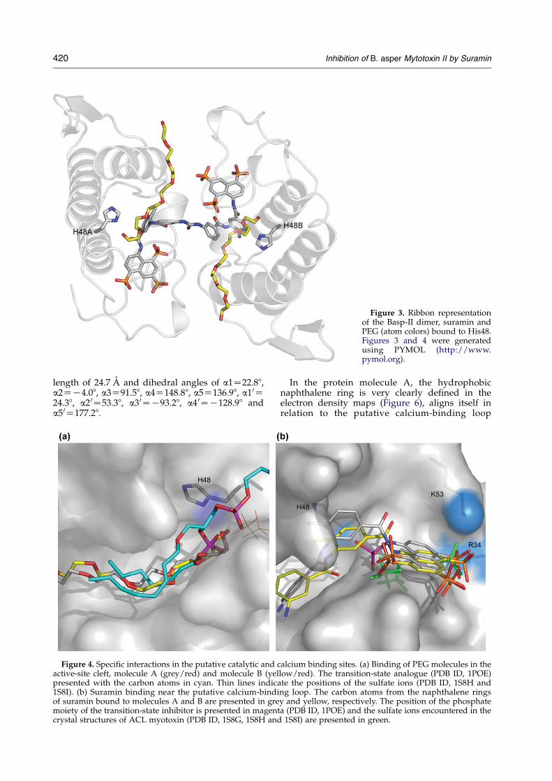
Figure 3. Ribbon representationof the Basp-II dimer, suramin andPEG (atom colors) bound to His48.Figures 3 and 4 were generatedusing PYMOL (http://www.pymol.org).
420 Inhibition of B. asper Mytotoxin II by Suramin
length of 24.7 A and dihedral angles of a1Z22.88,a2ZK4.08, a3Z91.58, a4Z148.88, a5Z136.98, a1 0Z24.38, a2 0Z53.38, a3 0ZK93.28, a4 0ZK128.98 anda5 0Z177.28.
Figure 4. Specific interactions in the putative catalytic and cactive-site cleft, molecule A (grey/red) and molecule B (yelpresented with the carbon atoms in cyan. Thin lines indica1S8I). (b) Suramin binding near the putative calcium-bindiof suramin bound to molecules A and B are presented in gremoiety of the transition-state inhibitor is presented in magentcrystal structures of ACL myotoxin (PDB ID, 1S8G, 1S8H and
In the protein molecule A, the hydrophobicnaphthalene ring is very clearly defined in theelectron density maps (Figure 6), aligns itself inrelation to the putative calcium-binding loop
alcium binding sites. (a) Binding of PEG molecules in thelow/red). The transition-state analogue (PDB ID, 1POE)te the positions of the sulfate ions (PDB ID, 1S8H andng loop. The carbon atoms from the naphthalene ringsy and yellow, respectively. The position of the phosphatea (PDB ID, 1POE) and the sulfate ions encountered in the1S8I) are presented in green.

Figure 5. Chemical structure and torsion angles of suramin. This Figure was generated using ChemSketch version 5.0(http://www.acdlabs.com/).
Inhibition of B. asper Mytotoxin II by Suramin 421
(residues 27–35) and is additionally stabilized byhydrogen bonds to Arg34 and Lys53 (Figures 4(b)and 6; and Table 1). The adjacent phenyl ringinteracts with hydrophobic amino acid residues inthe loop that links helix 2 with the b-wing (residues65–71), whereas the central phenyl ring is stabilizedby hydrophobic interactions with residues sur-rounding the active site and the N terminus. Thedistal or second naphthalene ring of suramin,which is less well defined in the electron densitymaps, binds in a similar fashion between loops C
and E to the second protein molecule interactingwith Arg34B and Lys53B (Table 1).
Discussion
Myonecrosis is a major event in envenomation bysnakes and compounds such as suramin, heparin,heparin-like glycosaminoglycans and related poly-anions inhibit the activity of myotoxic PLA2s bothin vitro and in vivo.31–35 A number of studies have
Figure 6. Electron density con-toured at 1.5s around the suraminmolecule. Hydrogen bondsbetween suramin and Arg34 andLys53 are in red (broken lines).

422 Inhibition of B. asper Mytotoxin II by Suramin
attempted to identify and to delineate the region orregions important for the expression of myotoxicityin PLA2s,
23 on the basis of sequence homology,36–38
charge distribution,39–41 hydrophobicity profiles,39
chemical modification,42,43 use of synthetic pep-tides,34,44,45 and site-directed mutagenesis.30,46
With the biochemical and structural informationcurrently available we are now able to probe thestructure–function relationship, and the mode ofaction and inhibition of Lys49 PLA2 analogues byaddressing the roles of the C-terminal region andthe nominal active site in myotoxicity.
Suramin and its inhibitory effects
Since the conformational flexibility of suramin isrestricted to rotations around the single bonds, thesimultaneous binding of the naphthalene rings tothe two protein molecules perturbs the putativecalcium-binding loop and the C-terminal extensions.
The interfacial recognition face (i-face) of myo-toxic Lys49 PLA2s contains a high density ofpositive charges, a structural feature that has beenassociated with the ability of these proteins tointeract with phospholipid bilayers.23,41 Theinduced-fit binding of suramin results in the burialof 102.1 A2 (total surface area of suraminZ335.1 A2), does not directly involve amino acidresidues in the putative catalytic site but modifiesthe surface charge significantly (Figure 7). Thischange in the charge on the i-face plays a crucialrole in the inhibition of myotoxicity, since Basp-IIwould now be unable to bind to the negativelycharged membrane target in muscle cells.
The heparin-binding site
The positions of the sulfonate groups of suraminand the sulfate ions encountered in the crystalstructure of Lys49 PLA2s25 were used as a guide tooverlay the atomic coordinates of heparin (PDB ID,
Figure 7. Surface charge distribution (red negative and bluBasp-IICsuramin complex in the same orientation.
1XMN). This leads us to speculate that this highlyconserved sulfate-binding site could represent theprimary heparin-binding site in Basp-II. In thisorientation, the heparin molecule could extendtowards the C-terminal region, interacting withresidues 115–129, which contain the motif B–B–X–B(where B denotes a basic amino acid) and a total ofsix basic residues out of 13.34 This is analogous tothe interaction in the suramin–bFGF complexmodel, where the induced-fit binding of suraminsterically blocks the receptor-binding region ofbFGF and competes for the binding of heparin.47
The role of the C terminus
The C-terminal region (residues 115–134) ofmyotoxic Lys49 PLA2s forms a cationic/hydro-phobic site that has been suggested to play a rolein the mediation of electrostatic interactions withnegatively charged acceptors, acting as a cytolyticmotif.34,44,45,48 A synthetic peptide corresponding toresidues 115–129 of Basp-II induces cytolysis,interferes with the interaction between heparinand Basp-II, and binds directly to radiolabeledheparin in solution.34 Although the i.m. injectionof this peptide failed to induce myotoxicity inmice,28,49 the corresponding analogous peptides ofrelated Lys49 PLA2s from Agkistrodon piscivoruspiscivorus45 and Agkistrodon contortrix laticinctus28
venoms are myonecrotic. Site-directed mutagenesishas further demonstrated the importance of specificresidues in the C-terminal region for the develop-ment of myotoxic activity in another Lys49 PLA2
from B. jararacussu venom, bothropstoxin-I.46 Sub-stitution of Lys and Arg residues with Ala in the Cterminus results in a significant loss of myotoxicactivity of the recombinant bothropstoxin-I.46
The crystal structure of Basp-II (PDB code, 1CLP)determined in the absence of a ligand results in theexposure of the hydrophobic residues in theC-terminal region, which are referred to collectively
e positive) of the monomers, (a) of Basp-II and (b) of the

Inhibition of B. asper Mytotoxin II by Suramin 423
as the hydrophobic knuckle (residues Leu121 andLeu124), due to the interaction of Lys122 with thepeptide bond formed between Cys29 and Gly30.25
Superpositioning the structures of this protein inthe ligand-free state and when bound to suraminresults in an overall rmsd of 0.6 A. The largestdeviations are observed in the putative calcium-binding loop (rmsd 0.9 A for residues 28–35), at thebase of the b-wing, which is generally disordered(rmsd greater than 1.5 A for residues 86–87) and inthe C-terminal region (rmsd between 0.9 A and1.8 A for residues 120–129). An analysis of thesedeviations indicates that the binding of suramindoes not alter the interactions that are responsiblefor maintaining the conformation of the hydro-phobic knuckle but results in a rigid-body shift ofthe motif formed by the putative calcium-bindingloop and C terminus.
In our X-ray model, the C-terminal region doesnot form part of the suramin-binding site of thedimer, but contributes significantly to the mainten-ance of the positive charge on the i-face, which isimportant for the binding of the myotoxin to thephospholipid bilayers. Thus, the C terminus,besides being involved in the development ofmyonecrosis, also likely plays a dual role bymaintaining the positive charge on the i-face andinteracting with the putative calcium-binding loop.
The putative active site
Lys49 PLA2s are capable of binding fattyacids,25,50 stearic acid,26 and PEG at the nominalcatalytic site. The induced-fit binding of suramin atthe surface does not completely block access to thenominal active site. Thus, the active site is stillaccessible to fatty acids, as evidenced by thepresence of PEG molecules bound to His48, whichoccupy positions analogous to lysophospholipids inthe structure of human group IIA PLA2 (PDB ID,1POE; Figure 4(a)) complexedwith the transition-stateanalogue (L1-O-octyl-2-heptylphosphonyl-sn-gly-cero-3-phosphoethanolamine). A direct relationshipbetween the active site and the C terminus in aLys49 PLA2 has been proposed by the hyperpolari-zation of the hydrogen bond formed by Lys122 NEand the peptide bond between residues Cys29 andGly30.25
Concluding remarks
These results suggest strongly that suramin is apotentially useful drug in the prevention of acutemuscle toxicity associated with Bothrops sp. snake-bite envenomation, since it is highly effective ininhibiting the myotoxic activity of Basp-II in vitroand in vivo, pre-incubated or administered aloneafter injection of the toxin. The structural resultsindicate that the relative orientation of the putativecalcium-binding loop and C-terminal regionstogether with the maintenance of the charge patternon the i-face could be important for myotoxicity inLys49 PLA2s. Due to the diversity of proteins that
suramin can bind to, it is tempting to speculate thatit may interact with other PLA2s from venoms andother sources including mammalian group IIAPLA2s, thus suggesting a wider pharmacologicalpotential for this inhibitor and these structuralresults should serve as the basis for the develop-ment of novel naphthalene sulfonate derivatives.
Materials and Methods
Basp-II and suramin
Basp-II was isolated by two cycles of cation-exchangechromatography on CM-Sephadex C-25, as described.51
Suramin was obtained from Sigma Chemical Co.(St. Louis, MO, USA). All other reagents were ofanalytical grade.
In vitro myotoxicity
In vitro myotoxicity experiments were performed atroom temperature.31,32 The extensor digitorum longusmuscles from mice were removed, blotted, weighedimmediately and then transferred to sample-collectingunits of 2.5 ml capacity, where they were superfusedcontinuously at a flow-rate of 3.0 ml/minute with aphysiological saline solution equilibrated with 95% (v/v)O2, 5% (v/v) CO2. Perfusing physiological saline solutioncontained 135 mM NaCl, 5 mM KCl, 2 mM CaCl2, 1 mMMgCl2, 15 mM NaHCO3, 1 mM NaH2PO4, 11 mM glu-cose. The final pH was adjusted to 7.3 after physiologicalsaline solution (PSS) equilibration with the gaseousmixture. After 60 minutes equilibration, Basp-II alone(25 mg/ml) or the Basp-II plus suramin (10 mM) wereadded to the solution containing the extensor digitorumlongus (EDL) muscles and then the perfusates werecollected and replaced with fresh PSS. The collectedsamples were stored at 4 8C. CK activity was determinedby using a diagnostic kit (Sigma Chemical Co., USA). Therate of CK release from the isolated muscles wasexpressed as the increase in CK release compared tocontrol values. Basal release rate refers to the enzyme lossfrom the muscles into the PSS medium during theequilibration period of perfusion (one hour), startingimmediately after the preparation had been mounted inthe sample-collection unit. The CK activity was expressedin international units (U), where 1 U is the amount thatcatalyzes the transformation of 1 mmol of substrate at25 8C. The rate of CK release from the isolated muscle wasexpressed as enzyme units released into the medium pergram per one hour of collection (U gK1 hK1).31,32
In vivo myotoxicity
Changes in plasma CK activity
Basp-II was dissolved in 0.1 ml of PSS and injected bythe i.m. route into the thighs of Swiss mice (20–25 g bodyweight). The amounts of toxin or suramin administeredwere adjusted taking into account the weight of eachanimal and injected in doses of 2.0 mg/g for the toxin and1.0 mg/g for suramin. Previous studies indicate that i.m.injection of 0.1 ml of PSS has no effect on plasma CKactivity.21,32 Two different experimental protocols wereused; in protocol A, Basp-II and suramin were incubatedfor 15 minutes at 37 8C, and then injected i.m. into each

424 Inhibition of B. asper Mytotoxin II by Suramin
animal. In protocol B, Basp-II was administered i.m. to theanimals 15 minutes before the intraperitoneal injection ofsuramin (1.0 mg/g). In both protocols, the final volume oftoxin injected, either alone or with suramin, was 0.1 ml.The animals were anesthetized lightly with diethyl etherimmediately before and two hours after the injection ofthe toxin for blood collection, in accordance withguidelines for care and use of laboratory animals.52
Plasma was separated by centrifugation and stored at4 8C for subsequent determination of CK activity asdescribed above.
Histological alterations
Mice, weighing 25.0(G5.0) g, were assembled into fourgroups of three mice each. Animals were anesthetizedwith ether, and subsequently injected with Basp-II(2.5 mg/g in 50 ml of PSS). Injection was performed justover the EDLmuscle of the right limb, as described.53 Thecontrol group was injected only with PSS in the right paw(50 ml). Two different experimental protocols were usedfor the study of suramin antimyotoxic effect. In the firstprotocol, the Basp-II was incubated with suramin (1 mg/kg) for 15 minutes prior to the injection of the mixture. Inthe second protocol, suramin (1 mg/kg) was injected i.v.15 minutes after the injection of Basp-II. The mice wereanesthetized with ether and sacrificed by cervicaldislocation 24 hours after the injection of the toxin. TheEDL muscles were dissected and fixed for two to threehours in 2.5% (v/v) glutaraldehyde, 4% (v/v) parafor-maldehyde in 0.1 M sodium cacodylate (pH 7.4). Theywere subsequently washed thrice in the same buffer andpost-fixed for one hour in 1% (w/v) osmium tetroxide.The tissue was then dehydrated in increasing concen-trations of acetone (30–100%, v/v) and embedded inPolybed 812 resin. Sections (400–600 nm) for lightmicroscopic examination were obtained using an RMCultramicrotome and stained with 1% (w/v) toluidineblue dye.
Crystallization and X-ray diffraction data collection
Basp-II was dissolved at a concentration of 10 mg/mlin 0.02 M Hepes (pH 7.5) and suramin was added at amolar ratio of 1:1.2. This complex was crystallized from asolution containing 0.1 M sodium acetate (pH 4.6), 15%(w/v) PEG 3350, 20% (v/v) isopropanol at 291 K by thehanging-drop, vapor-diffusion method as described.54
X-ray diffraction data were collected at 100 K from asingle crystal at a synchrotron radiation source (PCrbeamline, Laboratorio Nacional de Luz Sincrotron, LNLS,Campinas, Brazil) where the wavelength was set to1.427 A. Diffraction intensities were measured using aMARCCD detector (MAR Research), and the diffractiondata were integrated, reduced and processed using theDENZO/SCALEPACK suite of programs.55
Structure determination and refinement
The atomic coordinates of Basp-II24 (PDB ID, 1CLP)were utilized to solve the structure by molecularreplacement with the program package AMoRe.56 Non-crystallographic symmetry restraints were imposed in theearly cycles of refinement and the translation-libration-screw, positional and restrained isotropic B-factor refine-mentswere carried out usingREFMAC5,57 and the electrondensity maps were examined after each round of
refinement with TURBO-FRODO.58 The stereochemistryof the final structure was evaluated using PROCHECK.59
Protein Data Bank accession code
The atomic coordinates and structure factors of theBasp-IICsuramin complex have been deposited with theProtein Data Bank (accession code: 1Y4L).
Acknowledgements
This research was supported by grants fromFundacao de Amparo a Pesquisa do Estado de SaoPaulo (FAPESP), Structural Molecular Biology Net-work (SMOLBNet), Conselho Nacional de Desen-volvimento Cientifico e Tecnologico (CNPq),Coordenacao de Aperfeicoamento de Pessoal deNivel Superior (CAPES/DAAD) and Centros dePesquisa, Inovacao e Difusao (CEPID) to R.K.A.,Fundacao de Amparo a Pesquisa do Estado do Riode Janeiro (FAPERJ) and Programa de Apoio aNucles de Excelencia (PRONEX) to P.A.M., as wellas fromVicerrectoria de Investigacion, University ofCosta Rica, to B.L. and to J.M.G. M.T.M. is therecipient of a FAPESP doctoral fellowship.
References
1. Burch, T. A. & Ashburn, L. L. (1951). Experimentaltherapy of onchocerciasis with suramin and hetrazan;results of a three-year study. Am. J. Trop. Med. Hyg. 31,617–623.
2. Williamson, J. & Desowitz, R. S. (1956). Prophylacticactivity of suramin complexes in animal trypanoso-miasis. Nature, 177, 1074–1075.
3. Cherry, J. K. (1960). The treatment of onchocerciasis.East. Afr. Med. J. 37, 550–558.
4. Schneider, J. (1963). Treatment of human Africantrypanosomiasis. Bull. World Health Organ. 28, 763–786.
5. LaRocca, R. V., Cooper, M. R., Uhrich, M., Danesi, R.,Walther, M. M., Linehan, W. M. & Myers, C. E. (1991).Use of suramin in treatment of prostatic carcinomarefractory to conventional hormonal manipulation.Urol. Clin. North Am. 18, 123–129.
6. van Oosterom, A. T., ten Bokkel Huinink, W. W., vander Burg, M. E., Vermorken, J. B., Willemse, P. H. &Neijt, J. P. (1991). Phase II clinical trial of doxifluridinein patients with advanced ovarian cancer. Eur.J. Cancer, 27, 747–749.
7. Waltenberger, J., Mayr, U., Frank, H. & Hombach, V.(1996). Suramin is a potent inhibitor of vascularendothelial growth factor. A contribution to themolecular basis of its antiangiogenic action. J. Mol.Cell Cardiol. 28, 1523–1529.
8. Cadene, M., Duranton, J., North, A., Si-Tahar, M.,Chignard, M. & Bieth, J. G. (1997). Inhibition ofneutrophil serine proteinases by suramin. J. Biol.Chem. 272, 9950–9955.
9. Raj, P. A., Marcus, E. & Rein, R. (1998). Conformation-al requirements of suramin to target angiogenicgrowth factors. Angiogenesis, 2, 183–199.

Inhibition of B. asper Mytotoxin II by Suramin 425
10. Hermans, J. M., Haines, D. S., James, P. S. & Jones, R.(2003). Kinetics of inhibition of sperm beta-acrosinactivity by suramin. FEBS Letters, 544, 119–222.
11. Jennings, F. W., Rodgers, J., Bradley, B., Gettinby, G.,Kennedy, P. G. & Murray, M. (2002). Human Africantrypanosomiasis: potential therapeutic benefits of analternative suramin and melarsoprol regimen. Para-sitol. Int. 51, 381–388.
12. Fleck, S. L., Birdsall, B., Babon, J., Dluzewski, A. R.,Martin, S. R., Morgan, W. D. et al. (2003). Suramin andsuramin analogues inhibit merozoite surface protein-1 secondary processing and erythrocyte invasion bythe malaria parasite Plasmodium falciparum. J. Biol.Chem. 278, 47670–47677.
13. McCain, D. F., Wu, L., Nickel, P., Kassack, M. U.,Kreimeyer, A., Gagliardi, A. et al. (2004). Suraminderivatives as inhibitors and activators of protein-tyrosine phosphatases. J. Biol. Chem. 279, 14713–14725.
14. Fernandez-Tornero, C., Lozano, R. M., Redondo-Horcajo, M., Gomez, A. M., Lopez, J. C., Quesada, E.et al. (2003). Leads for development of new naphtha-lenesulfonate derivatives with enhanced antiangio-genic activity: crystal structure of acidic fibroblastgrowth factor in complex with 5-amino-2-naphtha-lene sulfonate. J. Biol. Chem. 278, 21774–21781.
15. Arruda, E. Z., Silva, N. M., Moraes, R. A. &Melo, P. A.(2002). Effect of suramin onmyotoxicity of some crotalidsnake venoms. Braz. J. Med. Biol. Res. 35, 723–726.
16. de Oliveira, M., Cavalcante, W. L., Arruda, E. Z.,Melo, P. A., Dal-Pai Silva, M. & Gallacci, M. (2003).Antagonism of myotoxic and paralyzing activities ofbothropstoxin-I by suramin. Toxicon, 42, 373–379.
17. Arni, R. K. & Ward, R. J. (1996). Phospholipase A2—astructural review. Toxicon, 34, 827–841.
18. Kini, R. M. (2003). Excitement ahead: structure,function and mechanism of snake venom phospho-lipase A2 enzymes. Toxicon, 42, 827–840.
19. Mebs, D. & Ownby, C. L. (1990). Myotoxic com-ponents of snake venoms: their biochemical andbiological activities. Pharm. Ther. 48, 223–236.
20. da Silva Giotto, M. T., Garratt, R. C., Oliva, G.,Mascarenhas, Y. P., Giglio, J. R., Cintra, A. C. et al.(1998). Crystallographic and spectroscopic character-ization of a molecular hinge: conformational changesin bothropstoxin I, a dimeric Lys49-phospholipase A2homologue. Proteins: Struct. Funct. Genet. 30, 442–454.
21. Melo, P. A., Nascimento, M. C., Mors, W. B. & Ownby,C. L. (1994). Inhibition of the myotoxic and hemor-rhagic activities of crotalid venoms by Eclipta prostrata(Asteraceae) extracts and constituents. Toxicon, 32,595–603.
22. Scott, D. L., Otwinowski, Z., Gelb, M. H. & Sigler, P. B.(1990). Crystal structure of bee-venom phospholipaseA2 in a complex with a transition-state analogue.Science, 250, 1563–1566.
23. Murakami, M. T. & Arni, R. K. (2003). A structurebased model for liposome disruption and the role ofcatalytic activity in myotoxic phospholipase A2s.Toxicon, 42, 903–913.
24. Arni, R. K., Ward, R. J., Gutierrez, J. M. & Tulinsky, A.(1995). Structure of a calcium-independent phospho-lipase-like myotoxic protein from Bothrops aspervenom. Acta Crystallog. sect. D, 51, 311–317.
25. Ambrosio, A. L., Nonato, M. C., Selistre-de-Araujo,H. S., Arni, R. K., Ward, R. J., Ownby, C. L. et al. (2005).A molecular mechanism for Lys49-phospholipase A2activity based on ligand-induced conformationalchange. J. Biol. Chem. 280, 7326–7335.
26. Watanabe, L., Soares, A. M., Ward, R. J., Fontes,
M. R. M. & Arni, R. K. (2005). Structural insights forfatty acid binding in a Lys49-phospholipase A(2):crystal structure of myotoxin II from Bothrops moojenicomplexed with stearic acid. Biochimie, 87, 161–167.
27. Lee, W. H., da Silva Giotto, M. T., Marangoni, S.,Toyama, M., Polikarpov, I. & Garrat, R. C. (2001).Structural basis for low catalytic activity in Lys49phospholipases A2—a hypothesis: the crystal struc-ture of piratoxin II complexed to fatty acid. Biochem-istry, 40, 28–36.
28. Lomonte, B., Angulo, Y. & Santamaria, C. (2003).Comparative study of synthetic peptides correspond-ing to region 115–129 in Lys49 myotoxic phospho-lipases A2 from snake venoms. Toxicon, 42, 307–312.
29. Lomonte, B., Angulo, Y. & Calderon, L. (2003). Anoverview of lysine-49 phospholipase A2 myotoxinsfrom crotalid snake venoms and their structuraldeterminants of myotoxic action. Toxicon, 42, 885–901.
30. Ward, R. J., Chioato, L., de Oliveira, A. H., Ruller, R. &Sa, J. M. (2002). Active-site mutagenesis of a Lys49-phospholipase A2: biological and membrane-disrupt-ing activities in the absence of catalysis. Biochem. J.362, 89–96.
31. Melo, P. A. & Suarez-Kurtz, G. (1988). Release ofcreatine kinase from skeletal muscles by Bothropsvenoms: heparin potentiation of inhibition by anti-venin. Braz. J. Med. Biol. Res. 21, 548–558.
32. Melo, P. A. & Suarez-Kurtz, G. (1988). Release ofsarcoplasmic enzymes from skeletal muscle byBothrops jararacussu venom: antagonism by heparinand by the serum of South American marsupials.Toxicon, 26, 87–95.
33. Lomonte, B., Tarkowski, A., Bagge, U. & Hanson,L. A. (1994). Neutralization of the cytolytic andmyotoxic activities of phospholipases A2 fromBothrops asper snake venom by glycosaminoglycansof the heparin/heparan sulfate family. Biochem.Pharmacol. 47, 1509–1518.
34. Lomonte, B., Moreno, E., Tarkowski, A., Hanson, L. A.& Maccarana, M. (1994). Neutralizing interactionbetween heparins and myotoxin II, a lysine 49phospholipase A2 from Bothrops asper snake venom.Identification of a heparin-binding and cytolytic toxinregion by the use of synthetic peptides and molecularmodeling. J. Biol. Chem. 269, 29867–29873.
35. Melo, P. A., Homsi-Brandeburgo, M. I., Giglio, J. R. &Suarez-Kurtz, G. (1993). Antagonism of the myotoxiceffects of Bothrops jararacussu venom and bothrop-stoxin by polyanions. Toxicon, 31, 285–291.
36. Krizaj, I., Turk, D., Ritonja, A. & Gubensek, F. (1989).Primary structure of ammodytoxin C further revealsthe toxic site of ammodytoxin. Biochim. Biophys. Acta,999, 198–202.
37. Selistre de Araujo, H. S., White, S. P. & Ownby, C. L.(1996). Sequence analysis of Lys49 phospholipase A2myotoxins: a highly conserved class of proteins. Arch.Biochem. Biophys. 326, 21–30.
38. Ward, R. J., Rodrigues Alves, A., Rugierro Neto, J.,Arni, R. K. & Casari, J. A. (1998). Sequence spaceanalysis of Lys49 phopholipases A2: clues towardsidentification of residues involved in a novel mech-anism of membrane damage and in myotoxicity.Protein Eng. 11, 285–294.
39. Kini, R. M. & Iwanaga, S. (1986). Structure-functionrelationships of phospholipases. II: charge densitydistribution and the myotoxicity of presynapticallyneurotoxic phospholipases. Toxicon, 24, 895–905.
40. Kini, R. M. & Evans, H. J. (1989). Role of cationicresidues in cytolytic activity: modification of lysine

426 Inhibition of B. asper Mytotoxin II by Suramin
residues in the cardiotoxin from Naja nigricollisvenom and correlation between cytolytic and anti-platelet activity. Biochemistry, 28, 9209–9216.
41. Falconi, M., Desideri, A. & Rufini, S. (2000).Membrane-perturbing activity of Viperidae myotox-ins: an electrostatic surface potential approach to apuzzling problem. J. Mol. Recogn. 13, 14–19.
42. Andriao-Escarso, S. H., Soares, A. M., Rodrigues,V. M., Angulo, Y., Diaz, C., Lomonte, B. et al. (2000).Myotoxic phospholipases A(2) in Bothrops snakevenoms: effect of chemical modifications on theenzymatic and pharmacological properties ofbothropstoxins from Bothrops jararacussu. Biochimie,82, 755–763.
43. Soares, A. M., Andriao-Escarso, S. H., Bortoleto, R. K.,Rodrigues-Simioni, L., Arni, R. K., Ward, R. J. et al.(2001). Dissociation of enzymatic and pharmacologi-cal properties of piratoxins-I and -III, two myotoxicphospholipases A2 from Bothrops pirajai snake venom.Arch. Biochem. Biophys. 387, 188–196.
44. Calderon, L. & Lomonte, B. (1998). Immunochemicalcharacterization and role in toxic activities of region115–129 of myotoxin II, a Lys49 phospholipase A2from Bothrops asper snake venom. Arch. Biochem.Biophys. 358, 343–350.
45. Nunez, C. E., Angulo, Y. & Lomonte, B. (2001).Identification of the myotoxic site of the Lys49phospholipase A(2) from Agkistrodon piscivorus pisci-vorus snake venom: synthetic C-terminal peptides fromLys49, but not from Asp49 myotoxins, exert mem-brane-damaging activities. Toxicon, 39, 1587–1594.
46. Chioato, L., Oliveira, A. H. C., Ruller, R., Sa, J. M. &Ward, R. J. (2002). Distinct sites for myotoxic andmembrane-damaging activities in the C-terminalregion of a Lys49-phospholipase A2. Biochem. J. 366,971–976.
47. Fernandez-Tornero, C., Lozano, R. M., Redondo-Horcajo, M., Gomez, A. M., Lopez, J. C., Quesada, E.et al. (2003). Leads for development of new naphtha-lenesulfonate derivatives with enhanced antiangio-genic activity: crystal structure of acidic fibroblastgrowth factor in complex with 5-amino-2-naphtha-lene sulfonate. J. Biol. Chem. 278, 21774–21781.
48. Paramo, L., Lomonte, B., Pizarro-Cerda, J.,Bengoechea, J. A., Gorvel, J. P. & Moreno, E. (1998).Bactericidal activity of Lys49 and Asp49 myotoxicphospholipases A2 from Bothrops asper snake
venom-synthetic Lys49 myotoxin II-(115–129)-peptideidentifies its bactericidal region. Eur. J. Biochem. 253,452–461.
49. Gutierrez, J. M. & Lomonte, B. (1997). PhospholipaseA2 myotoxins from Bothrops snake venoms. In VenomPhospholipase A2 Enzymes: Structure, Function andMechanism (Kini, R. M., ed.), p. 321, Wiley, Chichester.
50. Lee, W. H., Toyama, M. H., Soares, A. M., Giglio, J. R.,Marangoni, S. & Polikarpov, I. (1999). Crystallizationand preliminary X-ray diffraction studies of piratoxinIII, a D-49 phospholipase A2 from the venom ofBothrops pirajai. Acta Crystallog. sect. D, 55, 1229–1230.
51. Lomonte, B. & Gutierrez, J. M. (1989). A new muscledamaging toxin, myotoxin II, from the venom of thesnake Bothrops asper (terciopelo). Toxicon, 27, 725–733.
52. Mass, J., Heling, W. & Seeger, K. (1997). Anesthesia ofexperimental animals. In Drug Discovery and Evalu-ation: Pharmacological Assays (Vogel, H. G. & Vogel, W.,eds), p. 732, Spring-Verlag, Berlin.
53. Melo, P. A. & Ownby, C. L. (1999). Ability ofwedelolactone, heparin, and para-bromophenacylbromide to antagonize the myotoxic effects of twocrotaline venoms and their PLA2 myotoxins. Toxicon,37, 199–215.
54. Murakami, M. T., Gava, L. M., Zela, S. P., Arruda,E. Z., Melo, P. A., Gutierrez, J. M. & Arni, R. K. (2004).Crystallization and preliminary X-ray diffractionanalysis of suramin, a highly charged polysulfonatednapthylurea, complexed with a myotoxic PLA2from Bothrops asper venom. Biochim. Biophys. Acta,1703, 83–85.
55. Otwinowski, Z. & Minor, W. (1997). Processing ofX-ray diffraction data collection in oscillation mode.Methods Enzymol. 276, 307–326.
56. Navaza, J. (1994). AMoRe: an automated package formolecular replacement. Acta Crystallog. sect. A, 50,157–163.
57. Murshudov, G. N., Vagin, A. A. & Dodson, E. J. (1997).Refinement of macromolecular structures by themaximum-likelihood method. Acta Crystallog. sect.D, 53, 240–255.
58. Roussel, A. & Cambillau, C. (1989). Turbo-Frodo. InSilicon Graphics Geometry Partner Directory (SiliconGraphics, ed.), p. 77, Mountain View, CA.
59. Laskowski, R. A., MacArthur, M. W., Moss, D. S. &Thornton, J. M. (1993). PROCHECK: a program tocheck the stereochemical quality of protein structures.J. Appl. Crystallog. 26, 283–291.
Edited by I. Wilson
(Received 23 March 2005; received in revised form 23 April 2005; accepted 27 April 2005)