inhibition of ameloblastoma invasion in vitro and in vivo by inhibitor of metalloproteinase-2...
TRANSCRIPT

Inhibition of ameloblastoma invasion in vitro and in vivoby inhibitor of metalloproteinase-2 activity
Bin Zhang1,*, Jin Zhang
2,*, Hong-Zhang Huang
3, Wei-Liang Chen
1, Qian Tao
3, Dong-Lin Zeng
3, Lei-Tao
Zhang1, Jian-Hui Xu
1
1Department of Oral and Maxillofacial Surgery, The Second Affiliated Hospital, Sun Yat-Sen University, Guangzhou; 2Departmentof Internal Medicine, The Second Affiliated Hospital, Sun Yat-Sen University, Guangzhou; 3Department of Oral and Maxillofacialsurgery, Guanghua College of Stomatology, Sun Yat-sen University, Guangzhou, China
BACKGROUND: Ameloblastoma is an odontogenic
benign tumor characterized by local invasiveness and most
of its local recurrences clinically result from local invasion.
This study used matrix metalloproteinase-2 (MMP-2)
inhibitor I (MMP-2I) to investigate the role played by MMP-
2 activity in the local invasiveness of ameloblastoma.
METHODS: The cells and xenografts of ameloblastoma
were treated with MMP-2I and treatment group were
compared with the control group. In vitro, the invasive
activity of tumor cells was assayed in transwell cell cul-
ture chamber. Gelatinolytic activity of gelatinases and
MMP-2 ⁄ tissue inhibitor of matrix metalloproteinase
(TIMP-2) protein expression was detected using gelatin
zymography and flow cytometry. The cell viability and
adhesion were evaluated using methyl thiazol tetra-
zolium. In vivo, bilateral subrenal capsule xenograft
transplantation of ameloblastoma was performed in 10
nude mice and the invasion of ameloblastoma into the
renal parenchyma was observed.
RESULTS: Active-MMP-2 of conditioned media was sig-
nificantly lower in treatment group than in the control
group. Accordingly, potential of in vitro cell invasion,
adhesion and in vivo tumor invasion were also significantly
lower in the treatment group than in the control group.
CONCLUSIONS: Inhibitor of MMP-2 activity suppressed
the local invasive capability of ameloblastoma by
decreasing MMP-2 activity. MMP-2 activity is in relation
with invasive capacity of ameloblastoma.
J Oral Pathol Med (2009) 38: 731–736
Keywords: ameloblastoma; invasion; metalloproteinase-2 activ-
ity; metalloproteinase-2 inhibitor
Introduction
Ameloblastoma is the most common odontogenic tumorof the jaw. Although histologically benign, it is a locallyaggressive benign tumor with frequent local recurrencesand local invasions. For decades, a less aggressive policyconsisting of local curettage or marginal resection wasadopted for this kind of benign tumor. A high recurrentrate was reported according to this treatment policy (1).Patients suffered from multiple recurrences requiredrepeated surgery, and might still lose their masticatoryfunction and disfigure. Currently extensive surgery is themost acceptable treatment modality in the classicsolid ⁄multicystic ameloblastoma which also increasedpost-operative complication and functional impairment(2). For this reason, the treatment about ameloblastomais constantly challenging problem because of its localrecurrences and local invasions. Matrix metalloprotein-ases (MMPs) and zinc-dependent proteolytic enzymesplay an important role in matrix degradation during thetumor growth, invasion and tumor-induced angiogene-sis (3). Especially, MMP-2 can degrade type IV collagen,one of the major components of the basement mem-brane, resulting in the promotion of tumor invasion andmetastasis (4). For the past few years, MMP inhibitors(MMPIs) have demonstrated their ability to delaymalignant tumor growth and to block metastasis (5, 6).To know the ability for MMP-2 inhibitor to inhibit localinvasion of ameloblastoma and to verify whetherinvasive capacity of ameloblastoma has any directrelation with MMP-2 activity or not, we used MMP-2Inhibitor I (MMP-2I) to investigate the role playedby MMP-2 activity in the local invasiveness of amelo-blastoma.
Materials and methods
Samples used in all study were obtained after informedconsent and with the approval of the Sun Yet-senUniversity Ethics Committee. The sample of this studywas from the patient with multiple recurrences in
Correspondence: Professor H.-Z. Huang, Guanghua College ofStomatology, Sun Yat-Sen University, No. 56, Lingyuan RoadWest, Guangzhou City, Guang-dong 510055, China. Tel: +86 208133 2429, Fax: +86 20 8133 2853, E-mail: [email protected]*These two authors contributed equally to this work.Accepted for publication February 15, 2009
doi: 10.1111/j.1600-0714.2009.00771.x
J Oral Pathol Med (2009) 38: 731–736
ª 2009 The Authors. Journal compilation ª 2009 Blackwell Munksgaard Æ All rights reserved
interscience.wiley.com/journal/jop
Journal of
Oral Pathology & Medicine

zygomatic fossa and was the classic solid type (plexi-form) ameloblastoma confirmed in histopathology.In vitro, first of all, primary cell culture of ameloblas-
toma tissue was performed. Briefly, tumor tissues wereminced into 1 mm3 small pieces, and incubated overnightwith Dulbecco’s modified Eagle medium (DMEM;Invitrogen, Carlsbad, CA, USA) containing 1 mg ⁄mlcollagenase I (Invitrogen) at 37�C. Collagenase-digestedtissues were plated onto 35-mm dishes coated withcollagen I (Invitrogen) in DMEM containing 10% fetalcalf serum, 200 lg ⁄ml streptomycin and 200 IU ⁄mlpenicillin, and incubated at 37�C under a humidified5% CO2 atmosphere. When the cells became confluentin 12–14 days interval, they were split again and usedfor experiments. The epithelial origin of primary cellswas confirmed using immunocytochemistry with anti-cytokeratin and anti-vimentin primary antibodies
(Maixin, Fuzhou, China) and an SP Hypersensitive kit(Maxim Biotech, San Francisco, CA, USA) accordingto the manufacturer’s instructions. The cells weredivided into treatment and control groups according todifferent concentrations of MMP-2I (Cat. No. 444244;Calbiochem, San Diego, CA, USA), which is synonymsof cis-9-Octadecenoyl-N-hydroxylamide and potentselective inhibitor of MMP-2 (gelatinase A): the treat-ment group treated with 5 lg ⁄ml(groupM5) or 10 lg ⁄ml(group M10) MMP-2I, and the control group with0 lg ⁄ml MMP-2I (group M0, negative control). Flowcytometry withmonoclonal anti-MMP-2 or anti-TIMP-2(Maixin) primary antibody was employed to measure theexpression rate of MMP-2 or TIMP-2 using a flowcytometer (Becton Dickinson, Sunnyvale, CA, USA) inprimary cells, the control group and the treatment groupwere treated with MMP-2I for 36 h. After cells of thecontrol and the treatment group were cultured about3 days in serum-free cell culture medium DK-SFM (Cat.No. 10744; Gibco, Carlsbad, CA, USA), gelatinolyticactivity of gelatinases in 10 ll cell culture medium wasanalyzed by gelatin zymography according to the methodreported by Kleiner (7) and using an auto imaginganalysis system (Kontron IBAS2.0; Munich, Germany)in three groups. All optical density measurements weremade among samples on the same gel to ensurecomparability. All experiments were performed intriplicate.
In vitro cell viability and adhesion ability were evalu-ated using methyl thiazol tetrazolium (MTT; SigmaChemical Co., St Louis, Mo, USA). Briefly, cells wereseeded in the 96-well plate at a concentration of 1 · 104
cells ⁄well and incubated for 24 h and then the cells weretreated with MMP-2I for 6 days for cell viability assay.In cell adhesion assay, after 96-well microtiter plates werepre-coated with 20 mg ⁄ l fibronectin and incubated at4�C overnight, the wells were blocked with 2% bovineserum albumin (BSA) for 60 min at 37�C, the cells weredistributed to the wells (2 · 105 ⁄well) and were treatedwithMMP-2I for 20, 40, 60 and 90 min. On the indicatedtime thereafter, cells were incubated with 0.5 mg ⁄mlMTT. After 4 h, MTT crystals were dissolved indimethyl-sulfoxide, and the absorbance was measuredat 492 nm using an ELISA plate reader. Each assay wasperformed in quadruplicate.
In vitro cell invasion capacity was assayed in Transwellcell chambers (Costa, Cambrige, MA, USA), accordingto the method reported by Kido (8). Briefly, polycarbon-ate filters with an 8-lm pore size were pre-coated with5 lg of fibronectin in a volume of 50 ll on the lowersurface. The Matrigel (BD Biosciences, Bedford, MA,USA) was diluted to 100 lg ⁄ml with cold phosphate-buffered saline (PBS) and applied to the upper surface ofthe filters (5 lg ⁄ filter), and dried overnight under a hoodat room temperature. The coated filters were washedextensively in PBS, and then dried immediately beforeuse. The cells were harvested, washed, and resuspendedto a final concentration of 2 · 106 ⁄ml in DMEM with0.1% BSA. Cell suspensions (100 ll) with or withoutMMP-2I were added to the upper compartment andincubated for 20 h at 37�C in a 5%CO2 atmosphere. Thefilters were fixed with methanol and stained with Giemsa.The cells on the upper surface were removed by wipingwith cotton swabs. The cells invading the lower surfacethrough Matrigel were manually counted under a micro-scope at a magnification of ·320, and each assay wasperformed in quadruplicate.
In vivo tumor invasion capacity was assessed bysubrenal capsule xenograft transplant according theranking criteria reported by Wang (9). The BALB ⁄ cnude mice were from the animal experiment center ofSun Yat-Sen University, China. The care and treatmentof experimental animals were in accordance with the
Adhesion rate ¼ Average A492 nm value of MMP-2I group
Average A492 nm value of the control group� 100%:
Inhibitory rate of adhesion
¼ (Average A492 nm value of control group�Average A492 nm value of MMP-2I group)
Average A492 nm value of control group� 100%:
Inhibition of ameloblastoma invasion
Zhang et al.
732
J Oral Pathol Med

institutional guidelines. Briefly, the tumor tissues of ahuman ameloblastoma were cut into 1-mm3 pieces andwere transplanted to bilateral subrenal capsule in10 nude mice (5 weeks old) anesthetized with chloralhydrate. Three days later, the mice were randomlydivided into two groups (5 mice ⁄ group, namely 10xenografts ⁄ group). Experimental mice were treated withMMP-2I (50 mg ⁄ kg ⁄day) for 30 day by i.p. injection.Control mice were given the same solvent (concentrationwas the same as experimental mice). The mice werekilled by cervical dislocation after the treatment wasover. Autopsies were performed and all kidneys wereexcised, fixed, and embedded in paraffin, and examinedhistologically with hematoxylin and eosin stain. Evalu-ation criteria of invasive potential of ameloblastoma insubrenal capsule xenograft transplant: rank 0, tumortissue begins multiplicative division but does not invadeperipheral tissue; rank 1, tumor tissue proliferatesactively and begins to invade the interspace of renaltubule; rank 2, tumor cells invade renal parenchyma to10–20 cell layers and renal tubule does not occurdenaturation; rank 3, the area of invasion by tumorcells increases further and a great quantity renal tubulesoccur degenerative atrophy; rank 4, renal parenchyma isinvolved in invasion over 50% and nephridial tissueoccurs necrosis.
All measurement data are expressed as mean ± SD.The difference between the groups was examined usingthe one-way ANOVA in measurement data and chi-square in enumeration data (probabilities of <0.05 wereaccepted as significant). The Statistical Package for theScial Sciences (version 11.0; SPSS; Chicago, IL, USA)was used.
Results
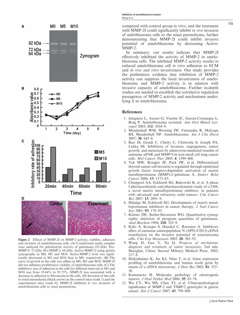
After ameloblastoma tissues were cultured for 18–36 h,ameloblastoma cells migrated out from ameloblastomatissue, presented membraniform growth, arrangementlike slabstone (Fig. 1A). Secondary cell culture showedlarge flattened or spindle-shaped morphology and grewvigorously (Fig. 1B). Immunocytochemistry showedthat the cells were immunopositive for CK andimmunonegative for vimentin (Fig. 1C), and indicatedthat the culture cells were epithelial origin. Flowcytometry showed MMP-2 immunopositive rate ofprimary cells were 22.3 ± 5.6 (Fig. 1D). Active-MMP-2 levels of conditioned media were significantly lower inM5 and M10 groups than in M0 group (127.7 ± 122.8and 86.0 ± 113.3 compared with 163.6 ± 128.7,P < 0.05), and there were no significant differences in
Pro-MMP-9 and Pro-MMP-2 among three groups(P > 0.05; Fig. 2A). There were no significant differ-ences in expression of MMP-2 and TIMP-2 protein inthe culture cells between M0 group and M5 and M10groups (MMP-2: 23.60 ± 8.90, 23.62 ± 3.46, 17.82 ±3.63; TIMP-2: 28.11 ± 9.48, 21.51 ± 3.03, 30.37 ±5.66, P > 0.05). The curve of growth indicated that thecells for all three groups grew vigorously. There was nosignificant difference among the groups (P > 0.05,Fig. 2B). The inhibitory rate of adhesion in the cellswas significantly higher in M5 and M10 groups than inM0 group (Fig. 2C, P < 0.05). The invasion rate wassignificantly lower in M5 and M10 groups than in M0group (161.9 ± 17.9 and 85.6 ± 12.7 compared with228.2 ± 27.7, P < 0.01). The inhibitory rate of inva-sion in M5 and M10 groups was 29.1% and 62.5%,respectively. The inhibitory of invasion was significantlyhigher in M10 group than in M5 group (P < 0.01).In vivo tumor invasion assay showed that invasive ratioof renal parenchyma was significantly lower in theexperimental mice than in the control mice (P<0.01).Invasion rank 0 was observed in two of 10 kidneys(20%) and invasion rank 1 in eight of 10 kidneys (80%)in the control mice, but invasion rank 0 was observed in9 ⁄ 10 (90%) and rank 1 in 1 ⁄ 10 (10%) in the experi-mental mice (Fig. 2D).
Discussion
Local invasion remains a major cause of frequentrecurrences and dissatisfactory treatment in ameloblas-toma clinically. Many studies have identified genetic andmolecular alterations in ameloblastoma (10, 11), but themechanisms underlying the local invasiveness of thisneoplasm have yet to be clarified. Reports from somestudies indicate that MMP-2 plays an important role inthe invasion of malignant tumors such as head and neckcarcinomas, gastric cancer, colon cancer, etc (4, 12).MMP-2 activity has been directly correlated with theaggressiveness of malignant tumor cells (13, 14). Todate, there are still few reports of studies on thecorrelation between MMP-2 activity and the localinvasiveness of human ameloblastoma. The purpose ofthis study was to determine whether MMP-2 activityinhibition can suppress the local invasiveness of humanameloblastoma. To accomplish this, we employedMMP-2I to inhibit MMP-2 activity and then detectedthe relationship between MMP-2 activity and invasionof ameloblastoma.
MMP-2 activity can be regulated by endogenousTIMP-2 and synthetical inhibitors (5, 6). We investigated
Invasion rate ¼ Average cell numbers invading the lower surface in treated group
Average cell numbers invading the lower surface in the control group� 100%:
Inhibitory rate of invasion
¼ (Average cell numbers invading the lower surface in the control group � Average cell numbers invading the lower surface in the treated group)
Average cell numbers invading the lower surface in the control group�100%:
Inhibition of ameloblastoma invasion
Zhang et al.
733
J Oral Pathol Med

the effect of MMP-2I on MMP-2 activity and MMP-2and TIMP-2 protein expression in ameloblastomacells, and found that MMP-2I could significantlydecrease the levels of Active-MMP-2 secreted byameloblastoma cells, but did not influence the expres-sion of MMP-2 ⁄TIMP-2 protein in ameloblastoma cells,indicating that the effect of MMP-2I on ameloblastomacells may be on account of direct inhibition of MMP-2activity. We also investigated the effect of MMP-2I onviability of ameloblastoma cells, and authenticate forthe first time that MMP-2I did not influence in vitroproliferative capability of ameloblastoma cells, demon-strating that the effect of MMP-2I on ameloblastomacells may not be associated with cellular proliferativecapability.
Active-MMP-2 can degrade type IV collagen of themajor components in the basement membrane andregulate the adhesion between cell and matrix, which arenecessary to tumor invasion (15, 16). We investigatedthe effect of MMP-2I on the adhesive properties ofameloblastoma cells, and found that MMP-2I couldinhibit tumor cell adhesion to fibronectin-coated sub-strates, indicating that MMP-2I may suppress the abilityof ameloblastoma cells to transgress normal tissueboundaries and disperse to the adjacent sites by theregulation of Active-MMP-2 on the adhesive capacitybetween cell and matrix. In vitro and in vivo invasionassay further showed that ameloblastoma cells invadingthe lower surface of the filter through Matrigel weresignificantly decreased in MMP-2I treated groups
A B
C
D
0
200
400
600
800
1000
SS
C-H
eigh
t
100 101 102 103 104
MMP-2FITC
Figure 1 Primary cell culture of ameloblastoma. (A) Primary culture ameloblastoma cells migrated out from ameloblastoma tissue (200·).(B) Subcultured ameloblastoma cells illustrate their vigorously growth in 2–4 passages, large flattened or spindle-shaped morphology (100·).(C) Immunocytochemistry confirmed that cytokeratin was immunopositive and vimentin was immunonegative in ameloblastoma cells (300·).(D) Matrix metalloproteinase (MMP)-2 immunopositive of ameloblastoma cells was detected by flow cytometry.
Inhibition of ameloblastoma invasion
Zhang et al.
734
J Oral Pathol Med
compared with control group in vitro, and the treatmentwith MMP-2I could significantly inhibit in vivo invasionof amloblastoma cells to the renal parenchyma, furtherdemonstrating that MMP-2I could inhibit invasivepotential of ameloblastoma by decreasing Active-MMP-2.
In summary, our results indicate that MMP-2Ieffectively inhibited the activity of MMP-2 in amelo-blastoma cells. The inhibited MMP-2 activity results inreduced ameloblastoma cell in vitro adhesion to ECMand in vivo and vitro invasiveness. Our study providesthe preliminary evidence that inhibition of MMP-2activity can suppress the local invasiveness of amelo-blastoma and MMP-2 activity is in relation withinvasive capacity of ameloblastoma. Further in-depthstudies are needed to establish the correlative regulationpassageway of MMP-2 activity and mechanisms under-lying it in ameloblastoma.
References
1. Junquera L, Ascani G, Vicente JC, Garcia-Consuegra L,Roig P. Ameloblastoma revisited. Ann Otol Rhinol Lar-yngol 2003; 112: 1034–9.
2. Mendenhall WM, Werning JW, Fernandes R, MalyapaRS, Mendenhall NP. Ameloblastoma. Am J Clin Oncol2007; 30: 645–8.
3. Rao JS, Gondi C, Chetty C, Chittivelu S, Joseph PA,Lakka SS. Inhibition of invasion, angiogenesis, tumorgrowth, and metastasis by adenovirus-mediated transfer ofantisense uPAR and MMP-9 in non-small cell lung cancercells. Mol Cancer Ther 2005; 4: 1399–408.
4. Yeh MW, Rougier JP, Park JW, et al. Differentiatedthyroid cancer cell invasion is regulated through epidermalgrowth factor receptor-dependent activation of matrixmetalloproteinase (MMP)-2 ⁄ gelatinase A. Endocr RelatCancer 2006; 13: 1173–83.
5. Chiappori AA, Eckhardt SG, Bukowski R, et al. A phaseI pharmacokinetic and pharmacodynamic study of s-3304,a novel matrix metalloproteinase inhibitor, in patientswith advanced and refractory solid tumors. Clin CancerRes 2007; 13: 2091–9.
6. Hidalgo M, Eckhardt SG. Development of matrix metal-loproteinase inhibitors in cancer therapy. J Natl CancerInst 2001; 93: 178–93.
7. Kleiner DE, Stetler-Stevenson WG. Quantitative zymog-raphy: detection of picogram quantities of gelatinases.Anal Biochem 1994; 218: 325–9.
8. Kido A, Krueger S, Haeckel C, Roessner A. Inhibitoryeffect of antisense aminopeptidase N (APN ⁄CD13) cDNAtransfection on the invasive potential of osteosarcomacells. Clin Exp Metastasis 2003; 20: 585–92.
9. Wang JJ, Gao Y, Xu Q. Progress of mechanism,diagnosis and treatment of tumor metastasis, 2nd edn.Shanghai, China: Second Military Medical Press, 2002;217–8.
10. Heikinheimo K, Jee KJ, Niini T, et al. Gene expressionprofiling of ameloblastoma and human tooth germ bymeans of a cDNA microarray. J Dent Res 2002; 81: 525–30.
11. Kumamoto H. Molecular pathology of odontogenictumors. J Oral Pathol Med 2006; 35: 65–74.
12. Wu CY, Wu MS, Chen YJ, et al. Clinicopathologicalsignificance of MMP-2 and TIMP-2 genotypes in gastriccancer. Eur J Cancer 2007; 43: 799–808.
A
B
C
D
Figure 2 Effects of MMP-2I on MMP-2 activity, viability, adhesionand invasion of ameloblastoma cells. (A) Conditioned media sampleswere analyzed for gelatinolytic activity of gelatinases (92 kDa, Pro-MMP-9; 72 kDa, Pro-MMP-2; 64 kDa, Active-MMP-2) using gelatinzymography in M0, M5 and M10. Active-MMP-2 level was signif-icantly decreased in M5 and M10 than in M0, respectively. (B) Thecurve of growth in the cells was affinis in M0, M5 and M10. MMP-2Idid not influence proliferative viability of ameloblastoma cells. (C) Theinhibitory rate of adhesion in the cells for different intervals in M5 andM10 was from 19.64% to 95.75%. MMP-2I was associated with adecrease in adhesion to fibronectin in the cells. (D) Invasion of the cellsto renal parenchyma (black arrow) in the control mice (rank 1) and theexperimental mice (rank 0). MMP-2I inhibited in vivo invasion ofameloblastoma cells to renal parenchyma.
Inhibition of ameloblastoma invasion
Zhang et al.
735
J Oral Pathol Med

13. Baum O, Hlushchuk R, Forster A, et al. Increasedinvasive potential and up-regulation of MMP-2 inMDA-MB-231 breast cancer cells expressing the beta3integrin subunit. Int J Oncol 2007; 30: 325–32.
14. Chu CS, Xue B, Tu C, et al. NRAGE suppressesmetastasis of melanoma and pancreatic cancer in vitroand in vivo. Cancer Lett 2007; 250: 268–75.
15. Kenny HA, Kaur S, Coussens LM, Lengyel E. The initialsteps of ovarian cancer cell metastasis are mediated byMMP-2 cleavage of vitronectin and fibronectin. J ClinInvest 2008; 118: 1367–79.
16. Ray JM, Stetler-Stevenson WG. The role of matrix metal-loproteases and their inhibitors in tumor invasion, metas-tasis and angiogenesis. Eur Respir J 1994; 7: 2062–72.
Acknowledgements
This work was supported by Grant No. 30471896 from the National
Natural Science Foundation of China, and Grant Nos 06021272 and
04300240 from the Guangdong Natural Science Foundation of China.
Inhibition of ameloblastoma invasion
Zhang et al.
736
J Oral Pathol Med