inflammatory mediators of cognitive impairment in bipolar disorder
TRANSCRIPT

Accepted Manuscript
Inflammatory mediators of cognitive impairment in bipolar disorder
Isabelle E. Bauer, Michaela C. Pasco, Bianca Wollenhaupt-Aguiar, Flavio Kapczinski,Jair C. Soares
PII: S0022-3956(14)00123-X
DOI: 10.1016/j.jpsychires.2014.04.017
Reference: PIAT 2362
To appear in: Journal of Psychiatric Research
Received Date: 19 November 2013
Revised Date: 17 April 2014
Accepted Date: 21 April 2014
Please cite this article as: Bauer IE, Pasco MC, Wollenhaupt-Aguiar B, Kapczinski F, Soares JC,Inflammatory mediators of cognitive impairment in bipolar disorder, Journal of Psychiatric Research(2014), doi: 10.1016/j.jpsychires.2014.04.017.
This is a PDF file of an unedited manuscript that has been accepted for publication. As a service toour customers we are providing this early version of the manuscript. The manuscript will undergocopyediting, typesetting, and review of the resulting proof before it is published in its final form. Pleasenote that during the production process errors may be discovered which could affect the content, and alllegal disclaimers that apply to the journal pertain.

MANUSCRIP
T
ACCEPTED
ACCEPTED MANUSCRIPT1
Inflammatory mediators of cognitive impairment in bipolar disorder
Running title: Inflammation and cognitive functioning
Isabelle E. Bauer - University of Texas Health Science Center at Houston, Department of
Psychiatry and Behavioral Sciences, 77054 Houston, TX, United States
Michaela C. Pascoe, Department of Clinical Neuroscience and Rehabilitation,Sahlgrenska
Academy at University of Gothenburg,Box 440,40530 Gothenburg,Sweden
Bianca Wollenhaupt-Aguiar - Laboratório de Psiquiatria Molecular, Instituto Nacional de
Ciência e Tecnologia – Translacional em Medicina (INCT), Hospital de Clínicas de Porto
Alegre, Programa de Pós-Graduação em Ciências Biológicas: Bioquímica, Universidade Federal
do Rio Grande do Sul, Porto Alegre, RS, Brazil
Flavio Kapczinski - Laboratório de Psiquiatria Molecular, Instituto Nacional de Ciência e
Tecnologia – Translacional em Medicina (INCT), Hospital de Clínicas de Porto Alegre,
Universidade Federal do Rio Grande do Sul, Porto Alegre, RS, Brazil
Jair C. Soares - University of Texas Health Science Center at Houston, Department of Psychiatry
and Behavioral Sciences, 77054 Houston, TX, United States

MANUSCRIP
T
ACCEPTED
ACCEPTED MANUSCRIPT2
Corresponding author:
Isabelle Bauer
University of Texas Health Science Center at Houston
Department of Psychiatry and Behavioral Science
1941 East Road
77054 Houston, TX
Email: [email protected]
Ph: (713) 486-2624
Declaration of interest
Dr Bauer, Dr Pascoe and Dr Wollenhaupt-Aguiar have no conflicts of interest
Professor Kapczinski has received grants/research support from Astra-Zeneca, Eli Lilly, Janssen-
Cilag, Servier, CNPq, CAPES, NARSAD and Stanley Medical Research Institute; has been a
member of the board of speakers for Astra-Zeneca, Eli Lilly, Janssen and Servier; and has served
as a consultant for Servier.
Professor J. C. Soares has received grants/research support from Forrest, BMS, Merck, Stanley
Medical Research Institute, NIH and has been a speaker for Pfizer and Abbott.

MANUSCRIP
T
ACCEPTED
ACCEPTED MANUSCRIPT3
Abstract
Objectives: Recent studies have pointed to neuroinflammation, oxidative stress and neurotrophic
factors as key mediators in the pathophysiology of mood disorders. Little is however known
about the cascade of biological episodes underlying the cognitive deficits observed during the
acute and euthymic phases of bipolar disorder (BD). The aim of this review is to assess the
potential association between cognitive impairment and biomarkers of inflammation, oxidative
stress and neurotrophic activity in BD.
Methods: Scopus (all databases), Pubmed and Ovid Medline were systematically searched with
no language or year restrictions, up to November 2013, for human studies that collected both
inflammatory markers and cognitive data in BD. Selected search terms were bipolar disorder,
depression, mania, psychosis, inflammatory, cognitive and neurotrophic.
Results: Ten human studies satisfied the criteria for consideration. The findings showed that
high levels of peripheral inflammatory-cytokine, oxidative stress and reduced brain derived
neurotrophic factor (BDNF) levels were associated with poor cognitive performance. The BDNF
val66met polymorphism is a potential vulnerability factor for cognitive impairment in BD.
Conclusions: Current data provide preliminary evidence of a link between the cognitive decline
observed in BD and mechanisms of neuroinflammation and neuroprotection. The identification
of BD specific inflammatory markers and polymorphisms in inflammatory response genes may
be of assistance for therapeutic intervention.
Keywords: neuroinflammation, oxidative stress, neurotrophin, cognitive functioning, bipolar
disorder
MANUSCRIP
T
ACCEPTED
ACCEPTED MANUSCRIPT4
Glossary
ACC = Anterior cingulate cortex
AMPH = d-amphetamine
ATP = adenosine triphosphate
BBB = Brain blood barrier
BD = Bipolar disorder
CAT = Catalase
CANTAB = Cambridge Neuropsychological Test Automated Battery
CPT = Continuous performance test
CRF= Corticotropin-releasing factor
CRP = C-reactive protein
DNA = Deoxyribonucleic acid
ELR = excellent lithium responders
ERK = Extracellular signal-regulated kinase
FAB = Frontal Assessment Battery
FEP = First episode psychosis
FTT = Finger tapping test

MANUSCRIP
T
ACCEPTED
ACCEPTED MANUSCRIPT5
GABA = Gamma-Aminobutyric acid
GPx = Glutathione peroxidase
GR = Glutathione reductase
GSH = Glutathione (GSH)
HC = Healthy control
HPA = Hypothalamic-pituitary-adrenal
IL = Interleukin
INF-α = Interferon-α
LPS = Lipopolysaccharide
MDA = Malondialdehyde
MDD = Major Depression Disorder
MINI = Mini International Neuropsychiatric Interview
MMSE = Mini-Mental State Examination
MRI = Magnetic resonance imaging
MRS = Magnetic resonance spectroscopy
Na+K+ATPase = Sodium-potassium adenosine triphosphatase pump
NFkB = Nuclear factor-kappa B

MANUSCRIP
T
ACCEPTED
ACCEPTED MANUSCRIPT6
NGF = Nerve growth factor
NO = Nitric oxide
NMDA = N-methyl-D-aspartic acid
NOS = Reactive nitrogen species
NPSH = Non-protein thiols
NT-3/NT-4 = Neurotrophin 3 or 4
O&NS = Oxidative and nitrosative stress
PANSS = Positive and negative syndrome scale
PET = Positron emission tomography
PG = Prostaglandins
PhSe2 = Diphenyldiselenide
RAVLT = Rey’s Auditory Verbal Leaning Test
RBANS = Repeatable Battery for the Assessment of Neuropsychological Status
ROS = Reactive oxygen species
sACC = Subgenual anterior cingulate cortex
SB = Sodium butyrate
fMRI = functional magnetic resonance imaging

MANUSCRIP
T
ACCEPTED
ACCEPTED MANUSCRIPT7
SCID= Structured Clinical Interview for DSM-IV Axis I Disorders
sMRI = structural magnetic resonance imaging
SOD = Superoxide dismutase
SSRI = Selective serotonin reuptake inhibitors
TAS = Total anti-oxidant status
TBARS = Thiobarbituric acid reactive substances
TNF = Tumour necrosis factor
WAIS = Wechsler Adult Intelligence Scale
WCST = Wisconsin Card Sorting Test

MANUSCRIP
T
ACCEPTED
ACCEPTED MANUSCRIPT8
Introduction
The mood symptoms of bipolar disorder (BD) are more often than not accompanied by verbal
and working memory deficits (1, 2), poor sustained attention (3) and reduced executive
functioning (4-6). Cognitive deficits persist during the euthymic phase of BD (7, 8) which
suggests that cognitive dysfunction may not be attributable to mood disturbance. In the last
decade an increasing number of papers have emphasized the roles of inflammation, oxidative
stress and related cellular degeneration in the pathophysiology of mood disorders (9-11). It is
however still unclear whether these mechanisms are associated with the risk of developing
cognitive impairment in patients diagnosed with BD.
BD is characterized by high peripheral levels of pro-inflammatory agents, such as interleukins
(in particular IL-6, IL-2R, IL-1beta), tumour necrosis factor (TNF-α) and cellular TNF-α
receptors (TNFR1) (12), and elevated pro-oxidative C-reactive protein (CRP) concentrations
(13-16). This increase in the peripheral inflammation is likely to be associated with elevated
neuroinflammation. Indeed cytokines penetrate the brain via leaky regions (e.g. choroid plexus)
and are associated with the increased expression of pro-inflammatory eicosanoids (prostaglandin
2 - PGE2), nitric oxide (NO) (17), TNF-α, IL-1β, reactive oxygen species as well as monocytes
and macrophages in the brain (17-19) (Figure 1). Alongside the increase in peripheral
inflammation, BD has been associated with a decrease in brain-derived neurotrophic factor
(BDNF) levels (20, 21). Neurotrophins, such as BDNF, are a group of secreted proteins that are
essential for neuron survival and synaptic functioning (22-25).
MANUSCRIP
T
ACCEPTED
ACCEPTED MANUSCRIPT9
-----
Figure 1 about here
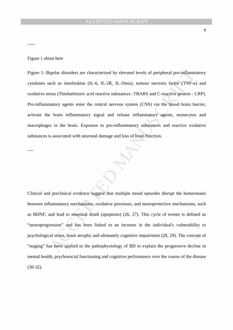
Figure 1: Bipolar disorders are characterized by elevated levels of peripheral pro-inflammatory
cytokines such as interleukins (IL-6, IL-2R, IL-1beta), tumour necrosis factor (TNF-α) and
oxidative stress (Thiobarbituric acid reactive substances -TBARS and C-reactive protein - CRP).
Pro-inflammatory agents enter the central nervous system (CNS) via the blood brain barrier,
activate the brain inflammatory signal and release inflammatory agents, monocytes and
macrophages in the brain. Exposure to pro-inflammatory substances and reactive oxidative
substances is associated with neuronal damage and loss of brain function.
----
Clinical and preclinical evidence suggest that multiple mood episodes disrupt the homeostasis
between inflammatory mechanisms, oxidative processes, and neuroprotective mechanisms, such
as BDNF, and lead to neuronal death (apoptosis) (26, 27). This cycle of events is defined as
“neuroprogression” and has been linked to an increase in the individual's vulnerability to
psychological stress, brain atrophy and ultimately cognitive impairment (28, 29). The concept of
“staging” has been applied to the pathophysiology of BD to explain the progressive decline in
mental health, psychosocial functioning and cognitive performance over the course of the disease
(30-32).

MANUSCRIP
T
ACCEPTED
ACCEPTED MANUSCRIPT10
Accordingly, neuroimaging studies show that individuals diagnosed with BD exhibit a
significant loss of gray matter volume and white matter integrity, which is likely related to
inflammatory processes such as apoptosis, cellular shrinkage, alterations in neurogenesis and
reduced gliogenesis (33). Recent neuroimaging studies have also identified a significant cortical
atrophy and enlargement of the ventricles in individuals who experienced multiple mood
episodes as compared to gender and age-matched healthy individuals (34, 35). Furthermore, an
inverse relationship between gray matter volumes and length of illness has also been reported
(36, 37).
In summary, chronic inflammation may lead to structural brain abnormalities and cognitive
deficits in individuals diagnosed with BD. However, to date, this hypothesis has not been
systematically reviewed. Thus, the purpose of this review is to assess the potential association
between cognitive impairment and biomarkers of inflammation, oxidative stress and
neurotrophic activity in individuals diagnosed with BD.
Literature search
Scopus (all databases), Pubmed and Ovid Medline were systematically searched with no
language or year restrictions, up to November 2013, for research articles addressing the
relationship between bipolar disorder, inflammation and cognition. Selected search terms were
‘bipolar disorder’, ‘depression’, ‘mania’, ‘psychosis’, ‘inflammatory’, ‘cognitive’ and
‘neurotrophic’ as occurring either anywhere in the article (for Pubmed and Ovid Medline) or in
the case of PUBMED, in the title, abstract or keywords only. The search engines listed above
were chosen because of their well-established accuracy and exhaustive search across

MANUSCRIP
T
ACCEPTED
ACCEPTED MANUSCRIPT11
multidisciplinary fields such as psychology, nutrition, biochemistry and medicine (38). Inclusion
was restricted to studies with clinical populations with a diagnosis of BD, studies were cognitive
functioning was assessed using pen and paper or computerized cognitive batteries, and where
inflammatory markers or polymorphisms of inflammatory genes using blood or other tissues
were quantified. Excluded studies included those using animal models, clinical populations with
neurological and cardiovascular diseases, children, adolescents, pregnant or lactating mothers,.
Exclusion criteria was defined by the following considerations. Cognition is affected by a range
of neurochemical mechanisms and cardiovascular parameters. During pregnancy and lactation, a
number of physiological changes take place and this physiological state could affect cognitive
performance. Finally, since the nervous system of children, and adolescents is still developing,
the relationship between inflammation and cognition in children cannot be equated to that
observed in a mature central nervous system. All data were extracted by a single, non-blinded,
reviewer (IB) to determine if studies met inclusion criteria and, in cases where this information
was not provided in abstracts, full texts were obtained. All papers identified were published in
English. No papers were identified prior to 2003. Duplicates, review articles and articles not
fulfilling the search criteria were removed (Figure 2).
------------------------
Figure 2 about here
Figure 2. PRISMA flowchart (38) showing the filtering process used to select the 10 studies included in the systematic review of studies investigating inflammatory markers and cognition in bipolar disorder
---------------------------

MANUSCRIP
T
ACCEPTED
ACCEPTED MANUSCRIPT12
Quality evaluation
Since there is no official instrument for the evaluation of observational studies in psychiatry and
inflammation we conducted a quality evaluation based on the Centre for Reviews and
Dissemination (CRD) Hierarchy of evidence (39) and a revised version of Ibrahim and
colleague’s quality evaluation scale (40). The CRD Hierarchy of evidence ranks study designs in
descending order of strength: 1. Experimental studies, 2. Quasi experimental studies, 3.
Controlled observational studies, 3a. Cohort studies, 3b. Case control studies, 4. Observational
studies without control groups, 5. Expert opinion based on theory, laboratory research or
consensus. The quality evaluation scale was composed of 6 items: 1) The clinical sample was
representative of the target population, 2) The control group was appropriately matched (e.g. by
age, gender) to the clinical sample 3) The authors conducted sample size calculations and/or
power analyses 4) The study used well-established measures of inflammation 5) The study used
well-established measures of cognitive functioning 6) The authors reported confidence intervals
and/or effect sizes of their findings. Each item was scored one point if the criterion was satisfied.
The overall quality score was calculated by adding the scores of all items.
Study Characteristics
We identified ten published clinical studies exploring the association between peripheral pro-
inflammatory cytokines, oxidative markers, neurotrophins and cognitive performance in
individuals diagnosed with BD. Two studies collected peripheral measures of oxidative stress
(CRP, RBANS) and peripheral pro-inflammatory cytokine measurements (IL-18, TNF). Eight
studies examined the relationship between BDNF levels or polymorphism and cognitive

MANUSCRIP
T
ACCEPTED
ACCEPTED MANUSCRIPT13
functioning (see Table 1). All studies were observational in nature and did not involve any anti-
inflammatory and/or antioxidant treatments. Cognitive performance in all studies comprised of
traditional pen-and-paper tests and computerized cognitive batteries. Table 1 summarizes the
study characteristics.
------
Table 1 about here
---------
Results of identified Studies
Pro-inflammatory cytokines, markers of oxidative stress and cognitive functioning
Peripheral serum CRP expression was negatively correlated with performance scores of
immediate memory, language, and attention, on the Repeatable Battery for the Assessment of
Neuropsychological Status (RBANS) in a study involving 107 individuals diagnosed with BD.
The authors interpreted these results as indicating that oxidative damage negatively affects
cognitive functioning in BD patients. However, as this study did not include a control population
it is unknown whether the relationship between CRP expression and cognitive performance is
specific to individuals diagnosed with BD, or whether it may also be observed in healthy controls
(41).
MANUSCRIP
T
ACCEPTED
ACCEPTED MANUSCRIPT14
Peripheral serum expression of the pro-inflammatory cytokine, TNF-α, was found to be
negatively correlated with accuracy on the delayed memory component on the Rey Auditory
Verbal Learning Test (RAVLT), in a study consisting of 54 medicated individuals diagnosed
with euthymic (absence of a depressive or manic cycle) BD type I. Furthermore, the expression
of two soluble TNF receptors (sTNFr1 and sTNFr2) was higher in euthymic BD individuals as
compared to healthy controls. (42). It is noteworthy that BD patients and healthy individuals did
not differ in terms of TNF-α levels. The authors concluded that this result may have been related
to the fast degradation of TNF-α in peripheral tissues (42). Further, the elevated production of
sTNF receptors may explain why cognitive deficits persist during the euthymic phase of BD (43,
44). Given that previous research shows that the production of sTNF receptors is catalyzed by
TNF-α (45), it is however unclear why the levels of sTNF receptors did not correlate with
cognitive performance in Doganavsargil‐Baysal et al.’s study (42).
At present, research regarding the relationship between inflammatory response and cognitive
performance in BD is extremely limited. The above studies however provide preliminary
evidence of the negative effects of pro-inflammatory and oxidative processes on high-order
cognitive abilities such as memory, attention and executive functioning.
Neurotrophins and cognitive functioning
In one study, middle-aged euthymic BD patients were found to have higher peripheral BDNF
expression than gender-matched healthy individuals. However, there was no significant
correlation between BDNF expression and the Mini-Mental State Examination (MMSE) and
Frontal Assessment Battery (FAB) scores (46). This negative finding may be due to the type of

MANUSCRIP
T
ACCEPTED
ACCEPTED MANUSCRIPT15
tests used to measure cognitive functioning. Indeed the MMSE and the FAB provide a short and
generic assessment of age-related cognitive decline (e.g. in Alzheimer’s and fronto-temporal
dementia) but are not sufficiently sensitive to detect mood-related cognitive changes (47, 48).
Furthermore, since the participants of this study were relatively young (M±SD: 50.88±9.11
years), they likely exhibited a high level of accuracy on these tests.
Dias et al. (2009) found that serum BDNF levels positively correlated with accuracy on a verbal
fluency task in individuals diagnosed with BD. It is important to emphasize that, contrary to the
findings of Barbosa et al., Dias et al. found no difference in BDNF expression between euthymic
BD and healthy individuals, in peripheral blood samples (49). Participants in this study were
medicated, which may have influenced BDNF expression and confounded results. In particular,
valproate-treated participants had higher BDNF levels, and lithium-treated participants lower
BDNF levels, when compared with non-medicated healthy volunteers (49). Additionally, this
study involved individuals with euthymic BD. While previous research shows that BDNF
expression can fluctuate during manic and depressive episodes (20, 50), previous research
demonstrated that, during the euthymic phase of BD, BDNF expression is comparable to that of
healthy volunteers (51).
Consistent with Dias et al.’s study (2009), Chou et al. did not find any difference in plasma
BDNF expression between euthymic BD and healthy controls. Furthermore, in the clinical
sample there was no significant correlation between BDNF expression and cognitive
performance (52). Since the mean illness duration was shorter (6 years) than that in Dias et al.’s
study (13 years) it could be hypothesized that illness duration counteracts the beneficial effects
of BDNF on cognitive performance. In another study poor lithium responders were found to
have lower BDNF levels compared to healthy controls. By contrast, excellent lithium responders

MANUSCRIP
T
ACCEPTED
ACCEPTED MANUSCRIPT16
(ELR) exhibited BDNF levels comparable to those of healthy controls, and performed better than
non-ELR on all tasks of the Cambridge Neuropsychological Test Automated Battery
(CANTAB), in particular the spatial working memory task (53). Thus, it could be hypothesized
that high BDNF levels counteract the cognitive decline observed in BD.
Previous research has focused on the relationship between the genotype of BDNF and cognitive
functioning. In particular, studies have associated the BDNF val66met polymorphism with BD
symptomatology. In this BDNF gene variation the valine (val) allele is replaced by the
methionine (met) allele at codon 66 (54). In the present review, we identified one study showing
that met carriers diagnosed with BD have smaller hippocampi volumes and larger ventricles than
val/val carriers. Moreover, met carriers were seen to encounter more difficulties in verbal fluency
and working memory tasks than the val/val group (55).
Additionally, Rybakowski et al. found that individuals with a BDNF val66met polymorphism
developed BD type 1 approximately 11 years earlier than val/val carriers and performed more
poorly on a test of executive functioning (Wisconsin Card Sorting Test - WCST) (56). A few
years later Rybakowski et al. found that the val/val genotype was associated with higher
accuracy on the N-back and WCST tests when compared with val/met and met/met genotypes
(57).
By contrast, another identified study, by Tramontina et al. found that val/val carriers diagnosed
with BD type I made more perseverative errors than val/met and met/met participants. Thus the
met allele was not seen to be associated with cognitive impairment in this study (58), possibly
due to the heterogeneous ancestry of Tramontina et al.’s participants (European, Amerindian and
African) as compared to the more homogenous European ancestry of participants involved in

MANUSCRIP
T
ACCEPTED
ACCEPTED MANUSCRIPT17
Rybakowski’s study. Alternatively, other factors such as the age of onset of the disease and the
severity of BD may have blunted the differences between met and val carriers, however the
influence of these variables were not explored.
Overall, current findings provide initial evidence of an association between decreased BDNF
levels and a high risk of cognitive decline in BD. Furthermore, the BDNF val66met
polymorphism appears to be a potential risk factor for cognitive impairment in BD.
Quality evaluation: findings
The quality and reliability of the 10 studies included in this review are shown in Table 2. The
CDR hierarchy of evidence was estimated to be 3-4, as the current studies are not randomized
cross-sectional studies with and without a control group. Given the observational nature of the
studies, the current findings provide little information on trends over time and do not investigate
possible causality link between inflammation and cognitive impairment in bipolar disorder.
Further, investigators were not blinded to the case/control status of their participants. This raises
the possibility that the knowledge of the diagnosis may have affected their testing style and
cognitive evaluation. The clinical populations were recruited in hospital settings and their
diagnosis was based on well-established clinical scales such as the SCID (59) and the Mini
International Neuropsychiatric inventory, (60) which indicates that the clinical profile of the
samples is a reliable representation of the bipolar illness. All studies used well-accepted
techniques to estimate inflammatory markers (e.g. ELISA assays) and widely used measures of
cognitive functioning (e.g. Repeatable Battery for the Assessment of Neuropsychological
Status). It could therefore be concluded that the current results provide an accurate description of

MANUSCRIP
T
ACCEPTED
ACCEPTED MANUSCRIPT18
the cognitive functioning and inflammatory response in BD patients. While the average sample
size was satisfactory as it ranged from medium to large (N > 30), only two studies reported
estimating the sample size based on power analyses. As a result, some of the studies may be
underpowered and report misleading findings. In addition, the lack of information on the effect
sizes and the confidence intervals of the statistical analyses limit the evaluation of the size of the
experimental effects of the findings. Other methodological flaws include the absence of a control
population in three studies and inadequate matching of the control population to the clinical
population in six studies. Moreover, given the current trend for overrepresentation of positive
studies in medicine and hard sciences (11), it is possible that a number of unpublished studies did
not find any link between inflammation and cognition in bipolar disorder.
---------------
Insert Table 2 here
-------------
Discussion
This review aimed to examine the literature exploring the relationship between the cognitive
deficits seen among individuals diagnosed with bipolar disorder, and markers of inflammation,
neurotrophins and oxidative damage. Thus, we conducted a systematic search of the human
literature on inflammation and cognition in BD. It is important to emphasize that this is a novel
approach as previous reviews have linked inflammation with mood symptoms, but have not
explored the relationship between peripheral markers of inflammation and cognitive impairment.

MANUSCRIP
T
ACCEPTED
ACCEPTED MANUSCRIPT19
We identified 10 observational studies. Of these studies 2 investigated the relationship between
pro-inflammatory cytokines (TNF-a) and markers of oxidative stress (CRP) and 8 investigated
the relationship between the neurotrophin BDNF, and cognitive functioning. The results of these
studies indicate that the cognitive deficits observed in individuals diagnosed with BD appear to
be associated with an increased inflammatory state and a decrease in the neurotropic factor,
BDNF. Despite the limited number of studies in this field and their heterogeneity in terms of
cognitive outcome measures, these studies provide preliminary evidence that an elevated
inflammatory state negatively affects frontotemporal cognitive abilities such as memory,
attention and executive functions, and indicate a need for further investigation.
Consistent with previous research (28), we identified one study showing that individuals
diagnosed with BD have reduced hippocampi volumes and larger ventricles. Importantly, this
was associated with more difficulties in verbal fluency and working memory tasks (55). Previous
authors have speculated that systemic toxicity and cognitive dysfunction are directly related to
the number of episodes suffered by the patient (32) and that peripheral cytokine and BDNF
expression could be potential markers of illness progression, thus corroborating the “staging”
hypothesis of BD (61). However, as all the studies identified in the present systematic review
were observational in nature, there appears to be no research regarding the relationship between
inflammatory markers and changes in cognitive measures over the course of the BD. A
longitudinal design study could be a suitable approach to explore the relationship between
peripheral biomarkers of inflammation, oxidative stress and neurotrophic activity. This type of
design may also help clarify the relationship between biomarkers and cognitive impairment at
different phases of the disease.

MANUSCRIP
T
ACCEPTED
ACCEPTED MANUSCRIPT20
None of the reviewed studies investigate the relationship between oxidative stress, mitochondrial
dysfunction and neuroprogression. However, a number of studies using animal models of mania
have observed increased levels of reactive oxygen species (ROS), a marker of mitochondrial
dysfunction (62). One study demonstrated increased lipid peroxidation, and a high number of
free radical, superoxide in submitochondrial particles of the prefrontal cortex and hippocampus
(2). A second study showed that repeated amphetamine exposure, which induces manic
symptomatology in animal models, increases levels of anti-oxidant enzymes, superoxide
dismutase (SOD) and catalase (CAT), in regional specific manner, in the prefrontal cortex,
hippocampus and striatum (3). The authors interpreted these results to reflect an imbalance
between SOD and CAT expression, potentially indicative of a predisposition to the generation of
ROS (63, 64). Increased oxidative stress has been associated with abnormalities in the
glutamatergic system and neuronal apoptosis. Neuronal apoptosis has been hypothesized to be a
progressive process that begins at synaptic terminals and dendrites and continues to the cell body
via apoptotic cascades (65, 66). The dynamic of neuronal cell death mechanisms may underlie
the decline in neurocognitive function observed over the course of the bipolar illness.
Additionally, clinical research indicates that BD is characterized by low levels of brain energy
metabolites such as creatine and high lactate and glutamate-related metabolite concentrations,
which are clinical markers of mitochondrial dysfunction (67), as assessed using magnetic
resonance spectroscopy (MRS)(68). Lactate accumulation may indicate a shift to anaerobic
glycolytic mechanisms, possibly due to inadequate energy production within the mitochondria
(68, 69). Anaerobic glycolysis produces less adenosine triphosphate (ATP) molecules than
aerobic glycosis, and reduced ATP production could lead to cerebral hypometabolism, brain
dysfunction and eventually cognitive impairment (70).

MANUSCRIP
T
ACCEPTED
ACCEPTED MANUSCRIPT21
A further limitation of the studies reviewed here is that they differ with respect to the estimation
of the peripheral levels of inflammatory biomarkers. Indeed, as illustrated in Table 1, the
majority of the studies measured the expression of cytokines, BDNF and antioxidants in serum,
plasma, or whole blood cells (a combination of serum, plasma and erythrocytes). For instance,
since plasma cells have a short turnover (71), they may be ideal to measure acute inflammation
(e.g. infection), but may not reflect a state of chronic inflammation. Erythrocytes and whole
blood cells (which have a turnover of approximately 12 weeks) (72) may therefore be better
indices of inflammatory markers in cell membranes and possibly the brain tissue. Further,
previous studies have shown that aminoacids and carbohydrate levels differ significantly
between plasma and serum (8, 73). In particular, BDNF levels are higher in serum compared to
plasma (40, 73, 74). The latter result is probably due to the release of BDNF from platelets to
serum during the coagulation process (40). Hence, finding of studies using serum BDNF levels
may not be comparable to those of studies using plasma BDNF levels. Quality evaluation of the
current studies reveals some methodological concerns in terms of research design and statistical
analyses, as earlier discussed. Hence, caution should be taken in the interpretation of the data
presented in this systematic review, regarding the relationship between inflammation and
cognition in bipolar disorder.
Surprisingly, none of the studies identified investigated the relationship between the
inflammatory response and the hypothalamic-pituitary-adrenal (HPA) axis activation. Indeed a
number of mood disorders present with abnormalities in the HPA axis, such as increased levels
of cortisol and corticotropin-releasing factor (CRF)(75). A potential explanation for the abnormal
HPA axis activity is that pro-inflammatory cytokines disrupt the glucocorticoid function and lead
to glucorticoid resistance, characterized by cortisol and CRF hypersecretion. In turn,

MANUSCRIP
T
ACCEPTED
ACCEPTED MANUSCRIPT22
glucocorticoid resistance initiates pro-inflammatory mechanisms and reduces peripheral BDNF
levels (73, 76). Both glucocorticoid resistance and inflammation have been associated with
depressed mood and cognitive difficulties (73). It is notable that in medicated BD patients the
glucocorticoid receptor antagonist, mifepristone improves mood and spatial working memory,
and to a lesser extent, verbal fluency and spatial recognition (74, 77). In particular, the
improvement in spatial memory appear to be related to the cortisol response to mifiprestone, not
mood changes (74). It could be speculated that mifiprestone inhibits the proinflammatory
response by regulating the HPA-axis activity, however this remains unknown as these studies did
not collect inflammatory markers. Taken together these findings provide a strong rationale for
the development of trials investigating the synergistic role of the inflammatory response and the
HPA-axis function in the pathophysiology of BD.
In conclusion, research in the field of cognition and inflammation in BD is still in its infancy and
additional work is needed to understand how pro-inflammatory processes affect brain function
and induce cognitive impairment. The identification of inflammatory markers and
polymorphisms in inflammatory response genes underlying cognitive decline will have important
implications for the development of new therapies targeting chronic inflammatory conditions
such as BD.
Acknowledgements:
This work was partly supported by the Stanley Medical Research Institute, NIH grant MH
085667 (JCS), and by the Pat Rutherford Jr. Chair in Psychiatry (UTHealth).

MANUSCRIP
T
ACCEPTED
ACCEPTED MANUSCRIPT23
References
1. Bearden CE, Glahn DC, Monkul ES, Barrett J, Najt P, Villarreal V, et al. Patterns of memory
impairment in bipolar disorder and unipolar major depression. Psychiatry research. 2006;142(2):139-50.
2. Diwadkar VA, Goradia D, Hosanagar A, Mermon D, Montrose DM, Birmaher B, et al. Working
memory and attention deficits in adolescent offspring of schizophrenia or bipolar patients: comparing
vulnerability markers. Progress in neuro-psychopharmacology & biological psychiatry. 2011;35(5):1349-
54.
3. Glahn DC, Bearden CE, Barguil M, Barrett J, Reichenberg A, Bowden CL, et al. The neurocognitive
signature of psychotic bipolar disorder. Biological psychiatry. 2007;62(8):910-6.
4. Quraishi S, Frangou S. Neuropsychology of bipolar disorder: a review. Journal of affective
disorders. 2002;72(3):209.
5. Najt P, Perez J, Sanches M, Peluso M, Glahn D, Soares JC. Impulsivity and bipolar disorder.
European neuropsychopharmacology. 2007;17(5):313-20.
6. Ancin I, Santos JL, Teijeira C, Sanchez-Morla EM, Bescos MJ, Argudo I, et al. Sustained attention
as a potential endophenotype for bipolar disorder. Acta Psychiatr Scand. 2010;122(3):235-45.
7. Robinson LJ, Thompson JM, Gallagher P, Goswami U, Young AH, Ferrier IN, et al. A meta-analysis
of cognitive deficits in euthymic patients with bipolar disorder. Journal of Affective Disorders.
2006;93(1–3):105-15.
8. Malhi GS, Ivanovski B, Hadzi-Pavlovic D, Mitchell PB, Vieta E, Sachdev P. Neuropsychological deficits and functional impairment in bipolar depression, hypomania and euthymia. Bipolar disorders.
2007;9(1-2):114-25.
9. Miller BJ, Buckley P, Seabolt W, Mellor A, Kirkpatrick B. Meta-analysis of cytokine alterations in
schizophrenia: clinical status and antipsychotic effects. Biological psychiatry. 2011;70(7):663-71.
10. Carey AN, Fisher DR, Joseph JA, Shukitt-Hale B. The ability of walnut extract and fatty acids to
protect against the deleterious effects of oxidative stress and inflammation in hippocampal cells. Nutr
Neurosci. 2013;16(1):13-20.
11. Turner EH. Publication Bias, with a Focus on Psychiatry: Causes and Solutions. CNS drugs.
2013;27(6):457-68.
12. Barbosa IG, Huguet RB, Sousa LP, Abreu MNS, Rocha NP, Bauer ME, et al. Circulating levels of GDNF in bipolar disorder. Neuroscience letters. 2011;502(2):103-6.
13. Kapczinski F, Dal-Pizzol F, Teixeira AL, Magalhaes PV, Kauer-Sant’Anna M, Klamt F, et al.
Peripheral biomarkers and illness activity in bipolar disorder. Journal of psychiatric research.
2011;45(2):156-61.
14. Kunz M, Ceresér KM, Goi PD, Fries GR, Teixeira AL, Fernandes BS, et al. Serum levels of IL-6, IL-10
and TNF-α in patients with bipolar disorder and schizophrenia: differences in pro-and anti-inflammatory
balance. Revista Brasileira de Psiquiatria. 2011;33(3):268-74.
15. Gimeno D, Kivimaki M, Brunner EJ, Elovainio M, De Vogli R, Steptoe A, et al. Associations of C-
reactive protein and interleukin-6 with cognitive symptoms of depression: 12-year follow-up of the
Whitehall II study. Psychological medicine. 2009;39(3):413. 16. Myint AM, Schwarz MJ, Steinbusch HW, Leonard BE. Neuropsychiatric disorders related to
interferon and interleukins treatment. Metabolic brain disease. 2009;24(1):55-68.
17. Capuron L, Dantzer R. Cytokines and depression: the need for a new paradigm. Brain Behav
Immun. 2003;17(1):119-24.
18. Minagar A, Alexander JS. Inflammatory Disorders of the Nervous System: Pathogenesis,
Immunology, and Clinical Management: Springer; 2005.

MANUSCRIP
T
ACCEPTED
ACCEPTED MANUSCRIPT24
19. Blank T, Prinz M. Microglia as modulators of cognition and neuropsychiatric disorders. Glia.
2013;61(1):62-70.
20. Cunha A, Frey BN, Andreazza AC, Goi JD, Rosa AR, Gonçalves CA, et al. Serum brain-derived
neurotrophic factor is decreased in bipolar disorder during depressive and manic episodes.
Neuroscience letters. 2006;398(3):215-9. 21. Bourne C, Aydemir Ö, Balanzá-Martínez V, Bora E, Brissos S, Cavanagh J, et al.
Neuropsychological testing of cognitive impairment in euthymic bipolar disorder: an individual patient
data meta-analysis. Acta Psychiatrica Scandinavica. 2013;128(3):149-62.
22. Ichim G, Tauszig-Delamasure S, Mehlen P. Neurotrophins and cell death. Experimental Cell
Research. 2012;318(11):1221-8.
23. Huang EJ, Reichardt LF. Neurotrophins: roles in neuronal development and function. Annual
review of neuroscience. 2001;24:677.
24. Buckley PF, Pillai A, Evans D, Stirewalt E, Mahadik S. Brain derived neurotropic factor in first-
episode psychosis. Schizophrenia research. 2007;91(1):1-5.
25. Jiang L, Shi Y, Wang L, Yang Z. The influence of orally administered docosahexaenoic acid on cognitive ability in aged mice. The Journal of Nutritional Biochemistry. 2009;20(9):735-41.
26. Fries GR, Pfaffenseller B, Stertz L, Paz AVC, Dargél AA, Kunz M, et al. Staging and
neuroprogression in bipolar disorder. Current psychiatry reports. 2012;14(6):667-75.
27. Berk M, Kapczinski F, Andreazza A, Dean O, Giorlando F, Maes M, et al. Pathways underlying
neuroprogression in bipolar disorder: focus on inflammation, oxidative stress and neurotrophic factors.
Neuroscience & biobehavioral reviews. 2011;35(3):804-17.
28. Berk M, Conus P, Kapczinski F, Andreazza AC, Yücel M, Wood SJ, et al. From neuroprogression to
neuroprotection: implications for clinical care. Med J Aust. 2010;193(4 Suppl):S36-40.
29. Kapczinski F, Vieta E, Andreazza AC, Frey BN, Gomes FA, Tramontina J, et al. Allostatic load in
bipolar disorder: implications for pathophysiology and treatment. Neuroscience & Biobehavioral Reviews. 2008;32(4):675-92.
30. Gama CS, Kunz M, Magalhães PV, Kapczinski F. Staging and neuroprogression in bipolar disorder:
a systematic review of the literature. Revista Brasileira de Psiquiatria. 2013;35(1):70-4.
31. Frodl T, Amico F. Is there an association between peripheral immune markers and
structural/functional neuroimaging findings? Progress in Neuro-Psychopharmacology and Biological
Psychiatry. 2013;3(48):295-303.
32. Kapczinski F, Dias VV, Kauer-Sant'Anna M, Frey BN, Grassi-Oliveira R, Colom F, et al. Clinical
implications of a staging model for bipolar disorders. Expert review of neurotherapeutics. 2009;9(7):957-
66.
33. Czéh B, Lucassen PJ. What causes the hippocampal volume decrease in depression? European archives of psychiatry and clinical neuroscience. 2007;257(5):250-60.
34. Pfaffenseller B, Gama CS, Kapczinski F, Duarte JA, Kunz M. Anatomical faces of neuroprogression
in bipolar disorder. Neuropsychiatry. 2012;2(4):279-80.
35. Strakowski SM, Adler CM, Almeida J, Altshuler LL, Blumberg HP, Chang KD, et al. The functional
neuroanatomy of bipolar disorder: a consensus model. Bipolar Disord. 2012;14(4):313-25.
36. Brambilla P, Harenski K, Nicoletti M, Mallinger AG, Frank E, Kupfer DJ, et al. Differential effects
of age on brain gray matter in bipolar patients and healthy individuals. Neuropsychobiology.
2001;43(4):242-7.
37. Frey BN, Zunta-Soares GB, Caetano SC, Nicoletti MA, Hatch JP, Brambilla P, et al. Illness duration
and total brain gray matter in bipolar disorder: evidence for neurodegeneration? Eur Neuropsychopharmacol. 2008;18(10):717-22.
38. Moher D LA, Tetzlaff J, Altman DG, The PRISMA Group. Preferred Reporting Items for Systematic
Reviews and Meta-Analyses: The PRISMA Statement. PLoS Med 2009;6(6):e1000097.

MANUSCRIP
T
ACCEPTED
ACCEPTED MANUSCRIPT25
39. Cochrane, Collaboration. Cochrane Reviewers’ Handbook. Version 4.2. 1.2003.
40. Ibrahim AK, Kelly SJ, Adams CE, Glazebrook C. A systematic review of studies of depression
prevalence in university students. Journal of psychiatric research. 2012;47(3):391-400.
41. Dickerson F, Stallings C, Origoni A, Vaughan C, Khushalani S, Yolken R. Elevated C-reactive
protein and cognitive deficits in individuals with bipolar disorder. Journal of Affective Disorders. 2013;150(2):456-59.
42. Doganavsargil-Baysal O, Cinemre B, Aksoy UM, Akbas H, Metin O, Fettahoglu C, et al. Levels of
TNF-α, soluble TNF receptors (sTNFR1, sTNFR2), and cognition in bipolar disorder. Human
Psychopharmacology: Clinical and Experimental. 2013;28(2):160-7.
43. Pattanayak RD, Sagar R, Mehta M. Neuropsychological performance in euthymic Indian patients
with bipolar disorder type I: Correlation between quality of life and global functioning. Psychiatry and
clinical neurosciences. 2012;66(7):553-63.
44. King DA, Caine ED. Cognitive impairment and major depression: beyond the pseudodementia
syndrome. Neuropsychological Assessment of Neuropsychiatric Disorders. I. Grant KMA, editor: New
York: Oxford University Press; 1996. 45. Grassi-Oliveira R, Brietzke E, Pezzi JC, Lopes RP, Teixeira AL, Bauer ME. Increased soluble tumor
necrosis factor-α receptors in patients with major depressive disorder. Psychiatry and clinical
neurosciences. 2009;63(2):202-8.
46. Barbosa IG, Rocha NP, Huguet RB, Ferreira RA, Salgado JV, Carvalho LA, et al. Executive
dysfunction in euthymic bipolar disorder patients and its association with plasma biomarkers. Journal of
affective disorders. 2012;137(1):151.
47. Gluhm S, Goldstein J, Loc K, Colt A, Van Liew C, Corey-Bloom J. Cognitive Performance on the
Mini-Mental State Examination and the Montreal Cognitive Assessment Across the Healthy Adult
Lifespan. Cognitive and Behavioral Neurology. 2013;26(1):1-5.
48. Boban M, Malojčić B, Mimica N, Vuković S, Zrilić I. The Frontal Assessment Battery in the Differential Diagnosis of Dementia. Journal of geriatric psychiatry and neurology. 2012;25(4):201-7.
49. Dias VV, Brissos S, Frey BN, Andreazza AC, Cardoso C, Kapczinski F. Cognitive function and serum
levels of brain-derived neurotrophic factor in patients with bipolar disorder. Bipolar disorders.
2009;11(6):663-71.
50. Goldstein BI, Collinger KA, Lotrich F, Marsland AL, Gill M-K, Axelson DA, et al. Preliminary
findings regarding proinflammatory markers and brain-derived neurotrophic factor among adolescents
with bipolar spectrum disorders. Journal of Child and Adolescent Psychopharmacology. 2011;21(5):479-
84.
51. Fernandes BS, Gama CS, Maria Ceresér K, Yatham LN, Fries GR, Colpo G, et al. Brain-derived
neurotrophic factor as a state-marker of mood episodes in bipolar disorders: a systematic review and meta-regression analysis. Journal of psychiatric research. 2011;45(8):995-1004.
52. Chou Y-H, Wang S-J, Lirng J-F, Lin C-L, Yang K-C, Chen C-K, et al. Impaired cognition in bipolar I
disorder: The roles of the serotonin transporter and brain-derived neurotrophic factor. Journal of
affective disorders. 2012;143:131-7.
53. Rybakowski JK, Suwalska A. Excellent lithium responders have normal cognitive functions and
plasma BDNF levels. The International Journal of Neuropsychopharmacology. 2010;13(05):617-22.
54. Craddock N, O’donovan M, Owen M. The genetics of schizophrenia and bipolar disorder:
dissecting psychosis. Journal of Medical Genetics. 2005;42(3):193-204.
55. Aas M, Haukvik UK, Djurovic S, Bergmann Ø, Athanasiu L, Tesli MS, et al. BDNF val66met
modulates the association between childhood trauma, cognitive and brain abnormalities in psychoses. Progress in Neuro-Psychopharmacology and Biological Psychiatry. 2013;46:181-8.

MANUSCRIP
T
ACCEPTED
ACCEPTED MANUSCRIPT26
56. Rybakowski JK, Borkowska A, Czerski PM, Skibińska M, Hauser J. Polymorphism of the
brain-derived neurotrophic factor gene and performance on a cognitive prefrontal test in bipolar
patients. Bipolar disorders. 2003;5(6):468-72.
57. Rybakowski JK, Borkowska A, Skibinska M, Szczepankiewicz A, Kapelski P, Leszczynska-
Rodziewicz A, et al. Prefrontal cognition in schizophrenia and bipolar illness in relation to Val66Met polymorphism of the brain-derived neurotrophic factor gene. Psychiatry and clinical neurosciences.
2006;60(1):70-6.
58. Tramontina JF, Yates D, Magalhães PVdS, Trentini C, Sant'Anna MK, Fries GR, et al. Brain-derived
neurotrophic factor gene val66met polymorphism and executive functioning in patients with bipolar
disorder. Revista Brasileira de Psiquiatria. 2009;31(2):136-40.
59. First MB, Spitzer RL, Gibbon M, Williams JB. Structured Clinical Interview for DSM-IV® Axis I
Disorders (SCID-I), Clinician Version, Administration Booklet: American Psychiatric Pub; 2012.
60. Sheehan DV, Lecrubier Y, Harnett Sheehan K, Janavs J, Weiller E, Keskiner A, et al. The validity of
the Mini International Neuropsychiatric Interview (MINI) according to the SCID-P and its reliability.
European Psychiatry. 1997;12(5):232-41. 61. Kauer-Sant'Anna M, Kapczinski F, Andreazza AC, Bond DJ, Lam RW, Young LT, et al. Brain-derived
neurotrophic factor and inflammatory markers in patients with early-vs. late-stage bipolar disorder. The
International Journal of Neuropsychopharmacology. 2009;12(04):447-58.
62. Murphy MP. Mitochondrial dysfunction indirectly elevates ROS production by the endoplasmic
reticulum. Cell metabolism. 2013;18(2):145-6.
63. Frey BN, Valvassori SS, Gomes KM, Martins MR, Dal-Pizzol F, Kapczinski F, et al. Increased
oxidative stress in submitochondrial particles after chronic amphetamine exposure. Brain Research.
2006;1097(1):224-9.
64. Frey BN, Valvassori SS, Réus GZ, Martins MR, Petronilho FC, Bardini K, et al. Changes in
antioxidant defense enzymes after d-amphetamine exposure: implications as an animal model of mania. Neurochemical research. 2006;31(5):699-703.
65. Mattson MP. Apoptosis in neurodegenerative disorders. Nature Reviews Molecular Cell Biology.
2000;1(2):120-30.
66. Mattson MP, Keller JN, Begley JG. Evidence for synaptic apoptosis. Experimental neurology.
1998;153(1):35-48.
67. Haas RH, Parikh S, Falk MJ, Saneto RP, Wolf NI, Darin N, et al. Mitochondrial disease: a practical
approach for primary care physicians. Pediatrics. 2007;120(6):1326-33.
68. Strakowski S. The bipolar brain: integrating neuroimaging and genetics: Oxford University Press;
2012.
69. Gigante AD, Bond DJ, Lafer B, Lam RW, Young LT, Yatham LN. Brain glutamate levels measured by magnetic resonance spectroscopy in patients with bipolar disorder: a meta-analysis. Bipolar
disorders. 2012;14(5):478-87.
70. Minuzzi L, Antônio G, Fonseca Moreira JC, Frey BN. Mitochondrial Dysfunction in Bipolar
Disorder: Lessons from Brain Imaging and Molecular Markers; Disfunción mitocondrial en el trastorno
bipolar: lecciones de las imágenes cerebrales y los marcadores moleculares. Rev colomb psiquiatr.
2011;40(supl. 1):166-82.
71. Slifka MK, Antia R, Whitmire JK, Ahmed R. Humoral Immunity Due to Long-Lived Plasma Cells.
Immunity. 1998;8(3):363-72.
72. Katan M, Deslypere J, Van Birgelen A, Penders M, Zegwaard M. Kinetics of the incorporation of
dietary fatty acids into serum cholesteryl esters, erythrocyte membranes, and adipose tissue: an 18-month controlled study. Journal of lipid research. 1997;38(10):2012-22.

MANUSCRIP
T
ACCEPTED
ACCEPTED MANUSCRIPT27
73. Pace TWW, Hu F, Miller AH. Cytokine-effects on glucocorticoid receptor function: Relevance to
glucocorticoid resistance and the pathophysiology and treatment of major depression. Brain Behav
Immun. 2007;21(1):9-19.
74. Watson S, Gallagher P, Porter RJ, Smith MS, Herron LJ, Bulmer S, et al. A randomized trial to
examine the effect of mifepristone on neuropsychological performance and mood in patients with bipolar depression. Biological psychiatry. 2012;72(11):943-9.
75. DeBattista C, Belanoff J. The use of mifepristone in the treatment of neuropsychiatric disorders.
Trends in Endocrinology & Metabolism. 2006;17(3):117-21.
76. Mackin P, Gallagher P, Watson S, Young AH, Ferrier IN. Changes in brain-derived neurotrophic
factor following treatment with mifepristone in bipolar disorder and schizophrenia. Australian and New
Zealand journal of psychiatry. 2007;41(4):321-6.
77. Young AH, Gallagher P, Watson S, Del-Estal D, Owen BM, Ferrier IN. Improvements in
neurocognitive function and mood following adjunctive treatment with mifepristone (RU-486) in bipolar
disorder. Neuropsychopharmacology. 2004;8(29):1538-45.

MANUSCRIP
T
ACCEPTED
ACCEPTED MANUSCRIPT
Acknowledgment
N/A

MANUSCRIP
T
ACCEPTED
ACCEPTED MANUSCRIPT
1
Table 1: Summary of studies that explored changes in inflammatory and oxidative stress biomarkers, and cognitive measures in individuals with bipolar disorder; BD = Bipolar disorder, HC = Healthy control Citation Subject
description
(diagnosis, gender
(M/F)
Age (Years) Design Duration of
illness
(years)
Antipsychotic/mood
stabilizer
medication (Yes or
No)
Cognitive measures Inflammatory
biomarker
Statistics Outcome
+ Evidence of a
link between
inflammation
and cognition
-no evidence of
a link between
inflammation
and cognition
Oxidative stress and cytokines
Dickerson et al. (2013)
Journal of Affective
Disorders
107 BD (31/76) 36.3±13.4 Observational
study
≈ 20 years Yes Repeatable Battery for
the Assessment of
Neuropsychological
status (RBANS)
Wechsler Adult
Intelligence Scale
(WAIS III), Trail
Making Test
Serum CRP Logistic
regression
+Lower
RBANS scores
in the High CRP
group compared
to the Low CRP
group
Doganavsargil-Baysal et
al. (2013)
Human
Psychopharmacology
54 euthymic BDI
(18/36)
18 HC (5/13)
BD:
39.46±11.62
HC
38.33±10.80
Observational
study
≈ 13 years Yes Wisconsin Card Sorting
Test (WCST), Rey’s
Auditory Verbal
Leaning Test (RAVLT)
Serum TNF Spearman’s
correlation, t-test
+BD have
higher levels of
sTNFr1 and
sTNFr2 than HC
+BD exhibit
lower cognitive
performance on

MANUSCRIP
T
ACCEPTED
ACCEPTED MANUSCRIPT
2
WCST and
RAVLT than
HC
+sTNFr2 levels
correlate
positively with
illness duration
Neurotrophin
Aas et al. (2013)
Progress in Neuro-
psychopharmacology
249 BD (122/127)
and 476 HC
BD:
20-40
HC: 34.79±10.25
Observational
study
Not
provided
Yes California Verbal
Learning test, letter
number sequencing,
digit span
forwards/backwards,
Color Word
Interference test, verbal
fluency, block design,
matrix reasoning,
vocabulary
BDNF gene
(not specified
whether serum
or plasma)
Regression model +val/met BDNF
gene carriers are
more vulnerable
to childhood
trauma sequels,
have smaller
hippocampal
volumes and
larger ventricles
Barbosa et al. (2012)
Journal of Affective
Disorders
25 euthymic BD
(8/17) vs 25 HC
(11/14)
BD:
50.88±9.11
HC:
48.04±7.08
Observational
study
27.88±11.8 Yes Mini-Mental State
Examination and
Frontal Assessment
Battery
Plasma BDNF
Mann-Whitney U
test, Spearman’s
correlation
+Higher BDNF
in the BD than
HC
-No correlation
between BDNF
and executive
functioning

MANUSCRIP
T
ACCEPTED
ACCEPTED MANUSCRIPT
3
Chou et al. (2012)
Journal of affective
disorders
23 BD (6/17) and
33 HC (12/21)
HC: 37.6±7.8
BD: 36.5±8.9
Observational
study
5.7±4.68 Yes Go/No-Go task of the
test for attentional
performance, Memory
(Wechsler Memory
scale – III), Word list,
Face test, Color trail
test, Wisconsin card
sorting test
Plasma BDNF t-test, ANCOVA,
correlation
-no difference in
BDNF levels
between BD and
HC
+reduced face
recognition and
accuracy on the
WCST in BD
compared to HC
Dias et al. (2009)
Bipolar disorders
65 euthymic BD
(24/41), 50 HC
BD: 37.8±10.51
HC:33.6±9.66
Observational
study
13.3±8.78 Yes Wechsler Memory
Scale, Wechsler
Intelligence Scale for
adults revised
Serum BDNF t-test, ANOVA, -No difference
in BDNF levels
between BD and
HC
+Positive
correlation
between BDNF
levels and
verbal fluency
in BD and HC
Rybarkowski et al. (2003)
Bipolar disorder
54 BD I (18/36) BD: 18-72 (mean
46 years)
Observational
study
16±12 Yes Wisconsin Card Sorting
Test
Whole blood
BDNF gene
t-test +the val/val
BDNF
polymorphism
exhibits better
cognitive
functioning than
the val/met
genotype

MANUSCRIP
T
ACCEPTED
ACCEPTED MANUSCRIPT
4
+the val allele
was associated
with an earlier
onset of illness
Rybarkowski et al. (2006)
Psychiatry and Clinical
Neurosciences
111 BD (37/74)
160 HC (gender
ratio N/A)
BD: 43.4±13.7
HC: 32.9±11.5
Observational
study
N/A N/A Wisconsin Card Sorting
Test, N-back test
Whole blood
BDNF gene
t-test, ANOVA +The percentage
of correct
reactions to the
N-back test and
accuracy on
WCST was
higher in the
val/val group
compared with
val/met and
met/met
Rybarkowski et al. (2010)
International Journal of
Neuropsychopharmacology
60 BD (25/35)
60 HC (25/35)
BD: 52.6±10.2
HC:52.1±13.6
Observational
study
22.2±10.8 Yes CANTAB Plasma BDNF
level
Mann-
Whitney/Kruskal-
Wallis ANOVA
+BD have lower
BD have poorer
cognitive
performance
and lower
BDNF plasma
levels than HC
+No difference
in plasma
BDNF levels
between ELR
and HC

MANUSCRIP
T
ACCEPTED
ACCEPTED MANUSCRIPT
5
Tramontina et al. (2009)
Revista Brasileira de
Psiquiatria
64 BD I (14/50) BD I: 42.3±11.1 Observational
study
N/A Yes Wisconsin Card Sorting
Test
BDNF gene
(not specified
whether serum
or plasma)
Mann-Whitney’s
U analysis,
ANCOVA
-The percentage
of non-
perseverative
errors was
higher in the
val/val group
-No association
between met
allele and
cognitive
functioning

MANUSCRIP
T
ACCEPTED
ACCEPTED MANUSCRIPT
Table 2. Quality assessment of the 10 studies included in the review. The Overall Quality Score was calculated by adding scores of items 1 to 6; CRD levels are: 1. Experimental studies, 2. Quasi-experimental studies,
3. Controlled observational studies, 3a. Cohort studies, 3b. Case control studies, 4. Observational studies without control groups, 5. Expert opinion based on theory, laboratory research or consensus; BD = Bipolar
disorder, HC = healthy control
Citation
Overall Quality
Score
(1-6)
CRD
Hierarchy of
evidence
(Levels 1-5)
Sample size
N < 30 small
N = 30-50
medium
N > 50 large
Item 1
Representative
sample
Item 2
Matched control
groups
Item 3
Power
analysis/Sample size
calculation
Item 4
Adequate
assessment of
inflammation
Item 5
Adequate
measure of
cognition
Item 6
Confidence
intervals (CI) or or
effect size (ES)
Criterion is fulfilled = 1
Criterion is not fulfilled = 0
Dickerson et al. (2013) 4 4 Large, 107 BD 1, patients were
recruited in a
psychiatric health
care program,
diagnosis was based
on SCID
0, No HC 0 1 1 1, CI, no ES
Doganavsargil-Baysal et
al. (2013)
6 3 Small/Medium
54 BDI
18 HC
1, patients were
recruited in an
outpatient
psychiatric clinic,
diagnosis was based
on DSM-IV TR
1, HC matched by age,
gender, educational
level
1 1 1 0, No CI, Estimated
ES = 1
Aas et al. (2013) 3 3 Large
249 BD
476 HC
1: Patients recruited
in hospitals,
diagnosis based on
DSM-IV criteria.
0, HC not
demographically
matched
0 1 1
0, No CI/ES
Barbosa et al. (2012) 4 3 Small, 25 BD
25 HC
1, patients recruited
in an out/inpatient
1, HC matched by age
and gender
0 1 1 0, No CI/ES

MANUSCRIP
T
ACCEPTED
ACCEPTED MANUSCRIPT
psychiatric clinic,
diagnosis based on
Mini International
Psychiatric
inventory
Chou et al. (2012) 4 3 Small
23 BD
33 HC
1: patients were
diagnosed based on
DSM-IV criteria.
1, age-matched HC 0 1
1 0, No CI/ES
Dias et al. (2009) 3 3 Medium
65 BD
50 HC
1, patients
diagnosed based on
DSM-IV criteria
and Mini
International
Psychiatric
inventory
0, HC not
demographically
matched
0 1 1 0, No CI/ES
Rybakowski et al. (2003) 3 4 Medium
54 BD I
1: Outpatients
recruited in
hospitals, diagnosis
based on DSM-IV
criteria.
0, No HC 0 1 1 0, No CI/ES
Rybakowski et al. (2006) 3 3 Large
111 BD
160 HC
1: inpatients
recruited in
hospitals, diagnosis
based on DSM-IV
criteria.
0, Not matched 0 1 1 0, No CI/ES
Rybakowski et al. (2010) 4 3 Large
60 BD
60 HC
1: Patients attending
an outpatient
lithium clinic,
1, age and gender-
matched HC
0 1 1 0, No CI/ES

MANUSCRIP
T
ACCEPTED
ACCEPTED MANUSCRIPT
diagnosis based on
DSM-IV criteria -
SCID
Tramontina et al. (2009) 4 4 Medium
64 BD I
1: outpatients
recruited in a
hospital setting,
diagnosis based on
DSM-IV criteria.
0, No HC 1 1 1 0, No CI/ES

MANUSCRIP
T
ACCEPTED
ACCEPTED MANUSCRIPT

MANUSCRIP
T
ACCEPTED
ACCEPTED MANUSCRIPT
Figure 2. PRISMA Flowchart showing the filtering process used to select the 10 studies included in the systematic review of studies investigating inflammatory markers and cognition in bipolar disorder
Records identified through Scopus, Pubmed and OvidMedline database
searching (n=222)
Scre
enin
g In
clud
ed
Elig
ibili
ty
Iden
tifi
cati
on
Records after duplicates removed (n =187)
Records screened (n = 187)
Records excluded (based on information in title
abstract, i.e. did not need to read full text) as review
articles, animal studies, neurological disorders mental disorders other
than BD (n = 154) Full-text articles assessed for eligibility
(n = 33)
Full-text articles excluded as included children and adolescents, did not use
pen and paper or computerized cognitive measures, absence of inflammatory markers
(n =23)
Studies included in qualitative synthesis
(n = 10)

MANUSCRIP
T
ACCEPTED
ACCEPTED MANUSCRIPT
Conflict of interest
Dr Bauer, Dr Pascoe and Dr Wollenhaupt-Aguiar have no conflicts of interest
Professor Kapczinski has received grants/research support from Astra-Zeneca, Eli Lilly, Janssen-
Cilag, Servier, CNPq, CAPES, NARSAD and Stanley Medical Research Institute; has been a
member of the board of speakers for Astra-Zeneca, Eli Lilly, Janssen and Servier; and has served
as a consultant for Servier.
Professor J. Soares has received grants/research support from Forrest, BMS, Merck, Stanley
Medical Research Institute, NIH and has been a speaker for Pfizer and Abbott.

MANUSCRIP
T
ACCEPTED
ACCEPTED MANUSCRIPT
Contributors
Isabelle Bauer managed and searched the literature and wrote the first draft of the manuscript. All authors contributed to and have approved the final manuscript.

MANUSCRIP
T
ACCEPTED
ACCEPTED MANUSCRIPT
Role of funding source
This work was partly supported by the Stanley Medical Research Institute, NIH grant MH
085667 (JCS), and by the Pat Rutherford Jr. Chair in Psychiatry (UTHealth).