inflammation-associated cancer development in digestive...
TRANSCRIPT
TitleInflammation-associated cancer development in digestiveorgans: mechanisms and roles for genetic and epigeneticmodulation.
Author(s) Chiba, Tsutomu; Marusawa, Hiroyuki; Ushijima, Toshikazu
Citation Gastroenterology (2012), 143(3): 550-563
Issue Date 2012-09
URL http://hdl.handle.net/2433/160134
Right
© 2012 AGA Institute. Published by Elsevier Inc.; この論文は出版社版でありません。引用の際には出版社版をご確認ご利用ください。This is not the published version. Pleasecite only the published version.
Type Journal Article
Textversion author
Kyoto University
- 1 -
Inflammation-Associated Cancer Development
in Digestive Organs: Mechanisms and Roles for Genetic and Epigenetic
Modulation-
Tsutomu Chiba*, Hiroyuki Marusawa* and Toshikazu Ushijima‡
*Department of Gastroenterology and Hepatology, Graduate School of Medicine, Kyoto
University, Kyoto, and ‡Division of Epigenomics, National Cancer Center Research
Institute, Tokyo, Japan
Abstract
Chronic inflammation, regardless of infectious agents, plays important roles in the
development of various cancers particularly in digestive organs, including Helicobacter
pylori (H. pylori)-associated gastric cancer, hepatitis C virus (HCV)-positive
hepatocellular carcinoma, and colitis-associated colon cancers. Cancer development is
characterized by stepwise accumulation of genetic and epigenetic alterations of various
proto-oncogenes and tumor suppressor genes. During chronic inflammation, infectious
agents such as H. pylori and HCV as well as intrinsic mediators of inflammatory
responses, including proinflammatory cytokines and reactive oxygen and nitrogen
species, can induce genetic and epigenetic changes, including point mutations, deletions,
duplications, recombinations, and methylation of various tumor-related genes through
various mechanisms. Furthermore, inflammation also modulates the expressions of
microRNAs that influence the production of several tumor-related mRNAs or proteins.
These molecular events induced by chronic inflammation work in concert to alter
important pathways involved in normal cellular function, and hence accelerate
inflammation-associated cancer development. Among these, recent studies highlighted
an important role of activation-induced cytidine deaminase, a nucleotide-editing enzyme
essential for somatic hypermutation and class-switch recombination of the
immunoglobulin gene, as a genomic modulator in inflammation-associated cancer
development.
Keywords: Inflammation; Cancer; H. pylori; HCV; Mutation Induction; Epigenetics;
DNA methylation; microRNA; Activation-Induced Cytidine Deaminase (AID).
- 2 -
Abbreviations used in this paper: 5-mC, 5-methyl cytosine; 8-OHdG,
8-hydroxydeoxyguanosine; A, adenine; AID, activation-induced cytidine deaminase;
APC, adenomatous polyposis coli; AUC, area under the curve; Basp1, brain abundant,
membrane attached signal protein 1; BCR, breakpoint cluster region; C, cytosine;
cagPAI, cytotoxin-associated gene pathogenicity island; CD40, cluster of differentiation
40- TNF receptor superfamily member 5; CDH1, cadherin-1; CDKN, cyclin-dependent
kinase inhibitor; COX-2, cyclooxygenase 2; Cxcl2, chemokine (C-X-C motif) ligand 2;
DNMT, DNA methyltransferase; DSS, dextran sulfate sodium; EGF, epidermal growth
factor; ERK, extracellular signal-regulated kinase; EZH2, enhancer of zeste homolog 2;
FIH-1, regulating factor inhibiting hypoxia inducible factor 1; FLNc, filamin Cγ; G,
guanine; H, histone; HBV, hepatitis B virus; HCC, hepatocellular carcinoma; HCV,
hepatitis C virus; HIF-1, hypoxia-inducible factor-1; IBD, inflammatory bowel disease;
IGH, immunoglobulin H; IKK, I-κB kinase; iNOS, inducible nitric oxide synthase;
IL-1β, interleukin-1β; IFN, interferon; JNK, c-Jun N-terminal kinase; Let-7, lethal-7;
Lphn2, latrophilin 2; LPS, lipopolysaccharide; Mafg, musculoaponeurotic fibrosarcoma
oncogene homolog; MALT, mucosa-associated lymphatic tissue; miRNA, microRNA;
MAPK, mitogen activated protein kinase; MBD4, methyl-CpG binding domain protein
4; miR-7, microRNA-7; MLH1,mutL homolog 1; MSH2, mutS homolog 2; MSH6,
mutS homolog 6; NF-κB, activation of transcription factor nuclear factor κB; NLRs,
nod-like receptors; PDCD4, programmed cell death 4; PSC, primary sclerosing
cholangitis; PTEN, phosphatase and tensin homolog; RASSF1A, RAS-association
domain family 1, isoform A; RB, retinoblastoma protein; RNS, reactive nitrogen
species; ROC, Receiver-operating characteristic; ROS, reactive oxygen species; SCID,
severe combined immunodeficiency; STAT3, signal transducer and activator of
transcription 3; SIRT1, sirtuin 1; STAT6, signal transducer and activator of transcription
6; SVR, sustained virological response; T, thymine; Th2, T helper 2 cell; THDB,
thrombomodulin; TET1, ten-eleven translocation 1; TLRs, toll-like receptors; TNF-α,
tumor necrosis factor-α; U, uracil; UNG, uracil DNA glycosylase; WEE1, mitosis
inhibitor protein kinase
- 3 -
Introduction
Nearly 150 years ago, Rudolf Virchow noted that inflammatory cells are present in
tumor tissues and that tumors develop at sites of chronic inflammation; he suggested
that chronic inflammation plays important roles in cancer development. Since then,
many clinical and epidemiological studies have confirmed a strong association between
inflammation and cancer1, 2
. For instance, epidemiological studies have shown that
approximately 10-15% of cancers were related to chronic infections with viruses,
bacteria or parasites3-7
, and moreover, that up to 25% of all cancers were associated with
chronic inflammation irrespective of the presence or absence of infection5-7
.
In inflammation-associated cancer development, in addition to infectious agents
such as Helicobacter pylori (H. pylori) and hepatitis C virus (HCV), many intrinsic
mediators of inflammation including proinflammatory cytokines, eicosanoids, growth
factors, and reactive oxygen species (ROS) and reactive nitrogen species (RNS) exert
important effects in cancer development through various mechanisms. These include
enhancement of cell growth and mobility, induction of angiogenesis, and inhibition of
apoptosis. However, a hallmark of cancer development is the stepwise accumulation of
various genetic and epigenetic alterations of the genome. Indeed, recent genome-wide
analysis of human cancer tissues revealed that a single cancer cell generally possesses
approximately 100 mutations in coding regions, 10-20 of which are known as “driver
genes” that contribute to cancer development8-10
, and moreover, that there are many
somatic gene rearrangements, including duplications, deletions and inversions in human
cancer genomes11, 12
. In addition to genetic alterations, recent studies have also shown
that chronic inflammation enhances epigenetic changes as represented by DNA
methylation13
. It is estimated that several hundreds to thousands of genes are methylated
in a cancer cell14
, and that aberrant DNA methylation is present even in
normal-appearing tissues, being involved in field cancerization13, 15, 16
.
Digestive organs are inhabited by many microorganisms and are infiltrated by many
immune cells in physiological and pathological conditions, and thus they are more or
less accompanied by certain levels of inflammation. Here, we review mechanisms of
how inflammation is involved in cancer development in digestive organs, particularly
focusing on the role of chronic inflammation in inducing genetic and epigenetic
changes.
Cancers in Digestive Organs Associated with Inflammation
- 4 -
Many cancers arise in digestive organs. Indeed, gastric cancer remains the third
leading cause of cancer death in men and the fifth in women, and colorectal cancer is
the third most commonly diagnosed cancer in men and the second most in women
world-wide17
. In addition, hepatocellular carcinoma (HCC) is one of the most frequent
malignancies and its incidence is increasing not only in an endemic area for the hepatitis
virus but also in the United States and other western countries18
. Digestive organs cover
a large part of the body surface in contact with the outer environment. Accordingly, they
are not only inhabited by many microorganisms but also exposed to ingested food or
chemical agents, and therefore infiltrated by many immune cells in pathological as well
as normal conditions, supporting the perpetuation of chronic inflammation. Therefore, it
is reasonable that many cancers in digestive organs are associated with inflammation.
The best examples of inflammation-associated cancer in humans are gastric cancer
and HCC. Since the discovery of H. pylori by Warren and Marshall in 198219
, it has
been well established that H. pylori-positive patients with chronic gastritis have a
significantly higher risk for gastric cancer than H. pylori-negative subjects20
, and
moreover, careful investigations have shown more than 95% positivity for H. pylori
infection in gastric cancer patients21
. On the other hand, hepatitis B virus (HBV) and
HCV infections account for approximately 60% and 33% of the total HCC cases in
developing countries and 23% and 20% in developed countries, respectively6, 22
, and the
majority of HCCs develop in patients who have chronic hepatitis or liver cirrhosis.
Other inflammation-associated cancers in digestive organs are colitic cancers developed
in patients with inflammatory bowel disease (IBD) or celiac disease 23-25
, primary
sclerosing cholangitis (PSC)-associated cholangiocarcinoma26
, primary biliary
cirrhosis-associated HCC27
, and Barrett’s cancer developed in patients with reflux
esophagitis28
. In addition, the incidence of pancreatic cancer in patients with chronic
pancreatitis is reported to be 4-8 times higher than in the general population29
, and more
strikingly, the incidence of pancreatic cancer in patients with hereditary pancreatitis is
53 times higher than in the normal population30
, indicating that chronic pancreatitis is a
risk for pancreatic cancer.
In addition to cancers, inflammation is also a risk for developing various
lymphomas in digestive organs. These include H. pylori-induced mucosa-associated
lymphatic tissue (MALT) lymphoma or plasmacytoma31, 32
, HCV-related lymphoma33
,
and lymphoma related to celiac disease34
.
Mechanisms for Inflammation-Associated Cancer Development
- 5 -
The inflammatory response is coordinated by a large range of mediators, which are
released from immune cells, mesenchymal cells and epithelial cells; these mediators
exert various functions in maintaining or resolving inflammation, and at the same time
are involved in cancer development. Among the mediators, cytokines play central roles
in diversifying the inflammatory process, and interleukin (IL)-1β, tumor necrosis
factor (TNF)-α , and IL-6 are known to be the major cytokines important for
inflammation and cancer development35-37
.
IL1-β and TNF-α act directly on epithelial cells to induce activation of
transcription factor nuclear factor-κB (NF-κB), a key transcription factor mediating
inflammation and cancer development36, 37
. NF-κB activation not only promotes
growth or suppresses apoptosis of epithelial cells but also stimulates the production of
growth factors and cytokines such as epidermal growth factor (EGF) and IL-6, enhances
cyclooxygenase (COX)2 induction, and increases ROS production38
. The induced
COX-2 subsequently has many functions, including enhancement of cell growth and
angiogenesis39
. ROS modifies protein function40
. IL-6 activates signal transducer and
activator of transcription 3 (STAT3) and thereby enhances cell growth, and stimulates
growth factor production, including the Reg protein41
. Interestingly, TNF-α and IL-6
often create a positive-feedback loop during cancer development42
.
At the same time, these cytokines also activate mitogen-activated protein kinase
(MAPK) cascades. For instance, TNF-α and IL-6 have been shown to activate the
extracellular signal-regulated kinase (ERK)/MAPK cascade, an important signaling
pathway involved in many processes in carcinogenesis including cell proliferation,
migration, and angiogenesis43, 44
. Similarly, IL1-β, TNF-α, and IL-6 all activate c-Jun
N-terminal kinase (JNK). Although JNKs are primarily attributed to proapoptotic cell
death or tumor suppression in response to inflammation or various stressors45
, JNK
activation, particularly JNK1, by proinflammatory cytokines has been reported to
contribute to inflammation-associated cancer development through cell death-induced
“compensatory proliferation”45-48
. In this regard, an interesting thing to note is that H.
pylori directly activates ERK/MAPK and JNK in human gastric cells via a type IV
secretion system-dependent mechanism49, 50
.
Thus, these mediators of inflammation form a complex of regulatory networks, and
appear to work in concert to enhance cancer development. However, for normal cells to
be eventually transformed and become cancer cells with clonal expansion, inflammation
has to damage cellular DNA, either genetically or epigenetically, leading to permanent
alteration within the genome.
- 6 -
Inflammation and Genetic Modulation
Cancer is a genetic disease resulting from stepwise accumulation of genetic and
epigenetic alterations that drives the progressive transformation of normal cells into
malignant derivatives51
. Inactivation of tumor-suppressor genes and/or activation of
oncogenes caused by somatic mutations, DNA copy number changes, or chromosomal
aberrations are widely detectable in human cancer cells. Among them, the tumor
suppressor TP53 gene is one of the most frequent targets for genetic alterations in many
human cancers52
. An important point to note is that TP53 mutations are frequently
present also in non-cancerous tissues with chronic inflammation before cancer
development. Indeed, multiple genetic changes in the TP53 gene have been detected in
various inflammatory tissues such as IBD53, 54
, Barrett’s esophagus55
and
HCV-associated chronic hepatitis56
. For example, by analyzing the individual crypt
mutation burden across plaques of the dysplasia, it was shown that mutations in TP53
genes could be identified in the majority of inflamed crypts of patients with ulcerative
colitis (UC)57
. Moreover, TP53 mutations are detectable at the frequencies of 4-15
nucleotides out of 104 nucleotides in the hepatocytes of the patients with chronic HCV
infecction56
. Normal mutation rates cannot account for such abundant genetic changes
that accumulate in inflamed epithelial cells, suggesting that certain molecular
mechanisms underlie such a large number of genetic alterations. Therefore, to
understand the mechanisms of inflammation-associated tumorigenesis, several possible
intrinsic mutagens responsible for genetic aberrations in the inflammatory condition
have been proposed. Among them, free radicals and intrinsic DNA mutator enzymes
appear to be important candidates in the setting of chronic inflammation (Figure 1).
Free radicals refer to any molecular species with one or more unpaired electron(s),
including ROS and RNS38
. Interestingly, increases in TP53 gene mutations at codons
247 and 248 are paralleled by an enhanced expression of nitric oxide synthase (iNOS)
in the inflamed lesions of the colonic tissues of patients with ulcerative colitis54
. HCV
infection also induces iNOS mRNA expression, thereby enhancing nitric oxide (NO)
production, which in turn results in DNA breaks and enhanced mutation frequencies58
.
Moreover, an increased level of NO accelerated spontaneous tumor development,
mostly lymphomas, in Trp53-deficient mouse model infected with Cryptosporidium
parvum59
.
In the inflammatory condition, cellular ROS levels are substantially elevated, and
nucleic acids exposed to ROS generate various modified bases such as oxidatively
altered purines and pyrimidines60
. These modified nucleic acids could induce the
- 7 -
putative DNA damage, including single- or double-stranded DNA breaks, DNA
intrastrand adducts, and DNA protein crosslinks61
. In addition, ROS alters the mismatch
repair function and allows mutations to accumulate in microsatellite sequences62
. It has
been well recognized that oncogene activation is capable of inducing genomic
instability in precancerous lesions as well as cancer cells63
. In this regard, ROS is also a
a putative mediators that links excessive activity of oncogene products and DNA
damage. For example, oncogene c-MYC overexpression results in DNA damage prior
to the S phase in association with the ROS induction in normal human fibroblasts64
.
These findings suggested that the cumulative situation of ROS production, a condition
of so-called oxidative stress, is involved in both the initiation and progression of
inflammation-associated cancers through the induction of genetic instability.
Importantly, the typical mutation pattern induced by oxidative stress cannot account
for a mutation signature observed in many human cancer tissues, particularly in
inflammation-associated cancers. Among the oxidized nucleosides, one of the
common products of free radical attack on DNA is 8-hydroxydeoxyguanine (8-OHdG),
which is considered to be a biomarker of oxidative stress65
. The typical pattern of
nucleotide alterations induced by 8-OHdG is guanine (G)/cytosine (C) to thymine
(T)/adenine (A) transversions, which have been observed in the RAS oncogene and
TP53 tumor suppressor gene in lung and liver cancers66, 67
. However, recent genome
wide analyses clearly demonstrated that G/C to T/A transversions account for a minor
proportion of the total mutations identified in human cancer cells, and instead C/G to
T/A transitions are the most prevalent mutation pattern in various cancer tissues,
including inflammation-associated cancers68
. Thus, it appears reasonable to assume that
there is an alternative mechanism that accounts for the most frequent mutational pattern,
C/G to T/A transitions, detected in many human cancer tissues.
Recently, several human enzymes that are capable of inducing nucleotide alterations
have been identified, providing a new avenue for understanding mutagenesis
mechanisms 69
. Among them, activation-induced cytidine deaminase (AID) is a well
defined molecule involved in DNA mutations in the human genome. Through its
enzymatic activity, AID can deaminate C on target DNA to produce a uracil (U), and
therefore turns a DNA C:G pair into a U:G mismatch. When DNA replication starts
before recognition by the repair system, a U:G mismatch gives rise to C/G to T/A
transition. Alternatively, recognition of a U:G mismatch by uracil-DNA-glycosylase
(UNG) or mutS homolog 2 (MSH2)/mutS homolog 6 (MSH6) heterodimer induces
mutations in the U:G mismatch or at the nearby A:T site (Figure 2). As a result, AID can
induce any type of mutations70
. Under physiological conditions, AID contributes to
- 8 -
generating antibody gene diversification in activated B lymphocytes by inducing
somatic hypermutation and class switch recombination of immunoglobulin gene71
. In
sharp contrast to the favorable function of AID in the immune system, the role of AID in
tumorigenesis through induction of genetic instability was first suggested in
hematopoietic malignancies. A number of studies have demonstrated that increased AID
expression in various neoplasms of the B lymphocytic lineage was associated with
unfavorable mutations and chromosomal translocations72, 73
. For instance, AID has been
shown to be responsible for the chromosomal breaks in c-MYC leading to a
c-MYC/immunoglobulin H (IGH) translocation in B cell lymphoma74
. Moreover, AID
induces breakpoint cluster region (BCR)-Abelson murine leukemia viral oncogene
homolog 1 (ABL1) mutations leading to Imatinib resistance in chronic myeloid leukemia
cells75
. Since the target of AID-mediated genotoxic effects was not restricted to
immunoglobulin genes and a variety of other genes also received the AID-mediated
mutations in B cells70
, it was not surprising that aberrant upregulation of AID induced
genetic alterations in various tumor-related genes, leading to the transformation of
hematopoietic cells.
As described, activation of NF-κB is induced in response to various inflammatory
stimulations, and is deeply involved in multiple processes of cancer initiation and
progression 36
. Interestingly, NF-κB is a major transcription factor for AID in B cells
that is activated through cluster of differentiation 40-TNF receptor superfamily member
5 (CD40) ligation by T cells76
, suggesting that AID might link NF-κB activation and
genetic instability in non-lymphoid cells in the setting of inflammation. In agreement
with this hypothesis, AID expression is induced in response to proinflammatory
cytokine stimulation via the NF-κB-dependent pathway in various epithelial cells
(Figure 3). In hepatocytes, AID expression is induced by TNF-α through the I-κB
kinase (IKK)-dependent NF-κB signaling pathway77
. Consistent with a previous
finding that the HCV core protein triggers the activation of NF-κB in hepatocytes78
,
the HCV core protein itself also up-regulates endogenous AID in cultured hepatocytes77
.
NF-κB-mediated induction of AID expression is not limited to hepatocytes. In human
gastric epithelial cells, AID expression is induced by TNF-α stimulation via activation
of NF-κB, but not detected in non-stimulated cells79
. More interestingly, aberrant AID
expression is induced by the infection of a pathogenic H. pylori strain, the
cytotoxin-associated gene pathogenicity island (CagPAI)-positive strain that is capable
of introducing bacterial virulence factors into the host cells through a type-IV secretion
system and activating NF-κB, indicating that both bacterial factors introduced into
epithelial cells and the inflammatory mediators such as TNF-α and IL-1β induced by
- 9 -
H. pylori infection cooperatively promote aberrant AID expression in H. pylori-infected
gastric mucosal cells. Similar to hepatocytes and gastric mucosal cells, TNF-α
stimulation resulted in upregulation of endogenous AID in human colonic cells via the
IKK-dependent NF-κB signaling pathway80
. In addition, IL-4 and IL-13, which are
involved in Th2 type immune response in IBD, induced aberrant AID expression in a
signal transducer and activator of transcription 6 (STAT6)-dependent manner in human
colonic epithelial cells80
. Of note, IL-4 is known to induce AID also in B cells71
.
Consistent with the in vitro analyses, aberrant AID expression is widely detectable
in not only various inflammation-associated cancer tissues but also in a variety of
inflamed epithelial tissues where tumorigenic risk is high, including chronic hepatitis
and cirrhosis caused by HCV infection56
, chronic gastritis caused by H. pylori
infection79
, IBD80
, PSC81
, and the columnar cell-lined Barrett’s esophagus82
.
The impact of AID expression in non-lymphoid epithelial cells was clarified using
both in vivo and in vitro systems with aberrant AID expression. Constitutive and
ubiquitous AID expression in transgenic mice induced lymphoma development via the
accumulation of somatic mutations in various non-immunoglobulin genes, including the
proto-oncogene c-Myc83
. More importantly, further phenotypic analyses revealed that
AID transgenic mice also develop neoplasia in epithelial tissues, including lung, liver
and stomach accompanied by the emergence of Trp53 mutations, indicating that
aberrant AID expression in epithelial cells can induce genetic instability leading to
cancer development83, 84
. It is widely recognized that the frequently mutated
tumor-related genes differ among different cancers. For instance, nucleotide alterations
in the K-RAS are detectable in almost all human pancreatic cancers85
, while it is
relatively low in other human tumors. Similarly, the c-MYC is a frequent target for
genetic alterations in human lung cancers, while its nucleotide alterations are rare in
hepatocellular carcinoma86
. However, the mechanisms underlying the accumulation of
organ-specific genomic changes in oncogenic pathways are not well known.
Interestingly, organ-specific changes in mutational profiles were observed in the
epithelial tissues of the AID transgenic mice. Indeed, the c-Myc gene was frequently
mutated in non-cancerous tissue of the lung, while K-ras gene mutations were
frequently detectable in gastric cancer developed in AID transgenic mice84
. Thus, the
organ-specific differences in the mutational profiles in AID transgenic mice suggest the
possibility that the target preference of AID-induced mutagenesis in different tissues
might contribute to the diversity of tissue-specific oncogenic pathways in various
epithelial organs.
In vitro analyses using human cultured cells with constitutive AID expression
- 10 -
revealed that TP53 mutations were frequently induced by AID genotoxic activity in
hepatocytes, and gastric, colonic, and bile duct epithelial cells77, 79-81
. Similar to the
TP53 gene, the cyclin-dependent kinase inhibitor (CDKN)-2B-CDKN2A locus was
identified as a target for AID-mediated genotoxic activity. The CDKN2B-CDKN2A
locus encodes the potent suppressor proteins, p16INK4a
, p15INK4b
, and p14ARF
, that
regulate the activities of the retinoblastoma protein (RB) and the TP53 transcription
factor. Aberrant AID expression preferentially induces somatic mutations at the
CDKN2B-CDKN2A locus in gastric epithelial cells and biliary cells81, 87
. Moreover,
comparative genomic hybridization analysis clearly demonstrated that constitutive AID
activation in cultured gastric epithelial cells caused submicroscopic deletions as
represented by copy number losses of various chromosomal loci, especially at the
CDKN2B-CDKN2A locus at 9p21. Copy number reduction of Cdkn2b-Cdkn2a was also
seen in the gastric mucosa of AID transgenic mice87
. In agreement with the preferential
deletions at the CDKN2B-CDKN2A locus in gastric epithelial cells by AID introduction,
AID expression was required for inducing DNA single-strand breaks in the CDKN2B
gene in leukemia cells88
, and furthermore, the deletion of the CDKN2B-CDKN2A locus
is frequently detectable in AID-expressing lymphoid blast crisis leukemia cells75
. These
findings suggest that AID can induce both mutations and deletions at the same gene
locus, and moreover, that the representative tumor-suppressor genes, TP53 and
CDKN2B-CDKN2A may be common targets for AID-mediated genotoxic effects in
various human tissues in the setting of inflammation.
Finally, a recent finding that a deficiency of endogenous AID reduced the incidence
of both accumulation of somatic mutations in the Trp53 gene and the development of
colitis-associated colorectal cancers further supports the critical role of AID in
inflammation-associated cancer development via its ability to induce genetic alterations
in tumor-related genes89
.
Inflammation and Epigenetic Modulation
Epigenetic modifications are DNA-associated modifications that are inherited upon
somatic cell replication, which include DNA methylation and histone modifications90
.
Coordinated changes of epigenetic modifications control development and tissue
differentiation, and erasure of epigenetic modifications is involved in reprogramming.
In somatic cells, DNA methylation is present in repetitive elements, CpG-sparse regions,
and in a very limited number of CpG islands91, 92
. DNA methylation of a CpG island in a
promoter region causes silencing of its downstream gene, whether it is a protein-coding
- 11 -
gene or a miRNA gene, by forming nucleosomes and thus possibly blocking access of
RNA polymerase II to the promoter93, 94
. In contrast, DNA methylation of a gene body is
often associated with increased gene expression91, 95
.
Histone modifications denote chemical modifications, such as acetylation,
methylation, and ubiquitination of lysine and arginine residues of histones, mainly H3
and H4 but also H2A and H2B93
. Specific histone modifications, such as acetylation of
histones H3 and H4 (H3Ac and H4Ac) and trimethylation of lysine 4 of histone H3
(H3K4 me3) are associated with active gene transcription. In contrast, di- and
trimethylation of H3 lysine 9 (H3K9 me2 and H3K9 me3) and trimethylation of H3
lysine 27 (H3K27 me3) are associated with gene repression. H3K9 me2 represses gene
transcription in concert with DNA methylation, while H3K27 me3 works independently
of DNA methylation96
. Trimethylation of H3 lysine 36 (H3K36 me3) is considered to
mark exonic regions of active genes. However, the mechanisms of how histone
modifications are inherited upon somatic cell replication remains unclear97
.
In cancer cells, the presence of regional hypermethylation and global
hypomethylation has been described98, 99
. Regional hypermethylation refers to aberrant
DNA methylation of promoter CpG islands physiologically kept unmethylated95, 100
. If
aberrant methylation is induced in a promoter CpG island, it consistently induces
silencing of its downstream gene90
. Many tumor-suppressor genes that have promoter
CpG islands, such as CDKN2A, mutL homolog 1 (MLH1), cadherin-1 (CDH1), and
RAS-association domain family 1, isoform A (RASSF1A), can be permanently
inactivated by aberrant DNA methylation as drivers, that have significant roles in cancer
development. At the same time, most of the aberrant DNA methylation of promoter
CpG islands are considered to be passengers, that play no role in carcinogenesis14
.
Several hundreds to thousands of promoter CpG islands are aberrantly methylated in a
cancer, and the number is too large for all of them to be drivers. Moreover, most of the
genes methylated in cancers are not expressed in normal tissues101, 102
, and such genes
are considered to be not involved in carcinogenesis. Global hypomethylation was shown
to be causally involved in carcinogenesis by inducing genomic instability103
. In addition,
induction of H3K27 me3 is considered to be an alternative mechanism to induce gene
silencing96
, and aberrant H3K27 me3 was observed in promoter regions consisting of
200-600 genes96, 104
. Again, the number is very large, and most are expected to be
passengers.
As inducers of aberrant DNA methylation, aging was first indicated105
, and chronic
inflammation was then suggested by the presence of aberrant DNA methylation of
specific tumor-suppressor genes in non-cancerous colonic mucosae of patients with
- 12 -
IBD106, 107
. Aberrant DNA methylation was present more frequently in liver tissues of
patients with HCC than in those with metastatic liver tumors108
. By measuring
methylation levels of passenger genes in gastric mucosae of H. pylori-infected
individuals, a very close association between H. pylori infection and high methylation
levels in gastric mucosa was demonstrated15
. Aberrant DNA methylation is particularly
prominent in chronic inflammation-associated cancers, such as gastric cancer, HCCs,
colitic cancer, cholangiocarcinoma, Barrett’s cancer, and pancreatic cancer13
. These
findings strongly indicated that the major inducer of aberrant DNA methylation is
chronic inflammation.
Levels of aberrant DNA methylation accumulated in normal-appearing tissues
correlate with the risk of gastric, colon, breast, and renal cancers15, 109-112
. Such
accumulation mainly involves passenger and driver genes to some extent, and is
considered to form an epigenetic field for cancerization (epigenetic field defect) (Figure
4)113
. Chronic inflammation-associated cancers are known to show multiple events,
which can be explained by the presence of a field defect in normal-appearing tissues.
Along with the accumulation of genetic alterations, an epigenetic field defect is deeply
involved in the development of inflammation-associated cancers. The degree of
epigenetic field defect can be easily measured using methylation levels of marker
genes114
, which are passenger genes in most cases and show relatively high methylation
levels in predisposed tissues113
.
Mechanistic studies, including cause and effect of accumulated aberrant DNA
methylation and chronic inflammation, were conducted using animal models. When H.
pylori-induced inflammation was suppressed by cyclosporine A in Mongolian gerbils,
induction of aberrant DNA methylation was markedly suppressed, while the number of
H. pylori in gastric mucosae was unaffected16
. This indicated that inflammation, not H.
pylori itself, is critical for induction of aberrant DNA methylation. Expression analysis
of inflammation-related genes showed that expression levels of Il1b, Nos, Tnf, and
chemokine (C-X-C motif) ligand 2 (Cxcl2) correlated with methylation levels in gastric
mucosae. H. pylori-induced inflammation was capable of inducing aberrant DNA
methylation, but not repeated induction of acute inflammation by ethanol or a high
sodium concentration115
. Il1β, Nos2 and Tnf were specifically upregulated by the H.
pylori-induced inflammation. Notably, in humans, a polymorphism of the IL1B
promoter was associated with not only gastric cancer susceptibility35
, but also the
presence of the CpG island methylation phenotype in gastric cancers116
.
Another animal model for methylation induction by chronic inflammation is mouse
colitis induced by administration of dextran sulfate sodium (DSS)117
. Aberrant DNA
- 13 -
methylation of multiple genes occurred in DSS-induced colitis mucosae before
induction of colon tumors, showing an epigenetic field118
. The induction of aberrant
DNA methylation was unaffected even in severe combined immunodeficiency (SCID)
mice that lacked T and B cells, suggesting that infiltrated macrophages might be critical
for methylation induction. Gene expression analysis in colonic mucosae in wild-type
and SCID mice showed that expression levels of Il1b, Nos, and Ifng were associated
with methylation induction in colonic mucosae. Taken together with the finding in the H.
pylori-infected gerbils, infiltration of macrophages and resulting secretion of Il-1β and
Tnf-α as well as production of active oxygen species are believed to be involved in
induction of aberrant DNA methylation in epithelial cells (Figure 1).
Several in vitro studies have been conducted to examine inflammatory signals that
lead to methylation induction in target cells. Treatment of insulinoma or blood cells with
IL-1β or a NO donor induced methylation of endogenous genes by increasing activity
of DNA methyltransferase(s) (DNMTs)119
. IL-6 induces DNMT1 transcription by
increasing its promoter activity and suppressing miR-148a and miR-152, both of which
target DNMT1120,121.
Although some studies suggested that DNA methylation is induced
by IL-1β or IL-6, the changes were marginal possibly because identification of
appropriate target CpG islands was difficult and the levels of increase were too small to
be detected by ordinary methods. Prostaglandin E2 treatment of cancer cell lines
increased DNMT1 and DNMT3B expression, and induced DNA methylation of specific
genes, which was also observed in vivo122
.
In contrast to in vitro studies, mRNA expression levels of Dnmt1, Dnmt3A, and
Dnmt3B were not increased in vivo, such as colonic mucosae with DSS-induced colitis 16
, and human gastric tissues with H. pylori infection123
. In line with these in vivo
findings, O'Hagan et al. recently showed in vitro that oxidative damage recruits
complexes containing DNMTs, a histone deacetylase (sirtuin 1, SIRT1), and histone
methyltransferase (enhancer of zeste homolog 2, EZH2) to damaged chromatin, and
induces DNA methylation124
. They also showed that, in ApcMin
mice infected with an
inflammation-inducing bacterium, Dnmt1 and Ezh2 are recruited to promoter CpG
islands of untranscribed or minimally transcribed genes. Promoter CpG islands with
H3K27 me3 and without RNA polymerase II are susceptible to DNA methylation
induction101, 102
.
Taken together, we can hypothesize a model for aberrant DNA methylation
induction in vivo (Figure 5). Inflammatory signals mainly from macrophages, such as
IL-1β, TNF-α, and IL-6, and oxidative stress, possibly produced by NO synthase, are
likely to recruit a complex with DNMT1 and EZH2 to promoter CpG islands with
- 14 -
H3K27 me3 flag and without protection by RNA polymerase II. Since DNA
methylation is harmful to a gene, aberrant DNA methylation is likely to be induced only
rarely and at scattered CpG sites within a CpG island (seeds of methylation)123
. Most
"seeds of methylation" are erased during cell replication, but can lead to dense
methylation of a CpG island at very low frequencies 125, 126
. If such dense methylation is
induced in a promoter CpG island of a tumor-suppressor gene, the tissue becomes
predisposed to carcinogenesis, and forms an epigenetic field defect.
In addition to aberrant DNA methylation of promoter CpG islands, cancer cells are
characterized by global DNA hypomethylation as well as aberrant hypomethylation of
oncogenes99, 127
. Gastric mucosa infected by H. pylori displays global
hypomethylation128
. In this regard, it is interesting to note that AID has been recently
shown to be involved in active DNA demethylation during fetal development129
.
Mechanistically, AID deaminates 5-methyl cytosine (5-mC) to yield T. This T would be
subsequently removed by either of the T:G mismatch-specific glycosylases, thymidine
DNA glycosylase or methyl-CpG binding domain protein 4 (MBD4). The resulting
abasic site would then be replaced by an unmethylated C via base excision repair
processes, resulting in DNA demethylation. Notably, AID participates in active
demethylation by 5-mC hydroxylase, ten-eleven translocation 1 (TET1), and subsequent
gene expression in the dentate gyrus of adult mouse brain130
. Thus, whether AID is
involved in DNA demethylation during cancer development is an interesting topic for
future studies 131
. The fact that AID targets the chromatin marked by H3K4 me3 histone
modification132
, in contrast to preferential DNA methylation at promoter CpG islands
with H3K27 me3 histone modification101, 102
, might suggest opposing mechanisms for
induction of DNA methylation and demethylation.
Inflammation and MicroRNA Modulation
MicroRNAs (miRNAs) are short noncoding RNAs that regulate the expression of
many target genes post-transcriptionally, and are thus involved in a variety of cellular
functions. Recent studies have revealed that miRNAs have important roles in cancer
development as either oncogenes or tumor suppressor genes by regulating various
cancer-related proteins or mRNA expressions133, 134
. Indeed, cancer cells are associated
with dysregulation of many miRNA expressions, which occurs through a variety of
mechanisms, such as genetic changes, epigenetic regulation, or altered expression of
transcription factors135
. On the other hand, miRNA expression is also altered in
inflammatory conditions, and such alterations in miRNA expression appear to play roles
- 15 -
not only in controlling chronic inflammation, but also in promoting cancer
development136, 137
. Many of the changes in miRNA expressions observed in
inflammatory tissues are derived from immune cells that may participate in
hematopoietic tumorigenesis138
. However, recent reports have shown that inflammation
also induces changes in cancer-related miRNAs in epithelial cells, suggesting a direct
link between alteration of miRNA expressions and inflammation-associated cancer
development139, 140
.
miRNA expressions in epithelial cells can be altered during inflammation through
various mechanisms such as NF-κB activation by toll-like receptors (TLRs) or
cytokine stimulation and STAT3 phosphorylation by IL-6 or other cytokines139-143
.
Among those, several miRNAs are identified as tumor suppressor miRNAs. miR-7
targets not only Egfr but also latrophilin (Lphn2), brain abundant, membrane attached
signal protein 1 (Basp1) and musculoaponeurotic fibrosarcoma oncogene homolog
(Mafg), and thus is considered to be a tumor suppressor miRNA142
. In a mouse model of
inflammation-associated cancer development, expression of miR-7 has been shown to
be inhibited by activated macrophages in Helicobacter-infected gastritis mucosa, being
involved in gastric cancer development, whereas it was increased in germ-free
conditions142
. Lethal-7 (Let-7), consisting of 12 members, targets the RAS family and
c-MYC 144, 145
, and genomic locations of let-7 family members are frequently deleted in
colon cancers and other solid cancers146
. NF-κB activation enhances Lin28B
transcription that causes posttranscriptional inhibition of let7 family member expression,
and let-7 directly inhibits IL-6 expression, a cytokine often produced in cancer cells.
Thus, reduction of let-7 expression by NF-κB activation appears to play a role in a
positive feedback loop for NF-κB activation through an increase of IL-6 in cancer cells 147
.
miR-155, a possible oncogenic miRNA, is involved in blood cell maturation,
immune responses and autoimmune disorders, and high expression of miR-155 is
associated with the development of myeloproliferative disorders148
. Recent studies have
revealed a direct link between elevation of miR-155 and tumor formation and
development in gastric and colon cancers148, 149
. miR-155 expression is induced by NF-
κ B, IFN-β and TLR stimulation150
, and thus enhanced by H. pylori and
lipopolysaccharadie (LPS) treatment151
. Recently, Croce et al.143
reported that
TNF-α/LPS stimulation enhances miR-155 expression in association with an increased
mutation rate. They also showed that miR-155 targets mitosis inhibitor protein kinase 1
(WEE1), which blocks cell-cycle progression, and therefore reasoned that reduction of
WEE1 by miR-155 allowed cell division to continue even in the presence of DNA
- 16 -
damage, leading to enhanced mutation induction. In another study, they also
demonstrated that miR-155 promotes gene mutations by down-regulating the core
mismatch repair proteins, hMSH2, hMSH6 and hMLH1152
. Of particular interest are the
recent reports showing that miR-155 negatively regulates AID in B cells. Teng et al.153
demonstrated that miR-155 is upregulated in B cells undergoing class-switch
recombination, and regulates the germinal center reaction by modulating AID.
Moreover, miR-155 has been suggested to inhibit MYC-IGH translocation by reducing
AID mRNA and protein in B cells 154
. Thus, although an inhibitory effect of miR-155 on
AID has not been examined in non-B cells, miR-155 may also have a tumor suppressor
function in epithelial cells by inhibiting AID production.
A miRNA expression pattern distinct from normal colonic mucosa has been found
in the colonic mucosa and colitic tumor of patients with IBD as well as mice with
colonic inflammation, including upregulation of miR-21 and miR-3 155
. miR-21 is one of
the most highly expressed miRNAs in colonic tissues of patients with ulcerative
colitis155
, and its expression is enhanced by LPS and IL-6 through STAT3 activation,
targeting key regulators of cell proliferation and apoptosis such as phosphatase and
tensin homolog (PTEN) and programmed cell death 4 (PDCD4)156
. Olaru et al.157
recently demonstrated that in colitic cancer development miR-31 expression increases in
a stepwise fashion from IBD to cancer, and that miR-31 directly targets regulating factor
inhibiting hypoxia inducible factor 1 (FIH-1), decreasing its repressor activity for
hypoxia-inducible factor 1 (HIF-1).
It is now evident that miRNAs exert various functions in inflammation-associated
cancer development. However, alterations of miRNA expression observed in
inflammatory tissues occur in both immune cells and epithelial cells. Accordingly, it is
important to dissect miRNA changes in the two cell types, as the patterns of the miRNA
changes are different between immune cells and epithelial cells. Further elucidation of
the changes of miRNA expression, particularly in epithelial cells, will facilitate our
understanding of the role of tumor-related miRNAs in inflammation-associated cancer
development.
Application to Cancer Prevention, Diagnostics, and Therapeutics
In order to prevent inflammation-associated cancer development, it is crucial to
cure or control inflammation. Indeed, it has been repeatedly demonstrated that
long-term therapy with anti-inflammatory drugs resulted in fewer appearances of
tumors158
. The best way to control chronic inflammation is, of course, to eliminate
- 17 -
causative infections. In other cases unrelated to infection such as IBD and PSC, one
approach is to block the action of key regulators of inflammation. In this regard, NF-κ
B or STAT3, and their activators TNF-α or IL-6, respectively, may be good targets for
suppressing the inflammatory response. However, since treatment usually needs to be
continued for long periods to control chronic inflammation, agents without serious side
effects with lower costs should be developed. For this purpose, many natural agents
derived from vegetables, fruits, spices, and their components have been tested. Among
them, curcumin, derived from yellow spice turmeric (Curcuma longa) has been used for
centuries, and has been shown to suppress NF-κB- as well as STAT3-regulated
inflammation159
, and thus can be administered safely over the long-term160
. Indeed, a
recent study showed that curcumin reduced TNF- α expression, prevented
cancer-associated weight loss, and induced apoptosis of tumors in patients with
colorectal cancer161
. Resveratrol, a natural polyphenolic, non-flavonoid antioxidant
found in grapes and other berries has been shown to have generalized inhibitory effects
on inflammation-related molecules such as NF-κB, COX2 and tyrosine kinases162
.
Recently, Resveratrol was found to alter the expression of many tumor-related
miRNAs163
. Similar types of agents may have the potential to both prevent and treat
cancers164
.
In contrast to controlling inflammatory mediators, blocking genetic modulation
appears to be difficult. One might consider inhibiting AID. However, because AID plays
a critical role in immunoglobulin maturation in B cells, specific targeting for AID in the
epithelial cells without affecting AID in B cells is critical. Control of epigenetic
modulation can be considered from two aspects: suppression of methylation induction
and reversal of induced methylation. Since induction of methylation is not essential in
adult somatic cells, control of this process is a promising approach to prevent chronic
inflammation-associated cancers. On the other hand, reversal of aberrant DNA
methylation is an attractive idea to repair an epigenetic field defect, but targeting only
aberrant DNA methylation without affecting physiological DNA methylation is
currently very difficult.
H. pylori eradication ameliorates chronic inflammation, and reduces the risk for
gastric cancer. However, it is apparent that eradication cannot completely resolve
chronic inflammation, as some patients develop gastric cancer even after successful
eradication165
. Likewise, some patients with chronic hepatitis or liver cirrhosis due to
HCV infection also develop HCC after obtaining sustained virological response
(SVR)166
. As such, when inflammation is not appropriately controlled or even when
inflammation is resolved after long-standing inflammation, accurate prediction for the
- 18 -
risk of developing cancers in the inflammatory tissues becomes important. As was
discussed, carcinogenesis is characterized by a stepwise accumulation of both genetic
and epigenetic changes. Importantly, previous data suggested that the extent of those
genetic and epigenetic modulations is paralleled with duration or severity of
inflammation15, 167
, and the degree of epigenetic field defect can be measured relatively
easily and accurately. Thus, both qualitative and quantitative detection of these genetic
and epigenetic changes in inflammatory tissues or tissues previously exposed to
inflammation may provide a good risk marker for inflammation-associated cancer
development. Indeed, epigenetic risk markers that can differentiate gastric mucosae of
cancer patients from those of healthy individuals with odds ratios between 12.7-36.0
have been isolated168, 169
, and a prospective study is now being conducted.
Conclusion
Many cancers in digestive organs develop in the background of chronic
inflammation. During chronic inflammation, a variety of mediators for inflammation
such as cytokines, growth factors, eicosanoids, ROS and NOS form complex networks
for not only maintaining or reducing inflammation but also promoting cell growth,
angiogenesis and inhibiting apoptosis. These events eventually merge into and result in
both genetic and epigenetic changes of the cellular genome, leading to
inflammation-associated cancer development. In particular, AID plays a crucial role in
inducing not only mutations, but also chromosomal aberrations during inflammation.
Moreover, signals from macrophages with resulting mislocalization of DNMTs appear
to be involved in the induction of epigenetic alterations.
Interestingly, epigenetic inactivation of MLH1 leads to accumulation of genetic
alterations170
. At the same time, recent studies have demonstrated that AID induces
DNA demethylation through its deaminating activity on methylated cytosines131
. Thus,
genetic and epigenetic events are mutually related and work in concert in the
development of inflammation-associated cancers.
- 19 -
References
1. Coussens LM, Werb Z. Inflammation and cancer. Nature 2002;420:860-867.
2. Walczak H. TNF and ubiquitin at the crossroads of gene activation, cell death,
inflammation, and cancer. Immunol Rev 2011;244:9-28.
3. Yeh JM, Goldie SJ, Kuntz KM, et al. Effects of Helicobacter pylori infection and
smoking on gastric cancer incidence in China: a population-level analysis of
trends and projections. Cancer Causes Control 2009;20:2021-2029.
4. Vennervald BJ, Polman K. Helminths and malignancy. Parasite Immunol
2009;31:686-696.
5. Balkwill F, Mantovani A. Inflammation and cancer: back to Virchow? Lancet
2001;357:539-545.
6. Hussain SP, Harris CC. Inflammation and cancer: an ancient link with novel
potentials. Int J Cancer 2007;121:2373-2380.
7. Mantovani A, Allavena P, Sica A, et al. Cancer-related inflammation. Nature
2008;454:436-444.
8. Stephens PJ, McBride DJ, Lin ML, et al. Complex landscapes of somatic
rearrangement in human breast cancer genomes. Nature 2009;462:1005-1010.
9. Pleasance ED, Stephens PJ, O'Meara S, et al. A small-cell lung cancer genome
with complex signatures of tobacco exposure. Nature 2010;463:184-190.
10. Sjoblom T, Jones S, Wood LD, et al. The consensus coding sequences of human
breast and colorectal cancers. Science 2006;314:268-274.
11. Pleasance ED, Cheetham RK, Stephens PJ, et al. A comprehensive catalogue of
somatic mutations from a human cancer genome. Nature 2010;463:191-196.
12. Kumar-Sinha C, Tomlins SA, Chinnaiyan AM. Recurrent gene fusions in
prostate cancer. Nat Rev Cancer 2008;8:497-511.
13. Ushijima T, Hattori N. Molecular Pathways: Involvement of Helicobacter
pylori-Triggered Inflammation in the Formation of an Epigenetic Field Defect,
and Its Usefulness as Cancer Risk and Exposure Markers. Clin Cancer Res
2012;18:923-929.
14. Ushijima T, Asada K. Aberrant DNA methylation in contrast with mutations.
Cancer Sci 2010;101:300-305.
15. Maekita T, Nakazawa K, Mihara M, et al. High levels of aberrant DNA
methylation in Helicobacter pylori-infected gastric mucosae and its possible
association with gastric cancer risk. Clin Cancer Res 2006;12:989-995.
16. Niwa T, Tsukamoto T, Toyoda T, et al. Inflammatory processes triggered by
- 20 -
Helicobacter pylori infection cause aberrant DNA methylation in gastric
epithelial cells. Cancer Res 2010;70:1430-1440.
17. Jemal A, Bray F, Center MM, et al. Global cancer statistics. CA Cancer J Clin
2011;61:69-90.
18. Altekruse SF, McGlynn KA, Reichman ME. Hepatocellular carcinoma incidence,
mortality, and survival trends in the United States from 1975 to 2005. J Clin
Oncol 2009;27:1485-1491.
19. Warren JR, Marshall B. Unidentified curved bacilli on gastric epithelium in
active chronic gastritis. Lancet 1983;1:1273-1275.
20. Uemura N, Okamoto S, Yamamoto S, et al. Helicobacter pylori infection and the
development of gastric cancer. N Engl J Med 2001;345:784-789.
21. Chiba T, Marusawa H, Seno H, et al. Mechanism for gastric cancer development
by Helicobacter pylori infection. J Gastroenterol Hepatol 2008;23:1175-1181.
22. Parkin DM. The global health burden of infection-associated cancers in the year
2002. Int J Cancer 2006;118:3030-3044.
23. Bernstein CN, Blanchard JF, Kliewer E, et al. Cancer risk in patients with
inflammatory bowel disease: a population-based study. Cancer 2001;91:854-862.
24. Askling J, Linet M, Gridley G, et al. Cancer incidence in a population-based
cohort of individuals hospitalized with celiac disease or dermatitis herpetiformis.
Gastroenterology 2002;123:1428-1435.
25. Elfstrom P, Granath F, Ye W, et al. Low risk of gastrointestinal cancer among
patients with celiac disease, inflammation, or latent celiac disease. Clin
Gastroenterol Hepatol 2012;10:30-36.
26. Patel T. Cholangiocarcinoma. Nat Clin Pract Gastroenterol Hepatol
2006;3:33-42.
27. Imam MH, Silveira MG, Sinakos E, et al. Long-term Outcomes of Patients With
Primary Biliary Cirrhosis and Hepatocellular Carcinoma. Clin Gastroenterol
Hepatol 2012;10:182-185.
28. Shaheen NJ, Richter JE. Barrett's oesophagus. Lancet 2009;373:850-861.
29. DiMagno EP, Reber HA, Tempero MA. AGA technical review on the
epidemiology, diagnosis, and treatment of pancreatic ductal adenocarcinoma.
American Gastroenterological Association. Gastroenterology 1999; 117: 1464-
1484.
30. Whitcomb DC, Applebaum S, Martin SP. Hereditary pancreatitis and pancreatic
carcinoma. Ann N Y Acad Sci 1999;880:201-209.
31. Wotherspoon AC, Doglioni C, Diss TC, et al. Regression of promary low-grade B
- 21 -
cell gastric lymphoma of mucosa-associated lymphoid tissue type after
eradication of Helicobacter pylori. Lancet 1993;342:575-577.
32. Kodama Y, Kawabata K, Yoshida S, et al. Malt lymphoma simulating an
extramedullary plasmacytoma of the stomach. Am J Med 1999;107:530-532.
33. Hartridge-Lambert SK, Stein EM, Markowitz AJ, et al. Hepatitis C and
non-hodgkin lymphoma: The clinical perspective. Hepatology 2012;55:634-641.
34. Smedby KE, Akerman M, Hildebrand H, et al. Malignant lymphomas in coeliac
disease: evidence of increased risks for lymphoma types other than
enteropathy-type T cell lymphoma. Gut 2005;54:54-59.
35. El-Omar EM, Carrington M, Chow WH, et al. Interleukin-1 polymorphisms
associated with increased risk of gastric cancer. Nature 2000;404:398-402.
36. Ben-Neriah Y, Karin M. Inflammation meets cancer, with NF-kappaB as the
matchmaker. Nat Immunol 2011;12:715-723.
37. Kuraishy A, Karin M, Grivennikov SI. Tumor promotion via injury- and
death-induced inflammation. Immunity 2011;35:467-477.
38. Hussain SP, Hofseth LJ, Harris CC. Radical causes of cancer. Nat Rev Cancer
2003;3:276-285.
39. Wang D, DuBois RN. Eicosanoids and cancer. Nat Rev Cancer
2010;10:181-193.
40. Ziech D, Franco R, Pappa A, et al. Reactive oxygen species (ROS)--induced
genetic and epigenetic alterations in human carcinogenesis. Mutat Res
2011;711:167-173.
41. Sekikawa A, Fukui H, Fujii S, et al. REG Ialpha protein mediates an
anti-apoptotic effect of STAT3 signaling in gastric cancer cells. Carcinogenesis
2008;29:76-83.
42. Kanda K, Komekado H, Sawabu T, et al. Nardilysin and ADAM proteases
promote gastric cancer cell growth by activating intrinsic cytokine signalling via
enhanced ectodomain shedding of TNF-alpha. EMBO Mol Med
2012;4:396-411.
43. Schievella AR, Chen JH, Graham JR, et al. MADD, a novel death domain protein
that interacts with the type I tumor necrosis factor receptor and activates
mitogen-activated protein kinase. J Biol Chem 1997;272:12069-12075.
44. Kamimura D, Ishihara K, Hirano T. IL6-signal transduction and its physiological
roles: the signal orchestration model. Rev Physiol Biochem Pharmacol
2003;149:1-38.
45. Chen F. JNK-induced apoptosis, compensatory growth, and cancer stem cells.
- 22 -
Cancer Res 2012;72:379-386.
46. Liu J, Yan J, Jiang S, et al. Site-specific ubiquitination is required for releaving the
transcription factor Miz1-mediated suppresion on TNFα-induced JNK activation
and inflammation. Proc Natl Acad Sci 2012;109:191-196.
47. Inokuchi S, Aoyama T, Miura K, et al. Disruption of TAK1 in hepatocytes causes
hepatic injury, inflammation, fibrosis and carcinogenesis. Proc Natl Acad Sci
USA 2010;107:844-849.
48. Chang Q, Zhang Y, Beezhold KJ, et al. Sustained JNK1 activation is associated
with altered histone H3 methylations in human liver cancer. J Hepatol 2009; 50:
323-333.
49. Higashi H, Tsutsumi R, Muto S, et al. SHP-2 tyrosine phosphatase as an
intracellular target of Helicobacter pylori CagA protein. Science
2002;295:683-686.
50. Snider JL, Allison C, Bellaire BH, et al. The beta1 integrin activates JNK
independent of CagA, and JNK actvation is required for Helicobacter pylori
CagA+-induced motility of gastric cancer cells. J Biol Chem 2008; 283:
13952-13963.
51. Hanahan D, Weinberg RA. The hallmarks of cancer. Cell 2000;100:57-70.
52. Joerger AC, Fersht AR. Structure-function-rescue: the diverse nature of common
p53 cancer mutants. Oncogene 2007;26:2226-2242.
53. Brentnall TA, Haggitt RC, Rabinovitch PS, et al. Risk and natural history of
colonic neoplasia in patients with primary sclerosing cholangitis and ulcerative
colitis. Gastroenterology 1996;110:331-338.
54. Hussain SP, Amstad P, Raja K, et al. Increased p53 mutation load in
noncancerous colon tissue from ulcerative colitis: a cancer-prone chronic
inflammatory disease. Cancer Res 2000;60:3333-3337.
55. Barrett MT, Sanchez CA, Prevo LJ, et al. Evolution of neoplastic cell lineages in
Barrett oesophagus. Nat Genet 1999;22:106-109.
56. Kou T, Marusawa H, Kinoshita K, et al. Expression of activation-induced
cytidine deaminase in human hepatocytes during hepatocarcinogenesis. Int J
Cancer 2007;120:469-476.
57. Leedham SJ, Graham TA, Oukrif D, et al. Clonality, founder mutations, and field
cancerization in human ulcerative colitis-associated neoplasia. Gastroenterology
2009; 136:542-550.
58. Machida K, Cheng KT, Sung VM, et al. Hepatitis C virus infection activates the
immunologic (type II) isoform of nitric oxide synthase and thereby enhances
- 23 -
DNA damage and mutations of cellular genes. J Virol 2004;78:8835-8843.
59. Hussain SP, He P, Subleski J, et al. Nitric oxide is a key component in
inflammation-accelerated tumorigenesis. Cancer Res 2008;68:7130-7136.
60. Demple B, Harrison L. Repair of oxidative damage to DNA: enzymology and
biology. Annu Rev Biochem 1994;63:915-948.
61. Federico A, Morgillo F, Tuccillo C, et al. Chronic inflammation and oxidative
stress in human carcinogenesis. Int J Cancer 2007;121:2381-2386.
62. Gasche C, Chang CL, Rhees J, et al. Oxidative stress increases frameshift
mutations in human colorectal cancer cells. Cancer Res 2001;61:7444-7448.
63. Halazonetis TD, Gorgoulis VG, Bartek J. An oncogene-induced DNA damage
model for cancer development. Science 2008;319:1352-1355.
64. Vafa O, Wade M, Kern S, et al. c-Myc can induce DNA damage, increase
reactive oxygen species, and mitigate p53 function: a mechanism for
oncogene-induced genetic instability. Mol Cell 2002;9:1031-1044.
65. Evans MD, Dizdaroglu M, Cooke MS. Oxidative DNA damage and disease:
induction, repair and significance. Mutat Res 2004;567:1-61.
66. Takahashi T, Nau MM, Chiba I, et al. p53: a frequent target for genetic
abnormalities in lung cancer. Science 1989;246:491-494.
67. Hsu IC, Metcalf RA, Sun T, et al. Mutational hotspot in the p53 gene in human
hepatocellular carcinomas. Nature 1991;350:427-428.
68. Greenman C, Stephens P, Smith R, et al. Patterns of somatic mutation in human
cancer genomes. Nature 2007;446:153-158.
69. Conticello SG. The AID/APOBEC family of nucleic acid mutators. Genome Biol
2008;9:229 (1-10).
70. Liu M, Duke JL, Richter DJ, et al. Two levels of protection for the B cell genome
during somatic hypermutation. Nature 2008;451:841-845.
71. Honjo T, Kinoshita K, Muramatsu M. Molecular mechanism of class switch
recombination: linkage with somatic hypermutation. Annu Rev Immunol
2002;20:165-196.
72. Greeve J, Philipsen A, Krause K, et al. Expression of activation-induced cytidine
deaminase in human B-cell non-Hodgkin lymphomas. Blood
2003;101:3574-3580.
73. Pasqualucci L, Guglielmino R, Houldsworth J, et al. Expression of the AID
protein in normal and neoplastic B cells. Blood 2004;104:3318-3325.
74. Robbiani DF, Bothmer A, Callen E, et al. AID is required for the chromosomal
breaks in c-myc that lead to c-myc/IgH translocations. Cell 2008;135:1028-1038.
- 24 -
75. Klemm L, Duy C, Iacobucci I, et al. The B cell mutator AID promotes B lymphoid
blast crisis and drug resistance in chronic myeloid leukemia. Cancer Cell
2009;16:232-245.
76. Nagaoka H, Tran TH, Kobayashi M, et al. Preventing AID, a physiological
mutator, from deleterious activation: regulation of the genomic instability that is
associated with antibody diversity. Int Immunol 2010;22:227-235.
77. Endo Y, Marusawa H, Kinoshita K, et al. Expression of activation-induced
cytidine deaminase in human hepatocytes via NF-kappaB signaling. Oncogene
2007;26:5587-5595.
78. Marusawa H, Hijikata M, Chiba T, et al. Hepatitis C virus core protein inhibits
Fas- and tumor necrosis factor alpha-mediated apoptosis via NF-kappaB
activation. J Virol 1999;73:4713-4720.
79. Matsumoto Y, Marusawa H, Kinoshita K, et al. Helicobacter pylori infection
triggers aberrant expression of activation-induced cytidine deaminase in gastric
epithelium. Nat Med 2007;13:470-476.
80. Endo Y, Marusawa H, Kou T, et al. Activation-induced cytidine deaminase links
between inflammation and the development of colitis-associated colorectal
cancers. Gastroenterology 2008;135:889-898.
81. Komori J, Marusawa H, Machimoto T, et al. Activation-induced cytidine
deaminase links bile duct inflammation to human cholangiocarcinoma.
Hepatology 2008;47:888-896.
82. Morita S, Matsumoto Y, Okuyama S, et al. Bile acid-induced expression of
activation-induced cytidine deaminase during the development of Barrett's
oesophageal adenocarcinoma. Carcinogenesis 2011;32:1706-1712.
83. Okazaki IM, Hiai H, Kakazu N, et al. Constitutive expression of AID leads to
tumorigenesis. J Exp Med 2003;197:1173-1181.
84. Morisawa T, Marusawa H, Ueda Y, et al. Organ-specific profiles of genetic
changes in cancers caused by activation-induced cytidine deaminase expression.
Int J Cancer 2008;123:2735-2740.
85. Almoguera C, Shibata D, Forrester K, et al. Most human carcinomas of the
exocrine pancreas contain mutant c-K-ras genes. Cell 1988;53:549-554.
86. Thorgeirsson SS, Grisham JW. Molecular pathogenesis of human hepatocellular
carcinoma. Nat Genet 2002;31:339-346.
87. Matsumoto Y, Marusawa H, Kinoshita K, et al. Up-regulation of
activation-induced cytidine deaminase causes genetic aberrations at the
CDKN2b-CDKN2a in gastric cancer. Gastroenterology 2010;139:1984-1994.
- 25 -
88. Feldhahn N, Henke N, Melchior K, et al. Activation-induced cytidine deaminase
acts as a mutator in BCR-ABL1-transformed acute lymphoblastic leukemia cells.
J Exp Med 2007;204:1157-1166.
89. Takai A, Marusawa H, Minaki Y, et al. Targeting activation-induced cytidine
deaminase prevents colon cancer development despite persistent colonic
inflammation. Oncogene 2012;31:1733-1742.
90. Jones PA, Baylin SB. The epigenomics of cancer. Cell 2007;128:683-692.
91. Rauch TA, Wu X, Zhong X, et al. A human B cell methylome at 100-base pair
resolution. Proc Natl Acad Sci USA 2009;106:671-678.
92. Lister R, Pelizzola M, Dowen RH, et al. Human DNA methylomes at base
resolution show widespread epigenomic differences. Nature 2009;462:315-322.
93. Li B, Carey M, Workman JL. The role of chromatin during transcription. Cell
2007;128:707-719.
94. Lin JC, Jeong S, Liang G, et al. Role of nucleosomal occupancy in the epigenetic
silencing of the MLH1 CpG island. Cancer Cell 2007;12:432-444.
95. Yamashita S, Hosoya K, Gyobu K, et al. Development of a novel output value
for quantitative assessment in methylated DNA immunoprecipitation-CpG island
microarray analysis. DNA Res 2009;16:275-286.
96. Kondo Y, Shen L, Cheng AS, et al. Gene silencing in cancer by histone H3
lysine 27 trimethylation independent of promoter DNA methylation. Nat Genet
2008;40:741-750.
97. Margueron R, Reinberg D. Chromatin structure and the inheritance of epigenetic
information. Nat Rev Genet 2010;11:285-296.
98. Feinberg AP, Tycko B. The history of cancer epigenetics. Nat Rev Cancer
2004;4:143-153.
99. Yoshida T, Yamashita S, Takamura-Enya T, et al. Alu and Satα hypomethylation
in Helicobacter pylori-infected gastric mucosae. Int J Cancer 2011;128:33-39.
100. Rauch TA, Zhong X, Wu X, et al. High-resolution mapping of DNA
hypermethylation and hypomethylation in lung cancer. Proc Natl Acad Sci USA
2008;105:252-257.
101. Takeshima H, Ushijima T. Methylation destiny: Moira takes account of histones
and RNA polymerase II. Epigenetics 2010;5:89-95.
102. Takeshima H, Yamashita S, Shimazu T, et al. The presence of RNA polymerase
II, active or stalled, predicts epigenetic fate of promoter CpG islands. Genome
Res 2009;19:1974-1982.
103. Chen RZ, Pettersson U, Beard C, et al. DNA hypomethylation leads to elevated
- 26 -
mutation rates. Nature 1998;395:89-93.
104. Enroth S, Rada-Iglesisas A, Andersson R, et al. Cancer associated epigenetic
transitions identified by genome-wide histone methylation binding profiles in
human colorectal cancer samples and paired normal mucosa. BMC cancer
2011;11:450.
105. Issa JP, Ottaviano YL, Celano P, et al. Methylation of the oestrogen receptor
CpG island links ageing and neoplasia in human colon. Nat Genet
1994;7:536-540.
106. Hsieh CJ, Klump B, Holzmann K, et al. Hypermethylation of the p16INK4a
promoter in colectomy specimens of patients with long-standing and extensive
ulcerative colitis. Cancer Res 1998;58:3942-3945.
107. Issa JP, Ahuja N, Toyota M, et al. Accelerated age-related CpG island
methylation in ulcerative colitis. Cancer Res 2001;61:3573-3577.
108. Kondo Y, Kanai Y, Sakamoto M, et al. Genetic instability and aberrant DNA
methylation in chronic hepatitis and cirrhosis--A comprehensive study of loss of
heterozygosity and microsatellite instability at 39 loci and DNA
hypermethylation on 8 CpG islands in microdissected specimens from patients
with hepatocellular carcinoma. Hepatology 2000;32:970-979.
109. Nakajima T, Maekita T, Oda I, et al. Higher methylation levels in gastric
mucosae significantly correlate with higher risk of gastric cancers. Cancer
Epidemiol Biomarkers Prev 2006;15:2317-2321.
110. Shen L, Kondo Y, Rosner GL, et al. MGMT promoter methylation and field
defect in sporadic colorectal cancer. J Natl Cancer Inst 2005;97:1330-1338.
111. Yan PS, Venkataramu C, Ibrahim A, et al. Mapping geographic zones of cancer
risk with epigenetic biomarkers in normal breast tissue. Clin Cancer Res
2006;12:6626-6636.
112. Arai E, Kanai Y, Ushijima S, et al. Regional DNA hypermethylation and DNA
methyltransferase (DNMT) 1 protein overexpression in both renal tumors and
corresponding nontumorous renal tissues. Int J Cancer 2006;119:288-296.
113. Ushijima T. Epigenetic field for cancerization. J Biochem Mol Biol
2007;40:142-150.
114. Shin CM, Kim N, Park JH, et al. Prediction of the risk for gastric ancer using
candidate methylation markers in the non-neoplastic gastric mucosae. J Pathol
2012;226:654-665.
115. Hur K, Niwa T, Toyoda T, et al. Insufficient role of cell proliferation in aberrant
DNA methylation induction and involvement of specific types of inflammation.
- 27 -
Carcinogenesis 2011;32:35-41.
116. Yoo EJ, Park SY, Cho NY, et al. Influence of IL1B polymorphism on CpG island
hypermethylation in Helicobacter pylori-infected gastric cancer. Virchows Arch
2010;456:647-652.
117. Rosenberg DW, Giardina C, Tanaka T. Mouse models for the study of colon
carcinogenesis. Carcinogenesis 2009;30:183-196.
118. Katsurano M, Niwa T, Yasui Y, et al. Early-stage formation of an epigenetic field
defect in a mouse colitis model, and non-essential roles of T- and B-cells in DNA
methylation induction. Oncogene 2012;31:342-351.
119. Hmadcha A, Bedoya FJ, Sobrino F, et al. Methylation-dependent gene silencing
induced by interleukin 1beta via nitric oxide production. J Exp Med
1999;190:1595-1604.
120. Hodge DR, Xiao W, Clausen PA, et al. Interleukin-6 regulation of the human
DNA methyltransferase (HDNMT) gene in human erythroleukemia cells. J Mol
Biol 2001;276:39508-39511.
121. Braconi C, Huang N, Patel T. MicroRNA-dependent regulation of DNA
methyltransferase-1 and tumor suppressor gene expression by interleukin-6 in
human malignant cholangiocytes. Hepatology 2010;51:881-890.
122. Xia D, Wang D, Kim SH, et al. Prostaglandin E(2) promotes intestinal tumor
growth via DNA methylation. Nat Med 2012;18:224-226.
123. Nakajima T, Yamashita S, Maekita T, et al. The presence of a methylation
fingerprint of Helicobacter pylori infection in human gastric mucosae. Int J
Cancer 2009;124:905-910.
124. O'Hagan HM, Wang W, Sen S, et al. Oxidative damage targets complexes
containing DNA methyltransferases, SIRT1, and polycomb members to
promoter CpG Islands. Cancer Cell 2011;20:606-619.
125. Stirzaker C, Song JZ, Davidson B, et al. Transcriptional gene silencing promotes
DNA hypermethylation through a sequential change in chromatin modifications
in cancer cells. Cancer Res 2004;64:3871-3877.
126. Ushijima T, Watanabe N, Shimizu K, et al. Decreased fidelity in replicating CpG
methylation patterns in cancer cells. Cancer Res 2005;65:11-17.
127. Ehrlich M. DNA hypomethylation in cancer cells. Epigenomics 2009;1:239-259.
128. Bae JM, Shin SH, Kwon HJ, et al. ALU and LINE-1 hypomethylations in
multistep gastric carcinogenesis and their prognostic implications. Int J Cancer
2012 (in press)
129. Cortellino S, Xu J, Sannai M, et al. Thymine DNA glycosylase is essential for
- 28 -
active DNA demethylation by linked deamination-base excision repair. Cell
2011;146:67-79.
130. Guo JU, Su Y, Zhong C, et al. Hydroxylation of 5-methylcytosine by TET1
promotes active DNA demethylation in the adult brain. Cell 2011;145:423-434.
131. Fritz EL, Papavasiliou N. Cytidine deaminases: AIDing DNA demethylation?
Genes Dev 2010;24:2107-2114.
132. Kato L, Begum NA, Burroughs AM, et al. Nonimmunoglobulin target loci of
activation-induced cytidine deaminase (AID) share unique features with
immunoglobulin genes. Proc Natl Acad Sci USA 2012;109:2479-2484.
133. Di Leva G, Croce CM. Roles of small RNAs in tumor formation. Trends Mol
Med 2010;16:257-267.
134. Croce CM. Causes and consequences of microRNA dysregulation in cancer. Nat
Rev Genet 2009;10:704-714.
135. Lu J, Getz G, Miska EA, et al. MicroRNA expression profiles classify human
cancers. Nature 2005;435:834-838.
136. Sonkoly E, Pivarcsi A. MicroRNAs in inflammation and response to injuries
induced by environmental pollution. Mutat Res 2011;717:46-53.
137. O'Connell RM, Rao DS, Baltimore D. MicroRNA Regulation of Inflammatory
Responses. Annu Rev Immunol 2012;30:295-312.
138. Zhao JL, Rao DS, Boldin MP, et al. NF-kappaB dysregulation in
microRNA-146a-deficient mice drives the development of myeloid malignancies.
Proc Natl Acad Sci USA 2011;108:9184-9189.
139. Padgett KA, Lan RY, Leung PC, et al. Primary biliary cirrhosis is associated
with altered hepatic microRNA expression. J Autoimmun 2009;32:246-253.
140. Wu F, Zikusoka M, Trindade A, et al. MicroRNAs are differentially expressed in
ulcerative colitis and alter expression of macrophage inflammatory peptide-2
alpha. Gastroenterology 2008;135:1624-1635.
141. Schetter AJ, Heegaard NH, Harris CC. Inflammation and cancer: interweaving
microRNA, free radical, cytokine and p53 pathways. Carcinogenesis
2010;31:37-49.
142. Kong D, Piao YS, Yamashita S, et al. Inflammation-induced repression of tumor
suppressor miR-7 in gastric tumor cells. Oncogene 2011 Dec 5 [Epub ahead of
print].
143. Tili E, Michaille JJ, Wernicke D, et al. Mutator activity induced by
microRNA-155 (miR-155) links inflammation and cancer. Proc Natl Acad Sci
USA 2011;108:4908-4913.
- 29 -
144. Johnson SM, Grosshans H, Shingara J, et al. RAS is regulated by the let-7
microRNA family. Cell 2005;120:635-647.
145. Akao Y, Nakagawa Y, Naoe T. let-7 microRNA functions as a potential growth
suppressor in human colon cancer cells. Biol Pharm Bull 2006;29:903-906.
146. Calin GA, Sevignani C, Dumitru CD, et al. Human microRNA genes are
frequently located at fragile sites and genomic regions involved in cancers. Proc
Natl Acad Sci USA 2004;101:2999-3004.
147. Iliopoulos D, Hirsch HA, Struhl K. An epigenetic switch involving NF-kappaB,
Lin28, Let-7 MicroRNA, and IL6 links inflammation to cell transformation. Cell
2009;139:693-706.
148. Tili E, Croce CM, Michaille JJ. miR-155: on the crosstalk between inflammation
and cancer. Int Rev Immunol 2009;28:264-284.
149. Volinia S, Calin GA, Liu CG, et al. A microRNA expression signature of human
solid tumors defines cancer gene targets. Proc Natl Acad Sci USA
2006;103:2257-2261.
150. O'Connell RM, Taganov KD, Boldin MP, et al. MicroRNA-155 is induced
during the macrophage inflammatory response. Proc Natl Acad Sci USA
2007;104:1604-1609.
151. Xiao B, Liu Z, Li BS, et al. Induction of microRNA-155 during Helicobacter
pylori infection and its negative regulatory role in the inflammatory response. J
Infect Dis 2009;200:916-925.
152. Valeri N, Gasparini P, Fabbri M, et al. Modulation of mismatch repair and
genomic stability by miR-155. Proc Natl Acad Sci USA 2010;107:6982-6987.
153. Teng G, Hakimpour P, Landgraf P, et al. MicroRNA-155 is a negative regulator
of activation-induced cytidine deaminase. Immunity 2008;28:621-629.
154. Dorsett Y, McBride KM, Jankovic M, et al. MicroRNA-155 suppresses
activation-induced cytidine deaminase-mediated Myc-Igh translocation.
Immunity 2008;28:630-638.
155. Pekow JR, Kwon JH. MicroRNAs in inflammatory bowel disease. Inflamm
Bowel Dis 2012;18:187-193.
156. Iliopoulos D, Jaeger SA, Hirsch HA, et al. STAT3 activation of miR-21 and
miR-181b-1 via PTEN and CYLD are part of the epigenetic switch linking
inflammation to cancer. Mol Cell 2010;39:493-506.
157. Olaru AV, Selaru FM, Mori Y, et al. Dynamic changes in the expression of
MicroRNA-31 during inflammatory bowel disease-associated neoplastic
transformation. Inflamm Bowel Dis 2011;17:221-231.
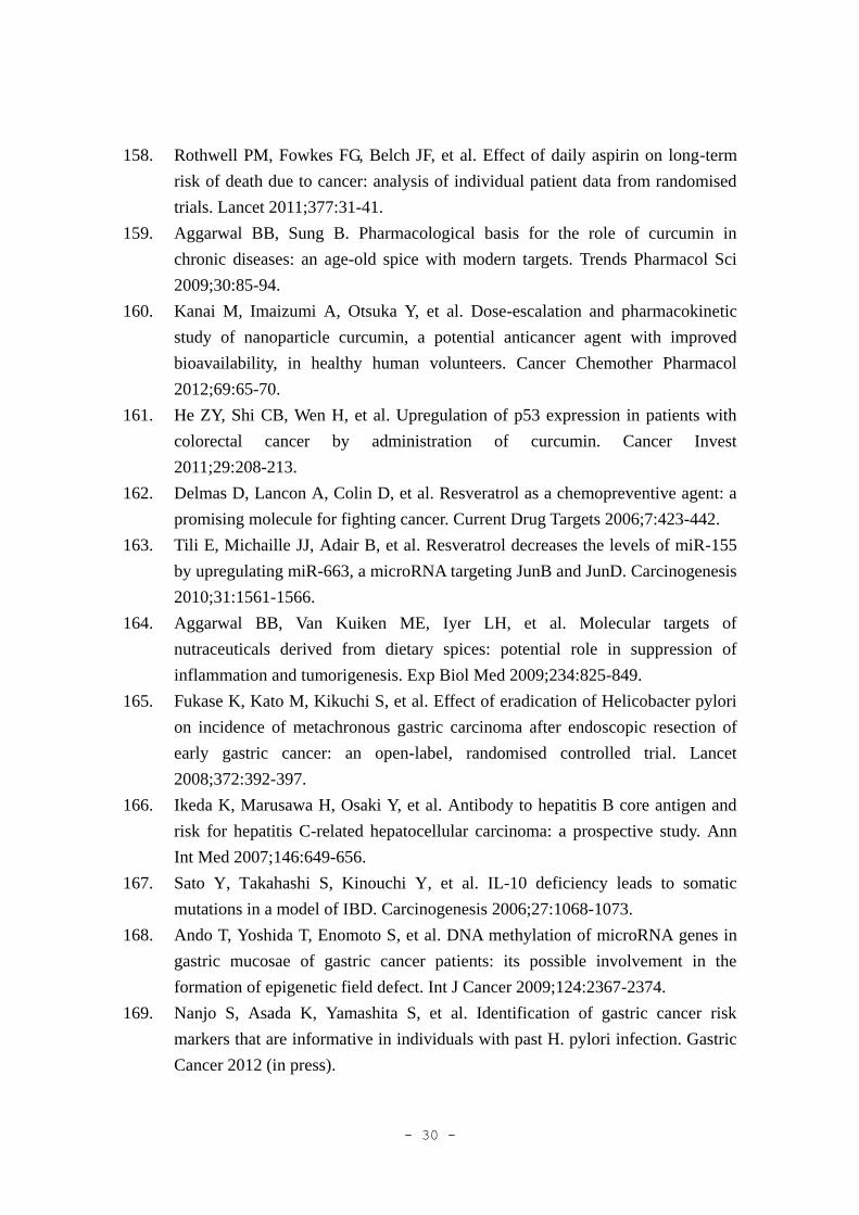
- 30 -
158. Rothwell PM, Fowkes FG, Belch JF, et al. Effect of daily aspirin on long-term
risk of death due to cancer: analysis of individual patient data from randomised
trials. Lancet 2011;377:31-41.
159. Aggarwal BB, Sung B. Pharmacological basis for the role of curcumin in
chronic diseases: an age-old spice with modern targets. Trends Pharmacol Sci
2009;30:85-94.
160. Kanai M, Imaizumi A, Otsuka Y, et al. Dose-escalation and pharmacokinetic
study of nanoparticle curcumin, a potential anticancer agent with improved
bioavailability, in healthy human volunteers. Cancer Chemother Pharmacol
2012;69:65-70.
161. He ZY, Shi CB, Wen H, et al. Upregulation of p53 expression in patients with
colorectal cancer by administration of curcumin. Cancer Invest
2011;29:208-213.
162. Delmas D, Lancon A, Colin D, et al. Resveratrol as a chemopreventive agent: a
promising molecule for fighting cancer. Current Drug Targets 2006;7:423-442.
163. Tili E, Michaille JJ, Adair B, et al. Resveratrol decreases the levels of miR-155
by upregulating miR-663, a microRNA targeting JunB and JunD. Carcinogenesis
2010;31:1561-1566.
164. Aggarwal BB, Van Kuiken ME, Iyer LH, et al. Molecular targets of
nutraceuticals derived from dietary spices: potential role in suppression of
inflammation and tumorigenesis. Exp Biol Med 2009;234:825-849.
165. Fukase K, Kato M, Kikuchi S, et al. Effect of eradication of Helicobacter pylori
on incidence of metachronous gastric carcinoma after endoscopic resection of
early gastric cancer: an open-label, randomised controlled trial. Lancet
2008;372:392-397.
166. Ikeda K, Marusawa H, Osaki Y, et al. Antibody to hepatitis B core antigen and
risk for hepatitis C-related hepatocellular carcinoma: a prospective study. Ann
Int Med 2007;146:649-656.
167. Sato Y, Takahashi S, Kinouchi Y, et al. IL-10 deficiency leads to somatic
mutations in a model of IBD. Carcinogenesis 2006;27:1068-1073.
168. Ando T, Yoshida T, Enomoto S, et al. DNA methylation of microRNA genes in
gastric mucosae of gastric cancer patients: its possible involvement in the
formation of epigenetic field defect. Int J Cancer 2009;124:2367-2374.
169. Nanjo S, Asada K, Yamashita S, et al. Identification of gastric cancer risk
markers that are informative in individuals with past H. pylori infection. Gastric
Cancer 2012 (in press).

- 31 -
170. Toyota M, Ahuja N, Ohe-Toyota M, et al. CpG island methylator phenotype in
colorectal cancer. Proc Natl Acad Sci USA 1999;96:8681-8686.
- 32 -
Table 1. Inflammation-associated cancers in digestive organs
___________________________________________________________
Inflammation- Underling inflammation
associated cancer
__________________________________________________________
Barrett’s cancer Reflux esophagitis
Gastric cancer H. pylori-induced chronic gastritis
Colitic cancer Inflammatory bowel disease
Celiac disease
HCC HCV and HBV chronic hepatitis
Primary biliary cirrhosis
Cholangiocarcinoma Primary sclerosing cholangitis
Pancreatic cancer Chronic pancreatitis
Hereditary pancreatitis
Lymphoma H. pylori-induced MALT lymphoma
HCV-associated lymphoma
Celiac disease-associated lymphoma
___________________________________________________________
HCC; hepatocellular carcinoma
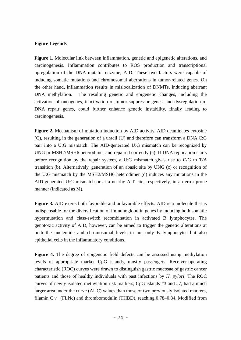
- 33 -
Figure Legends
Figure 1. Molecular link between inflammation, genetic and epigenetic alterations, and
carcinogenesis. Inflammation contributes to ROS production and transcriptional
upregulation of the DNA mutator enzyme, AID. These two factors were capable of
inducing somatic mutations and chromosomal aberrations in tumor-related genes. On
the other hand, inflammation results in mislocalization of DNMTs, inducing aberrant
DNA methylation. The resulting genetic and epigenetic changes, including the
activation of oncogenes, inactivation of tumor-suppressor genes, and dysregulation of
DNA repair genes, could further enhance genetic instability, finally leading to
carcinogenesis.
Figure 2. Mechanism of mutation induction by AID activity. AID deaminates cytosine
(C), resulting in the generation of a uracil (U) and therefore can transform a DNA C:G
pair into a U:G mismatch. The AID-generated U:G mismatch can be recognized by
UNG or MSH2/MSH6 heterodimer and repaired correctly (a). If DNA replication starts
before recognition by the repair system, a U:G mismatch gives rise to C/G to T/A
transition (b). Alternatively, generation of an abasic site by UNG (c) or recognition of
the U:G mismatch by the MSH2/MSH6 heterodimer (d) induces any mutations in the
AID-generated U:G mismatch or at a nearby A:T site, respectively, in an error-prone
manner (indicated as M).
Figure 3. AID exerts both favorable and unfavorable effects. AID is a molecule that is
indispensable for the diversification of immunoglobulin genes by inducing both somatic
hypermutation and class-switch recombination in activated B lymphocytes. The
genotoxic activity of AID, however, can be aimed to trigger the genetic alterations at
both the nucleotide and chromosomal levels in not only B lymphocytes but also
epithelial cells in the inflammatory conditions.
Figure 4. The degree of epigenetic field defects can be assessed using methylation
levels of appropriate marker CpG islands, mostly passengers. Receiver-operating
characteristic (ROC) curves were drawn to distinguish gastric mucosae of gastric cancer
patients and those of healthy individuals with past infections by H. pylori. The ROC
curves of newly isolated methylation risk markers, CpG islands #3 and #7, had a much
larger area under the curve (AUC) values than those of two previously isolated markers,
filamin Cγ (FLNc) and thrombomodulin (THBD), reaching 0.78–0.84. Modified from
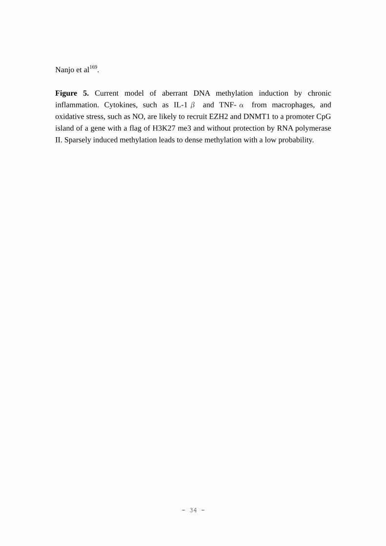
- 34 -
Nanjo et al169
.
Figure 5. Current model of aberrant DNA methylation induction by chronic
inflammation. Cytokines, such as IL-1β and TNF-α from macrophages, and
oxidative stress, such as NO, are likely to recruit EZH2 and DNMT1 to a promoter CpG
island of a gene with a flag of H3K27 me3 and without protection by RNA polymerase
II. Sparsely induced methylation leads to dense methylation with a low probability.

Inflammation
ROS AID
Dysregulated DNA repair
Tumor Suppressor Inactivation
Oncogene Activation
Carcinogenesis
Mutations Chromosomal Aberrations
Figure 1.
Mislocalization of DNMT1 ?
Aberrant DNA methylation
Chromosomal instability
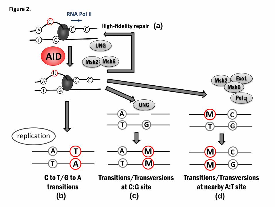
UNG
Msh2 Msh6
High-fidelity repair
AID
RNA Pol II
replication
C to T/G to A
transitions
T
A
Figure 2.
UNG
Exo1 Msh2 Msh6
Pol ղ
Transitions/Transversions
at C:G site
T
A
G
T
A M
Transitions/Transversions
at nearby A:T site
G
M
A M
(a)
(b) (c) (d)
T
T
M
M
C
C
G
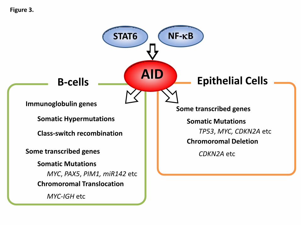
Epithelial Cells B-cells
NF-kB
AID
Somatic Hypermutations
Class-switch recombination
Chromoromal Translocation
Figure 3.
STAT6
Immunoglobulin genes
Some transcribed genes
Somatic Mutations
MYC, PAX5, PIM1, miR142 etc
MYC-IGH etc
Chromoromal Deletion
Some transcribed genes
Somatic Mutations
TP53, MYC, CDKN2A etc
CDKN2A etc
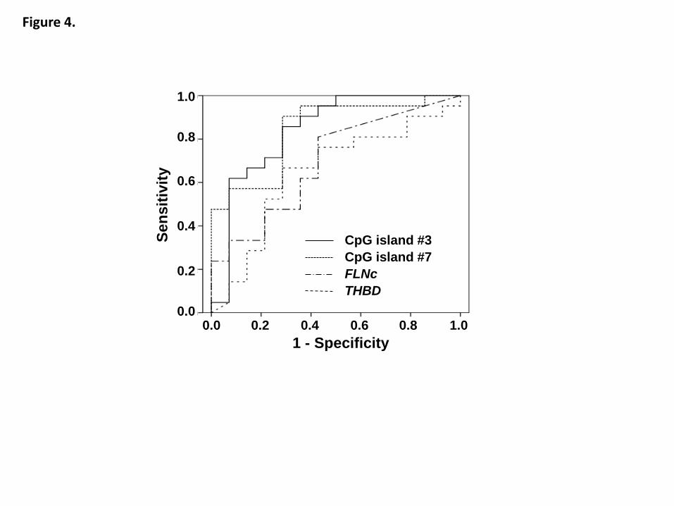
Figure 4.
1 - Specificity
Se
nsit
ivit
y
0.0 0.2 0.4 0.6 0.8 1.0 0.0
0.2
0.4
0.6
0.8
1.0
CpG island #3
CpG island #7
FLNc
THBD
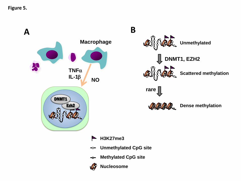
Figure 5.
Macrophage
TNFa
IL-1b
Ezh2
DNMT1
NO
A B Unmethylated
Scattered methylation
Dense methylation
H3K27me3
Unmethylated CpG site
Methylated CpG site
Nucleosome
DNMT1, EZH2
rare