infection, musculoskeletal (msk) · musculoskeletal (msk) infection ... deep musculoskeletal...
TRANSCRIPT

CLINICAL PATHWAY
Page 1 of 16
MUSCULOSKELETAL (MSK) INFECTION
Algorithm 1. Orthopedic Follow-up for MSK Infection
MSK Infection
Osteomyelitis Septic joint
Non-operative
Operative:
Return in 2 weeks
for wound check
1st visit:
2 weeks wound
check
3rd visit: 6 months
x-rays
2nd visit: 3 months
x-rays
If septic hip
4th visit: 2 years x-
rays (AP pelvis, frog,
lateral)
3rd visit:
1 year x-rays
2nd visit:
3 months x-rays
If infection is
adjacent to physes
4th visit: 1 year x-rays
(bilateral hips to
ankles standing AP)
Inclusion Criteria:
· Patients 6 months to 18 years
· Patients with suspicion of acute (less
than 2 weeks) deep musculoskeletal
infection, osteomyelitis, septic
arthritis, pyomyostis
Exclusion Criteria:
·Infants (less than 6 months)
·Post-op infection or foreign bodies
·Infections from penetrating trauma
·Chronic infection
·Medically complex children

CLINICAL PATHWAY
Page 2 of 16
Algorithm 2. Musculoskeletal Infection Diagnostic
Patient arrives with pain in bone, joint or muscle, and/or
refusal to walk, weight bear, or use limb
Evaluation by ED Attending
(See Clinical Assessment section)
History and Clinical Evaluation
considered High Risk for MSK
Infection
History and Clinical Evlauation
considered Low Risk for MSK
Infection (fracture, symptoms
resolve, no fever, mild pain,
alternative cause)
Order appropriate 2-view plain films
of area, CBC, ESR, CRP and blood
culture
Usual ED
care
Inclusion Criteria:
· Patients 6 months to 18 years
· Patients with suspicion of acute (less
than 2 weeks) deep musculoskeletal
infection, osteomyelitis, septic
arthritis, pyomyostis
Exclusion Criteria:
·Infants (less than 6 months)
·Post-op infection or foreign bodies
·Infections from penetrating trauma
·Chronic infection
·Medically complex children
Surgical intervention indicated if:
· Microbial etiology unknown (i.e. blood
cultures negative)
· Infection of a joint suspected
· Drainable abscess or fluid collection
found on clinical exam or imaging,
especially if patient has signs of toxic
shock syndrome (i.e., hypotension,
“sunburn” rash, peeling skin, etc.)
Does patient have decreased use/pain of extremity or
spine and or inability to bear weight AND at least one of
the following:
1. Positive blood culture
2. And/or abnormal plain films
3. And/or abnormal labs
a. Abnormal white blood cell count (>12K)
b. Abnormal ESR (>40)
c. CRP (>2 mg/dL)
4. And/or fever >38.5ºC
Suspected septic knee:
· ED aspiration, interpretation and
disposition.
· If suspected bacterial (>50K cell
count, + Gram stain, clinically
consistent), proceed as in non-
knee MSK infection.
· If epidemiology supports,
consider Lyme disease (send
serology) and consider ortho/
rheum/ID consults as
appropriate
Can patient be
safely discharged
to home?
Does patient
have reliable
follow-up?
Can patient take
oral fluids and pain
meds?
Discharge to home with
PCP follow-up or an
orthopedic consult in the
morning
Proceed to inpatient
algorithm for inpatient
management and ED
management while
awaiting admission/
OR
· Admit
· Consult Orthopedics
· Consider ID consult
· Withhold antibiotics
until seen by
Orthopedics/ID
· Emergent orthopedic evaluation
· Consider ID consult either in ED or on
Ward
· Consider further imaging as in imaging studies section.
Orthopedics to order MRIs from ED, advise on order for ward.
· Biopsy/aspiration to establish microbial etiology (unless blood
culture positive for likely pathogen), for therapeutics (to prevent
rupture into contiguous joint, for example) and abscess discovery.
· Send source culture (NO SWABS), Gram stain and pathology on
all cases (exception for patients with positive blood culture for likely
pathogen). For joints, send cell count, glucose, protein, synovial
pathology and fluid to save from suspected source.
· If orthopedics cannot access, discuss with interventional radiology.
NoYes
Yes
Yes
Yes
No
No
+/-
No
CLINICAL PATHWAY
Page 3 of 16
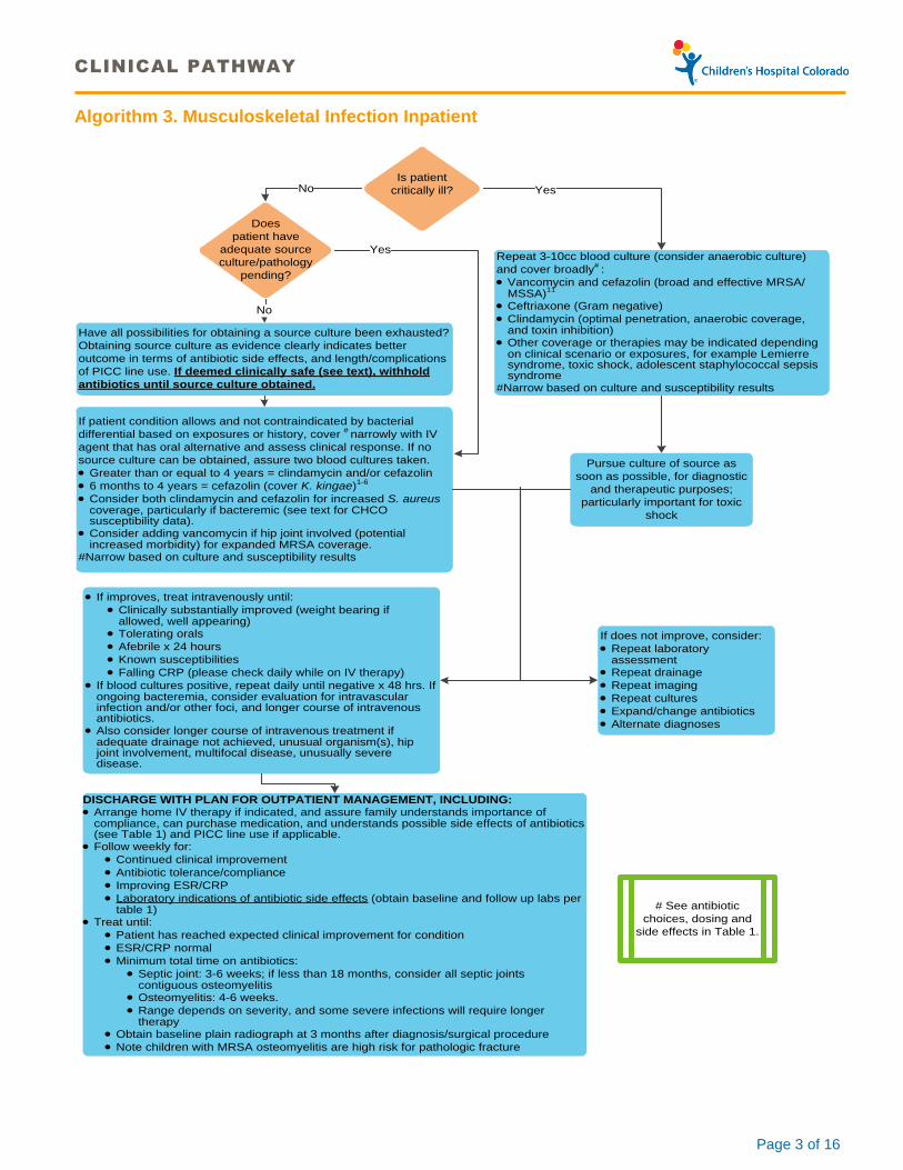
Algorithm 3. Musculoskeletal Infection Inpatient
Is patient
critically ill?
Does
patient have
adequate source
culture/pathology
pending?
Repeat 3-10cc blood culture (consider anaerobic culture)
and cover broadly# :
· Vancomycin and cefazolin (broad and effective MRSA/MSSA)
11
· Ceftriaxone (Gram negative)
· Clindamycin (optimal penetration, anaerobic coverage, and toxin inhibition)
· Other coverage or therapies may be indicated depending on clinical scenario or exposures, for example Lemierre syndrome, toxic shock, adolescent staphylococcal sepsis syndrome
#Narrow based on culture and susceptibility results
Have all possibilities for obtaining a source culture been exhausted?
Obtaining source culture as evidence clearly indicates better
outcome in terms of antibiotic side effects, and length/complications
of PICC line use. If deemed clinically safe (see text), withhold
antibiotics until source culture obtained.
If patient condition allows and not contraindicated by bacterial
differential based on exposures or history, cover # narrowly with IV
agent that has oral alternative and assess clinical response. If no
source culture can be obtained, assure two blood cultures taken.
· Greater than or equal to 4 years = clindamycin and/or cefazolin
· 6 months to 4 years = cefazolin (cover K. kingae)1-6
· Consider both clindamycin and cefazolin for increased S. aureus coverage, particularly if bacteremic (see text for CHCO susceptibility data).
· Consider adding vancomycin if hip joint involved (potential increased morbidity) for expanded MRSA coverage.
#Narrow based on culture and susceptibility results
· If improves, treat intravenously until:
· Clinically substantially improved (weight bearing if allowed, well appearing)
· Tolerating orals
· Afebrile x 24 hours
· Known susceptibilities
· Falling CRP (please check daily while on IV therapy)
· If blood cultures positive, repeat daily until negative x 48 hrs. If ongoing bacteremia, consider evaluation for intravascular infection and/or other foci, and longer course of intravenous antibiotics.
· Also consider longer course of intravenous treatment if adequate drainage not achieved, unusual organism(s), hip joint involvement, multifocal disease, unusually severe disease.
DISCHARGE WITH PLAN FOR OUTPATIENT MANAGEMENT, INCLUDING:· Arrange home IV therapy if indicated, and assure family understands importance of
compliance, can purchase medication, and understands possible side effects of antibiotics (see Table 1) and PICC line use if applicable.
· Follow weekly for:
· Continued clinical improvement
· Antibiotic tolerance/compliance
· Improving ESR/CRP
· Laboratory indications of antibiotic side effects (obtain baseline and follow up labs per table 1)
· Treat until:
· Patient has reached expected clinical improvement for condition
· ESR/CRP normal
· Minimum total time on antibiotics:
· Septic joint: 3-6 weeks; if less than 18 months, consider all septic joints contiguous osteomyelitis
· Osteomyelitis: 4-6 weeks.
· Range depends on severity, and some severe infections will require longer therapy
· Obtain baseline plain radiograph at 3 months after diagnosis/surgical procedure
· Note children with MRSA osteomyelitis are high risk for pathologic fracture
# See antibiotic
choices, dosing and
side effects in Table 1.
Pursue culture of source as
soon as possible, for diagnostic
and therapeutic purposes;
particularly important for toxic
shock
If does not improve, consider:
· Repeat laboratory assessment
· Repeat drainage
· Repeat imaging
· Repeat cultures
· Expand/change antibiotics
· Alternate diagnoses
No Yes
No
Yes

CLINICAL PATHWAY
Page 4 of 16
SUMMARY
DIAGNOSIS
· Clinical Assessment
o Vital signs on admission
o Observation and/or history for limited use or immobility of extremity/spine, gait disturbance/limp, inability to
bear weight, pain, travel history/exposures, fever greater than or equal to 38.5ºC
o Physical examination for the presence of limited range of motion, tenderness, swelling, warmth at site, erythema, psoas sign, fever
· Initial Evaluation and Labs
o Complete blood count (CBC) with differential, C-reactive protein (CRP), Erythrocyte sedimentation rate (ESR), Blood culture x 2 (per Children’s Hospital Colorado protocol)
o If knee/elbow: Athrocentesis, interpretation and disposition in ED. Arthrocentesis performed by ED or Orthopedics (if ED physician, consider Ortho consult prior to arthrocentesis).
o If suspected bacterial (greater than or equal to 50,000 cell count, organisms seen on Gram stain, or if presentation is clinically consistent), proceed with MSK CCG
o If travel/cell count supports, consider Lyme disease (send serology) and consider Orthopedics/Rheumatology/ Infectious Disease (ID) consults
o Suspected deep seated bones or joints (e.g. shoulder, hip): Obtain labs/imaging; consult Orthopedics
o Source Evaluation: Send purulent material (aspirate, not a swab) for Gram stain and bacterial culture, and if possible tissue/synovium for pathology. If joint fluid, send for bacterial culture, Gram stain, cell count, differential and fluid to hold. If unusual case or exposures, consult ID for further testing/culturing recommendations.
· Initial Imaging Studies
o Plain radiographs (all patients): Radiographs can be insensitive for the evaluation of acute soft tissue and osseous infection, but if diagnostic may avoid further imaging; soft tissue swelling, though nonspecific, may be an early finding of MSK infection.
o Ultrasound: Hip ultrasound may be helpful in the setting of equivocal physical exam findings for hip effusion. Consider ordering if a negative hip ultrasound would avoid further imaging and/or aspiration.
o Additional Imaging (e.g. MRI or CT) as directed by ORTHOPEDIC CONSULTANT AND RADIOLOGY
TREATMENT
· Surgical intervention indicated if:
o Microbial etiology unknown (i.e. blood cultures negative)
o Infection of a joint suspected
o Drainable abscess or fluid collection found on clinical exam or imaging, especially if patient has signs of toxic shock syndrome (i.e., hypotension, “sunburn” rash, peeling skin, etc.)
· Antibiotic Therapy (Table 1)
o Withhold antimicrobial therapy until source cultures obtained unless patient unstable
ALGORITHM 1. Orthopedic Follow up for MSK Infection ALGORITHM 2. Musculoskeletal Infection Diagnostic Algorithm ALGORITHM 3. Musculoskeletal Infection Inpatient Algorithm TABLE 1. Antibiotics and Monitoring for Patients with Musculoskeletal Infections

CLINICAL PATHWAY
Page 5 of 16
TABLE OF CONTENTS
Algorithm 1. Orthopedic Follow up for MSK Infection
Algorithm 2. MSK Infection Diagnostic Algorithm
Algorithm 3. MSK Infection Inpatient Algorithm
Summary
Clinical Assessment
Initial Evaluation and Labs
Initial Imaging Studies
Procedures | Interventions
Consultations to Consider
Initial Therapies | Emergency Department (ED) and Inpatient Unit
Admission Criteria
Change to Oral Antibiotics | Discharge Criteria
Discharge Planning and Follow-up
Patient | Caregiver Education
Table 1. Antibiotics and Monitoring for Patients with MSK Infection
References
Clinical Improvement Team
TARGET POPULATION
Inclusion Criteria
· 6 months to 18 years
· With suspicion of acute (less than 2 weeks) deep musculoskeletal infection; osteomyelitis, septic arthritis, pyomyositis
Exclusion Criteria
· Postoperative infection or foreign bodies
· Infections from penetrating trauma
· Chronic infection
· Infants (less than 6months), as they may have: 1) other pathogens, 2) multifocal disease, and 3) poor oral antibiotic absorption
· Medically complex children
KEY TREATMENT PRINCIPLES
Indicated:
· Laboratory workup including complete blood count (CBC) with differential, C-reactive protein (CRP), erythrocyte sedimentation rate (ESR), and blood cultures1

CLINICAL PATHWAY
Page 6 of 16
· Radiographic imaging3
· Source culture prior to antibiotics in non-critical patients
· Avoidance of PICC line placement in most patients
Not routinely indicated:
· Antibiotics prior to source culture for well-appearing children
· MRI (unless orthopedic consult deems necessary)
· PICC/central line placement
CLINICAL ASSESSMENT
1. Vital signs on admission
2. Observation and/or history for:
o Limited used or immobility of extremity or spine
o Gait disturbance/Limp
o Inability to bear weight
o Pain
o Fever greater than 38.5ºC
o Travel and exposures
3. Physical examination for the presence of:
o Limited range of motion
o Tenderness
o Swelling
o Warmth at site
o Erythema
o Psoas sign
o Fever
INITIAL EVALUATION AND LABS
1. Complete blood count (CBC) with differential
2. C-reactive protein (CRP)
3. Erythrocyte sedimentation rate (ESR)
4. Blood culture x 2 (per Children’s Hospital Colorado protocol)
5. If knee/elbow: Athrocentesis, interpretation and disposition in ED. Arthrocentesis performed by ED or Orthopedics (if ED physician, consider Ortho consult prior to arthrocentesis).
o If suspected bacterial (greater than or equal to >50,000 cell count, + Gram stain, or presentation clinically consistent), proceed with MSK CCG.
o If travel/cell count supports, consider Lyme disease (send serology) and consider Orthopedics/Rheumatology /Infectious Disease consults.
CLINICAL PATHWAY
Page 7 of 16
6. Suspected deep seated bone or joints (e.g. shoulder, hip): Obtain labs/imaging; consult Orthopedics
INITIAL IMAGING STUDIES
· Plain radiographs (all patients): Radiographs can be insensitive for the evaluation of acute soft tissue and osseous infection, but if diagnostic may avoid further imaging; soft tissue swelling, though nonspecific, may be an early finding of MSK infection.
· Ultrasound: Hip ultrasound may be helpful in the setting of equivocal physical exam findings for hip effusion. Consider ordering if a negative hip ultrasound would avoid further imaging and/or aspiration.
Additional Imaging as directed by ORTHOPEDIC CONSULTANT AND RADIOLOGY
· MRI: The orthopedic team is to place all MRI orders in EPIC from the ED. MRI should not be ordered until Orthopedics has evaluated patient. This is to assure the correct exam is ordered in the appropriate time frame.
MRI ordering process:
Critically ill patients needing EMERGENT exam:
· 7am-10pm: Call radiologist to approve a sedated or non-sedated emergent exam. Please contact MSK radiologist through lead tech (7-5993) from 7am-4pm. Please contact evening radiologist through the lead tech (7-5993) from 4pm-10pm.
· 10pm-7am: if an MRI should not wait for earliest sedated or non-sedated slot please have Orthopedic attending call the CT tech (7-8645), who will call in an MRI tech to perform an emergent exam.
Weekdays (7am-10pm), patients needing URGENT (not critical/emergent) exam:
· 7 a.m. to 4 p.m.: Call radiologist to schedule exam, goal is to perform within 10 hours.
· 4 p.m. to 10 p.m.: Call evening radiologist through lead tech (7-5993) to schedule exam, goal is to perform during evening shift if slot available, otherwise in following AM as below after 10pm.
· After 10 p.m. NON-SEDATED patient: Please have Orthopedic resident, fellow, PA or attending call the overnight CT Tech (7-8645), who will schedule and place patient in a 6:30am slot.
· After 10 p.m. SEDATED patient: Please have Orthopedic resident, fellow, PA, or attending call overnight CT tech (7-8645), who will schedule and place patient in a 7:30am sedated slot. Anesthesiology will accommodate sedated MRI add-on by sending swing shift anesthesiologist if necessary.
Weekends and holidays, patients needing URGENT (not critical/emergent) exam
· 7am-10pm: Call on-call radiologist through lead tech (7-5993) to schedule exam. Goal is to perform the MRI within 10 hours.
· After 10pm NON-SEDATED patient: Please have Orthopedic resident call the overnight CT tech (7-8645), who will schedule and place patient in a 7:30 am slot.
· After 10 p.m. SEDATED patient: Please have Orthopedic resident call overnight CT tech (7-8645), who will schedule and place patient in the earliest sedated slot. If sedated MRI slot is outside the 10 hour goal, Orthopedic attending should call anesthesia to find first available anesthesiologist.
PROCEDURES | INTERVENTIONS (Emergency Department/Operating Room/Interventional Radiology)
Biopsy/aspiration to establish microbial etiology, for therapeutics (to prevent rupture into contiguous joint, for example) and abscess discovery. Evidence clearly indicates better outcome in terms of antibiotic side effects, and length/complications of PICC line use13. Send pus (not a swab) for Gram stain and bacterial culture, and if possible tissue/synovium for pathology. If joint fluid, send for bacterial culture, Gram stain, cell count, differential and fluid to hold. If unusual case or exposures, consult Infectious Disease for further testing/culturing recommendations.
CLINICAL PATHWAY
Page 8 of 16
CONSULTATIONS TO CONSIDER
Seek primary attending’s approval prior to consulting specialist, primary team to coordinate consult communication.
Orthopedics
Orthopedics prefers to be consulted on all confirmed and probable musculoskeletal infections as soon as suspected, and prior to advanced imaging, as orthopedics can facilitate and will order timely MRI.
Infectious Disease
Infectious Diseases prefers to be consulted on all confirmed and probable musculoskeletal infections, particularly if ID to follow as an outpatient or upon unit transfer. Consultation should occur per inpatient teams, unless early contact preferred by ED team.
Rheumatology
· Polyarthritis
· Suspicion or history of JRA, SLE, rheumatic fever, post-strep arthritis or psoriasis
· Chronic joint effusion
INITIAL THERAPIES | EMERGENCY DEPARTMENT AND INPATIENT UNIT
1. Pain control administered per Emergency Department/primary team
2. All patients are to be treated intravenously initially
3. Source culture should be obtained prior to starting antibiotics, unless blood culture positive or patient’s clinical status too unwell. Goal is source culture by end of day after admission, in orthopedic trauma room if needed. If time slots are an issue, call orthopedic trauma to help facilitate.
4. If blood culture positive, cover according to suspected organism (Table 1)
5. If patient condition allows and not contraindicated by bacterial differential based on exposures or history, cover narrowly with IV agent with oral alternative and follow cultures and assess clinical response.
o Greater than or equal to 4 years = clindamycin and/or cefazolin
o 6 months to 4 years = cefazolin (cover K. kingae)1-6
o Consider both clindamycin and cefazolin for increased S. aureus coverage, particularly if bacteremic. Per internal data from 2009-2014 isolates, if culture positive (80%), 81% of isolates are S. aureus; of those, 93% were susceptible to clindamycin, 88% were susceptible to cefazolin, 100% susceptible to combination.
o Consider adding vancomycin if hip joint involved (potential increased morbidity) for expanded MRSA coverage.
6. If critically ill, repeat appropriate volume blood culture (consider anaerobic blood culture for Lemierre syndrome) and cover broadly; multiple antibiotics may be indicated:
o Vancomycin and cefazolin (broad and effective MRSA/MSSA)11
o Ceftriaxone (Gram negative)
o Clindamycin (optimal penetration, anaerobic coverage, and toxin inhibition)
7. Other coverage or therapies may be indicated depending on clinical scenario or exposures, for example Lemierre syndrome, toxic shock, adolescent staphylococcal sepsis syndrome19-23
8. Narrow based on culture and susceptibility results

CLINICAL PATHWAY
Page 9 of 16
ADMISSION CRITERIA
· Admit all patients with suspected and confirmed acute musculoskeletal infections unless indicated otherwise by Pediatric Orthopedics or ID.
· Prior to admission at South Campus, consult orthopedic surgery to discuss best admission location for patient’s needs. If the patient requires transfer to Anschutz Medical Campus for expedited care, efforts should be made to obtain the MRI at South Campus prior to transfer if this can be obtained without a delay in care.
CHANGE TO ORAL ANTIBIOTICS | DISCHARGE CRITERIA
· If improves, treat intravenously and inpatient until:5,8,15,24-30
o Clinically substantially improved (weight bearing if allowed, improved motion of infected joint, well appearing)
o Tolerating orals
o Afebrile x 24 hours
o Known susceptibilities indicating there is an appropriate oral alternative to intravenous therapy
o Falling CRP
· If blood cultures positive, repeat daily post initiation of antibiotics until negative x 48 hours; if ongoing bacteremia, consider evaluation for intravascular infection and/or other foci, and longer course of intravenous antibiotics17.
· Also consider longer course of intravenous treatment if: adequate drainage not achieved, unusual organism(s), hip joint involvement, multifocal disease, unusually severe disease, doubt about oral absorption or compliance.
If condition does not improve, consider:
· Repeat laboratory assessment
· Repeat imaging31
· Repeat drainage
· Repeat cultures
· Expand/change antibiotics
· Alternate diagnoses
DISCHARGE PLANNING AND FOLLOW-UP
1. Arrange home IV therapy if indicated, and assure family: 1. understands importance of compliance, 2. is able to
purchase medication, and 3. understands possible side effects of antibiotics (Table 1) and PICC line use if applicable.
2. Treat until:
o Patient has reached expected clinical improvement for condition
o ESR/CRP normal
o Minimum total time on antibiotics26,28,29,36:
o Septic joint: 3 to 6 weeks; if less than 18 months, consider all septic joints contiguous osteomyelitis
o Osteomyelitis: 4 to 6 weeks
o Range depends on severity, and some severe infections will require longer therapy
3. Infectious diseases or primary care follow up
o Follow each 1-2 weeks for 35:
o Continued clinical improvement

CLINICAL PATHWAY
Page 10 of 16
o Antibiotic tolerance/compliance
o Improving ESR/CRP
o Laboratory indications of antibiotic side effects (obtain baseline and follow up labs per Table 1)
4. Orthopedic follow up (Algorithm 1.)
o Surgical patients should follow up at two weeks post operatively
o Obtain baseline plain radiograph at three months
o Remaining follow up after three months will be determined at discretion of orthopedics
o Note children with MRSA osteomyelitis are high risk for pathologic fracture37, children with hip infection at risk if avascular necrosis
PARENT | CAREGIVER EDUCATION
It is suggested that parent/caregiver education contain the following information:
· Education on obtaining and taking antibiotics
· Pain control measures
· Return precautions and contact information for orthopedics and infectious diseases
· Education regarding antibiotic side effects
· PICC line training and education (including risk of infection) if applicable

CLINICAL PATHWAY
Page 11 of 16
TABLE 1. Antibiotics and Monitoring for Patients with Musculoskeletal Infections
(Other antibiotics may be indicated based on culture results) Antimicrobial Stewardship at Children’s Hospital Colorado, Sarah Parker and Jason Child 2014
C
efa
zoli
n
(IV
)
Ce
ph
ale
xin
(P
O)
Ce
fota
xim
e
(IV
)
Ce
ftri
ax
on
e
(IV
)
Va
nco
my
cin
4 (
IV)
Cli
nd
am
yci
n 3
(IV
or
PO
)
Am
pic
illi
n
(IV
)
Am
ox
icil
lin
(P
O)
Daily amount (in mg/kg/day)
100-150 mg/kg/day divided Q6-8H
100-150 mg/kg/day divided QID
150-200 mg/kg/day divided Q6-8H
100 mg/kg/day divided Q12-24H
60-80 mg/kg/day divided Q6H
40 mg/kg/day IV, divided Q6-8H PO, divided TID
200mg/kg/day IV, divided Q6H
90mg/kg/day PO, divided TID
Single daily amount maximum for MSK infection
8000 mg divided Q6H
4000 mg divided QID
8000 mg divided Q6H
2000 mg divided Q12-24H
6000 mg divided Q8H (adjust dose with levels, can use continuous, consult ID)
2700 mg IV (IV, divided Q 8H) 1800 mg PO (PO, divided TID)
8000mg IV divided Q6H
2000-4000 mg PO divided TID
Organism
MSSA2 + + 2 2 + +
MRSA + +3
S. pyogenes (Group A strep)
+ + + + + + + +
S. pneumoniae + + + + + + + +
Kingella kingae5
+ + + + + +
Side Effects1
Diarrhea, including C. difficile colitis
+ + + + + + + +
Bone marrow suppression
+ + + + + + + +
Rash + + + + + + + +
Stevens Johnson Syndrome
+ + + + + + + +
Drug fever + + + + + + + +
Nephrotoxicity, Interstitial nephritis
+ + + + + +
Nephrotoxicity, other
+
Elevated transaminases
+ +
Labs to monitor for infection resolution and side effects
1CBC, CRP
or ESR,
BUN, Cr
1CBC, CRP
or ESR,
BUN, Cr
1CBC, CRP
or ESR,
BUN, Cr
1CBC, CRP
or ESR,
BUN, Cr,
LFTs
1CBC, CRP or
ESR, BUN, Cr,
vanc trough
1CBC, CRP or
ESR, BUN, Cr,
LFTs
1CBC, CRP or
ESR, BUN, Cr
1CBC, CRP or
ESR, BUN, Cr
1 All patients on antibiotics for MSK infection should be followed with a weekly CBC, ESR or CRP. There are additional labs specific to the antibiotic, for example: urinalysis and BUN/creatinine screen for renal function and interstitial nephritis, CBC for neutropenia. Clinically patients should be followed for signs of allergy including rash, for diarrhea (any antibiotic can cause Clostridium difficile colitis), for fevers (for severe allergy and line infection, recurrent infection), for compliance and other complaints. All antibiotics can cause anaphylaxis. Side effects listed are most common, but do not represent all side effects.
CLINICAL PATHWAY
Page 12 of 16
2 Although cefotaxime and ceftriaxone are often listed as having activity against MSSA, in general, antistaphylococcal penicillins (such as nafcillin) or first generation cephalosporins (such as cefazolin) are the preferred therapy. 3 The use of clindamycin for MRSA depends on local susceptibility patterns and, if available, susceptibility testing. It is important that the microbiology lab perform a “D-test” or equivalent for inducible clindamycin resistance. At Children’s Hospital Colorado, D test is routinely performed and our MSSA and MRSA isolates are 86% susceptible. We feel it is reasonable to use as first line in non-critically ill patients greater than 4 years of age (below 4 years, include K. kingae coverage). 4 Nafcillin, vancomycin and penicillin can be given by continuous infusion; discuss with ID/pharmacy. 5Kingella kingae is a predominant cause of bone and joint infection in the 6 month to less than 4 year age group, but is
difficult to culture. Unless microbial cause is known, it should be empirically covered. 92% of K. kingae disease is in
children aged 6 to 29 months; 1% is in children < 6 months, and 4% in children ≥ 4 years. It predominantly causes
septic arthritis, but can also cause isolated osteomyelitis and tenosynovitis; it generally has a milder presentation than
S. aureus.
CLINICAL PATHWAY
Page 13 of 16
REFERENCES
1. Basmaci R, Ilharreborde B, Lorrot M, Bidet P, Bingen E, Bonacorsi S. Predictive score to discriminate Kingella kingae from Staphylococcus aureus arthritis in France. The Pediatric Infectious Disease Journal 2011;30:1120-1; author reply 1-2.
2. Basmaci R, Lorrot M, Bidet P, et al. Comparison of clinical and biologic features of Kingella kingae and Staphylococcus aureus arthritis at initial evaluation. The Pediatric infectious disease journal 2011;30:902-4.
3. Chometon S, Benito Y, Chaker M, et al. Specific real-time polymerase chain reaction places Kingella kingae as the most common cause of osteoarticular infections in young children. The Pediatric Infectious Disease Journal 2007;26:377-81.
4. Goergens ED, McEvoy A, Watson M, Barrett IR. Acute osteomyelitis and septic arthritis in children. Journal of Paediatrics and Child Health 2005;41:59-62.
5. Thomsen I, Creech CB. Advances in the diagnosis and management of pediatric osteomyelitis. Current Infectious Disease Reports 2011;13:451-60.
6. Yagupsky P, Porsch E, St Geme JW, 3rd. Kingella kingae: an emerging pathogen in young children. Pediatrics 2011;127:557-65.
7. Caird MS, Flynn JM, Leung YL, Millman JE, D'Italia JG, Dormans JP. Factors distinguishing septic arthritis from transient synovitis of the hip in children. A prospective study. The Journal of Bone and Joint Surgery American Volume 2006;88:1251-7.
8. Kocher MS, Mandiga R, Murphy JM, et al. A clinical practice guideline for treatment of septic arthritis in children: efficacy in improving process of care and effect on outcome of septic arthritis of the hip. The Journal of Bone and Joint Surgery American, Volume 2003;85-A:994-9.
9. Kocher MS, Mandiga R, Zurakowski D, Barnewolt C, Kasser JR. Validation of a clinical prediction rule for the differentiation between septic arthritis and transient synovitis of the hip in children. The Journal of Bone and Joint Surgery American volume 2004;86-A:1629-35.
10. Sultan J, Hughes PJ. Septic arthritis or transient synovitis of the hip in children: the value of clinical prediction algorithms. The Journal of Bone and Joint Surgery British volume 2010;92:1289-93.
11. McConeghy KW, Bleasdale SC, Rodvold KA. The empirical combination of vancomycin and a beta-lactam for Staphylococcal bacteremia. Clin Infect Dis 2013;57:1760-5.
12. Paakkonen M, Kallio MJ, Kallio PE, Peltola H. Sensitivity of erythrocyte sedimentation rate and C-reactive protein in childhood bone and joint infections. Clinical Orthopaedics and Related Research 2010;468:861-6.
13. Ceroni D, Regusci M, Pazos JM, Saunders CT, Kaelin A. Risks and complications of prolonged parenteral antibiotic treatment in children with acute osteoarticular infections. Acta orthopaedica Belgica 2003;69:400-4.
14. Jaramillo D. Infection: musculoskeletal. Pediatric Radiology 2011;41 Suppl 1:S127-34. 15. Copley LA. Pediatric musculoskeletal infection: trends and antibiotic recommendations. The Journal of the
American Academy of Orthopaedic Surgeons 2009;17:618-26. 16. Gutierrez K. Bone and joint infections in children. Pediatric Clinics of North America 2005;52:779-94, vi. 17. Liu C, Bayer A, Cosgrove SE, et al. Clinical practice guidelines by the infectious diseases society of america for
the treatment of methicillin-resistant Staphylococcus aureus infections in adults and children. Clinical Infectious Diseases: an official publication of the Infectious Diseases Society of America 2011;52:e18-55.
18. Peltola H, Unkila-Kallio L, Kallio MJT, the Finnish Study G. Simplified Treatment of Acute Staphylococcal Osteomyelitis of Childhood. Pediatrics 1997;99:846-50.
19. Riordan T. Human infection with Fusobacterium necrophorum (Necrobacillosis), with a focus on Lemierre's syndrome. Clinical Microbiology Reviews 2007;20:622-59.
20. Gonzalez BE, Mon RA. Staphylococcus aureus infections in adolescents. Adolescent medicine: state of the art reviews 2010;21:318-31, x.
21. Ferguson MA, Todd JK. Toxic shock syndrome associated with Staphylococcus aureus sinusitis in children. The Journal of Infectious Diseases 1990;161:953-5.
22. Todd J, Fishaut M, Kapral F, Welch T. Toxic-shock syndrome associated with phage-group-I Staphylococci. Lancet 1978;2:1116-8.
23. Todd JK. Toxic shock syndrome - evolution of an emerging disease. Advances in experimental medicine and biology 2011;697:175-81.
24. Bachur R, Pagon Z. Success of short-course parenteral antibiotic therapy for acute osteomyelitis of childhood. Clinical Pediatrics 2007;46:30-5.
25. Ballock RT, Newton PO, Evans SJ, Estabrook M, Farnsworth CL, Bradley JS. A comparison of early versus late conversion from intravenous to oral therapy in the treatment of septic arthritis. Journal of Pediatric Orthopedics 2009;29:636-42.
26. Jagodzinski NA, Kanwar R, Graham K, Bache CE. Prospective evaluation of a shortened regimen of treatment for acute osteomyelitis and septic arthritis in children. Journal of Pediatric Orthopedics 2009;29:518-25.

CLINICAL PATHWAY
Page 14 of 16
27. Peltola H, Paakkonen M, Kallio P, Kallio MJ, Osteomyelitis-Septic Arthritis Study G. Short- versus long-term antimicrobial treatment for acute hematogenous osteomyelitis of childhood: prospective, randomized trial on 131 culture-positive cases. The Pediatric Infectious Disease Journal 2010;29:1123-8.
28. Syrogiannopoulos GA, Nelson JD. Duration of antimicrobial therapy for acute suppurative osteoarticular infections. Lancet 1988;1:37-40.
29. Weichert S, Sharland M, Clarke NM, Faust SN. Acute haematogenous osteomyelitis in children: is there any evidence for how long we should treat? Current opinion in infectious diseases 2008;21:258-62.
30. Zaoutis T, Localio AR, Leckerman K, Saddlemire S, Bertoch D, Keren R. Prolonged intravenous therapy versus early transition to oral antimicrobial therapy for acute osteomyelitis in children. Pediatrics 2009;123:636-42.
31. Courtney PM, Flynn JM, Jaramillo D, Horn BD, Calabro K, Spiegel DA. Clinical indications for repeat MRI in children with acute hematogenous osteomyelitis. Journal of Pediatric Orthopedics 2010;30:883-7.
32. Esposito S, Noviello S, Leone S, et al. Outpatient parenteral antibiotic therapy (OPAT) in different countries: a comparison. International journal of antimicrobial agents 2004;24:473-8.
33. Maraqa NF, Gomez MM, Rathore MH. Outpatient parenteral antimicrobial therapy in osteoarticular infections in children. Journal of Pediatric Orthopedics 2002;22:506-10.
34. Rathore MH. The unique issues of outpatient parenteral antimicrobial therapy in children and adolescents. Clinical infectious diseases : an official publication of the Infectious Diseases Society of America 2010;51 Suppl 2:S209-15.
35. Tice AD, Rehm SJ, Dalovisio JR, et al. Practice guidelines for outpatient parenteral antimicrobial therapy. IDSA guidelines. Clinical infectious diseases : an official publication of the Infectious Diseases Society of America 2004;38:1651-72.
36. Howard-Jones AR, Isaacs D. Systematic review of systemic antibiotic treatment for children with chronic and sub-acute pyogenic osteomyelitis. Journal of paediatrics and child health 2010;46:736-41.37. Belthur MV, Birchansky SB, Verdugo AA, et al. Pathologic fractures in children with acute Staphylococcus aureus osteomyelitis. The Journal of Bone and Joint Surgery American volume 2012;94:34-42.

CLINICAL PATHWAY
Page 15 of 16
Clinical pathways are intended for informational purposes only. They are current at the date of publication and are reviewed on a regular basis to align with the best available evidence. Some information and links may not be available to external viewers. External viewers are encouraged to consult other available sources if needed to confirm and supplement the content presented in the clinical pathways. Clinical pathways are not intended to take the place of a physician’s or other health care provider’s advice, and is not intended to diagnose, treat, cure or prevent any disease or other medical condition. The information should not be used in place of a visit, call, consultation or advice of a physician or other health care provider. Furthermore, the information is provided for use solely at your own risk. CHCO accepts no liability for the content, or for the consequences of any actions taken on the basis of the information provided. The information provided to you and the actions taken thereof are provided on an “as is” basis without any warranty of any kind, express or implied, from CHCO. CHCO declares no affiliation, sponsorship, nor any partnerships with any listed organization, or its respective directors, officers, employees, agents, contractors, affiliates, and representatives.
CLINICAL IMPROVEMENT TEAM MEMBERS
Sarah Parker, MD | Infectious Disease | Jaime Stewart, MD | Radiology Travis Heare, MD | Pediatric Orthopedics Jason Stoneback, MD | Pediatric Orthopedics Nathan Donaldson, DO | Pediatric Orthopedics Jenny Reese ,MD | Hospitalist Rakesh Mistry, MD | Emergency Medicine Justin Searns, MD | Infectious Disease Maggie Leyendecker, RN | Pediatric Orthopedics Heather Heizer, PA | Infectious Disease Jason Child, PharmD | Clinical Pharmacy Leigh Anne Bakel, MD | Hospitalist/Clinical Effectiveness Sarah Nickels, PhD | Clinical Effectiveness
APPROVED BY
Clinical Pathways and Measures Committee – April 18, 2016
MANUAL/DEPARTMENT Clinical Care Guidelines/Quality
ORIGINATION DATE June 19, 2012
LAST DATE OF REVIEW OR REVISION April 18, 2016
APPROVED BY
Lalit Bajaj, MD, MPH Medical Director, Clinical Effectiveness
REVIEW | REVISION SCHEDULE
Scheduled for full review on April 18, 2020

CLINICAL PATHWAY
Page 16 of 16