infection and development of plasmodiophora brassicae in resistant and...
TRANSCRIPT
This article was downloaded by: [McGill University Library]On: 18 September 2013, At: 06:08Publisher: Taylor & FrancisInforma Ltd Registered in England and Wales Registered Number: 1072954 Registered office: Mortimer House,37-41 Mortimer Street, London W1T 3JH, UK
Canadian Journal of Plant PathologyPublication details, including instructions for authors and subscription information:http://www.tandfonline.com/loi/tcjp20
Infection and development of Plasmodiophorabrassicae in resistant and susceptible canola cultivarsA. Deora a b , B. D. Gossen a & M. R. McDonald ba Agriculture and Agri-Food Canada, 107 Science Place, Saskatoon, SK S7N 0X2, Canadab Department of Plant Agriculture, University of Guelph, Guelph, ON, N1G 2W1, CanadaAccepted author version posted online: 08 May 2012.Published online: 27 Jun 2012.
To cite this article: A. Deora , B. D. Gossen & M. R. McDonald (2012) Infection and development of Plasmodiophorabrassicae in resistant and susceptible canola cultivars, Canadian Journal of Plant Pathology, 34:2, 239-247, DOI:10.1080/07060661.2012.681071
To link to this article: http://dx.doi.org/10.1080/07060661.2012.681071
PLEASE SCROLL DOWN FOR ARTICLE
Taylor & Francis makes every effort to ensure the accuracy of all the information (the “Content”) containedin the publications on our platform. However, Taylor & Francis, our agents, and our licensors make norepresentations or warranties whatsoever as to the accuracy, completeness, or suitability for any purpose of theContent. Any opinions and views expressed in this publication are the opinions and views of the authors, andare not the views of or endorsed by Taylor & Francis. The accuracy of the Content should not be relied upon andshould be independently verified with primary sources of information. Taylor and Francis shall not be liable forany losses, actions, claims, proceedings, demands, costs, expenses, damages, and other liabilities whatsoeveror howsoever caused arising directly or indirectly in connection with, in relation to or arising out of the use ofthe Content.
This article may be used for research, teaching, and private study purposes. Any substantial or systematicreproduction, redistribution, reselling, loan, sub-licensing, systematic supply, or distribution in anyform to anyone is expressly forbidden. Terms & Conditions of access and use can be found at http://www.tandfonline.com/page/terms-and-conditions
Can. J. Plant Pathol. (2012), 34(2): 239–247
Genetics and resistance/Génétique et résistance
Infection and development of Plasmodiophora brassicaein resistant and susceptible canola cultivars
A. DEORA1,2 , B. D. GOSSEN1 AND M. R. MCDONALD2
1Agriculture and Agri-Food Canada, 107 Science Place, Saskatoon, SK S7N 0X2, Canada2Department of Plant Agriculture, University of Guelph, Guelph, ON N1G 2W1, Canada
(Accepted 27 March 2012)
Abstract: Commercial cultivars of canola with resistance to clubroot (Plasmodiophora brassicae) have been registered recently in Canada.However, little is known about how and when resistance is expressed. Time series assessments of root hair infection and cortical infectionwere made in four cultivars that differed in reaction to two pathotypes, P3 and P6 (Williams’ system), of P. brassicae. The cultivars were‘45H29’ (resistant), ‘InVigor 5030’ (moderately resistant), ‘46A76’ (susceptible) and ‘45H21’ (susceptible to P3, resistant to P6). Forassessment of root hair infection (RHI), seedlings were harvested at 4, 8 and 12 days after inoculation (DAI). For assessment of corticalcolonization (% root area occupied by the pathogen), plants were harvested at 16, 22 and 28 DAI. In compatible interactions (susceptiblecultivar × virulent pathotype), RHI developed quickly and was slower in intermediate and incompatible interactions. The maximum RHI forboth compatible and incompatible interactions was 65–70%; maximum RHI was slightly but significantly lower (59%) in the intermediateinteraction type. At 28 DAI, cortical colonization was high in ‘46A76’ (P3 = 45%, P6 = 35%), intermediate in ‘InVigor 5030’ (P3 = 23%,P6 = 16%), and no colonization (0%) was observed for either pathotype in ‘45H29’. In ‘45H21’, cortical colonization by P3 was high (35%),but inoculation with P6 resulted in no colonization. In the compatible interactions, cortical colonization by P3 was consistently higher than byP6. There were small differences in the pattern of RHI associated with resistance, but resistance was most clearly expressed during secondaryinfection and development.
Keywords: Brassica napus, clubroot, mechanism of resistance, non-specific resistance, pathotype-specific resistance, primary infection,secondary infection
Résumé: Des cultivars commerciaux de canola, résistants à la hernie (Plasmodiophora brassicae), ont été enregistrés récemment au Canada.Toutefois, on ne sait pas vraiment comment et quand s’exprime la résistance. L’évaluation sur séries temporelles de l’infection de radicelles etde l’infection corticale a été effectuée chez quatre cultivars qui réagissaient différemment aux pathotypes P3 et P6 (système de William) de P.brassicae. Les cultivars étaient le ‘45H29’ (résistant), le ‘InVigor 5030’ (moyennement résistant), le ‘46A76’ (réceptif) et le ‘45H21’ (réceptifà l’égard de P3 et résistant à P6). Pour évaluer l’infection des radicelles (RHI), les semis ont été récoltés à 4, 8 et 12 jours après l’inoculation(JAI). Pour évaluer la colonisation corticale (pourcentage de la surface des racines envahie par l’agent pathogène), les plants ont été récoltés à16, 22 et 28 JAI. Sur le plan des interactions compatibles (cultivar réceptif × pathotype virulent), la RHI s’est développée rapidement, maisplus lentement au cours des interactions intermédiaires et incompatibles. Le taux maximum de RHI pour les interactions compatibles etincompatibles était de 65 à 70 %. Quant aux interactions de type intermédiaire, le taux maximum était légèrement, mais significativement plusbas (59 %). À 28 JAI, le taux de colonisation corticale chez ‘46A76’ était intense (P3 = 45 %, P6 = 35 %), moyen chez ‘InVigor 5030’(P3 = 23 %, P6 = 16 %) et nul (0 %) chez ‘45H29’ : aucun des deux pathotypes ne l’avait colonisé. Chez ‘45H21’, le taux de colonisationcorticale par P3 était élevé (35 %), mais aucune colonisation n’a résulté de l’inoculation avec P6. Sur le plan des interactions compatibles, lacolonisation par P3 était invariablement plus élevée que celle par P6. Il y avait de faibles différences dans le mode de la RHI associée à larésistance, par ailleurs celle-ci s’exprimait très nettement durant l’infection secondaire et son développement.
Correspondence to: B. D. Gossen. E-mail: [email protected]
ISSN: 0706-0661 print/ISSN 1715-2992 onlineThe contribution of Bruce Gossen is authored as part of his employment by Agriculture and Agri-Food Canada (AAFC), and copyright is asserted in the contribution by Her Majestythe Queen in Right of Canada. Dr Deora and Professor McDonald hereby waive their right to any copyright in the Article but not their right to be named as co-authors of the Article.http://dx.doi.org/10.1080/07060661.2012.681071
Dow
nloa
ded
by [
McG
ill U
nive
rsity
Lib
rary
] at
06:
08 1
8 Se
ptem
ber
2013
A. Deora et al. 240
Mots clés: Brassica napus, hernie, infection primaire, infection secondaire, mécanisme de résistance, résistance non spécifique, résistanceverticale
Introduction
Plasmodiophora brassicae Woronin is a soil-borne pro-tist that causes clubroot disease (Woronin, 1878) on manymembers of the Brassicacae family. The pathogen’s lifecycle involves two infection phases in a susceptible host(Ingram & Tommerup, 1972). In the primary infectionphase (root hair infection, RHI), resting spores in thesoil germinate to produce primary zoospores that infectroot hairs. Once inside the root hairs, the pathogen devel-ops into primary plasmodia that produce zoosporangia.Mature zoosporangia liberate secondary zoospores thatinitiate the secondary infection phase by infecting theroot cortex. Once inside the root, secondary zoosporesdevelop into secondary plasmodia that produce restingspores. This process is accompanied by prolific cell divi-sion and hypertrophy in the host (Ludwig-Müller &Schuller, 2008), resulting in formation of club-shapedgalls on the roots. The tissue disruption associated withlarge clubs inhibits nutrient and water transport withinthe plant, which can affect plant growth and production(Manzanares-Dauleux et al., 2006; Dixon, 2009).
In Canada, clubroot has historically been importantonly on Brassica vegetables, but the recent discoveryand rapid spread of clubroot on canola (Brassica napusL.) in Alberta poses a threat to the canola industry inwestern Canada (Howard et al., 2010). The pathotypesthat are present differ in various regions of Canada, butpathotype 3 (P3) predominates in Alberta, and P6 pre-dominates in Ontario and British Columbia (Strelkovet al., 2006). A few fungicides (e.g. cyazofamid andfluazinam) are known to reduce clubroot severity in veg-etable Brassicas (Tanaka et al., 1999; Pest ManagementRegulatory Agency, 2008), but they are not economicalfor use against clubroot on canola. Similarly, approachessuch as liming are not economical in the extensive pro-duction system employed for canola in western Canada(Hwang et al., 2011c).
The most effective approach for managing clubrootis through the use of resistant cultivars (Diederichsenet al., 2009). Clubroot-resistant cultivars of vegetableBrassicas reduce clubroot severity by inhibiting the pri-mary or secondary phases of infection (Tanaka et al.,2006; Donald et al., 2008). The most widely used genesfor clubroot resistance in Brassica vegetable crops wereinitially derived from European fodder turnip (B. rapaL.) (Diederichsen et al., 2009). However, resistance fromthis source has broken down quickly in several vegetable
cultivars (Tanaka et al., 1998; Osaki et al., 2008), and inthe winter rapeseed (B. napus) cultivar ‘Mendel’ in theUK (Harling & Oxley, 2007).
The timing of expression of resistance to clubroot hasbeen examined in vegetable Brassicas such as radish(Raphanus sativus L.) (Kroll et al., 1983), Chinese cab-bage (B. rapa L. subsp. pekinensis) (Tanaka et al., 2006)and cauliflower (B. oleracea var. botrytis L.) (Donaldet al., 2008), as well as in Arabidopsis thaliana L.(Kobelt et al., 2000) and European Clubroot Differential-06 (ECD-06, B. napus) (Morgner, 1995). Most of thesestudies assessed the development of a single pathotypeof P. brassicae in resistant vs. susceptible hosts, or twopathotypes of P. brassicae differing in pathogenicityon one cultivar (susceptible to one pathotype, resis-tant to the other). Primary infection has been reportedto occur in every cultivar of a host species (and inmany non-host species), independent of their suscepti-bility reaction. However, the extent of cortical infec-tion and colonization often differs substantially amongthese hosts (Diederichsen et al., 2009), which mayreflect differences in response among host genotypes,among pathotypes, or even among specific genes forresistance.
Breeding efforts have recently led to the commer-cialization of several resistant spring canola cultivars inCanada. The first cultivar released was ‘45H29’ (PioneerHi-Bred, ON, Canada). The source of the resistance genesused in these resistant cultivars is proprietary knowl-edge, and the durability of this resistance is not known.At present, almost no information is available on themechanism of resistance in the resistant cultivars. Hwanget al. (2011b) demonstrated that root hair infection in theresistant cultivar ‘45H29’ occurred initially at a lower fre-quency than in a susceptible cultivar, but reached the samemaximum level at 12 days after inoculation. The currentstudy was initiated to determine when clubroot resis-tance against the two predominant pathotypes in Canada,P3 and P6, is expressed in canola cultivars. Canola culti-vars representing a range of susceptibility and specificityto P3 and P6 (susceptible, moderately resistant and highlyresistant) were selected based on the results of field trialsin clubroot-infested sites in Ontario and Alberta (unpub-lished). The progression of primary and secondary infec-tion and development of P. brassicae was examined usingstaining and light microscopy and symptom developmentwas assessed in a growth room trial.
Dow
nloa
ded
by [
McG
ill U
nive
rsity
Lib
rary
] at
06:
08 1
8 Se
ptem
ber
2013
Clubroot development in resistant/susceptible canola 241
Materials and methods
Pathogen and plant materials
Clubbed roots of canola infected with pathotype 3(P3) were collected from a commercial field nearEdmonton, AB where P3 is predominant (provided by V.Manolii, University of Alberta, Edmonton, AB, Canada).Clubbed roots of cabbage infected with pathotype 6(P6) were collected from the Muck Crops ResearchStation of the University of Guelph, Holland Marsh, ON,Canada where only P6 has been identified. At both sites,the roots were collected in the autumn of 2008 and storedat −4◦C until required.
Four cultivars of canola were selected for the studyto represent a range of reactions to P3 and P6, based onsusceptibility in field trials at clubroot-infested sites inAlberta and Ontario. Three of the cultivars were fromPioneer Hi-Bred, Caledon, ON: ‘45H29’ – resistant (noclubbing) to both P3 and P6; ‘46A76’ – susceptible(severe clubbing) to both P3 and P6; and ‘45H21’ – sus-ceptible to P3 and resistant to P6. The remaining cultivarwas from Bayer CropScience, ON, Canada: ‘InVigor5030’. It is considered to be moderately resistant (MR,less severe clubbing than in the susceptible reaction) toboth P3 and P6. This ‘moderate resistance’ is similar tothe term ‘partial resistance’ used by Alix and co-workers(2007).
In this study, a compatible interaction refers to acultivar × pathotype pairing where the cultivar is highlysusceptible to the particular pathotype, resulting in rapiddevelopment of large clubs on the root. An incompatibleinteraction refers to a cultivar × pathotype interaction thatresults in no clubbing or very small and infrequent clubs.When the cultivar is moderately resistant (i.e. ‘InVigor5030’) the interaction results in smaller clubs and alower incidence of clubbed roots relative to a susceptiblecultivar.
Primary infection
A trial to assess root hair infection and development ofP. brassicae in each cultivar × pathotype interaction wasconducted under controlled environment conditions fol-lowing the method of Donald & Porter (2004). Briefly,each seedling was grown in a 5-mL pipette tip filled withsterilized sand and held as a group of three in a 50-mLFalcon tube. The plants were maintained in a growth roomat 25/20◦C (day/night), with 16-h photoperiod and a lightintensity of 300 µmol·m−2·s−1. Temperature and rela-tive humidity were monitored using a HOBO datalogger(model U 10-003, Onset, Pocasset, MA). The plants were
watered twice a day and fertilized with a nutrient solu-tion composed of 0.025% NPK (20 : 20 : 20) and 0.025%MgSO4 two to three times per week. The water or fertil-izer solution was adjusted to pH 6.0 using glacial aceticacid (95%, Acros Organics, NJ) before application.
Resting spores were extracted from the stored clubsand a suspension of 1 × 10⁶ resting spores mL−1 ofeach pathotype was prepared. Each 7-day-old seedlingwas inoculated with 1 mL of the spore suspensionapplied at the base of the stem. The plants werewatered twice a day with deionized water adjusted topH 6.0 using glacial acetic acid, to create favourableconditions for clubroot infection and development. A non-inoculated control was treated with pH adjusted deionizedwater only.
The seedlings were harvested at 4, 8 and 12 days afterinoculation (DAI) and assessed using a slight modifica-tion of the method of Sharma et al. (2011a). The rootswere rinsed in running water, fixed in acetic acid: ethanol(1 : 1, v : v) solution, and stained with aniline blue solu-tion (125 ppm) for 1 min. The percent root hair infectionwas assessed on 100 root hairs per seedling at 1 cm belowthe hypocotyls using a compound microscope at 250×magnification. To assess the progression of root hair infec-tion, the incidence of plasmodia, sporangia and dehiscedsporangia, which were deemed to be the most importantdevelopmental stages, was evaluated within each infectedroot hair. These developmental stages were differentiatedbased on morphological features and staining characteris-tics. A plasmodium is the amoeboid form of the pathogenthat develops immediately following the initial infectionof each root hair. When a mature plasmodium differenti-ates into sporangia, angular cleavage lines divide the cyto-plasm into round sporangia. The presence of these angularlines was used to differentiate plasmodia (no lines) fromimmature sporangia. When mature, the cytoplasm of thesporangia differentiates into secondary zoospores that aresubsequently released, leaving empty sporangial walls(dehisced sporangia) (Aist & Williams, 1971; Ingram &Tommerup, 1972). Plasmodia and sporangia stained asdark blue in aniline blue, while the dehisced sporangiaappeared as clusters of light blue bubbles (rings) withinroot hairs.
The study was conducted in a factorial randomizedcomplete block design with four replicates and threeplants per experimental unit. The two factors were cultivarand pathotype. The entire trial was repeated.
Secondary infection
A time-series trial was conducted to quantify secondaryinfection and development over time in each cultivar ×
Dow
nloa
ded
by [
McG
ill U
nive
rsity
Lib
rary
] at
06:
08 1
8 Se
ptem
ber
2013
A. Deora et al. 242
pathotype interaction. The plants were grown under thesame temperature and light conditions as in the primaryinfection trial. Individual seedlings were grown in tallplastic pots (19-cm-high conetainers, Stuewe and Sons,Inc, Corvallis, OR) filled with autoclaved (121◦C for30 min) sand. A 5-mL suspension of 1 × 10⁶ restingspores mL−1 was applied at the base of each 8-day-oldseedling; the control received water only. The experimentwas laid out in a split-plot design with four replicationsand one plant per replicate. The main plots were cultivar ×pathotype and the subplots were sampling date (16, 22,28 DAI). The entire trial was repeated.
The roots were harvested at 16, 22 and 28 DAI. Rootswere prepared, sectioned, stained and assessed using themethod of Sharma et al. (2011b). Briefly, a portion ofthe tap root, about 0.5 mm thick, was cut at 1 cmbelow the hypocotyl. The root hairs were removed fromthe root piece prior to sampling using a feather scalpel(Feather Safety Razor Co. Ltd., Osaka, Japan) and theroot section was fixed in an acetone : ethanol solution(1 : 1, v : v) for 48 h. Samples were treated with neutralbuffered formalin twice for 45 min and then dehydratedwith an increasing isopropanol series (70, 95 and 100%)and embedded in paraffin. A 4-µm thick cross-sectionwas obtained from each root section using a microtome(Leica 2255, Germany). The cross-sections from each ofthe four replicates (plants) of a treatment were placedon a single slide glass and stained in 0.5% methyleneblue for 5 min. Five photos per section were taken at250× magnification using a compound light microscope,and the proportion (%) of the total root area occupiedby pathogen tissue in each field of view was quantifiedusing image analysis (Assess software version 2.0, APSPress, Minneapolis, MN). A total of 20 fields of view pertreatment were assessed in the trial. The entire trial wasrepeated.
Symptom development
To assess clubroot incidence and severity for eachcultivar × pathotype interaction, a trial was conductedunder the same conditions as in the secondary infectionstudy except that the experiment was a factorial ran-domized complete block design with four replicates and10 plants per experimental unit. The two factors werecultivar and pathotype, plus a non-inoculated control.At 6 weeks after inoculation, the plants were uprooted,the roots were washed, and clubroot severity was assessedusing a 0–3 scale, where plants were divided into classesaccording to the extent of clubbing; 0 = no clubbing, 1< 1/3 of root clubbed, 2 = 1/3 to 2/3 of roots clubbed,and 3 > 2/3 of roots clubbed (Xue et al., 2008 based onKuginuki et al., 1999). The trial was repeated.
Clubroot incidence was calculated as the proportion(%) of plants showing clubroot symptoms. A diseaseseverity index (DSI) was calculated using the followingequation (Kobringer & Hagedorn, 1983):
DSI = �[(class no.) (no. plants in each class)]
(total no. plants per sample) (no. classes − 1)× 100
The height (cm) of each plant at 6 wk after inoculationwas measured from hypocotyl to plant apex and the meanheight per replicate was calculated. The developmentalstage of each plant was assessed as follows: vegetative(no reproductive structures), buds, flowers or pods, andthe proportion of plants in each stage was calculated foreach experimental unit.
Statistical analysis
Statistical analysis was performed using SAS softwareversion 9.2. To minimize the potential heterogeneityof variance, an arcsine transformation was performedon the percent root hair infection data. However, foruniformity of presentation, non-transformed means arepresented. A mixed model analysis of variance wasconducted for total root hair infection, the proportionof infected root hairs containing specific developmen-tal stages (plasmodia, sporangia, dehisced sporangia),clubroot incidence (%) and disease severity index.Cultivar, pathotype and their interaction were treated asfixed factors, and replicate and repetition as random fac-tors. For the cortical infection study, cultivar, pathotype,sampling date and their interactions were fixed factors andreplicate and repetition were random factors. For each ofthe variables, there was little or no effect of repetition,so the data were pooled across repetition for presentation.Means were separated using Tukey’s test. Differences aresignificant at P ≤ 0.05 unless otherwise specified.
Single degree of freedom contrasts were utilized toexamine several types of response to treatment in thesetrials. For primary infection, the largest differences inthe pattern of response to cultivar and pathotype werebetween assessments at 4 and 12 DAI, so contrastswere used to assess disease progress between these twodates. Similarly, differences in root hair infection, corti-cal colonization and developmental stage of the pathogenwere assessed among reaction types, i.e. compatible(‘45H21’ × P3, ‘46A76’ × P3 or P6), intermediate(‘InVigor 5030’ × P3 or P6), or incompatible (‘45H21’ ×P6, ‘45H29’ × P3 or P6). Contrasts were also used toassess the impact of clubroot on plant height and growthstage among reaction types compared with non-inoculatedcontrols, and to compare the effect of P3 and P6 acrosscultivars.
Dow
nloa
ded
by [
McG
ill U
nive
rsity
Lib
rary
] at
06:
08 1
8 Se
ptem
ber
2013
Clubroot development in resistant/susceptible canola 243
Results
Primary infection
There was a cultivar × pathotype × time interaction inanalysis of variance for root hair infection (RHI) and foreach of the developmental stages: plasmodia, sporangiaand dehisced sporangia. The incidence of RHI across allof the interactions and dates of assessment was higher forP3 than for P6 at 4 DAI (51 vs. 47) and 8 DAI (60 vs.56), but not at 12 DAI (64 vs. 62). The mean incidenceincreased steadily over time, from 50% at 4 DAI, to 58%at 8 DAI and 63% at 12 DAI.
Given the complex interactions in the dataset, clubrootreaction type (compatible, intermediate, incompatible)was selected to provide more meaningful grouping fordata analysis. There were small differences in the pat-tern of increase among the reaction types. At 4 DAI,RHI was higher in the compatible interactions than inthe intermediate and incompatible interactions (Table 1).There were no differences between the intermediate andincompatible interactions for the individual developmen-tal stages or total RHI (Table 1). At 8 DAI, RHI hadreached 68% (the maximum observed in the study) inthe compatible interactions, but levels were slightly lowerin the incompatible and intermediate interactions (datanot shown). At 12 DAI, RHI in the compatible andincompatible interactions differed only slightly (68 vs.63%), and both values were slightly higher than in theintermediate interaction (57%) (Table 1). The pattern ofresponse over time among the developmental stages indi-cates that pathogen development occurred more slowly inthe intermediate and incompatible interactions than in thecompatible interaction (Table 1).
Secondary infection
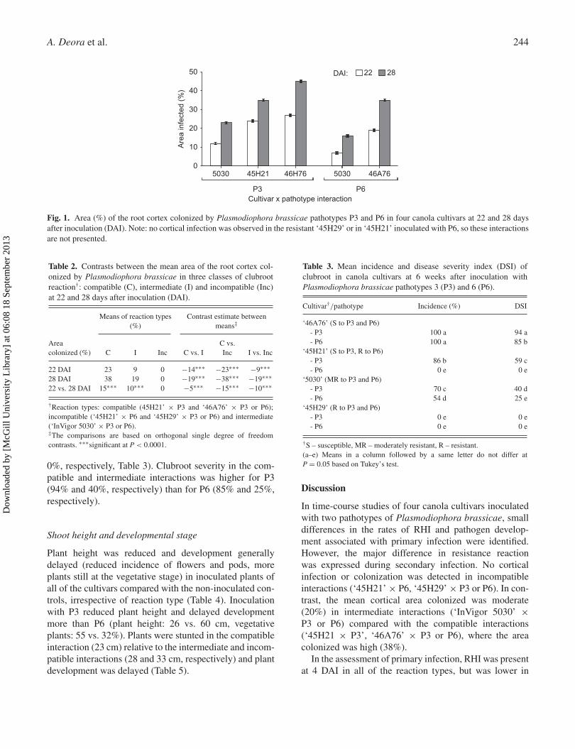
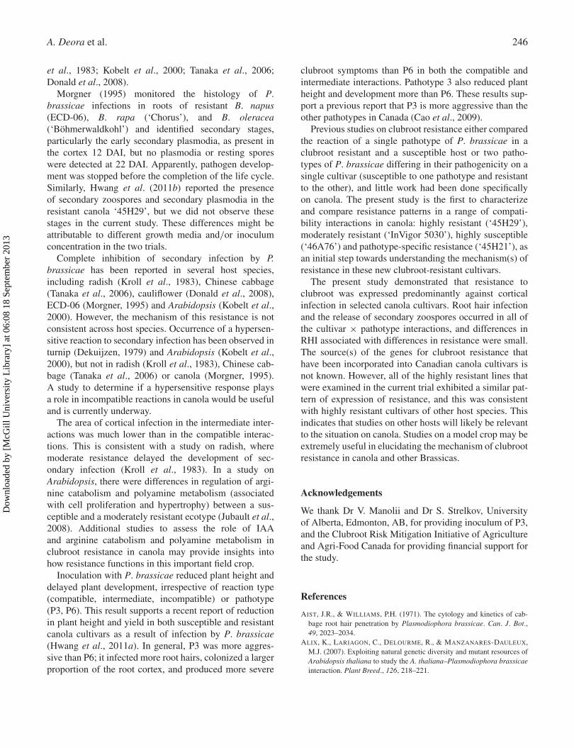
No cortical colonization was observed at 16 DAI in anyof the cultivar × pathotype interactions, so that sam-pling date was not included in the data analyses. Forthe remaining sampling dates, the impact of cultivar,pathotype, sampling date and their interactions were allsignificant for cortical colonization (Fig. 1). Therefore,the dataset was again grouped by clubroot reaction typefor further analyses. No infection or pathogen develop-ment was observed in the root cortex at any samplingdate in the incompatible interactions. At 22 DAI, col-onization by the pathogen was observed in the corticalcells in all of the compatible and intermediate interac-tions (Fig. 1). Roots of plants with intermediate resistancebore primarily vegetative plasmodia, while in the com-patible interactions, immature resting spores were alsoobserved. Disorganization of tissue structure resulting
Table 1. Contrasts between the mean incidence (%) of devel-opmental stages of Plasmodiophora brassicae during root hairinfection (RHI) for three classes of clubroot reaction†: compati-ble (C), intermediate (I) and incompatible (Inc), at 4 and 12 daysafter inoculation (DAI).
Means of reactiontypes (%)
Contrast estimates betweenmeans‡
Developmentalstages C I Inc C vs. I C vs. Inc I vs. Inc
Plasmodia4 DAI 43 35 33 −8∗∗∗ −10∗∗∗ −212 DAI 31 32 33 1 2∗ 14 vs. 12 DAI −12∗∗∗ −3 0 9∗∗∗ 12∗∗∗ 3
Sporangia4 DAI 10 5 4 −5∗∗∗ −6∗∗∗ −112 DAI 18 16 15 −2∗ −3∗ −14 vs. 12 DAI 8∗∗∗ 11∗∗∗ 11∗∗∗ 3∗ 3∗ 0
Dehisced sporangia4 DAI 6 3 3 −3∗ −3∗ 012 DAI 19 9 15 −10∗∗∗ −4∗ 6∗∗∗4 vs. 12 DAI 13∗∗∗ 6∗∗∗ 12∗∗∗ −7∗∗∗ −1 6∗∗∗
Total RHI4 DAI 59 43 40 −16∗∗∗ −19∗∗∗ −312 DAI 68 57 63 −11∗∗∗ −5∗ 6∗∗∗4 vs. 12 DAI 9∗∗∗ 14∗∗∗ 23∗∗∗ 5∗∗∗ 14∗∗∗ 9∗∗∗
†Reaction types: compatible (‘45H21’ × P3, ‘46A76’ × P3 or P6); incom-patible (‘45H21’ × P6, ‘45H29’ × P3 or P6) and intermediate (‘InVigor5030’ × P3 or P6).‡The comparisons are based on orthogonal single degree of freedomcontrasts. ∗,∗∗∗significant at P < 0.05 and 0.0001, respectively.
from abnormal cell division (hyperplasia) and enlarge-ment (hypertrophy) of cortical cells was observed in theseinteractions. At 28 DAI, most of the plasmodia had com-pleted development into resting spores in the intermediateand compatible interactions.
There was more cortical colonization in the compati-ble interaction than in the intermediate interaction at both22 and 28 DAI, and the extent of colonization increasedbetween the two sampling dates for both reaction types(Table 2). The mean cortical colonization in the compat-ible and intermediate interactions was slightly higher forP3 than for P6; 20% vs. 13% at 22 DAI and 34% vs. 25%at 28 DAI (Fig. 1).
Symptom development
The effects of cultivar, pathotype and their interactionon clubroot incidence and severity were all significant.Clubroot incidence was highest in the compatible inter-actions (mean = 95%), intermediate in the intermediateinteractions (mean = 62%) and there were no clubrootsymptoms in the incompatible interactions (mean = 0%).The pattern was similar for severity (mean = 79, 33 and
Dow
nloa
ded
by [
McG
ill U
nive
rsity
Lib
rary
] at
06:
08 1
8 Se
ptem
ber
2013
A. Deora et al. 244
5030 45H21
22DAI: 28
46H76 5030 46A76
50
40
30
20
10
0
Are
a in
fect
ed (
%)
Cultivar x pathotype interactionP3 P6
Fig. 1. Area (%) of the root cortex colonized by Plasmodiophora brassicae pathotypes P3 and P6 in four canola cultivars at 22 and 28 daysafter inoculation (DAI). Note: no cortical infection was observed in the resistant ‘45H29’ or in ‘45H21’ inoculated with P6, so these interactionsare not presented.
Table 2. Contrasts between the mean area of the root cortex col-onized by Plasmodiophora brassicae in three classes of clubrootreaction†: compatible (C), intermediate (I) and incompatible (Inc)at 22 and 28 days after inoculation (DAI).
Means of reaction types(%)
Contrast estimate betweenmeans‡
Areacolonized (%) C I Inc C vs. I
C vs.Inc I vs. Inc
22 DAI 23 9 0 −14∗∗∗ −23∗∗∗ −9∗∗∗28 DAI 38 19 0 −19∗∗∗ −38∗∗∗ −19∗∗∗22 vs. 28 DAI 15∗∗∗ 10∗∗∗ 0 −5∗∗∗ −15∗∗∗ −10∗∗∗
†Reaction types: compatible (45H21’ × P3 and ‘46A76’ × P3 or P6);incompatible (‘45H21’ × P6 and ‘45H29’ × P3 or P6) and intermediate(‘InVigor 5030’ × P3 or P6).‡The comparisons are based on orthogonal single degree of freedomcontrasts. ∗∗∗significant at P < 0.0001.
0%, respectively, Table 3). Clubroot severity in the com-patible and intermediate interactions was higher for P3(94% and 40%, respectively) than for P6 (85% and 25%,respectively).
Shoot height and developmental stage
Plant height was reduced and development generallydelayed (reduced incidence of flowers and pods, moreplants still at the vegetative stage) in inoculated plants ofall of the cultivars compared with the non-inoculated con-trols, irrespective of reaction type (Table 4). Inoculationwith P3 reduced plant height and delayed developmentmore than P6 (plant height: 26 vs. 60 cm, vegetativeplants: 55 vs. 32%). Plants were stunted in the compatibleinteraction (23 cm) relative to the intermediate and incom-patible interactions (28 and 33 cm, respectively) and plantdevelopment was delayed (Table 5).
Table 3. Mean incidence and disease severity index (DSI) ofclubroot in canola cultivars at 6 weeks after inoculation withPlasmodiophora brassicae pathotypes 3 (P3) and 6 (P6).
Cultivar†/pathotype Incidence (%) DSI
‘46A76’ (S to P3 and P6)- P3 100 a 94 a- P6 100 a 85 b
‘45H21’ (S to P3, R to P6)- P3 86 b 59 c- P6 0 e 0 e
‘5030’ (MR to P3 and P6)- P3 70 c 40 d- P6 54 d 25 e
‘45H29’ (R to P3 and P6)- P3 0 e 0 e- P6 0 e 0 e
†S – susceptible, MR – moderately resistant, R – resistant.(a–e) Means in a column followed by a same letter do not differ atP = 0.05 based on Tukey’s test.
Discussion
In time-course studies of four canola cultivars inoculatedwith two pathotypes of Plasmodiophora brassicae, smalldifferences in the rates of RHI and pathogen develop-ment associated with primary infection were identified.However, the major difference in resistance reactionwas expressed during secondary infection. No corticalinfection or colonization was detected in incompatibleinteractions (‘45H21’ × P6, ‘45H29’ × P3 or P6). In con-trast, the mean cortical area colonized was moderate(20%) in intermediate interactions (‘InVigor 5030’ ×P3 or P6) compared with the compatible interactions(‘45H21 × P3’, ‘46A76’ × P3 or P6), where the areacolonized was high (38%).
In the assessment of primary infection, RHI was presentat 4 DAI in all of the reaction types, but was lower in
Dow
nloa
ded
by [
McG
ill U
nive
rsity
Lib
rary
] at
06:
08 1
8 Se
ptem
ber
2013
Clubroot development in resistant/susceptible canola 245
Table 4. Effect of inoculation with Plasmodiophora brassicaepathotypes P3 and P6 (relative to a non-inoculated control) on plantheight and growth stage (% of plants) of four canola cultivars at6 weeks after inoculation, under controlled conditions.
Cultivar P3 P6 Control
Plant height (cm)‘45H21’ 25∗∗∗ 34∗ 38‘45H29 34∗ 32∗ 36‘InVigor 5030’ 26∗∗∗ 30∗∗∗ 35‘46A76’ 20∗∗∗ 23∗∗∗ 34Mean 26∗∗∗ 30∗∗∗ 36
Vegetative (%)‘45H21’ 81∗∗∗ 8 4‘45H29’ 8 8 0‘InVigor 5030’ 53∗∗∗ 43∗∗∗ 0‘46A76’ 78∗∗∗ 71∗∗∗ 4Mean 55∗∗∗ 32∗∗∗ 2
Buds (%)‘45H21’ 13∗ 10 5‘45H29’ 13∗ 7 4‘InVigor 5030’ 37∗∗∗ 34∗∗∗ 3‘46A76’ 14 19∗ 5Mean 19∗∗∗ 18∗∗∗ 4
Flowers (%)‘45H21’ 0∗ 29∗ 10‘45H29’ 34 31 28‘InVigor 5030’ 2∗∗∗ 8∗∗∗ 42‘46A76’ 0∗∗∗ 0∗∗∗ 31Mean 9∗∗∗ 17∗∗∗ 28
Pods (%)‘45H21’ 6∗∗∗ 53∗∗∗ 81‘45H29’ 45∗∗∗ 55∗ 68‘InVigor 5030’ 8∗∗∗ 15∗∗∗ 55‘46A76’ 8∗∗∗ 10∗∗∗ 60Mean 17∗∗∗ 33∗∗∗ 66
∗,∗∗∗Value differs from the non-inoculated control at P < 0.05 and 0.0001,respectively, based on a single df contrast.
Table 5. Contrasts between means plant height and growth stage(% of plants) among reaction types†: compatible (C), intermediate(I) and incompatible (Inc).
Means ofreaction types Contrast estimate between means‡
C I Inc C vs. I C vs. Inc I vs. Inc
Height (cm) 23 28 33 5∗∗∗ 10∗∗∗ 5∗∗∗Vegetative (%) 77 48 8 −29∗∗∗ −69∗∗∗ −40∗∗∗Buds (%) 15 36 10 21∗∗∗ −5∗ −26∗∗∗Flowers (%) 0 5 31 5 31∗∗∗ 26∗∗∗Pods (%) 8 11 51 3 43∗∗∗ 40∗∗∗
†Reaction types: compatible (‘45H21’ × P3 and ‘46A76’ × P3 or P6);incompatible (‘45H21’ × P6 and ‘45H29’ × P3 or P6) and intermediate(‘InVigor 5030’ × P3 or P6).‡The comparisons are based on orthogonal single degree of freedomcontrasts. ∗,∗∗∗significant at P < 0.05 and 0.0001, respectively.
the incompatible and intermediate interactions than in thecompatible interactions. However, total RHI in the incom-patible interactions increased over time, so that by 12 DAIit was similar (64%) to the compatible interaction (68%).A similar pattern was observed for individual develop-mental stages of the pathogen: development was initiallyslower in the incompatible and intermediate interactionsthan in the compatible interactions, but development pro-gressed slightly more quickly in the incompatible inter-action over time. This supports the results of previousstudies on canola (Hwang et al., 2011b) and Chinesecabbage (Tanaka et al., 2006), which showed that pri-mary infection developed later in resistant (‘45H29’) thansusceptible cultivars. In comparison, the RHI in interme-diate interactions remained lower (57%) at 12 DAI thanin the compatible or incompatible interactions. Similarto RHI, fewer (9%) sporangia dehisced in the intermedi-ate interactions. We conclude that, across several canolagenotypes and two pathotypes, RHI was slightly delayedin the incompatible interaction and slightly reduced in theintermediate interactions.
Even though differences in RHI in compatible andincompatible interactions were small, primary infectionmay play an important role in expression of resistance.In a recent study, high levels of cortical infection occurredin ryegrass inoculated with secondary zoospores of P.brassicae, but no cortical infection occurred in rye-grass where RHI occurred before exposure to secondaryzoospores (Feng et al., 2012). Although primary infec-tion is a non-specific phenomenon that can occur in bothhost species and in plants that do not develop any clubrootsymptoms (Ludwig-Müller et al., 1999), this indicatesthat primary infection may have a role in recognitionand induction of resistance. Another study showed thatRHI is related to the levels of indole acetic acid (IAA),a phytohormone that influences clubroot development.In cabbage, the level of IAA initially increased in suscep-tible cultivars and then declined, but continued to increaseeven after 14 DAI in resistant cultivars (Ludwig-Mülleret al., 1993).
In the present study, cortical infection was completelyinhibited in all of the incompatible interactions. Cultivar‘45H29’ was resistant to both P3 and P6 and ‘45H21’was resistant to only P6. This differential response topathotype indicates that the genetics of resistance toP3 and P6 differ in these two cultivars. However, the tim-ing of action of the resistance genes in these cultivarsappears to be similar, because the pathogen was com-pletely inhibited at the cortical infection stage. This indi-cates that high levels of resistance in canola are expressedin a manner similar to that observed in other hosts (Kroll
Dow
nloa
ded
by [
McG
ill U
nive
rsity
Lib
rary
] at
06:
08 1
8 Se
ptem
ber
2013
A. Deora et al. 246
et al., 1983; Kobelt et al., 2000; Tanaka et al., 2006;Donald et al., 2008).
Morgner (1995) monitored the histology of P.brassicae infections in roots of resistant B. napus(ECD-06), B. rapa (‘Chorus’), and B. oleracea(‘Böhmerwaldkohl’) and identified secondary stages,particularly the early secondary plasmodia, as present inthe cortex 12 DAI, but no plasmodia or resting sporeswere detected at 22 DAI. Apparently, pathogen develop-ment was stopped before the completion of the life cycle.Similarly, Hwang et al. (2011b) reported the presenceof secondary zoospores and secondary plasmodia in theresistant canola ‘45H29’, but we did not observe thesestages in the current study. These differences might beattributable to different growth media and/or inoculumconcentration in the two trials.
Complete inhibition of secondary infection by P.brassicae has been reported in several host species,including radish (Kroll et al., 1983), Chinese cabbage(Tanaka et al., 2006), cauliflower (Donald et al., 2008),ECD-06 (Morgner, 1995) and Arabidopsis (Kobelt et al.,2000). However, the mechanism of this resistance is notconsistent across host species. Occurrence of a hypersen-sitive reaction to secondary infection has been observed inturnip (Dekuijzen, 1979) and Arabidopsis (Kobelt et al.,2000), but not in radish (Kroll et al., 1983), Chinese cab-bage (Tanaka et al., 2006) or canola (Morgner, 1995).A study to determine if a hypersensitive response playsa role in incompatible reactions in canola would be usefuland is currently underway.
The area of cortical infection in the intermediate inter-actions was much lower than in the compatible interac-tions. This is consistent with a study on radish, wheremoderate resistance delayed the development of sec-ondary infection (Kroll et al., 1983). In a study onArabidopsis, there were differences in regulation of argi-nine catabolism and polyamine metabolism (associatedwith cell proliferation and hypertrophy) between a sus-ceptible and a moderately resistant ecotype (Jubault et al.,2008). Additional studies to assess the role of IAAand arginine catabolism and polyamine metabolism inclubroot resistance in canola may provide insights intohow resistance functions in this important field crop.
Inoculation with P. brassicae reduced plant height anddelayed plant development, irrespective of reaction type(compatible, intermediate, incompatible) or pathotype(P3, P6). This result supports a recent report of reductionin plant height and yield in both susceptible and resistantcanola cultivars as a result of infection by P. brassicae(Hwang et al., 2011a). In general, P3 was more aggres-sive than P6; it infected more root hairs, colonized a largerproportion of the root cortex, and produced more severe
clubroot symptoms than P6 in both the compatible andintermediate interactions. Pathotype 3 also reduced plantheight and development more than P6. These results sup-port a previous report that P3 is more aggressive than theother pathotypes in Canada (Cao et al., 2009).
Previous studies on clubroot resistance either comparedthe reaction of a single pathotype of P. brassicae in aclubroot resistant and a susceptible host or two patho-types of P. brassicae differing in their pathogenicity on asingle cultivar (susceptible to one pathotype and resistantto the other), and little work had been done specificallyon canola. The present study is the first to characterizeand compare resistance patterns in a range of compati-bility interactions in canola: highly resistant (‘45H29’),moderately resistant (‘InVigor 5030’), highly susceptible(‘46A76’) and pathotype-specific resistance (‘45H21’), asan initial step towards understanding the mechanism(s) ofresistance in these new clubroot-resistant cultivars.
The present study demonstrated that resistance toclubroot was expressed predominantly against corticalinfection in selected canola cultivars. Root hair infectionand the release of secondary zoospores occurred in all ofthe cultivar × pathotype interactions, and differences inRHI associated with differences in resistance were small.The source(s) of the genes for clubroot resistance thathave been incorporated into Canadian canola cultivars isnot known. However, all of the highly resistant lines thatwere examined in the current trial exhibited a similar pat-tern of expression of resistance, and this was consistentwith highly resistant cultivars of other host species. Thisindicates that studies on other hosts will likely be relevantto the situation on canola. Studies on a model crop may beextremely useful in elucidating the mechanism of clubrootresistance in canola and other Brassicas.
Acknowledgements
We thank Dr V. Manolii and Dr S. Strelkov, Universityof Alberta, Edmonton, AB, for providing inoculum of P3,and the Clubroot Risk Mitigation Initiative of Agricultureand Agri-Food Canada for providing financial support forthe study.
References
AIST, J.R., & WILLIAMS, P.H. (1971). The cytology and kinetics of cab-bage root hair penetration by Plasmodiophora brassicae. Can. J. Bot.,49, 2023–2034.
ALIX, K., LARIAGON, C., DELOURME, R., & MANZANARES-DAULEUX,M.J. (2007). Exploiting natural genetic diversity and mutant resources ofArabidopsis thaliana to study the A. thaliana–Plasmodiophora brassicaeinteraction. Plant Breed., 126, 218–221.
Dow
nloa
ded
by [
McG
ill U
nive
rsity
Lib
rary
] at
06:
08 1
8 Se
ptem
ber
2013
Clubroot development in resistant/susceptible canola 247
CAO, T., MANOLII, V.P., HWANG, S.F., HOWARD, R.J., & STRELKOV, S.E.(2009). Virulence and spread of Plasmodiophora brassicae [clubroot] inAlberta, Canada. Can. J. Plant Pathol., 31, 321–329.
DEKUIJZEN, H.M. (1979). Electron microscopic studies on the root hairsand cortex of a susceptible and a resistant variety of Brassica campestrisinfected with Plasmodiophora brassicae. Eur. J. Plant Pathol., 85, 1–17.
DIEDERICHSEN, E., FRAUEN, M., LINDERS, E.G.A., HATAKEYAMA, K., &HIRAI, M. (2009). Status and perspectives of clubroot resistance breedingin Crucifer crops. J. Plant Growth Regul., 28, 265–281.
DIXON, G.R. (2009). The occurrence and economical impact ofPlasmodiophora brassicae and clubroot disease. J. Plant Growth Regul.,28, 194–202.
DONALD, E.C., & PORTER, I.J. (2004). A sand-solution culture techniqueused to observe the effect of calcium and pH on root hair and corti-cal stages of infection by Plasmodiophora brassicae. Australas. PlantPathol., 33, 585–589.
DONALD, E.C., JAUDZEMS, E.C., & PORTER, I.J. (2008). Pathology ofcortical invasion by Plasmodiophora brassicae in clubroot resistant andsusceptible Brassica oleracea hosts. Plant Pathol., 57, 201–209.
FENG, J., XIAO, Q., HWANG, S.F., STRELKOV, S.E., & GOSSEN, B.D.(2012). Infection of canola by secondary zoospores of Plasmodiophorabrassicae produced on a nonhost (ryegrass). Eur. J. Plant Pathol. 132,309–315.
HARLING, R., & OXLEY, S. (2007). Clubroot disease of oilseed rapeand other Brassicae crops. Tech. Note Scott. Agr. Coll. (TN602). ISBN:1854828908.
HOWARD, R.J., STRELKOV, S.E., & HARDING, M.W. (2010). Clubroot ofcruciferous crops – new perspectives on an old disease. Can. J. PlantPathol., 32, 43–57.
HWANG, S.F., AHMED, H.U., STRELKOV, S.E., GOSSEN, B.D.,TURNBULL, G.D., PENG, G., & HOWARD, R.J. (2011a). Seedlingage and inoculum density affect clubroot severity and seed yield incanola. Can. J. Plant Sci., 91, 183–190.
HWANG, S.F., AHMED, H.U., ZHOU, Q., STRELKOV, S.E., GOSSEN, B.D.,PENG, G., & TURNBULL G.D. (2011b). Influence of cultivar resistanceand inoculum density on root hair infection of canola (Brassica napus)by Plasmodiophora brassicae. Plant Pathol., 60, 820–829.
HWANG, S.F., STRELKOV, S.E., GOSSEN, B.D., TURNBULL, G.D.,AHMED, H.U., & MANOLII, V.P. (2011c). Soil treatments and amend-ments for amelioration of clubroot on canola. Can. J Plant Sci., 91,999–1010.
INGRAM, D.S., & TOMMERUP, I.C. (1972). The life history ofPlasmodiophora brassicae Woron. Proc. R. Soc. Lond. [Biol], 180,103–112.
JUBAULT, M., HAMON, C., GRAVOT, A., LARIAGON, C., DELOURME,R., BOUCHEREAU, A., & MANZANARES-DAULEUX, M.J. (2008).Differential regulation of root arginine catabolism and polyaminemetabolism in clubroot-susceptible and partially resistant Arabidopsisgenotypes. Plant Physiol., 146, 2008–2019.
KOBELT, P., SIEMENS, J., & SACRISTÁN, M.D. (2000). Histological char-acterisation of the incompatible interaction between Arabidopsis thalianaand the obligate biotrophic pathogen Plasmodiophora brassicae. Mycol.Res., 104, 220–225.
KOBRINGER, K.M., & HAGEDORN, D.J. (1983). Determination of bean rootrot potential in vegetable production fields of Wisconsin’s Central Sands.Plant Dis., 67, 177–178.
KROLL, T.K., LACY, G.H., & MOORE, L.D. (1983). A quantitative descrip-tion of the colonization of susceptible and resistant radish plants byPlasmodiophora brassicae. J. Phytopathol., 108, 97–105.
KUGINUKI, Y., YOSHIKAWA, H., & HIRAI, M. (1999). Variation in viru-lence of Plasmodiophora brassicae in Japan tested with clubroot-resistantcultivars of Chinese cabbage (Brassica rapa L. ssp. pekinensis). Eur. J.Plant Pathol., 105, 327–332.
LUDWIG-MÜLLER, J., & SCHULLER, A. (2008). What can we learn fromclubroots: alterations in host roots and hormone homeostasis caused byPlasmodiophora brassicae. Eur. J. Plant Pathol., 121, 291–302.
LUDWIG-MÜLLER, J., BENDEL, U., THERMANN, P., RUPPEL, M.,EPSTEIN, E., & HILGENBERG, W. (1993). Concentrations of indole-3-acetic acid in plants of tolerant and susceptible varieties of Chinesecabbage infected with Plasmodiophora brassicae Woron. New Phytol.,125, 763–769.
LUDWIG-MÜLLER, J., BENNETT, R.N., KIDDLE, G., IHMIG, S., RÜPPEL,M., & HILGENBERG, W. (1999). The host range of Plasmodiophorabrassicae and its relationship to endogenous glucosinolate content. NewPhytol., 141, 443–458.
MANZANARES-DAULEUX, M.J., ROCHERIEUX, J., ALIX, K., GLORY, P.,GIBOULOT, A., LARIAGON, C., et al. (2006). Deciphering the hostmechanisms of resistance in the Brassicaceae/Plasmodiophora brassicaepathosystem. Acta Hort., 706, 317–322.
MORGNER, M. (1995). Mikroskopische Untersuchungen der Reaktionenempfindlicher und resistenter Genotypen verschiedener Brassica-Artengegenüber Plasmodiophora brassicae (Wor.). PhD dissertation, Institutfür Angewandte Genetik, Freie Universität Berlin, Germany.
OSAKI, K., TANAKA, S., & ITO, S. (2008). Pathogenicity ofPlasmodiophora brassicae populations from small, spheroid, resistant-type clubroot galls on roots of clubroot-resistant cultivars of Chinesecabbage (Brassica rapa L. subsp. pekinensis). J. Gen. Plant Pathol., 74,242–245.
PEST MANAGEMENT REGULATORY AGENCY (2008). RegistrationDecision: Fluazinam. Health Canada. ISBN: 978-1-100-10510-9 (978-1-100-10511-6).
SHARMA, K., GOSSEN, B.D., & MCDONALD M.R. (2011a). Effect of tem-perature on primary infection by Plasmodiophora brassicae and initiationof clubroot symptoms. Plant Pathol., 60, 830–838.
SHARMA, K., GOSSEN, B.D., & MCDONALD, M.R. (2011b). Effect of tem-perature on cortical infection by Plasmodiophora brassicae and clubrootseverity. Phytopathology, 101, 1424–1432.
STRELKOV, S.E., TEWARI, J.P., & SMITH-DEGENHARDT, E. (2006).Characterization of Plasmodiophora brassicae populations from Alberta,Canada. Can. J. Plant Pathol., 28, 467–474.
TANAKA, S., FUJIYAMA, S., SHIGEMORI, S., NAKAYAMA, A., ITO, S.,& KAMEYA, I.M. (1998). Pathogenesis of isolates of Plasmodiophorabrassicae from Japan. 1. Race and pathogenesis in clubroot resistantcultivars. Proc. Assoc. Plant Prot. Kyushu, 44, 15–19.
TANAKA, S., KOCHI, S., KUNITA, H., ITO, S. & KAMEYA-IWAKI,M. (1999). Biological mode of action of the fungicide, flusulfamide,against Plasmodiophora brassicae (clubroot). Eur. J. Plant Pathol., 105,577–584.
TANAKA, S., MIDO, H., & ITO, S. (2006). Colonization by two isolatesof Plasmodiophora brassicae with differing pathogenicity on a clubroot-resistant cultivar of Chinese cabbage (Brassica rapa L. subsp. pekinensis).J. Gen. Plant Pathol., 72, 205–209.
WORONIN, M.S. (1878). Plasmodiophora brassicae, the cause of cabbagehernia. Translated by C. Chupp (1934). Phytopathological Classic No. 4.St. Paul, MN: American Phytopathological Society.
XUE, S., CAO, T, HOWARD, R.J., HWANG, S.F., & STRELKOV, S.E.(2008). Isolation and variation in virulence of single-spore isolates ofPlasmodiophora brassicae from Canada. Plant Dis., 92, 456–462.
Dow
nloa
ded
by [
McG
ill U
nive
rsity
Lib
rary
] at
06:
08 1
8 Se
ptem
ber
2013