increased fatty acid uptake and altered fatty acid...
TRANSCRIPT
Increased Fatty Acid Uptake and Altered Fatty AcidMetabolism in Insulin-Resistant Muscle of ObeseZucker RatsLorraine Patricia Turcotte, Jason Richard Swenberger, Michelle Zavitz Tucker, and Alice Jane Yee
Altered muscle fatty acid (FA) metabolism may contrib-ute to the presence of muscle insulin resistance in thegenetically obese Zucker rat. To determine whether FAuptake and disposal are altered in insulin-resistantmuscle, we measured palmitate uptake, oxidation, andincorporation into di- and triglycerides in isolated rathindquarters, as well as muscle plasma membrane fattyacid–binding protein (FABPPM) content of lean (n 5 16,fa/1) and obese (n 5 15, fa/fa) Zucker rats (12 weeksof age). Hindquarters were perfused with 7 mmol/lglucose, 1,000 mmol/l albumin-bound palmitate, and al-bumin-bound [1-14C]palmitate at rest (no insulin). Glu-cose uptake was 42% lower in the obese than in the leanrats and indicated the presence of muscle insulin resis-tance. Fractional and total rates of palmitate uptakewere 42 and 74% higher in the obese than in the leanrats and were associated with higher muscle FABPPM
content (r2 5 0.69, P < 0.05). The percentage of palmi-tate oxidized was not significantly different betweengroups. FA disposal to storage was altered according tofiber type. When compared with lean rats, the rate oftriglyceride synthesis in red muscle was 158% higher inobese rats, and the rate of palmitate incorporation intodiglycerides in white muscle was 93% higher in obeserats. Pre- and postperfusion muscle triglyceride levelswere higher in both red and white muscles of the obeserats. These results show that increased FA uptake andaltered FA disposal to storage may contribute to thedevelopment of muscle insulin resistance in obeseZucker rats. Diabetes 50:1389–1396, 2001
Insulin-resistant states, such as obesity, are charac-terized by hyperlipidemia and elevated triglyceridestores (1,2). Whereas hyperlipidemia and elevatedtriglyceride stores may be caused in part by an
oversupply of fatty acids (FAs) released from adiposetissue (3), it could also be caused by an alteration in thedisposal of FA, especially in muscle tissue. As suggestedby the presence of an inverse relationship between insulinsensitivity and triglyceride content in muscle, an alteration
in the inherent capacity of the muscle to take up anddispose of an FA load could be critical to the developmentof muscle insulin resistance (4,5). However, studiesinvestigating the direct effects of insulin resistance onFA metabolism in muscle are scarce, and results areequivocal.
The genetically obese Zucker rat has been used exten-sively to study the effects of insulin resistance on musclemetabolism because it exhibits many of the pathophysio-logical alterations observed in obese humans, namelysevere obesity, hypertriglyceridemia, hyperinsulinemia,and chronic muscle insulin resistance (1–3). Althoughdefects in glucose metabolism have been well documentedin this model of insulin resistance, changes in FA metab-olism have only been inferred from changes in carbohy-drate metabolism, thus leading to equivocal conclusions(2,6,7). Some researchers have suggested that muscle FAoxidation was increased (2,7) in obese Zucker rats,whereas others have suggested that muscle FA oxidationwas decreased (6). Therefore, it is unclear whether muscleFA oxidation is altered with the presence of muscle insulinresistance. Furthermore, because FA kinetics were notmeasured directly in those studies, it was not possible todetermine whether the measured changes in FA utilizationwere caused by alterations in FA uptake, cellular FAdisposal, or both.
It has recently become evident that alterations in FAuptake could be of primary importance in the regulation ofFA utilization in muscle (8–10). Indeed, evidence suggeststhat at least part of the uptake of FA in muscle may becarrier-mediated and that fatty acid transporter proteinslocated in the plasma membranes are an integral compo-nent of this transport system (11,12). Thus, with thissystem, control of FA uptake would be possible at thetransport step, and the content of FA-transporter proteinsat the plasma membrane would be critical. Fatty acid–binding protein (FABPPM) is among the several proteinsthat have been identified as putative FA-transport proteins(11–13). Whereas the specific role of FABPPM in a putativetrans-sarcolemmal transport process has not been clearlyidentified, the protein has been shown to be present inmuscle, and its expression in muscle has been shown to bemodified by exposure to physiological stimuli associatedwith changes in FA utilization (13–17). Therefore, if mus-cle FA uptake is altered by the presence of muscle insulinresistance, this could be associated with concomitantchanges in the content of FABPPM. Conversely, the pres-ence of insulin resistance could be caused in part by
From the Department of Kinesiology and University of Southern CaliforniaDiabetes Center, University of Southern California, Los Angeles, Califonia.
Address correspondence and reprint requests to Lorraine P. Turcotte,Ph.D., Department of Kinesiology, University of Southern California, 3560Watt Way, PED 107, Los Angeles, CA 90089. E-mail: [email protected].
Received for publication 21 September 2000 and accepted in revised form 5March 2001.
CM, crude membrane; DGAT, diacylglycerol acyltransferase; FA, fatty acid;FABPPM, fatty acid–binding protein; FAT, fatty acid transporter; FFA, freefatty acid; GPAT, glycerol-3-phosphate acyltransferase; PM, plasma mem-brane; TBST, Tris-buffered saline with Tween.
DIABETES, VOL. 50, JUNE 2001 1389
metabolic impairments in the biosynthetic and oxidativepathways of FA disposal.
Thus, the purpose of this study was to determinewhether basal FA metabolism is impaired in insulin-resistant muscle by measuring palmitate uptake and dis-posal in the perfused hindlimbs of obese and lean Zuckerrats. The ability of the muscle to take up FA was alsoassessed by measuring FABPPM content. FA disposal wasassessed by the measurement of palmitate oxidation andincorporation into muscle di- and triglycerides.
RESEARCH DESIGN AND METHODS
Animal preparation. Lean (fa/1, n 5 16) and obese (fa/fa, n 5 15) femaleZucker rats (12 weeks of age) were housed in pairs, were maintained on a12:12-h light-dark cycle, and received regular rat diet and water ad libitum. Theobese rats were significantly heavier than the lean rats (407.5 6 7.2 vs. 207.5 65.5 g, respectively, P , 0.05).Hindquarter perfusion. A total of 10 lean and 9 obese rats were anesthetizedwith ketamine/Rompun (80 and 12 mg/kg body wt, respectively) and preparedsurgically for hindquarter perfusion as previously described (16,18). Beforeinsertion of the perfusion catheters, heparin (150 IU) was administered intothe inferior vena cava. The rats were killed with an intracardial injection ofketamine/Rompun immediately before the catheters were inserted, and thepreparation was placed in a perfusion apparatus, essentially as described byRuderman and colleagues (16,18).
The initial perfusate (200 ml) consisted of Krebs-Henseleit solution, 1- to2-day-old washed bovine erythrocytes (30% hematocrit), 5% bovine serumalbumin (Cohn fraction V; Sigma, St. Louis, MO), 7 mmol/l glucose, 1,000mmol/l albumin-bound palmitate, and 5 mCi of albumin-bound [1-14C] palmi-tate (ICN Pharmaceuticals, Costa Mesa, CA). This concentration of palmitatewas chosen because it is within the physiological range for Zucker rats (3). Tominimize the influence of confounding factors related to the presence ofinsulin and to allow us to make conclusions about inherent alterations causedby the presence of muscle insulin resistance, we chose to perfuse the hind-quarters without insulin. The perfusate (37°C) was continuously gassed witha mixture of 95% O2/5% CO2, which yielded arterial pH values of 7.3–7.4 andarterial PCO2 and PO2 values of typically 37–39 and 170–180 Torr, respectively,in both the lean and obese rats. Mean perfusion pressures were 121 6 7 and129 6 8 mmHg during unilateral hindquarter perfusion in the lean and obeserats, respectively.
The first 25 ml of perfusate that passed through the hindquarter werediscarded, and then the perfusate was recirculated at a flow of 10 ml/min (0.55and 0.59 ml z min21 z g21 perfused muscle in lean and obese rats, respectively).After an equilibration period of 20 min, the left superficial fast-twitch white(predominantly type IIb) sections and the deep fast-twitch red (predominantlytype IIa) sections of the gastrocnemius muscles as well as the plantaris musclewere taken out and freeze-clamped with aluminum clamps precooled in liquidN2. The left iliac vessels were then tied off, and a clamp was fixed tightlyaround the proximal part of the leg to prevent bleeding. The right leg was thenperfused at rest for 40 min at a perfusate flow of 5 ml/min (0.27 and 0.29 ml zmin21 z g21 perfused muscle in lean and obese rats, respectively). Arterial andvenous perfusate samples for the analysis of [14C]–free fatty acid (FFA) and14CO2 radioactivities were taken after 20, 30, and 40 min of perfusion. Arterialand venous perfusate samples for PCO2, PO2, pH, and hemoglobin determina-tions were taken after 15 and 30 min of perfusion. At the end of the 40-minperfusion period, muscle samples from the right leg of the animal were takenand treated as previously described. The exact muscle mass perfused wasdetermined by infusing a colored solution of methyl blue into the arterialcatheter and weighing the colored muscle mass at the end of the perfusions(16). The red and white gastrocnemius muscles were used for di- andtriglyceride analyses, and the plantaris muscle was used for FABPPM contentanalysis. The analysis of FABPPM content in plantaris muscle could only beperformed in crude membrane fractions because of the small muscle samplesize. However, this was done so that FABPPM content could be correlated withFA uptake data in muscles from the same hindquarter.FABPPM content. A total of six lean and six obese rats were anesthetizedwith ketamine/Rompun, decapitated, and the white and red portions of thegastrocnemius muscles of both legs immediately removed and trimmed of fatand connective tissues. Plasma membrane fractions were prepared fresh aspreviously described (15,16,19). Briefly, the muscles were minced thoroughlywith scissors, diluted fourfold in Tris-15% sucrose buffer with 0.1 mmol/lphenylmethylsulfonyl fluoride, 10 mmol/l ethylene glycol-bis(b-aminoethyl-ether)-N,N,N9,N9-tetraacetic acid, and 10 mg/ml trypsin inhibitor made freshdaily (pH 7.5), and homogenized by one 10-s burst with a Polytron homoge-
nizer on level 6. The homogenates were filtered through a multi-filter system,and the filtered homogenates were centrifuged at 100,000g for 1 h. The pelletswere resuspended in Tris-15% sucrose buffer and a small aliquot of theresulting suspension, termed the “crude-membrane (CM) fractions,” wasretained for analysis. The remaining suspension of CM fractions was layeredonto continuous sucrose gradients (35–70%), which were centrifuged at120,000g for 2 h using a Beckman SW28 swing-out rotor. The plasma mem-brane (PM) layers were harvested, washed in Tris buffer, and centrifuged at100,000g for 1 h. The pellets were reconstituted in a small volume of Trisbuffer and frozen in liquid nitrogen for analysis. To assess the purity of the PMpreparations, and the protein content and activity of the mitochondrialmembrane and PM marker enzyme, succinate dehydrogenase (20) and 59-nucleotidase (21), respectively, were measured and compared among the CMand PM fractions. Protein content was measured with the commerciallyavailable Bio-Rad (Richmond, CA) microassay procedure. The PM fractionsisolated from the red and white gastrocnemius muscles as well as the CMfractions isolated from the plantaris muscles were analyzed for FABPPM
content by Western blotting.Blood and muscle sample analysis. Arterial and venous perfusate sampleswere analyzed for glucose, lactate, glycerol, and FFA concentrations as well asfor [14C]-FFA and 14CO2 radioactivities (16). Samples for glucose and lactatewere kept on ice and analyzed using YSI glucose and lactate analyzers (YellowSprings Instruments, Yellow Springs, OH). Samples for FFA and glycerol wereput in 200 mmol/l ethylene glycol-bis(b-aminoethyl ether)-N,N,N9,N9-tetraace-tic acid (pH 7) and centrifuged, and the supernatant was frozen until analyzedspectrophotometrically using the WAKO NEFA-C test (Biochemical Diagnos-tics, Edgewood, NY) and the enzymatic glycerol kinase method (Sigma),respectively. Because the FFA concentration was low in the absence of addedpalmitate (,80 mmol/l) and because palmitate was the only FA added,measured FFA concentrations were taken to equal palmitate concentrations.
To determine plasma palmitate radioactivity, duplicate 100-ml aliquots ofthe perfusate plasma were mixed with liquid scintillation fluid (BudgetSolve,Research Product International, Mount Prospect, IL) and counted in a Tri-carbliquid scintillation counter (model 4000 CA; United Technologies Packard,Downers Grove, IL), as previously described (16). The liberation and collec-tion of 14CO2 from the blood were performed within 4–5 min of anaerobiccollection (2 ml) as previously described (16). Perfusate samples for thedetermination of PCO2, PO2, pH, and hemoglobin were collected anaerobically,placed on ice, and measured within 5 min of collection by an ABL5 acid-baselaboratory (Radiometer America, Westlake, OH) and spectrophotometricallywith Drabkin’s reagent (Sigma, St. Louis, MO), respectively.
Muscle triglyceride concentration was determined as glycerol residuesafter extraction and separation of the muscle samples as previously described(16). Briefly, lipids were extracted from powdered muscle samples bycentrifugation at 1,000g in 2:1 chloroform:methanol solution and 4 mmol/lmagnesium chloride. The organic extract was evaporated and reconstituted inchloroform, and silicic acid was added for the removal of phospholipids bycentrifugation. The resulting supernatant was evaporated, saponified in eth-anolic potassium hydroxide for 30 min at 70°C, and centrifuged with 0.15 mol/lmagnesium sulfate. The final supernatant was analyzed for glycerol spectro-photometrically by the enzymatic glycerol kinase method (Sigma). To mea-sure the incorporation of [14C]palmitate into muscle di- and triglycerides,lipids from the extracted organic layer were separated by liquid chromatog-raphy as previously described (16).
For Western blot analysis, solubilized PM proteins (30 mg) or CM proteins(10 mg) were separated by SDS-PAGE on a 12% resolving gel and transferredelectrophoretically to a polyvinylidene difluoride membrane (15,16). Themembrane was blocked in 1% bovine serum albumin in Tris-buffered salinewith Tween (TBST) (500 mmol/l NaCl, 20 mmol/l Tris, 0.05% Tween-20, pH 7.5)for 2 h (23°C), rinsed twice with TBST, and incubated at 4°C overnight with apolyclonal antibody to the rat hepatic FABPPM (1:2,000) developed in our lab(8). After further washing with TBST, the membrane was incubated with125I-goat anti-rabbit IgG for 2 h at 23°C. The membrane was then washed anddried, and the density of the bands were quantified using a phosphorImager(Molecular Dynamics, Sunnyvale, CA). A rat liver CM preparation was used asstandard, and results were expressed as relative density units. To determinewhether there were any differences in FABPPM protein content betweengroups, we measured and compared band density from PM fractions of eitherred or white skeletal muscles isolated from lean and obese rats. In all cases,multiple gels were analyzed.Calculations and statistics. Fractional uptake was calculated as the differ-ence in radioactivity between the arterial and venous perfusate samplesdivided by the radioactivity in the arterial sample (16). Palmitate delivery wascalculated by multiplying the perfusate plasma flow by the arterial perfusateplasma palmitate concentration. Palmitate uptake was calculated by multiply-ing the plasma palmitate delivery by the fractional uptake (16). Percent
FATTY ACID METABOLISM AND INSULIN RESISTANCE
1390 DIABETES, VOL. 50, JUNE 2001
palmitate oxidation was calculated by dividing the total amount of radioac-tivity recovered as 14CO2 by the total amount of radioactivity that was takenup by the muscles (16). Total palmitate oxidation was calculated by multiply-ing palmitate uptake by the percent oxidation. Both percent and totalpalmitate oxidation were corrected for label fixation by using the acetatecorrection factor of 1.9. This correction factor was estimated from hindquar-ter perfusions with [1-14C]acetate (n 5 4) and found to be similar to thecorrection factors estimated by our study and others (16,22). Uptake andrelease of substrates and uptake of oxygen across the hindquarter werecalculated by multiplying perfusate flow by the arteriovenous difference inconcentration and were expressed per gram of perfused muscle, which wasmeasured to be 8.7 6 0.8 and 4.2 6 0.7% (18.3 6 0.5 vs. 16.7 6 0.4 g, respec-tively, P . 0.05) of body weight for unilateral hindquarter perfusion in leanand obese rats, respectively. Palmitate accumulation into muscle di- or tri-glycerides was calculated as the radioactivity accumulated in each lipidfraction and was expressed per milligram of wet muscle weight. Triglyceridesynthesis rate was calculated as the amount of [14C]palmitate incorporatedinto the triglyceride fraction divided by the specific activity of the perfusateplasma [14C]palmitate and corrected for time (23). The arterial- and venous-specific activities for palmitate did not vary over time and were not signifi-cantly different between the lean (52.9 6 6.8 and 50.1 6 6.1 mCi/mmol,respectively, P . 0.05) and obese (50.3 6 2.8 and 44.9 6 2.4 mCi/mmol,respectively, P . 0.05) rats. Total muscle triglyceride was calculated byestimating the contributions of red and white muscles to the perfused musclemass (24). Statistical evaluation of the data was done using analysis ofvariance with Newman-Keul’s test for post hoc multiple comparisons, whenappropriate. Pearson product-moment correlations were computed whenapplicable. In all instances, an a of 0.05 was used to determine significance.
RESULTS
Palmitate metabolism. As dictated by the protocol,perfusate palmitate concentration and delivery to thehindquarter did not vary over time and were not signifi-cantly different between the lean and obese groups(1,031.9 6 43.4 mmol/l and 186.9 6 10.9 nmol z min21 z g21
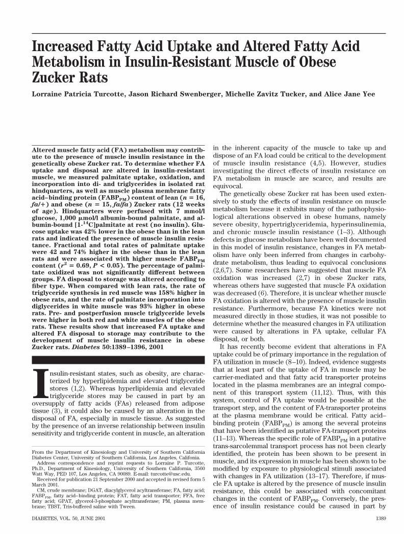
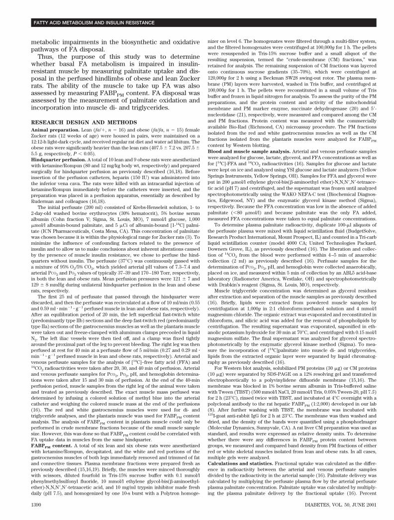
vs. 1,103.7 6 27.3 mmol/l and 211.2 6 5.9 nmol z min21 zg21, respectively, P . 0.05). The fractional and totaluptake of palmitate did not vary during 40 min of perfusionand were 42 and 74% higher (P , 0.05) in the obese(0.067 6 0.005 and 14.7 6 1.1 nmol z min21 z g21, respec-tively) than in the lean (0.047 6 0.004 and 8.5 6 0.6 nmolz min21 z g21, respectively) group, respectively (Fig. 1).Whereas the percentage of palmitate oxidized was notsignificantly different between groups (28.1 6 3.9 and32.6 6 6.0% for the lean and obese groups, respectively,P . 0.05), the total rate of palmitate oxidized was 65%higher in the obese than in the lean group (4.3 6 0.8vs. 2.6 6 0.5 nmol z min21 z g21, respectively, P , 0.05)(Fig. 1).Substrate exchange across the hindquarter. Restingoxygen uptake did not vary over time and was not signif-icantly different between the lean and obese groups(18.5 6 1.4 and 23.9 6 1.9 mmol z g21 z h21, respectively,P . 0.05). Arterial perfusate glucose concentrations didnot vary significantly over time and were not significantlydifferent between the lean and obese groups (7.0 6 0.2 and7.0 6 0.1 mmol/l, respectively, P . 0.05). Glucose uptakedid not change significantly over time but was found to be40% lower in the obese than in the lean group (6.2 6 0.7 vs.10.3 6 0.9 mmol z g21 z h21, respectively, P , 0.05) (Fig.2A). Arterial perfusate lactate concentration was on aver-age 23–28% higher in the obese than in the lean group andincreased by 32–37% during 40 min of perfusion in both thelean (1.09 6 0.04 to 1.49 6 0.08 mmol/l, P , 0.05) andobese (1.39 6 0.12 to 1.83 6 0.14 mmol/l, P , 0.05) groups.Lactate release decreased by 26–31% during 40 min ofperfusion in both the lean (5.7 6 1.3 to 4.2 6 0.8 mmol z g21
z h21) and obese (8.8 6 0.9 to 6.1 6 0.8 mmol z g21 z h21)groups and was 38–60% higher in the obese than in thelean group (Fig. 2B). Arterial perfusate glycerol concen-tration was 71–92% higher in the obese than in the leangroup and increased by 37–43% over time in both the lean(94.0 6 23.1 to 129.3 6 26.2 mmol/l, P , 0.05) and obese(171.5 6 42.1 to 244.4 6 29.2 mmol/l, P , 0.05) groups.Glycerol release did not change significantly over time ineither group but was 129–358% higher in the obese than inthe lean group (0.99 6 0.14 vs. 0.30 6 0.09 mmol z g21 z h21,respectively, P , 0.05) (Fig. 2C).Western blot analysis. As previously shown, the PMfractions isolated from the red and white skeletal musclesin both the lean and obese rats were enriched by 10- to12-fold in the specific activity of 59-nucleotidase relative totheir respective CM fractions (10,20) (Table 1). For eachmuscle group, protein yield and 59-nucleotidase activitywere not significantly different between the obese and leangroups. 59 Nucleotidase activity was found to be higher inthe red than in the white muscle in both the lean and obeserats. However, this did not affect the more importantcomparisons between the lean and obese groups. Succi-nate dehydrogenase activity in the PM fractions was notdetectable, thus indicating that contamination from mito-chondrial membrane proteins was negligible. These re-sults are consistent with those of previous reports usingsimilar PM-isolation procedures (3,38) and demonstrate
FIG. 1. Palmitate uptake (A) and oxidation (B) in perfused rathindquarters of lean and obese Zucker rats. Data are means 6 SE of10 lean and 9 obese Zucker rats. Because there were no significantchanges in values measured after 20, 30, and 40 min of perfusion,average values were used for each rat. M, lean group; ■, obese group.*P < 0.05 compared with the lean group.
L.P. TURCOTTE AND ASSOCIATES
DIABETES, VOL. 50, JUNE 2001 1391
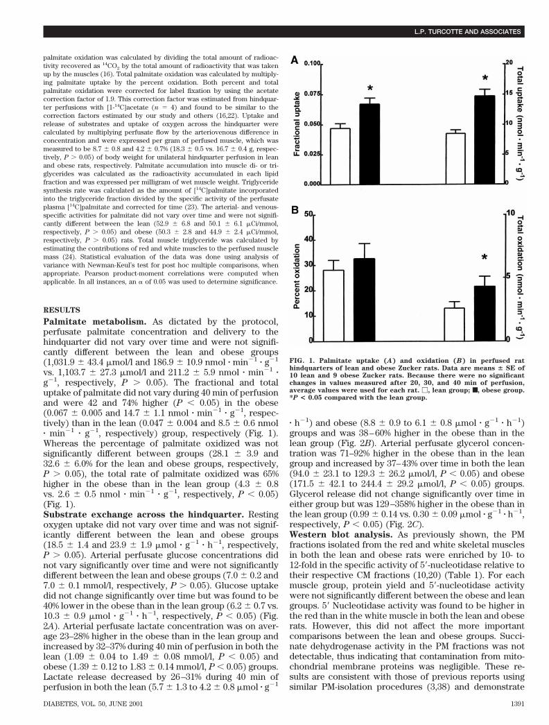
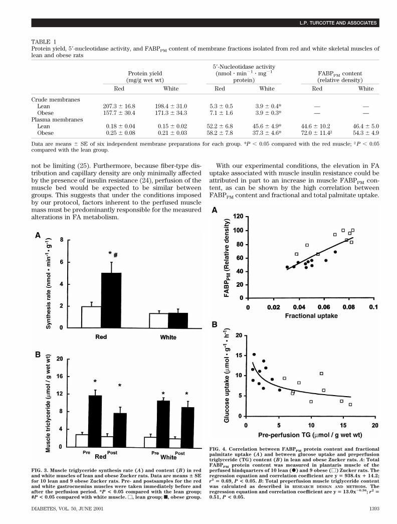
the integrity of our PM preparation. Scanning densitome-try of multiple gels revealed that compared with the leangroup, FABPPM content in the obese group was signifi-cantly higher by 61 6 5% in red skeletal muscle but notdifferent (17 6 3%, P . 0.05) in white skeletal muscle(Table 1).Muscle metabolites. Pre- and postperfusion triglycerideconcentrations were found to be higher in the obese than
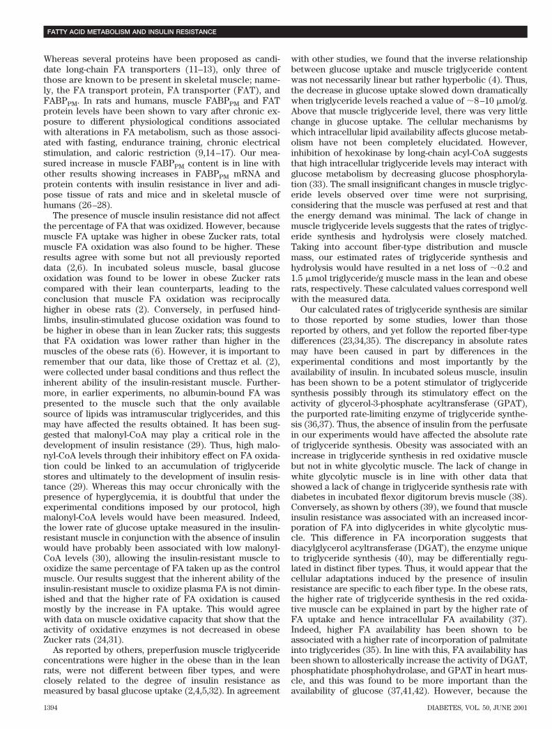
in the lean group in both the red and white gastrocnemiusmuscles (Fig. 3B). In both groups of rats, there were nosignificant decreases in the triglyceride concentration overtime in both the red and white gastrocnemius muscles.Both fiber type and obesity affected the triglyceride syn-thesis rate (Fig. 3A). In the obese group, the rate of tri-glyceride synthesis was 2.6-fold higher in the red than inthe white gastrocnemius muscle, whereas no differencewas found between fiber types in the lean group (Fig. 3A).In the red gastrocnemius muscle, the rate of triglyceridesynthesis was 158% higher in the obese than in the leangroup. In the white gastrocnemius muscle, the rate ofpalmitate incorporation into diglycerides was 93% higherin the obese than in the lean group (2.7 6 0.3 vs. 1.4 6 0.3dpm/mg, P , 0.05), whereas no difference was foundbetween fiber types.
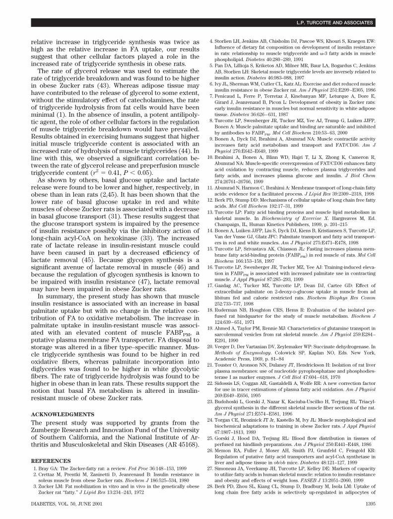
FABPPM content in plantaris muscle correlated posi-tively with fractional (y 5 938.4x 1 14.2; r2 5 0.69, P ,0.05) (Fig. 4A) and total (y 5 3.7x 1 24.8; r2 5 0.69, P ,0.05) palmitate uptake. The relationships between hind-quarter glucose uptake and preperfusion muscle triglycer-ide concentration in both red (y 5 12.9x20.34; r2 5 0.45,P , 0.05) and white (y 5 10.5x20.24; r2 5 0.34, P , 0.05)gastrocnemius muscles as well as total preperfusion tri-glyceride concentration (y 5 13.0x20.36; r2 5 0.51, P ,0.05) (Fig. 4B) were exponential and found to be signifi-cant. Linear correlations between these variables werealso found to be significant but generally lower (r2 5 0.43,0.37, and 0.49, respectively). Glycerol release was posi-tively correlated with total preperfusion triglyceride con-tent (y 5 7.2x 1 1.6; r2 5 0.41, P , 0.05).
DISCUSSION
Our results show that the presence of muscle insulinresistance in obese Zucker rats was associated with alter-ations in muscle FA metabolism as evidenced by anincrease in FA uptake and a change in cellular FA disposal.Muscle insulin resistance was associated with an increasein fractional and total palmitate uptake, and this wasassociated with an increase in the content of muscleFABPPM. In the absence of insulin, the relative distributionof FA disposal to oxidation was not changed by thepresence of muscle insulin resistance. However, underthose conditions, the distribution of FA to storage wasmodified according to fiber type. Thus, the rate of palmi-tate incorporation into triglycerides was increased in redmuscle, and that of diglycerides was increased in whitemuscle. Muscle insulin resistance was associated withhigher preperfusion muscle triglyceride levels in both redand white muscles, and this was associated with anincreased rate of triglyceride hydrolysis during the perfu-sion. These results show that, even in the absence ofinsulin, insulin-resistant muscle demonstrates an in-creased ability to take up FA from plasma and an altereddisposal of FA that is fiber-type specific.
With the use of the hindlimb-perfusion system, plasmaFA availability, blood flow, and capillary density are allfactors that could have some impact on changes in muscleFA metabolism. In this experiment, plasma FA availabilityand blood flow were not different between groups. Thus,the calculated rate of plasma FA delivery to the musclewas not different between groups and was high enough to
FIG. 2. Glucose uptake (A), lactate release (B), and glycerol release(C) in perfused rat hindquarters of lean and obese Zucker rats. Dataare means 6 SE of 10 lean and 9 obese Zucker rats. F, lean group; M,obese group. *P < 0.05 compared with the lean group; #P < 0.05represents a time effect.
FATTY ACID METABOLISM AND INSULIN RESISTANCE
1392 DIABETES, VOL. 50, JUNE 2001
not be limiting (25). Furthermore, because fiber-type dis-tribution and capillary density are only minimally affectedby the presence of insulin resistance (24), perfusion of themuscle bed would be expected to be similar betweengroups. This suggests that under the conditions imposedby our protocol, factors inherent to the perfused musclemass must be predominantly responsible for the measuredalterations in FA metabolism.
With our experimental conditions, the elevation in FAuptake associated with muscle insulin resistance could beattributed in part to an increase in muscle FABPPM con-tent, as can be shown by the high correlation betweenFABPPM content and fractional and total palmitate uptake.
FIG. 3. Muscle triglyceride synthesis rate (A) and content (B) in redand white muscles of lean and obese Zucker rats. Data are means 6 SEfor 10 lean and 9 obese Zucker rats. Pre- and postsamples for the redand white gastrocnemius muscles were taken immediately before andafter the perfusion period. *P < 0.05 compared with the lean group;#P < 0.05 compared with white muscle. M, lean group; f, obese group.
FIG. 4. Correlation between FABPPM protein content and fractionalpalmitate uptake (A) and between glucose uptake and preperfusiontriglyceride (TG) content (B) in lean and obese Zucker rats. A: TotalFABPPM protein content was measured in plantaris muscle of theperfused hindquarters of 10 lean (F) and 9 obese (M) Zucker rats. Theregression equation and correlation coefficient are y 5 938.4x 1 14.2;r2 5 0.69, P < 0.05. B: Total preperfusion muscle triglyceride contentwas calculated as described in RESEARCH DESIGN AND METHODS. Theregression equation and correlation coefficient are y 5 13.0x20.36; r2 50.51, P < 0.05.
TABLE 1Protein yield, 59-nucleotidase activity, and FABPPM content of membrane fractions isolated from red and white skeletal muscles oflean and obese rats
Protein yield(mg/g wet wt)
59-Nucleotidase activity(nmol z min21 z mg21
protein)FABPPM content(relative density)
Red White Red White Red White
Crude membranesLean 207.3 6 16.8 198.4 6 31.0 5.3 6 0.5 3.9 6 0.4* — —Obese 157.7 6 30.4 171.3 6 34.3 7.1 6 1.6 3.9 6 0.3* — —
Plasma membranesLean 0.18 6 0.04 0.15 6 0.02 52.2 6 6.8 45.6 6 4.9* 44.6 6 10.2 46.4 6 5.0Obese 0.25 6 0.08 0.21 6 0.03 58.2 6 7.8 37.3 6 4.6* 72.0 6 11.4† 54.3 6 4.9
Data are means 6 SE of six independent membrane preparations for each group. *P , 0.05 compared with the red muscle; †P , 0.05compared with the lean group.
L.P. TURCOTTE AND ASSOCIATES
DIABETES, VOL. 50, JUNE 2001 1393
Whereas several proteins have been proposed as candi-date long-chain FA transporters (11–13), only three ofthose are known to be present in skeletal muscle; name-ly, the FA transport protein, FA transporter (FAT), andFABPPM. In rats and humans, muscle FABPPM and FATprotein levels have been shown to vary after chronic ex-posure to different physiological conditions associatedwith alterations in FA metabolism, such as those associ-ated with fasting, endurance training, chronic electricalstimulation, and caloric restriction (9,14–17). Our mea-sured increase in muscle FABPPM content is in line withother results showing increases in FABPPM mRNA andprotein contents with insulin resistance in liver and adi-pose tissue of rats and mice and in skeletal muscle ofhumans (26–28).
The presence of muscle insulin resistance did not affectthe percentage of FA that was oxidized. However, becausemuscle FA uptake was higher in obese Zucker rats, totalmuscle FA oxidation was also found to be higher. Theseresults agree with some but not all previously reporteddata (2,6). In incubated soleus muscle, basal glucoseoxidation was found to be lower in obese Zucker ratscompared with their lean counterparts, leading to theconclusion that muscle FA oxidation was reciprocallyhigher in obese rats (2). Conversely, in perfused hind-limbs, insulin-stimulated glucose oxidation was found tobe higher in obese than in lean Zucker rats; this suggeststhat FA oxidation was lower rather than higher in themuscles of the obese rats (6). However, it is important toremember that our data, like those of Crettaz et al. (2),were collected under basal conditions and thus reflect theinherent ability of the insulin-resistant muscle. Further-more, in earlier experiments, no albumin-bound FA waspresented to the muscle such that the only availablesource of lipids was intramuscular triglycerides, and thismay have affected the results obtained. It has been sug-gested that malonyl-CoA may play a critical role in thedevelopment of insulin resistance (29). Thus, high malo-nyl-CoA levels through their inhibitory effect on FA oxida-tion could be linked to an accumulation of triglyceridestores and ultimately to the development of insulin resis-tance (29). Whereas this may occur chronically with thepresence of hyperglycemia, it is doubtful that under theexperimental conditions imposed by our protocol, highmalonyl-CoA levels would have been measured. Indeed,the lower rate of glucose uptake measured in the insulin-resistant muscle in conjunction with the absence of insulinwould have probably been associated with low malonyl-CoA levels (30), allowing the insulin-resistant muscle tooxidize the same percentage of FA taken up as the controlmuscle. Our results suggest that the inherent ability of theinsulin-resistant muscle to oxidize plasma FA is not dimin-ished and that the higher rate of FA oxidation is causedmostly by the increase in FA uptake. This would agreewith data on muscle oxidative capacity that show that theactivity of oxidative enzymes is not decreased in obeseZucker rats (24,31).
As reported by others, preperfusion muscle triglycerideconcentrations were higher in the obese than in the leanrats, were not different between fiber types, and wereclosely related to the degree of insulin resistance asmeasured by basal glucose uptake (2,4,5,32). In agreement
with other studies, we found that the inverse relationshipbetween glucose uptake and muscle triglyceride contentwas not necessarily linear but rather hyperbolic (4). Thus,the decrease in glucose uptake slowed down dramaticallywhen triglyceride levels reached a value of ;8–10 mmol/g.Above that muscle triglyceride level, there was very littlechange in glucose uptake. The cellular mechanisms bywhich intracellular lipid availability affects glucose metab-olism have not been completely elucidated. However,inhibition of hexokinase by long-chain acyl-CoA suggeststhat high intracellular triglyceride levels may interact withglucose metabolism by decreasing glucose phosphoryla-tion (33). The small insignificant changes in muscle triglyc-eride levels observed over time were not surprising,considering that the muscle was perfused at rest and thatthe energy demand was minimal. The lack of change inmuscle triglyceride levels suggests that the rates of triglyc-eride synthesis and hydrolysis were closely matched.Taking into account fiber-type distribution and musclemass, our estimated rates of triglyceride synthesis andhydrolysis would have resulted in a net loss of ;0.2 and1.5 mmol triglyceride/g muscle mass in the lean and obeserats, respectively. These calculated values correspond wellwith the measured data.
Our calculated rates of triglyceride synthesis are similarto those reported by some studies, lower than thosereported by others, and yet follow the reported fiber-typedifferences (23,34,35). The discrepancy in absolute ratesmay have been caused in part by differences in theexperimental conditions and most importantly by theavailability of insulin. In incubated soleus muscle, insulinhas been shown to be a potent stimulator of triglyceridesynthesis possibly through its stimulatory effect on theactivity of glycerol-3-phosphate acyltransferase (GPAT),the purported rate-limiting enzyme of triglyceride synthe-sis (36,37). Thus, the absence of insulin from the perfusatein our experiments would have affected the absolute rateof triglyceride synthesis. Obesity was associated with anincrease in triglyceride synthesis in red oxidative musclebut not in white glycolytic muscle. The lack of change inwhite glycolytic muscle is in line with other data thatshowed a lack of change in triglyceride synthesis rate withdiabetes in incubated flexor digitorum brevis muscle (38).Conversely, as shown by others (39), we found that muscleinsulin resistance was associated with an increased incor-poration of FA into diglycerides in white glycolytic mus-cle. This difference in FA incorporation suggests thatdiacylglycerol acyltransferase (DGAT), the enzyme uniqueto triglyceride synthesis (40), may be differentially regu-lated in distinct fiber types. Thus, it would appear that thecellular adaptations induced by the presence of insulinresistance are specific to each fiber type. In the obese rats,the higher rate of triglyceride synthesis in the red oxida-tive muscle can be explained in part by the higher rate ofFA uptake and hence intracellular FA availability (37).Indeed, higher FA availability has been shown to beassociated with a higher rate of incorporation of palmitateinto triglycerides (35). In line with this, FA availability hasbeen shown to allosterically increase the activity of DGAT,phosphatidate phosphohydrolase, and GPAT in heart mus-cle, and this was found to be more important than theavailability of glucose (37,41,42). However, because the
FATTY ACID METABOLISM AND INSULIN RESISTANCE
1394 DIABETES, VOL. 50, JUNE 2001
relative increase in triglyceride synthesis was twice ashigh as the relative increase in FA uptake, our resultssuggest that other cellular factors played a role in theincreased rate of triglyceride synthesis in obese rats.
The rate of glycerol release was used to estimate therate of triglyceride breakdown and was found to be higherin obese Zucker rats (43). Whereas adipose tissue mayhave contributed to the release of glycerol to some extent,without the stimulatory effect of catecholamines, the rateof triglyceride hydrolysis from fat cells would have beenminimal (1). In the absence of insulin, a potent antilipoly-tic agent, the role of other cellular factors in the regulationof muscle triglyceride breakdown would have prevailed.Results obtained in exercising humans suggest that higherinitial muscle triglyceride content is associated with anincreased rate of hydrolysis of muscle triglycerides (44). Inline with this, we observed a significant correlation be-tween the rate of glycerol release and preperfusion muscletriglyceride content (r2 5 0.41, P , 0.05).
As shown by others, basal glucose uptake and lactaterelease were found to be lower and higher, respectively, inobese than in lean rats (2,45). It has been shown that thelower rate of basal glucose uptake in red and whitemuscles of obese Zucker rats is associated with a decreasein basal glucose transport (31). These results suggest thatthe glucose transport system is impaired by the presenceof insulin resistance possibly via the inhibitory action oflong-chain acyl-CoA on hexokinase (33). The increasedrate of lactate release in insulin-resistant muscle couldhave been caused in part by a decreased efficiency oflactate removal (45). Because glycogen synthesis is asignificant avenue of lactate removal in muscle (46) andbecause the regulation of glycogen synthesis is known tobe impaired with insulin resistance (47), lactate removalmay have been impaired in obese Zucker rats.
In summary, the present study has shown that muscleinsulin resistance is associated with an increase in basalpalmitate uptake but with no change in the relative con-tribution of FA to oxidative metabolism. The increase inpalmitate uptake in insulin-resistant muscle was associ-ated with an elevated content of muscle FABPPM, aputative plasma membrane FA transporter. FA disposal tostorage was altered in a fiber type–specific manner. Mus-cle triglyceride synthesis was found to be higher in redoxidative fibers, whereas palmitate incorporation intodiglycerides was found to be higher in white glycolyticfibers. The rate of triglyceride hydrolysis was found to behigher in obese than in lean rats. These results support thenotion that basal FA metabolism is altered in insulin-resistant muscle of obese Zucker rats.
ACKNOWLEDGMENTS
The present study was supported by grants from theZumberge Research and Innovation Fund of the Universityof Southern California, and the National Institute of Ar-thritis and Musculoskeletal and Skin Diseases (AR 45168).
REFERENCES
1. Bray GA: The Zucker-fatty rat: a review. Fed Proc 36:148–153, 19992. Crettaz M, Prentki M, Zaninetti D, Jeanrenaud B: Insulin resistance in
soleus muscle from obese Zucker rats. Biochem J 186:525–534, 19803. Zucker LM: Fat mobilization in vitro and in vivo in the genetically obese
Zucker rat “fatty.” J Lipid Res 13:234–243, 1972
4. Storlien LH, Jenkins AB, Chisholm DJ, Pascoe WS, Khouri S, Kraegen EW:Influence of dietary fat composition on development of insulin resistancein rats: relationship to muscle triglyceride and v-3 fatty acids in musclephospholipid. Diabetes 40:280–289, 1991
5. Pan DA, Lillioja S, Kriketos AD, Milner MR, Baur LA, Bogardus C, JenkinsAB, Storlien LH: Skeletal muscle triglyceride levels are inversely related toinsulin action. Diabetes 46:983–988, 1997
6. Ivy JL, Sherman WM, Cutler CL, Katz AL: Exercise and diet reduced muscleinsulin resistance in obese Zucker rat. Am J Physiol 251:E299–E305, 1986
7. Penicaud L, Ferre P, Terretaz J, Kinebanyan MF, Leturque A, Dore E,Girard J, Jeanrenaud B, Picon L: Development of obesity in Zucker rats:early insulin resistance in muscles but normal sensitivity in white adiposetissue. Diabetes 36:626–631, 1987
8. Turcotte LP, Swenberger JR, Tucker MZ, Yee AJ, Trump G, Luiken JJFP,Bonen A: Muscle palmitate uptake and binding are saturable and inhibitedby antibodies to FABPPM. Mol Cell Biochem 210:53–63, 2000
9. Bonen A, Dyck DJ, Ibrahimi A, Abumrad NA: Muscle contractile activityincreases fatty acid metabolism and transport and FAT/CD36. Am J
Physiol 276:E642–E649, 199910. Ibrahimi A, Bonen A, Blinn WD, Hajri T, Li X, Zhong K, Cameron R,
Abumrad NA: Muscle-specific overexpression of FAT/CD36 enhances fattyacid oxidation by contracting muscle, reduces plasma triglycerides andfatty acids, and increases plasma glucose and insulin. J Biol Chem
274:26761–26766, 199911. Abumrad N, Harmon C, Ibrahimi A: Membrane transport of long-chain fatty
acids: evidence for a facilitated process. J Lipid Res 39:2309–2318, 199812. Berk PD, Stump DD: Mechanisms of cellular uptake of long chain free fatty
acids. Mol Cell Biochem 192:17–31, 199913. Turcotte LP: Fatty acid binding proteins and muscle lipid metabolism in
skeletal muscle. In Biochemistry of Exercise X. Hargreaves M, Ed.Champaign, IL, Human Kinetics Publishers, 1999, p. 201–215
14. Bonen A, Luiken JJFP, Liu S, Dyck DJ, Kiens B, Kristiansen S, Turcotte LP,Van der Vusse GJ, Glatz JFC: Palmitate transport and fatty acid transport-ers in red and white muscles. Am J Physiol 275:E471–E478, 1998
15. Turcotte LP, Srivastava AK, Chiasson JL: Fasting increases plasma mem-brane fatty acid-binding protein (FABPPM) in red muscle of rats. Mol Cell
Biochem 166:153–158, 199716. Turcotte LP, Swenberger JR, Tucker MZ, Yee AJ: Training-induced eleva-
tion in FABPPM is associated with increased palmitate use in contractingmuscle. J Appl Physiol 87:285–293, 1999
17. Gazdag AC, Tucker MZ, Turcotte LP, Dean DJ, Cartee GD: Effect ofextracellular palmitate on 2-deoxy-D-glucose uptake in muscle from adlibitum fed and calorie restricted rats. Biochem Biophys Res Comm
252:733–737, 199818. Ruderman NB, Houghton CRS, Hems R: Evaluation of the isolated per-
fused rat hindquarter for the study of muscle metabolism. Biochem J
124:639–651, 197119. Ahmed A, Taylor PM, Rennie MJ: Characteristics of glutamine transport in
sarcolemmal vesicles from rat skeletal muscle. Am J Physiol 259:E284–E291, 1990
20. Veeger D, Der Vartanian DV, Zeylemaker WP: Succinate dehydrogenase. InMethods of Enzymology. Colowick SP, Kaplan NO, Eds. New York,Academic Press, 1969, p. 81–84
21. Touster O, Aronson NN, Dulaney JT, Hendrickson H: Isolation of rat liverplasma membranes: use of nucleotide pyrophosphatase and phosphodies-terase I as marker enzymes. J Cell Biol 47:604–618, 1970
22. Sidossis LS, Coggan AR, Gastaldelli A, Wolfe RR: A new correction factorfor use in tracer estimations of plasma fatty acid oxidation. Am J Physiol
269:E649–E656, 199523. Budohoski L, Gorski J, Nazar K, Kaciuba-Uscilko H, Terjung RL: Triacyl-
glycerol synthesis in the different skeletal muscle fiber sections of the rat.Am J Physiol 271:E574–E581, 1996
24. Torgan CE, Brozinick JT Jr, Kastello M, Ivy JL: Muscle morphological andbiochemical adaptations to training in obese Zucker rats. J Appl Physiol
67:1807–1813, 198925. Gorski J, Hood DA, Terjung RL: Blood flow distribution in tissues of
perfused rat hindlimb preparations. Am J Physiol 250:E441–E448, 198626. Memon RA, Fuller J, Moser AH, Smith PJ, Grunfeld C, Feingold KR:
Regulation of putative fatty acid transporters and acyl-CoA synthetase inliver and adipose tissue in ob/ob mice. Diabetes 48:121–127, 1999
27. Simoneau JA, Veerkamp JH, Turcotte LP, Kelley DE: Markers of capacityto utilize fatty acids in human skeletal muscle: relation to insulin resistanceand obesity and effects of weight loss. FASEB J 13:2051–2060, 1999
28. Berk PD, Zhou SL, Kiang CL, Stump D, Bradbury M, Isola LM: Uptake oflong chain free fatty acids is selectively up-regulated in adipocytes of
L.P. TURCOTTE AND ASSOCIATES
DIABETES, VOL. 50, JUNE 2001 1395
Zucker rats with genetic obesity and non-insulin-dependent diabetesmellitus. J Biol Chem 272:8830–8835, 1997
29. Ruderman NB, Dean D: Malonyl CoA, long chain fatty acyl CoA and insulinresistance in skeletal muscle. J Basic Clin Physiol Pharmacol 9:295–308,1998
30. Saha AK, Vavvas D, Kurowski TG, Apazidis A, Witters LA, Shafrir E,Ruderman NB: Malonyl-CoA regulation in skeletal muscle: its link to cellcitrate and the glucose-fatty acid cycle. Am J Physiol 272:E641–E648, 1997
31. Ivy JL, Brozinick JT Jr, Torgan CE, Kastello GM: Skeletal muscle glucosetransport in obese Zucker rats after exercise training. J Appl Physiol
66:2635–2641, 198932. Spriet LL, Heigenhauser GJF, Jones NL: Endogenous triacylglycerol utili-
zation by rat skeletal muscle during tetanic stimulation. J Appl Physiol
60:410–415, 198633. Thompson AL, Cooney GJ: Acyl-CoA inhibition of hexokinase in rat and
human skeletal muscle is a potential mechanism of lipid-induced insulinresistance. Diabetes 49:1761–1765, 2000
34. Hopp JF, Palmer WK: Electrical stimulation alters fatty acid metabolism inisolated skeletal muscle. J Appl Physiol 68:2473–2481, 1990
35. Dyck DJ, Peters SJ, Glatz J, Gorski J, Keizer H, Kiens B, Liu S, Richter EA,Spriet LL, Van der Vusse GJ, Bonen A: Functional differences in lipidmetabolism in resting skeletal muscle of various fiber types. Am J Physiol
272:E340–E351, 199736. Muoio DM, Seefeld K, Witters LA, Coleman RA: AMP-activated kinase
reciprocally regulates triacylglycerol synthesis and fatty acid oxidation inliver and muscle: evidence that sn-glycerol-3-phosphate acyltransferase isa novel target. Biochem J 338:783–791, 1999
37. Stam H, Schoonderwoerd K, Hulsmann WC: Synthesis, storage and degra-dation of myocardial triglycerides. Basic Res Cardiol 82:19–28, 1987
38. Hopp JF, Palmer WK: Effect of glucose and insulin on triacylglycerolmetabolism in isolated normal and diabetic skeletal muscle. Metabolism
40:223–225, 199139. Cooper DR, Watson JE, Dao ML: Decreased expression of protein kinase-C
a, b, and e in soleus muscle of Zucker obese (fa/fa) rats. Endocrinology
133:2241–2247, 199340. Coleman RA, Lewin TM, Muoio DM: Physiological and nutritional regula-
tion of enzymes of triacylglycerol synthesis. Annu Rev Nutr 20:77–103,2000
41. Schoonderwoerd K, Van der Kraaij T, Hulsmann WC, Stam H: Hormonesand triacylglycerol metabolism under normoxic and ischemic conditions.Mol Cell Biochem 88:129–137, 1989
42. Haagsman HP, Van Golde LMG: Synthesis and secretion of very lowdensity lipoproteins by isolated rat hepatocytes in suspension: role ofdiacylglycerol acyltransferase. Arch Biochem Biophys 208:395–402, 1981
43. Dyck DJ, Spriet LL: Elevated muscle citrate does not reduce carbohydrateutilization during tetanic stimulation. Can J Physiol Pharmacol 72:117–125, 1994
44. Standl E, Lotz N, Dexel T, Janka HU, Kolb HJ: Muscle triglycerides indiabetic subjects: effect of insulin deficiency and exercise. Diabetologia
18:463–469, 198045. Kemmer FW, Berger M, Herberg L, Gries FA, Wirdeier A, Becker K:
Glucose metabolism in perfused skeletal muscle. Biochem J 178:733–741,1979
46. McDermott JC, Bonen A: Glyconeogenic and oxidative lactate utilization inskeletal muscle. Can J Physiol Pharmacol 70:142–149, 1992
47. Beck-Nielsen H: Mechanisms of insulin resistance in non-oxidative glucosemetabolism: the role of glycogen synthase. J Basic Clin Physiol Pharma-
col 9:255–279, 1998
FATTY ACID METABOLISM AND INSULIN RESISTANCE
1396 DIABETES, VOL. 50, JUNE 2001