inactivation of hepatitis a virus and norovirus surrogate in suspension and on food-contact...
TRANSCRIPT

lable at ScienceDirect
Food Microbiology 28 (2011) 568e572
Contents lists avai
Food Microbiology
journal homepage: www.elsevier .com/locate/ fm
Inactivation of hepatitis A virus and norovirus surrogate in suspension andon food-contact surfaces using pulsed UV light (pulsed light inactivationof food-borne viruses)
Julie Jean*, Rocío Morales-Rayas, Marie-Natacha Anoman, Safaa Lamhoujeb 1
Institut des Nutraceutiques et des Aliments Fonctionnels, Université Laval, Québec, Québec G1K 7P4, Canada
a r t i c l e i n f o
Article history:Received 24 August 2010Received in revised form16 November 2010Accepted 17 November 2010Available online 24 November 2010
Keywords:MNVHAVPulsed UV lightVirus inactivationFood-contact surfaces
* Corresponding author. Tel.: þ1 418 656 2131x138E-mail address: [email protected] (J. Jean).
1 Safaa Lamhoujeb works now at Bureau of MicroOttawa, Ontario.
0740-0020/$ e see front matter � 2010 Elsevier Ltd.doi:10.1016/j.fm.2010.11.012
a b s t r a c t
This study was conducted to evaluate the inactivation of murine norovirus (MNV-1) and hepatitis A virus(HAV) by pulsed ultraviolet (UV) light. MNV-1 was used as a model for human norovirus. Viralsuspensions of about 106 PFU/ml were exposed to pulses of UV light for different times and at differentdistances in a Xenon Steripulse device (model RS-3000C). Inactivation studies were also carried out on1-cm2 stainless steel and polyvinyl chloride disks with 105 PFU/ml. Inactivation of MNV-1 and HAV at10.5 cm from the UV source was greater on inert surfaces than in suspension. The presence of organicmatter (fetal bovine serum) reduced the effectiveness of pulsed light both in suspension and on surfaces.However, 2-s treatment in the absence of FBS completely inactivated (5 log reduction) the viral load atdifferent distances tested, whether in suspension (MNV-1) or on disks (MNV-1 and HAV). The sametreatment in the presence of fetal bovine serum (5%) allowed a reduction of about 3 log. This studyshowed that short duration pulses represent an excellent alternative for inactivation of food-borneviruses. This technology could be used to inactivate viruses in drinking water or on food-handlingsurfaces.
� 2010 Elsevier Ltd. All rights reserved.
1. Introduction
Several groups of viruses may be transmitted to humansthrough contaminated food, the environment, water, or person-to-person contact (Richards, 2001). Among these, norovirus (NoV)is currently recognized as the most common human food-bornepathogen in terms of the number of outbreaks and persons affected(Mead et al., 1999; Moore et al., 2004). According to epidemiolog-ical estimates in the USA, NoV accounted for over 60% of cases, 33%of hospitalizations and 7% of deaths among all diseases attributableto food-borne pathogens (Mead et al., 1999). The study anddetection of NoV are based on molecular methods and culturablesurrogates. Until 2004, feline calicivirus (FCV) was considered themost suitable model for NoV studies. However, FCV is a respiratoryvirus with low tolerance to acidic pH, in contrast with entericviruses (Doultree et al., 1999). Murine norovirus (MNV-1) hastherefore emerged as a more suitable model for NoV studies dueto its more similar characteristics such as genetic organization
49; fax: þ1 418 656 3353.
bial Hazards, Health Canada,
All rights reserved.
(Wobus et al., 2006) and transmission by the fecal-oral route(Wobus et al., 2004).
Together with NoV, hepatitis A virus (HAV) infections constituteone of the leading causes of food-borne disease outbreaks, partic-ularly in developing countries (Halliday et al., 1991; Koopmans andDuizer, 2004; Mead et al., 1999). The availability of vaccine andimprovements in sanitation and living conditions have contributedto a significant decrease in the incidence of HAV cases in developedcountries and among populations of high socio-economic status indeveloping countries (Normann et al., 2008). However, interna-tional travellers are still under at risk of infection due to the lengthof stay, living conditions, and the incidence of HAV infection in theregion visited (Keystone and Hershey, 2008). In addition, theCenters for Disease Control and Prevention (CDC) recorded 5683cases of HAV infection including 14 hepatitis A-related deaths in theUSA in 2004 (CDC, 2006), many of which were believed due to foodconsumption behaviours. Ready-to-eat products that have been incontact with contaminated surfaces are among the high-riskproducts. The prevalence of viruses on different food surfaces hasbeen linked to their high stability in the environment. In a foodpreparation setting, surfaces can be contaminated by food handlerswith poor personal hygiene, which can lead to the transfer of thevirus to various food products (D’Souza et al., 2006).
J. Jean et al. / Food Microbiology 28 (2011) 568e572 569
In termsof food safety, different foodpreservationprocesses suchas heating, high-pressure processing, dehydration, freezing and theaddition of preservatives have been used to reduce the incidence offood-borne illnesses due to viruses and other pathogens. However,many of these preservation techniques affect the organolepticquality of foods and reduce their nutritional value by degradingcertain components (Elmnasser et al., 2007). In this context, pulsedlight could represent an excellent alternative or complement toconventional thermal or chemical destruction, thus ensuring safefoods with satisfactory nutritional and organoleptic qualities. Thepulsed light method was proposed as a novel non-thermal tech-nology for food preservation or packaging material decontamina-tion by PurePulse Technology Inc. in San Diego, California (Dunnet al., 1995). It is based on short and high peak-energy light pulseswith a large spectrum of wavelengths (Elmnasser et al., 2007). Asreported by Dunn et al. (1995), pulsed light systems can producea spectrum that is 20,000 times more intense than sunlight at sealevel and contains some ultraviolet wavelengths that are removedfrom sunlight by the filtering effect of the Earth’s atmosphere.Moreover, pulsed light uses relatively low energy input to producehigh peak-power dissipation, thus providing an economical inacti-vation method (Rowan et al., 1999). Some studies have reported theefficacy of pulsed UV light against a broad spectrum of food-relatedmicroorganisms including bacteria (Rowan et al., 1999), someviruses (Lamont et al., 2007; Roberts and Hope, 2003) and spores(Jun et al., 2003). However, it has not yet been tested against themost common food-borne viruses such as NoV and HAV. Thepurpose of the present study was therefore to investigate theeffectiveness of pulsed UV treatment for the inactivation of MNV-1and HAV, in suspension and on food-contact surfaces.
2. Materials and methods
2.1. Viral propagation and titration
MNV-1 was kindly provided by Dr. Christiane E. Wobus at theUniversity of MichiganMedical School, USA and HAV strain HM-175was obtained from S. Bidawid, Bureau of Microbial Hazards, HealthCanada, Ottawa, Ontario. HAV strain HM-175 was propagated inFRhK-4 cells as described by Mbithi et al. (1992, 1991) and MNV-1was propagated in RAW 264.7 macrophage-like cells according toWobus et al. (2004). Both cell lines were grown in Dulbecco’sModified Eagle’s Medium (Cat. no. 319-030-CL, Wisent Inc.,St-Bruno, Québec) supplemented with 10% (v/v) fetal bovine serum(FBS, cat. no. 0800150, Wisent Inc.), 1% (v/v) 200 mM L-glutaminesolution (Cat. no. 609-065-EL, Wisent Inc.), 1% (v/v) non-essentialamino acids solution (Cat. no. 321-01-EL, Wisent Inc.), 1% (v/v) 1 MHEPES (Cat. no. 330-050-EL, Wisent Inc.) and 1% (v/v) penicillin/streptomycin solution (Cat. no. 450-201-EL, Winsent Inc.) at 37 �Cand 5% CO2. Determination of viral titters for viral stocks and afterinactivation tests was done as described by Mbithi et al. (1991)and Wobus et al. (2004) respectively for HAV and MNV-1. Briefly,MNV-1 titer was determined by assaying serial dilutions in dupli-cate in 12-well tissue culture plates containing 1.5 � 106 cells perwell. After inoculation with 150 ml of viral dilution, plates wereincubated at 37 �C and shaken gently every 15 min for 90 min. 1 mlof overlay agar (1.5% ultra-pure agarose, cat. no. 15510-027, Invi-trogen, Burlington, Ontario) supplemented with 2% (v/v) FBS wasthen added and the culture plates were incubated at 37 �C with 5%CO2. After 48 h, the overlay was removed carefully and the mono-layer was fixed with 3.7% formaldehyde in 0.85% saline overnight.Finally, the monolayer was stained with 0.1% crystal violet in 0.85%saline for plaque counting (Sattar et al., 1989). The HAV plaqueassay was similar to the MNV-1 assay, with minor modifications.
Briefly, 200 ml of inoculum were added to a cell monolayer andplates were incubated 90 min at 37 �C with rocking every 10 min.Agarose supplemented with 2% FBS was then overlaid (2 ml) andplates were incubated at 37 �C in the presence of 5% CO2 for eightdays. Plaque counting was carried out after staining with crystalviolet as described for MNV-1 (Sattar et al., 1989).
2.2. Pulsed UV treatment
Pulsed light treatments for HAV and MNV-1 inactivation werecarried out in a SteriPulse-RS-3000C sterilization research bench-top system (Xenon Corp., MA, USA). The device consists of anelectrical unit, a lamp (converting electrical energy into radiantenergy generating pulses of light), a cooling system, a controlmodule and a treatment chamber. The xenon lamp emitteda wavelength spectrum ranging from 200 to 1100 nm. The systemprovides three pulses per second with an irradiance of 1.27 J/cm2
and electrical power of 505 J per pulse. Samples were set up in thetreatment chamber and given pulses for 0.1, 0.6, 1 and 2 s (corre-sponding respectively to 1, 2, 3 and 6 pulses) at distances of 6, 8and 10.5 cm from the lamp. Some 3-s treatments were also donewhere indicated. The theoretical fluence F received by each samplewas calculated as follows: For disks, F¼ fluence rate � exposuretime � divergence factor (the divergence factor being L2/[L þ r]2
where L is the perpendicular distance from the UV lamp to the disksurface and r is the position of the disk relative to line L) andfor studies in suspension, F¼ fluence rate � exposure time �divergence factor � reflection factor (0.975) (Bolton and Linden,2003).
2.3. Inactivation studies
Inactivation of viruses using pulsed UV light was determinedin suspension and on food-contact surfaces. Viral suspensions(approximately 106 PFU/ml) were prepared in PBS and in PBS con-taining 5% FBS as organic matter. An absorption spectrum for PBS andPBS-5% FBS were determined with an Agilent 8453AUVeVis spectro-photometer (Agilent Technologies Inc., Mississauga, ON) as describedby Uesugi and Moraru (2009). The absorbance measurements wereperformed in triplicate for the entire spectral range of the pulsed lighttreatment (200e1100 nm). For exposure in suspension, 1 ml of viraldilution (in PBS with or without protein) was placed in a 24-wellmicroplate (Sarstedt Inc., USA). Unexposed viral solutions of MNV-1and HAVwere included in each treatment as positive control samples.After treatment, samples were kept refrigerated at 4 �C until thetitration step by plaque assay, which was carried out the same day.
For studies on inert surfaces, two types of surfaces widely usedin homes and in food processing industries were selected, stainlesssteel and polyvinyl chloride (PVC). Discs 11.5 mm in diameter and2.2 mm thick were cleaned with 10% sodium hypochlorite, rinsedthoroughly with sterile water, cleaned with 70% ethanol and rinsedagain with sterile water. Finally, stainless steel discs were auto-claved at 121 �C for 20 min while each side of the PVC disks wasdecontaminated by conventional UV light for 10 min in a laminarflow hood before inoculation. The viral suspension (50 ml) wasdeposited on the center of each disk at a titer of approximately105 PFU/ml for both MNV-1 and HAV. The experimentallycontaminated surfaces were then allowed to dry for 10 min at roomtemperature in a laminar flow hood and placed in 12-well tissueculture plates (Sarstedt Inc., USA). After treatment with pulsed UV,each disk was placed in a tube containing 500 ml of 0.05 M glycine/10% tryptose phosphate (pH 9) and sonicated (40 kHz, UltrasonicCleanser Branson 200, Danbury, CT, USA) for 10 min to elute thevirus. After sonication, surfaces were then rinsed repeatedly using
0
1
2
3
4
5
6
7
0.1 (0.004) 0.6 (0.027) 1 (0.045) 2 (0.091)
Pulses (s)
oitcudergo
Ln
a cd
b
a
c*
b
d*
a
c*
b d*
a c*
b
d*
0
1
2
3
4
5
6
7
0.1 (0.002) 0.6 (0.017) 1 (0.02) 2 (0.05)
Pulses (s)
oitcudergo
Ln
aa
a
bb
bc*c*
d
c*
d* d*
d*
ca
b
0
1
2
3
4
5
6
7
0.1 (0.007) 0.6 (0.043) 1 (0.073) 2 (0.146)
Pulse (s)
oitcudergo
Ln
HAV PBS
MNV-1 PBS
HAV 5% FBS
MNV-1 5% FBS
a
b
c
a
a
a
b
b
c* c* c*
d*d*
d*
d
b
A
B
C
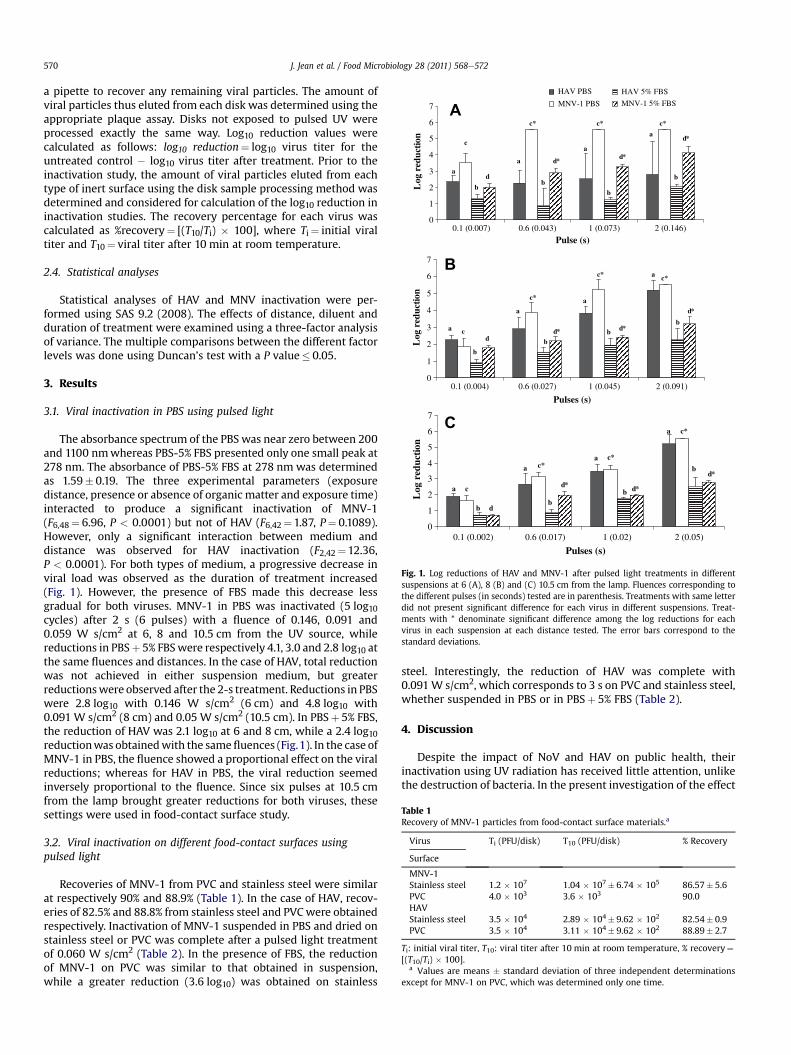
Fig. 1. Log reductions of HAV and MNV-1 after pulsed light treatments in differentsuspensions at 6 (A), 8 (B) and (C) 10.5 cm from the lamp. Fluences corresponding tothe different pulses (in seconds) tested are in parenthesis. Treatments with same letterdid not present significant difference for each virus in different suspensions. Treat-ments with * denominate significant difference among the log reductions for eachvirus in each suspension at each distance tested. The error bars correspond to thestandard deviations.
Table 1Recovery of MNV-1 particles from food-contact surface materials.a
Virus Ti (PFU/disk) T10 (PFU/disk) % Recovery
Surface
MNV-1Stainless steel 1.2 � 107 1.04 � 107� 6.74 � 105 86.57� 5.6PVC 4.0 � 103 3.6 � 103 90.0HAVStainless steel 3.5 � 104 2.89 � 104� 9.62 � 102 82.54� 0.9PVC 3.5 � 104 3.11 � 104� 9.62 � 102 88.89� 2.7
Ti: initial viral titer, T10: viral titer after 10 min at room temperature, % recovery¼[(T10/Ti) � 100].
a Values are means � standard deviation of three independent determinationsexcept for MNV-1 on PVC, which was determined only one time.
J. Jean et al. / Food Microbiology 28 (2011) 568e572570
a pipette to recover any remaining viral particles. The amount ofviral particles thus eluted from each disk was determined using theappropriate plaque assay. Disks not exposed to pulsed UV wereprocessed exactly the same way. Log10 reduction values werecalculated as follows: log10 reduction¼ log10 virus titer for theuntreated control � log10 virus titer after treatment. Prior to theinactivation study, the amount of viral particles eluted from eachtype of inert surface using the disk sample processing method wasdetermined and considered for calculation of the log10 reduction ininactivation studies. The recovery percentage for each virus wascalculated as %recovery¼ [(T10/Ti) � 100], where Ti¼ initial viraltiter and T10¼ viral titer after 10 min at room temperature.
2.4. Statistical analyses
Statistical analyses of HAV and MNV inactivation were per-formed using SAS 9.2 (2008). The effects of distance, diluent andduration of treatment were examined using a three-factor analysisof variance. The multiple comparisons between the different factorlevels was done using Duncan’s test with a P value� 0.05.
3. Results
3.1. Viral inactivation in PBS using pulsed light
The absorbance spectrum of the PBS was near zero between 200and 1100 nmwhereas PBS-5% FBS presented only one small peak at278 nm. The absorbance of PBS-5% FBS at 278 nm was determinedas 1.59� 0.19. The three experimental parameters (exposuredistance, presence or absence of organic matter and exposure time)interacted to produce a significant inactivation of MNV-1(F6,48¼ 6.96, P < 0.0001) but not of HAV (F6,42¼1.87, P¼ 0.1089).However, only a significant interaction between medium anddistance was observed for HAV inactivation (F2,42¼12.36,P < 0.0001). For both types of medium, a progressive decrease inviral load was observed as the duration of treatment increased(Fig. 1). However, the presence of FBS made this decrease lessgradual for both viruses. MNV-1 in PBS was inactivated (5 log10cycles) after 2 s (6 pulses) with a fluence of 0.146, 0.091 and0.059 W s/cm2 at 6, 8 and 10.5 cm from the UV source, whilereductions in PBSþ 5% FBSwere respectively 4.1, 3.0 and 2.8 log10 atthe same fluences and distances. In the case of HAV, total reductionwas not achieved in either suspension medium, but greaterreductionswere observed after the 2-s treatment. Reductions in PBSwere 2.8 log10 with 0.146 W s/cm2 (6 cm) and 4.8 log10 with0.091 W s/cm2 (8 cm) and 0.05 W s/cm2 (10.5 cm). In PBSþ 5% FBS,the reduction of HAV was 2.1 log10 at 6 and 8 cm, while a 2.4 log10reductionwas obtainedwith the samefluences (Fig.1). In the case ofMNV-1 in PBS, the fluence showed a proportional effect on the viralreductions; whereas for HAV in PBS, the viral reduction seemedinversely proportional to the fluence. Since six pulses at 10.5 cmfrom the lamp brought greater reductions for both viruses, thesesettings were used in food-contact surface study.
3.2. Viral inactivation on different food-contact surfaces usingpulsed light
Recoveries of MNV-1 from PVC and stainless steel were similarat respectively 90% and 88.9% (Table 1). In the case of HAV, recov-eries of 82.5% and 88.8% from stainless steel and PVCwere obtainedrespectively. Inactivation of MNV-1 suspended in PBS and dried onstainless steel or PVC was complete after a pulsed light treatmentof 0.060 W s/cm2 (Table 2). In the presence of FBS, the reductionof MNV-1 on PVC was similar to that obtained in suspension,while a greater reduction (3.6 log10) was obtained on stainless
steel. Interestingly, the reduction of HAV was complete with0.091W s/cm2, which corresponds to 3 s on PVC and stainless steel,whether suspended in PBS or in PBSþ 5% FBS (Table 2).
4. Discussion
Despite the impact of NoV and HAV on public health, theirinactivation using UV radiation has received little attention, unlikethe destruction of bacteria. In the present investigation of the effect
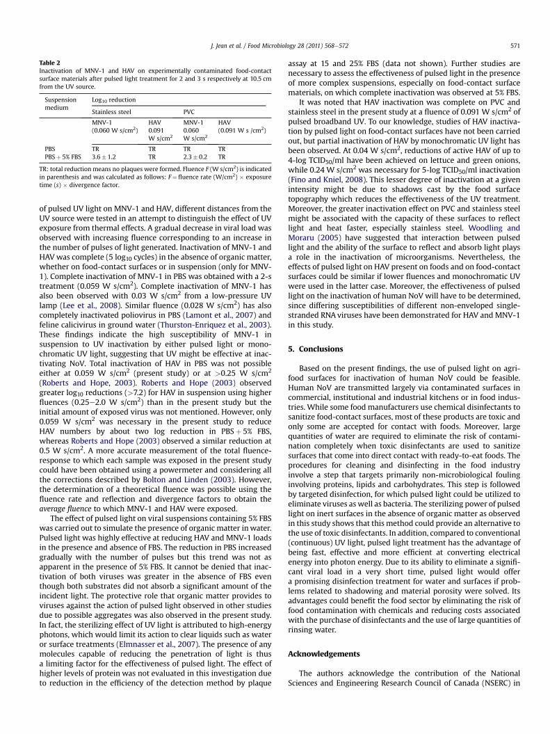
Table 2Inactivation of MNV-1 and HAV on experimentally contaminated food-contactsurface materials after pulsed light treatment for 2 and 3 s respectively at 10.5 cmfrom the UV source.
Suspensionmedium
Log10 reduction
Stainless steel PVC
MNV-1(0.060 W s/cm2)
HAV0.091W s/cm2
MNV-10.060W s/cm2
HAV(0.091 W s /cm2)
PBSPBSþ 5% FBS
TR3.6� 1.2
TRTR
TR2.3� 0.2
TRTR
TR: total reduction means no plaques were formed. Fluence F (W s/cm2) is indicatedin parenthesis and was calculated as follows: F¼ fluence rate (W/cm2) � exposuretime (s) � divergence factor.
J. Jean et al. / Food Microbiology 28 (2011) 568e572 571
of pulsed UV light on MNV-1 and HAV, different distances from theUV source were tested in an attempt to distinguish the effect of UVexposure from thermal effects. A gradual decrease in viral load wasobserved with increasing fluence corresponding to an increase inthe number of pulses of light generated. Inactivation of MNV-1 andHAVwas complete (5 log10 cycles) in the absence of organic matter,whether on food-contact surfaces or in suspension (only for MNV-1). Complete inactivation of MNV-1 in PBS was obtained with a 2-streatment (0.059 W s/cm2). Complete inactivation of MNV-1 hasalso been observed with 0.03 W s/cm2 from a low-pressure UVlamp (Lee et al., 2008). Similar fluence (0.028 W s/cm2) has alsocompletely inactivated poliovirus in PBS (Lamont et al., 2007) andfeline calicivirus in ground water (Thurston-Enriquez et al., 2003).These findings indicate the high susceptibility of MNV-1 insuspension to UV inactivation by either pulsed light or mono-chromatic UV light, suggesting that UV might be effective at inac-tivating NoV. Total inactivation of HAV in PBS was not possibleeither at 0.059 W s/cm2 (present study) or at >0.25 W s/cm2
(Roberts and Hope, 2003). Roberts and Hope (2003) observedgreater log10 reductions (>7.2) for HAV in suspension using higherfluences (0.25e2.0 W s/cm2) than in the present study but theinitial amount of exposed virus was not mentioned. However, only0.059 W s/cm2 was necessary in the present study to reduceHAV numbers by about two log reduction in PBSþ 5% FBS,whereas Roberts and Hope (2003) observed a similar reduction at0.5 W s/cm2. A more accurate measurement of the total fluence-response to which each sample was exposed in the present studycould have been obtained using a powermeter and considering allthe corrections described by Bolton and Linden (2003). However,the determination of a theoretical fluence was possible using thefluence rate and reflection and divergence factors to obtain theaverage fluence to which MNV-1 and HAV were exposed.
The effect of pulsed light on viral suspensions containing 5% FBSwas carried out to simulate the presence of organic matter inwater.Pulsed light was highly effective at reducing HAV and MNV-1 loadsin the presence and absence of FBS. The reduction in PBS increasedgradually with the number of pulses but this trend was not asapparent in the presence of 5% FBS. It cannot be denied that inac-tivation of both viruses was greater in the absence of FBS eventhough both substrates did not absorb a significant amount of theincident light. The protective role that organic matter provides toviruses against the action of pulsed light observed in other studiesdue to possible aggregates was also observed in the present study.In fact, the sterilizing effect of UV light is attributed to high-energyphotons, which would limit its action to clear liquids such as wateror surface treatments (Elmnasser et al., 2007). The presence of anymolecules capable of reducing the penetration of light is thusa limiting factor for the effectiveness of pulsed light. The effect ofhigher levels of protein was not evaluated in this investigation dueto reduction in the efficiency of the detection method by plaque
assay at 15 and 25% FBS (data not shown). Further studies arenecessary to assess the effectiveness of pulsed light in the presenceof more complex suspensions, especially on food-contact surfacematerials, on which complete inactivation was observed at 5% FBS.
It was noted that HAV inactivation was complete on PVC andstainless steel in the present study at a fluence of 0.091 W s/cm2 ofpulsed broadband UV. To our knowledge, studies of HAV inactiva-tion by pulsed light on food-contact surfaces have not been carriedout, but partial inactivation of HAV by monochromatic UV light hasbeen observed. At 0.04 W s/cm2, reductions of active HAV of up to4-log TCID50/ml have been achieved on lettuce and green onions,while 0.24 W s/cm2 was necessary for 5-log TCID50/ml inactivation(Fino and Kniel, 2008). This lesser degree of inactivation at a givenintensity might be due to shadows cast by the food surfacetopography which reduces the effectiveness of the UV treatment.Moreover, the greater inactivation effect on PVC and stainless steelmight be associated with the capacity of these surfaces to reflectlight and heat faster, especially stainless steel. Woodling andMoraru (2005) have suggested that interaction between pulsedlight and the ability of the surface to reflect and absorb light playsa role in the inactivation of microorganisms. Nevertheless, theeffects of pulsed light on HAV present on foods and on food-contactsurfaces could be similar if lower fluences and monochromatic UVwere used in the latter case. Moreover, the effectiveness of pulsedlight on the inactivation of human NoV will have to be determined,since differing susceptibilities of different non-enveloped single-stranded RNA viruses have been demonstrated for HAV and MNV-1in this study.
5. Conclusions
Based on the present findings, the use of pulsed light on agri-food surfaces for inactivation of human NoV could be feasible.Human NoV are transmitted largely via contaminated surfaces incommercial, institutional and industrial kitchens or in food indus-tries. While some food manufacturers use chemical disinfectants tosanitize food-contact surfaces, most of these products are toxic andonly some are accepted for contact with foods. Moreover, largequantities of water are required to eliminate the risk of contami-nation completely when toxic disinfectants are used to sanitizesurfaces that come into direct contact with ready-to-eat foods. Theprocedures for cleaning and disinfecting in the food industryinvolve a step that targets primarily non-microbiological foulinginvolving proteins, lipids and carbohydrates. This step is followedby targeted disinfection, for which pulsed light could be utilized toeliminate viruses as well as bacteria. The sterilizing power of pulsedlight on inert surfaces in the absence of organic matter as observedin this study shows that this method could provide an alternative tothe use of toxic disinfectants. In addition, compared to conventional(continuous) UV light, pulsed light treatment has the advantage ofbeing fast, effective and more efficient at converting electricalenergy into photon energy. Due to its ability to eliminate a signifi-cant viral load in a very short time, pulsed light would offera promising disinfection treatment for water and surfaces if prob-lems related to shadowing and material porosity were solved. Itsadvantages could benefit the food sector by eliminating the risk offood contamination with chemicals and reducing costs associatedwith the purchase of disinfectants and the use of large quantities ofrinsing water.
Acknowledgements
The authors acknowledge the contribution of the NationalSciences and Engineering Research Council of Canada (NSERC) in
J. Jean et al. / Food Microbiology 28 (2011) 568e572572
the form of financial support. They also thank Dr. Stephen Davidsfor critical reading of the manuscript.
References
Bolton, J.R., Linden, K.G., 2003. Standardization of methods for fluence (UV dose)determination in bench-scale UV experiments. J. Environ. Eng. 129, 209e215.
Centers for Disease Control and Prevention (CDC), 2006. Hepatitis SurveillanceReport No. 61. US Department of Health and Human Services, Atlanta, GA.
Doultree, J.C., Druce, J.D., Birch, C.J., Bowden, D.S., Marshall, J.A., 1999. Inactivation offeline calicivirus, a Norwalk virus surrogate. J. Hosp. Infect. 41, 51e57.
Dunn, J., Ott, T., Clark, W., 1995. Pulsed-light treatment of food and packaging. FoodTechnol. 49, 95e98.
D’Souza, D.H., Sair, A., Williams, K., Papafragkou, E., Jean, J., Moore, C., Jaykus, L.,2006. Persistence of caliciviruses on enviromnental surfaces and their transferto food. Int. J. Food Microbiol. 108, 84e91.
Elmnasser, N., Guillou, S., Leroi, F., Orange, N., Bakhrouf, A., Federighi, M., 2007.Pulsed-light system as a novel food decontamination technology: a review. Can.J. Microbiol. 53, 813e821.
Fino, V.R., Kniel, K.E., 2008. UV light inactivation of hepatitis A virus, Aichi virus, andfeline calicivirus on strawberries, green onions, and lettuce. J. Food Prot. 71,908e913.
Halliday, M.L., Kang, L.Y., Zhou, T.K., Hu, M.D., Pan, Q.C., Fu, T.Y., Huang, Y.S., Hu, S.L.,1991. An epidemic of hepatitis A attributable to the ingestion of raw clams inShanghai, China. J. Infect. Dis. 164, 852e859.
Jun, S., Irudayaraj, J., Demirci, A., Geiser, D., 2003. Pulsed UV-light treatment ofcorn meal for inactivation of Aspergillus niger spores. Int. J. Food Sci. Technol.38, 883e888.
Keystone, J.S., Hershey, J.H., 2008. The underestimated risk of hepatitis A andhepatitis B: benefits of an accelerated vaccination schedule. Int. J. Infect. Dis. 12,3e11.
Koopmans, M., Duizer, E., 2004. Foodborne viruses: an emerging problem. Int. J.Food Microbiol. 90, 23e41.
Lamont, Y., Rzezutka, A., Anderson, J.G., MacGregor, S.J., Given, M.J., Deppe, C.,Cook, N., 2007. Pulsed UV-light inactivation of poliovirus and adenovirus. Lett.Appl. Microbiol. 45, 564e567.
Lee, J., Zoh, K., Ko, G., 2008. Inactivation and UV disinfection of murine noroviruswith TiO2 under various environmental conditions. Appl. Environ. Microbiol.74, 2111e2117.
Mbithi, J.N., Springthorpe, V.S., Boulet, J.R., Sattar, S.A., 1992. Survival of hepatitis Avirus on human hands and its transfer on contact with animate and inanimatesurfaces. J. Clin. Microbiol. 30, 757e763.
Mbithi, J.N., Springthorpe, V.S., Sattar, S.A., 1991. Effect of relative humidity andair temperature on survival of hepatitis A virus on environmental surfaces.Appl. Environ. Microbiol. 57, 1394e1399.
Mead, P.S., Slutsker, L., Dietz, V., McCaig, L.F., Bresee, J.S., Shapiro, C., Griffin, P.M.,Tauxe, R.V., 1999. Food-related illness and death in the United States. Emerg.Infect. Dis. 5, 607e625.
Moore, C., Clark, E.M., Gallimore, C.I., Corden, S.A., Gray, J.J., Westmoreland, D., 2004.Evaluation of a broadly reactive nucleic acid sequence based amplification assayfor the detection of noroviruses in faecal material. J. Clin. Virol. 29, 290e296.
Normann, A., Badur, S., Onel, D., Kilic, A., Sidal, M., Larouze, B., Massari, V., Muller, J.,Flehmig, B., 2008. Acute hepatitis A virus infection in Turkey. J. Med. Virol. 80,785e790.
Richards, G.P., 2001. Enteric virus contamination of foods through industrial prac-tices: a primer on intervention strategies. J. Ind. Microbiol. Biotechnol. 27,117e125.
Roberts, P., Hope, A., 2003. Virus inactivation by high intensity broad spectrumpulsed light. J. Virol. Methods 110, 61e65.
Rowan, N.J., MacGregor, S.J., Anderson, J.G., Fouracre, R.A., McIlvaney, L., Farish, O.,1999. Pulsed-light inactivation of food-related microorganisms. Appl. Environ.Microbiol. 65, 1312e1315.
Sattar, S.A., Springthorpe, V.S., Karim, Y., Loro, P., 1989. Chemical disinfection of non-porous inanimate surfaces experimentally contaminated with four humanpathogenic viruses. Epidemiol. Infect. 102, 493e505.
Thurston-Enriquez, J.A., Haas, C.N., Jacangelo, J., Riley, K., Gerba, C.P., 2003. Inacti-vation of feline calicivirus and adenovirus type 40 by UV radiation. Appl.Environ. Microbiol. 69, 577e582.
Uesugi, A.R., Moraru, C.I., 2009. Reduction of Listeria on ready-to-eat sausages afterexposure to a combination of pulsed light and nisin. J. Food Prot. 72, 347e353.
Wobus, C.E., Thackray, L.B., Virgin, H.W., 2006. Murine norovirus:a model system tostudy norovirus biology and pathogenesis. J. Virol. 80, 5104e5112.
Wobus, C.E., Karst, S.M., Thackray, L.B., Chang, K.O., Sosnovtsev, S.V., Belliot, G.,Krug, A., Mackenzie, J.M., Green, K.Y., Virgin, H.W., 2004. Replication of nor-ovirus in cell culture reveals a tropism for dendritic cells and macrophages.PLoS Biol. 2, 2076e2084.
Woodling, S.E., Moraru, C.I., 2005. Influence of surface topography on the effec-tiveness of pulsed light treatment for the inactivation of Listeria innocua onstainless-steel surfaces. J. Food Sci. 70, 345e351.