in vivoimaging of pyrrole-imidazole polyamides with ... · in vivoimaging of pyrrole-imidazole...
TRANSCRIPT
In vivo imaging of pyrrole-imidazole polyamides withpositron emission tomographyDaniel A. Harki*, Nagichettiar Satyamurthy†, David B. Stout†, Michael E. Phelps†‡, and Peter B. Dervan*‡
*Division of Chemistry and Chemical Engineering, California Institute of Technology, Pasadena, CA 91125; and †Crump Institute for Molecular Imaging,Department of Molecular and Medical Pharmacology, David Geffen School of Medicine, University of California, Los Angeles, CA 90095
Contributed by Peter B. Dervan, June 30, 2008 (sent for review April 8, 2008)
The biodistribution profiles in mice of two pyrrole-imidazole poly-amides were determined by PET. Pyrrole-imidazole polyamides area class of small molecules that can be programmed to bind a broadrepertoire of DNA sequences, disrupt transcription factor-DNAinterfaces, and modulate gene expression pathways in cell cultureexperiments. The 18F-radiolabeled polyamides were prepared byoxime ligation between 4-[18F]-fluorobenzaldehyde and a hydrox-ylamine moiety at the polyamide C terminus. Small animal PETimaging of radiolabeled polyamides administered to mice revealeddistinct differences in the biodistribution of a 5-ring �-linkedpolyamide versus an 8-ring hairpin, which exhibited better overallbioavailability. In vivo imaging of pyrrole-imidazole polyamides byPET is a minimum first step toward the translation of polyamide-based gene regulation from cell culture to small animal studies.
biodistribution � fluorine-18 � oxime � radiosynthesis
The development of chemical agents to regulate aberrant geneexpression constitutes a promising strategy for treating hu-
man disease. These compounds function in cells by inhibiting thetranslation of mRNA gene products or by antagonizing tran-scription through direct DNA binding (1, 2). Pyrrole-imidazolepolyamides are a class of cell permeable oligomers programmedto bind DNA sequence specifically with affinities similar totranscription factors (2–4). Encoded by pairs of aromatic aminoacids N-methylpyrrole (Py) and N-methylimidazole (Im) (2),polyamides have been shown to target a wide range of discreteDNA sequences (5), localize to the nucleus in cell culture (6, 7),and access chromatin (8, 9). Polyamides can regulate endoge-nous gene expression through the disruption of DNA bindingproteins in the promoters of selected genes. The binding ofhypoxia-inducible factor (HIF-1�) to the hypoxia response ele-ment (HRE) has been antagonized by polyamides, resulting inthe decreased expression of HIF-1�-regulated genes, includingVEGF (10, 11). Androgen receptor (AR) binding to androgenresponse elements has also been inhibited by polyamides, yield-ing decreased expression levels of AR-regulated genes, such asprostate-specific antigen (12). A polyamide designed to inhibitactivating protein-1 binding in the promoter of TGF-�1 facili-tated decreased levels of TGF-�1 expression in vitro and in vivo(rats) (13). Gene activation has been achieved following poly-amide treatment of a transcriptionally repressed cell culturemodel of Friedreich’s ataxia (a trinucleotide repeat disease)(14). Given these promising biological activities in cell culture,tissue biodistribution studies of polyamides administered toanimals is highly warranted. In vivo imaging of polyamides indiscrete tissues would be of importance for the further devel-opment of polyamide-mediated gene regulation in animal mod-els, a pivotal step toward clinical applications.
A new paradigm in early drug discovery has been the utiliza-tion of PET (15, 16). PET is an analytical imaging technologythat measures real-time biodistribution of a positron-emittingprobe administered to a subject. Through appropriate selectionof the radiolabeled probe, a variety of biochemical processesrelevant to human disease can be studied by PET (17, 18).Outside of the clinic, small animal PET is widely used in basic
biomedical research and development, providing quantitativeassays for the study of gene expression, metabolism and signaltransduction, and drug biodistribution (15, 17–19). Preclinicaldrug screening by PET has been exploited for elucidating smallmolecule permeability to the blood-brain barrier (20, 21), and inhuman microdosing studies of experimental therapeutics duringearly clinical development (15, 16). In addition, the low-massdose of radiolabeled PET probes (ie, high specific activitycompounds injected at nanomolar concentrations, yielding pico-moles of probe/g tissue) allows for detailed biodistributionanalysis of target drugs without perturbing normal biologicalprocesses (17, 18).
In this study, we evaluated the real-time biodistribution oftwo polyamides of different classes, a 5-ring �-linked 1(ImPy�ImPy�Im�-C3-18F) and an 8-ring hairpin 3 [CtPyPyIm-(R)H2N�-PyImPyPy-C3-18F], in mice with PET imaging [�,�-alanine; Ct, 3-chlorothiophene-2-carboxylic acid; (R)H2N�,(R)-2,4-diaminobutyric acid]. Oxime ligation of radiolabeled4-[18F]-f luorobenzaldehyde (6) with hydroxylamine-function-alized 8 and 9 facilitated the rapid radiosynthesis of 1 and 3.The DNA binding affinity of analogous 19F-functionalized 2and 4 was determined by quantitative DNase I footprinting,and the stability of the oxime linkage was evaluated in vitro andin vivo. PET/CT analysis of 1 and 3 in normal C57 micerevealed considerable liver uptake for both polyamides, withrapid gastrointestinal (GI) tract clearance for �-linked 1.Interestingly, hairpin 3 exhibited substantially longer bioavail-ability, with minimal GI or renal clearance after 2 hours.
ResultsRadiosynthesis. 4-[18F]-f luorobenzaldehyde (6) was used as aradiochemical synthon (22–25) for the preparation of 18F-labeled1 and 3 (Fig. 1). Compound 6 was prepared by nucleophilicf luorination of trimethylammonium benzaldehyde derivative 5at elevated temperature in the presence of the K18F/Kryptofix[2.2.2] complex (Scheme 1) (25). Aniline accelerated oximeligation (26) facilitated the conjugation of 6 to hydroxylamine-functionalized 8 and 9 in radiochemical yields of 12% (for 1) and7% (for 3). Both syntheses were completed (material was HPLCpurified) at �100 min after end of bombardment (EOB).
DNase I Footprinting. Quantitative DNase I footprint titrationswere performed with 19F-functionalized 2 and 4 to assess if theaminooxy-linked label 7 affects DNA binding. The bindingaffinity of 2 was measured on the 5�-32P-PCR fragment fromplasmid pJWP-17 (Fig. 2) (27). 2 bound the designed match site
Author Contributions: D.A.H., N.S., D.B.S., M.E.P., and P.B.D. designed research; D.A.H.,N.S., and D.B.S. performed research; D.A.H., N.S., D.B.S., M.E.P., and P.B.D. analyzed data;and D.A.H., N.S., D.B.S., M.E.P., and P.B.D. wrote the paper.
The authors declare no conflict of interest.
‡To whom correspondence may be addressed. E-mail: [email protected] [email protected].
This article contains supporting information online at www.pnas.org/cgi/content/full/0806308105/DCSupplemental.
© 2008 by The National Academy of Sciences of the USA
www.pnas.org�cgi�doi�10.1073�pnas.0806308105 PNAS � September 2, 2008 � vol. 105 � no. 35 � 13039–13044
MED
ICA
LSC
IEN
CES
Dow
nloa
ded
by g
uest
on
May
15,
202
0
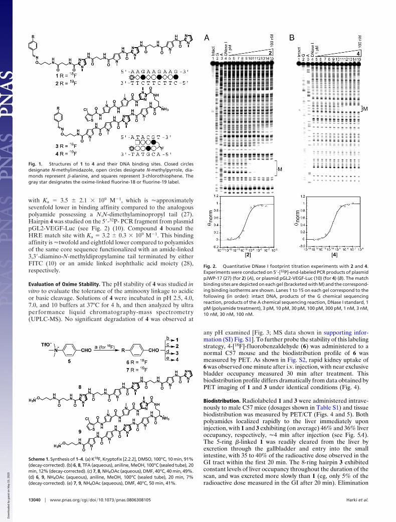
with Ka � 3.5 � 2.1 � 109 M�1, which is �approximatelysevenfold lower in binding affinity compared to the analogouspolyamide possessing a N,N-dimethylaminopropyl tail (27).Hairpin 4 was studied on the 5�-32P- PCR fragment from plasmidpGL2-VEGF-Luc (see Fig. 2) (10). Compound 4 bound theHRE match site with Ka � 3.2 � 0.3 � 109 M�1. This bindingaffinity is �twofold and eightfold lower compared to polyamidesof the same core sequence functionalized with an amide-linked3,3�-diamino-N-methyldipropylamine tail terminated by eitherFITC (10) or an amide linked isophthalic acid moiety (28),respectively.
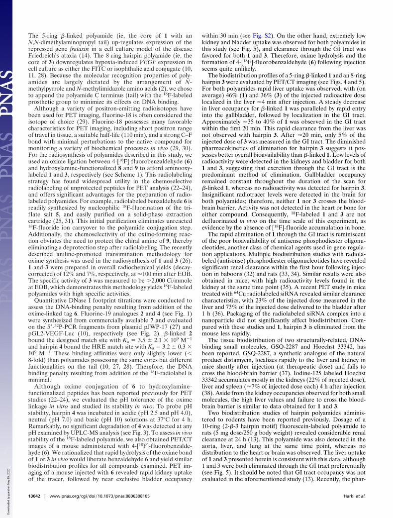
Evaluation of Oxime Stability. The pH stability of 4 was studied invitro to evaluate the tolerance of the aminooxy linkage to acidicor basic cleavage. Solutions of 4 were incubated in pH 2.5, 4.0,7.0, and 10 buffers at 37°C for 4 h, and then analyzed by ultraperformance liquid chromatography-mass spectrometry(UPLC-MS). No significant degradation of 4 was observed at
any pH examined [Fig. 3; MS data shown in supporting infor-mation (SI) Fig. S1]. To further probe the stability of this labelingstrategy, 4-[18F]-f luorobenzaldehyde (6) was administered to anormal C57 mouse and the biodistribution profile of 6 wasmeasured by PET. As shown in Fig. S2, rapid kidney uptake of6 was observed one minute after i.v. injection, with near exclusivebladder occupancy measured 30 min after treatment. Thisbiodistribution profile differs dramatically from data obtained byPET imaging of 1 and 3 under identical conditions (Fig. 4).
Biodistribution. Radiolabeled 1 and 3 were administered intrave-nously to male C57 mice (dosages shown in Table S1) and tissuebiodistribution was measured by PET/CT (Figs. 4 and 5). Bothpolyamides localized rapidly to the liver immediately uponinjection, with 1 and 3 exhibiting (on average) 46% and 36% liveroccupancy, respectively, �4 min after injection (see Fig. 5A).The 5-ring �-linked 1 was readily cleared from the liver byexcretion through the gallbladder and entry into the smallintestine, with 35 to 40% of the radioactive dose observed in theGI tract within the first 20 min. The 8-ring hairpin 3 exhibitedconstant levels of liver occupancy throughout the duration of thescan, and was excreted more slowly than 1 (eg, only 5% of theradioactive dose measured in the GI after 20 min). Elimination
Fig. 2. Quantitative DNase I footprint titration experiments with 2 and 4.Experiments were conducted on 5�-[32P]-end-labeled PCR products of plasmidpJWP-17 (27) (for 2) (A), or plasmid pGL2-VEGF-Luc (10) (for 4) (B). The matchbinding sites are depicted on each gel (bracketed with M) and the correspond-ing binding isotherms are shown. Lanes 1 to 15 on each gel correspond to thefollowing (in order): intact DNA, products of the G chemical sequencingreaction, products of the A chemical sequencing reaction, DNase I standard, 1pM (polyamide treatment), 3 pM, 10 pM, 30 pM, 100 pM, 300 pM, 1 nM, 3 nM,10 nM, 30 nM, 100 nM.
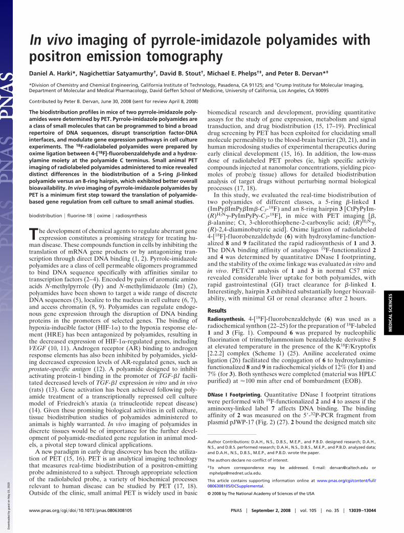
Fig. 1. Structures of 1 to 4 and their DNA binding sites. Closed circlesdesignate N-methylimidazole, open circles designate N-methylpyrrole, dia-monds represent �-alanine, and squares represent 3-chlorothiophene. Thegray star designates the oxime-linked fluorine-18 or fluorine-19 label.
Scheme 1. Synthesis of 1–4. (a) K18F, Kryptofix [2.2.2], DMSO, 100°C, 10 min, 91%(decay-corrected). (b) 6, 8, TFA (aqueous), aniline, MeOH, 100°C (sealed tube), 20min, 12% (decay-corrected). (c) 7, 8, NH4OAc (aqueous), DMF, 40°C, 40 min, 49%.(d) 6, 9, NH4OAc (aqueous), aniline, MeOH, 100°C (sealed tube), 20 min, 7%(decay-corrected). (e) 7, 9, NH4OAc (aqueous), DMF, 40°C, 50 min, 41%.
13040 � www.pnas.org�cgi�doi�10.1073�pnas.0806308105 Harki et al.
Dow
nloa
ded
by g
uest
on
May
15,
202
0

through the GI is favored over bladder excretion for bothcompounds. A constant level of 1 was observed in the gallbladderthroughout the entire PET scan, whereas no significant levels of3 were detected. Neither polyamide exhibited significant activityin the brain, heart, or bone.
Dosimetry. Biodistribution data obtained for 1 and 3 in mice wereused to estimate the dosimetry values for a human model. Thedose-limiting organ for 1 was the small intestine, with the urinarybladder wall, gallbladder, liver, and upper intestines possessingconsiderable radioactive doses (Table S2). For 3 the dose-limiting organ was the kidneys, with significant radioactive dosenoted in the gallbladder, small intestine, liver, urinary bladderwall, and upper and lower intestines (Table S3).
DiscussionRecently, the use of PET in early drug development has gainedconsiderable attention (15, 16). In this study, we assessed the
tissue bioavailability and overall biodistribution of two 18F-labeled polyamides of different classes (1 and 3) in mice usingsmall animal PET.
The polyamide cores selected for biodistribution analysis inthis study exhibit promising biological activities in cell culture.
Fig. 3. pH stability analysis of 4. 4 was incubated in pH 2.5 (A), pH 4.0 (B), pH7.0 (C), and pH 10 (D) buffers at 37°C for 4 h. Samples were neutralized byaddition of aqueous NH4OAc containing 9-aminoacridine (standard), andthen analyzed by UPLC-MS. DMI (1,3-dimethyl-2-imidazolidinone) (15%, vol/vol) was used as a cosolvent in the incubation and neutralization steps.Chromatograms shown (A–D) are UV detection at 254 nm. See Fig. S1 for MSdata.
Fig. 4. PET/CT images of mice administered 18F-labeled 1 (images A–C) and 3 (images D–F). Time-points shown are 1 min (A and D), 30 min (B and E), and 2 h(C and F) after injection.
Fig. 5. Biodistribution of 18F-labeled 1 (shown in black) and 3 (shown in gray)in mice by PET analysis. (A) Liver (open circles) versus GI (closed squares)occupancy for 1 and 3 over 2 h. (B and C) Remaining in vivo occupancy of 1 (B)and 3 (C) in mice, excluding the liver and GI values shown in A.
Harki et al. PNAS � September 2, 2008 � vol. 105 � no. 35 � 13041
MED
ICA
LSC
IEN
CES
Dow
nloa
ded
by g
uest
on
May
15,
202
0
The 5-ring �-linked polyamide (ie, the core of 1 with anN,N-dimethylaminopropyl tail) up-regulates expression of therepressed gene frataxin in a cell culture model of the diseaseFriedreich’s ataxia (14). The 8-ring hairpin polyamide (ie, thecore of 3) downregulates hypoxia-induced VEGF expression incell culture as either the FITC or isophthalic acid conjugate (10,11, 28). Because the molecular recognition properties of poly-amides are largely dictated by the arrangement of N-methylpyrrole and N-methylimidazole amino acids (2), we choseto append the polyamide C terminus (tail) with the 18F-labeledprosthetic group to minimize its effects on DNA binding.
Although a variety of positron-emitting radioisotopes havebeen used for PET imaging, f luorine-18 is often considered theisotope of choice (29). Fluorine-18 possesses many favorablecharacteristics for PET imaging, including short positron rangeof travel in tissue, a suitable half-life (110 min), and a strong C–Fbond with minimal perturbations to the native compound formonitoring a variety of biochemical processes in vivo (29, 30).For the radiosynthesis of polyamides described in this study, weused an oxime ligation between 4-[18F]-f luorobenzaldehyde (6)and hydroxylamine-functionalized 8 and 9 to afford aminooxy-labeled 1 and 3, respectively (see Scheme 1). This radiolabelingstrategy has found widespread utility in the chemoselectiveradiolabeling of unprotected peptides for PET analysis (22–24),and offers significant advantages for the preparation of radio-labeled polyamides. For example, radiolabeled benzaldehyde 6 isreadily synthesized by nucleophilic 18F-fluorination of the tri-f late salt 5, and easily purified on a solid-phase extractioncartridge (25, 31). This initial purification eliminates unreacted18F-fluoride ion carryover to the polyamide conjugation step.Additionally, the chemoselectivity of the oxime-forming reac-tion obviates the need to protect the chiral amine of 9, therebyeliminating a deprotection step after radiolabeling. The recentlydescribed aniline-promoted transimination methodology foroxime synthesis was used in the radiosynthesis of 1 and 3 (26).1 and 3 were prepared in overall radiochemical yields (decay-corrected) of 12% and 7%, respectively, at �100 min after EOB.The specific activity of 3 was measured to be �2,000 Ci/mmoleat EOB, which demonstrates this methodology yields 18F-labeledpolyamides with high specific activities.
Quantitative DNase I footprint titrations were conducted toassess the DNA-binding penalty resulting from addition of theoxime-linked tag 6. Fluorine-19 analogues 2 and 4 (see Fig. 1)were synthesized from commercially available 7 and evaluatedon the 5�-32P-PCR fragments from plasmid pJWP-17 (27) andpGL2-VEGF-Luc (10), respectively (see Fig. 2). �-linked 2bound the designed match site with Ka � 3.5 � 2.1 � 109 M�1
and hairpin 4 bound the HRE match site with Ka � 3.2 � 0.3 �109 M�1. These binding affinities were only slightly lower (�8-fold) than polyamides possessing the same cores but differentfunctionalities on the tail (10, 27, 28). Therefore, the DNAbinding penalty resulting from addition of the 18F-radiolabel isminimal.
Although oxime conjugation of 6 to hydroxylamine-functionalized peptides has been reported previously for PETstudies (22–24), we evaluated the pH tolerance of the oximelinkage in vitro and studied its stability in vivo. To probe pHstability, hairpin 4 was incubated in acidic (pH 2.5 and pH 4.0),neutral (pH 7.0) and basic (pH 10) solutions at 37°C for 4 h.Remarkably, no significant degradation of 4 was detected at anypH examined by UPLC-MS analysis (see Fig. 3). To assess in vivostability of the 18F-labeled polyamide, we also obtained PET/CTimages of a mouse administered with 4-[18F]-f luorobenzalde-hyde (6). We rationalized that rapid hydrolysis of the oxime bondof 1 or 3 in vivo would liberate benzaldehyde 6 and yield similarbiodistribution profiles for all compounds examined. PET im-aging of a mouse injected with 6 revealed rapid kidney uptakeof the tracer, followed by near exclusive bladder occupancy
within 30 min (see Fig. S2). On the other hand, extremely lowkidney and bladder uptake was observed for both polyamides inthis study (see Fig. 5), and clearance through the GI tract wasfavored for both 1 and 3. Therefore, oxime hydrolysis and theformation of 4-[18F]-f luorobenzaldehyde (6) following injectionseems quite unlikely.
The biodistribution profiles of a 5-ring �-linked 1 and an 8-ringhairpin 3 were evaluated by PET/CT imaging (see Figs. 4 and 5).For both polyamides rapid liver uptake was observed, with (onaverage) 46% (1) and 36% (3) of the injected radioactive doselocalized in the liver �4 min after injection. A steady decreasein liver occupancy for �-linked 1 was paralleled by rapid entryinto the gallbladder, followed by localization in the GI tract.Approximately �35 to 40% of 1 was observed in the GI tractwithin the first 20 min. This rapid clearance from the liver wasnot observed with hairpin 3. After �20 min, only 5% of theinjected dose of 3 was measured in the GI tract. The diminishedpharmacokinetics of elimination for hairpin 3 suggests it pos-sesses better overall bioavailability than �-linked 1. Low levels ofradioactivity were detected in the kidneys and bladder for both1 and 3, suggesting that excretion through the GI tract is thepredominant method of elimination. Gallbladder occupancyremained constant throughout the duration of the scan for�-linked 1, whereas no radioactivity was detected for hairpin 3.Insignificant radiotracer levels were detected in the brain forboth polyamides; therefore, neither 1 nor 3 crosses the blood-brain barrier. Activity was not detected in the heart or bone foreither compound. Consequently, 18F-labeled 1 and 3 are notdefluorinated in vivo on the time scale of this experiment, asevidence by the absence of [18F]-f luoride accumulation in bone.
The rapid elimination of 1 through the GI tract is reminiscentof the poor bioavailability of antisense phosphodiester oligonu-cleotides, another class of chemical agents used in gene regula-tion applications. Multiple biodistribution studies with radiola-beled (antisense) phosphodiester oligonucleotides have revealedsignificant renal clearance within the first hour following injec-tion in baboons (32) and rats (33, 34). Similar results were alsoobtained in mice, with high radioactivity levels found in thekidney at the same time point (35). A recent PET study in micetreated with 64Cu radiolabeled siRNA revealed similar clearancecharacteristics, with 23% of the injected dose measured in theliver and 73% of the injected dose delivered to the bladder after1 h (36). Packaging of the radiolabeled siRNA complex into ananoparticle did not significantly affect biodistribution. Com-pared with these studies and 1, hairpin 3 is eliminated from themouse less rapidly.
The tissue biodistribution of two structurally-related, DNA-binding small molecules, GSQ-2287 and Hoechst 33342, hasbeen reported. GSQ-2287, a synthetic analogue of the naturalproduct distamycin, localizes rapidly to the liver and kidney inmice shortly after injection (at therapeutic dose) and fails tocross the blood-brain barrier (37). Iodine-125 labeled Hoechst33342 accumulates mostly in the kidneys (22% of injected dose),liver and spleen (�7% of injected dose each) 4 h after injection(38). Aside from the kidney occupancies observed for both smallmolecules, the high liver values and failure to cross the blood-brain barrier is similar to data obtained for 1 and 3.
Two biodistribution studies of hairpin polyamides adminis-tered to rodents have been reported previously. Dosage of a10-ring (2-�-3 hairpin motif) f luorescein-labeled polyamide torats (5 mg dose/250 g body weight) revealed considerable renalclearance at 24 h (13). This polyamide was also detected in theaorta, liver, and lung at the same time point, whereas nodistribution to the heart or brain was observed. The liver uptakeof 1 and 3 presented herein is consistent with this data, although1 and 3 were both eliminated through the GI tract preferentially(see Fig. 5). It should be noted that GI tract occupancy was notevaluated in the aforementioned study (13). Recently, the phar-
13042 � www.pnas.org�cgi�doi�10.1073�pnas.0806308105 Harki et al.
Dow
nloa
ded
by g
uest
on
May
15,
202
0
macokinetics of a hairpin polyamide-chlorambucil conjugatedosed to mice (500 nmoles per mouse) was evaluated at twotime-points (39). Predominant occupancy of the polyamide-alkylator conjugate was observed in the lung, spleen, smallintestine, and pancreas at 2 and 24 h, as measured by liquidchromatography-mass spectrometry analysis (39). Eliminationthrough the GI tract parallels the results obtained with 1 and 3in this study. Interestingly, minimal liver occupancy was mea-sured with the hairpin polyamide-alkylator conjugate at 2 h (39),whereas 3 is predominantly localized in the liver at the same timepoint.
Although both of the above-mentioned studies provideinsights into polyamide bioavailability (13, 39), the necessity ofanimal killing and organ extraction permits only single time-point measurements to be conducted per animal. The meth-odology presented in this study permits examinations ofpolyamide biodistribution at multiple time points in the sameanimal, allowing detailed pharmacokinetic measurements tobe performed in only a few hours. An example of the utility ofthis technology is shown in Fig. 5. As previously mentioned,hairpin 3 exhibits greater liver occupancy than �-linked 1 at2 h. However, data obtained from PET imaging of 1 and 3 (seeFig. 5 A) reveals 1 to possess greater overall liver occupancythan hairpin 3 only minutes after injection. 1 is then rapidlycleared from the liver and distributed into the GI tract,whereas the level of hairpin 3 in the liver remains constantthroughout the scan. The differences in the pharmacokineticsof elimination between 1 and 3 is readily observed by PETimaging, whereas classical, single time-point methods forstudying compound biodistribution may fail to identify suchsubtle disparities.
PET imaging of 18F-labeled 1 and 3 reveals distinct differences intissue bioavailability between the two classes of polyamides, andconstitutes the first bioavailability study of polyamides by thistechnology. In addition, a robust oxime-based radiolabeling pro-cedure for the general preparation of 18F-labeled polyamides hasbeen described. These advances constitute a new tool for the furtherdevelopment of polyamide technology toward clinical applications,allowing candidate polyamides to be screened for favorable bio-availability properties during early chemical development.
Materials and MethodsPolyamide Synthesis. Protocols and Schemes S1–S4 for the synthesis of 18F-labeled polyamides, fluorine-19 standards, and radiolabeling precursors canbe found in the SI Experimental Details. Analytical HPLC characterization of18F-labeled compounds is shown in Fig. S3.
Determination of DNA Binding Affinities. Quantitative DNase I footprint titra-tion experiments were conducted on 5�-32P-PCR fragments from plasmidpJWP-17 (27) (2) and pGL2-VEGF-Luc (10) (4). Detailed experimental protocolshave been reported previously (40).
pH Stability Analysis. Individual polyamide-buffer solutions were prepared bydissolving polymaride 4 (15 �l, 375 �M solution in DMI) in pH 2.5, 4.0, 7.0, or10 buffers (85 �l, 20-mM high performance capillary electrophoresis grade
buffers from Fluka) or distilled and deionized water (see Fig. S1). The poly-amide solutions were shaken at 37°C for 4 h. Samples were neutralized byaddition of a solution (100 �l) consisting of aqueous NH4OAc (100 mM, pH 6.5;85%, vol/vol), DMI (15%, vol/vol), and 9-aminoacridine (5.0 �M, Fluka) asinternal standard for UPLC-MS analysis. Samples were vortexed, sonicatedbriefly (1 min), vortexed again, then immediately frozen in liquid nitrogen.Samples were stored at �80°C until analyzed. UPLC-MS analysis was per-formed on a Waters Acquity UPLC-LCT Premiere XE TOF-MS (ESI) system (seeFig. S1). A Waters Acquity UPLC BEH C18 column (2.1 � 50 mm, 1.7 �m) wasused, with the mobile phase consisting of a gradient of MeCN (containing0.1% formic acid) in formic acid (0.1%, aqueous). UV analysis was measured at254 nm.
PET/CT Imaging. PET/CT imaging was conducted on male C57 mice under anapproved protocol by the University of California at Los Angeles AnimalResearch Committee. Mice were kept warm, under 1 to 2% isoflurane gasanesthesia, and positioned using an imaging chamber. Fluorine-18-labeled 1and 3 and control 4-[18F]-fluorobenzaldehyde (6) were injected into the tailvein (dosages shown in Table S1). Data were acquired using a Siemens Pre-clinical Solutions microPET Focus 220 and microCAT II CT systems. PET datawere acquired for 2 or 3 h and reconstructed using filtered back projectioninto 22 or 28 frames, respectively. PET images are �1.8-mm resolution, 0.4-mmvoxel size. CT images are a low dose 400-�m resolution acquisition with200-�m voxel size. Images were coregistered and regions drawn using AMIDEsoftware (Andreas Loening, amide.sourceforge.net, Version 0.8.16).
Calculation of Biodistribution Values. The biodistribution graphs contain thepercentage of total dose in each organ over 2 h. Liver values are based on aregion drawn away from the gallbladder, with the mean value multiplied bythe estimated weight derived from 4.5% of the mouse body weight (valuefrom The Jackson Laboratory Web site). Kidney, heart and brain totals arefrom regions drawn on the PET images, and verified using the weights basedon literature values as percent body weight (assuming density equals 1, thusvolume equals weight). GI (1.75 g) and gallbladder (20 mg) weights are basedon literature search estimated organ weights for a 25-g mouse.
Calculation of Dosimetry Values. The total number of disintegrations wascalculated for each organ and the total body. The data were then extended to7.5 h after injection to estimate the majority of the total number of disinte-grations (�4 half-lives for physical decay, accounting for �95% of all disin-tegrations). No bladder voiding or bowel movements were assumed. Usingthe residency time equivalent measure, dosimetry was calculated usingOLINDA software (Version 1.0, Vanderbilt University, 2003). The adult malehuman model was used.
OLINDA is not designed for using PET data in mice to make human predic-tions. There are no S table values for the small organ sizes and distances inmice, so these data are preliminary and only provide a guide for estimatinglimiting dosage for potential human use and should be followed with humandosimetry to determine the proper dose limitations.
ACKNOWLEDGMENTS. We thank the staffs of the Biomedical Cyclotron Fa-cility and the Crump Preclinical Imaging Center at the University of California,Los Angeles for helpful discussions and support, and Dr. Mona Shahgholi(California Institute of Technology) for assistance with UPLC-MS analysis. Thiswork was supported by National Institutes of Health Grants GM27681, R01-EB001943, and R24 CA 92865; National Science Foundation Chemistry Re-search Instrumentation and Facilities Program Grant CHE-0541745; and theDepartment of Energy Cooperative Agreement DE-FC03-02ER63420. D.A.H.thanks the Friedreich’s Ataxia Research Alliance and the California Tobacco-Related Disease Research Program (16FT-0055) for postdoctoral fellowships.
1. Hannon GJ, Rossi JJ (2004) Unlocking the potential of the human genome with RNAinterference. Nature 431:371–378.
2. Dervan PB, Edelson BS (2003) Recognition of the DNA minor groove by pyrrole-imidazole polyamides. Curr Opin Struct Biol 13:284–299.
3. Trauger JW, Baird EE, Dervan PB (1996) Recognition of DNA by designed ligands atsubnanomolar concentrations. Nature 382:559–561.
4. Dervan PB (2001) Molecular recognition of DNA by small molecules. Bioorg Med Chem9:2215–2235.
5. Hsu CF, et al. (2007) Completion of a programmable DNA-binding small moleculelibrary. Tetrahedron 63:6146–6151.
6. Best TP, Edelson BS, Nickols NG, Dervan PB (2003) Nuclear localization of pyrrole-imidazole polyamide-fluorescein conjugates in cell culture. Proc Natl Acad Sci USA100:12063–12068.
7. Edelson BS, et al. (2004) Influence of structural variation on nuclear localizationof DNA-binding polyamide-fluorophore conjugates. Nucleic Acids Res 32:2802–2818.
8. Edayathumangalam RS, Weyermann P, Gottesfeld JM, Dervan PB, Luger K (2004)Molecular recognition of the nucleosomal ‘‘supergroove.’’ Proc Natl Acad Sci USA101:6864–6869.
9. Dudouet B, et al. (2003) Accessibility of nuclear chromatin by DNA binding polyamides.Chem Biol 10:859–867.
10. Olenyuk BZ, et al. (2004) Inhibition of vascular endothelial growth factor with asequence-specific hypoxia response element antagonist. Proc Natl Acad Sci USA101:16768–16773.
11. Nickols NG, Jacobs CS, Farkas ME, Dervan PB (2007) Modulating hypoxia-inducibletranscription by disrupting the HIF-1-DNA interface. ACS Chem Biol 2:561–571.
12. Nickols NG, Dervan PB (2007) Suppression of androgen receptor-mediated gene ex-pression by a sequence-specific DNA-binding polyamide. Proc Natl Acad Sci USA104:10418–10423.
13. Matsuda H, et al. (2006) Development of gene silencing pyrrole-imidazole polyamidetargeting the TGF-beta1 promoter for treatment of progressive renal diseases. J AmSoc Nephrol 17:422–432.
Harki et al. PNAS � September 2, 2008 � vol. 105 � no. 35 � 13043
MED
ICA
LSC
IEN
CES
Dow
nloa
ded
by g
uest
on
May
15,
202
0
14. Burnett R, et al. (2006) DNA sequence-specific polyamides alleviate transcriptioninhibition associated with long GAA●TTC repeats in Friedreich’s ataxia. Proc Natl AcadSci USA 103:11497–11502.
15. Bergstrom M, Grahnen A, Langstrom B (2003) Positron emission tomography micro-dosing: a new concept with application in tracer and early clinical drug development.Eur J Clin Pharmacol 59:357–366.
16. Weber WA, Czernin J, Phelps ME, Herschman HR (2008) Technology insight: novelimaging of molecular targets is an emerging area crucial to the development oftargeted drugs. Nat Clin Pract Oncol 5:44–54.
17. Phelps ME (2000) Positron emission tomography provides molecular imaging of bio-logical processes. Proc Natl Acad Sci USA 97:9226–9233.
18. Phelps ME (2000) PET: The merging of biology and imaging into molecular imaging.J Nucl Med 41:661–681.
19. Phelps ME (2002) Molecular imaging with positron emission tomography. Annu RevNucl Part Sci 52:303–338.
20. Lee CM, Farde L (2006) Using positron emission tomography to facilitate CNS drugdevelopment. Trends Pharmacol Sci 27:310–316.
21. Burns HD, et al. (1999) Positron emission tomography neuroreceptor imaging as a toolin drug discovery, research and development. Curr Opin Chem Biol 3:388–394.
22. Poethko T, et al. (2004) Two-step methodology for high-yield routine radiohalogena-tion of peptides: 18F-labeled RGD and octreotide analogs. J Nucl Med 45:892–902.
23. Poethko T, et al. (2004) Chemoselective pre-conjugate radiohalogenation of unprotectedmono- and multimeric peptides via oxime formation. Radiochim Acta 92:317–327.
24. Schottelius M, et al. (2004) First 18F-labeled tracer suitable for routine clinical imagingof sst receptor-expressing tumors using positron emission tomography. Clin. CancerRes 10:3593–3606.
25. Toyokuni T, et al. (2003) Synthesis of a new heterobifunctional linker, N-[4-(aminooxy)butyl]maleimide, for facile access to a thiol-reactive 18F-labeling agent.Bioconjugate Chem 14:1253–1259.
26. Dirksen A, Hackeng TM, Dawson PE (2006) Nucleophilic catalysis of oxime ligation.Angew Chem Int Ed 45:7581–7584.
27. Puckett JW, et al. (2007) Quantitative microarray profiling of DNA-binding molecules.J Am Chem Soc 129:12310–12319.
28. Nickols NG, Jacobs CS, Farkas ME, Dervan PB (2007) Improved nuclear localization ofDNA-binding polyamides. Nucleic Acids Res 35:363–370.
29. Lasne M-C, et al. (2002) Chemistry of beta-emitting compounds based on fluorine-18.Top Curr Chem 222:203–258.
30. Wester HJ (2003) in Handbook of Nuclear Chemistry, eds Vertes A, Nagy S, Klenesar Z(Kluwer Academic Publishers, The Netherlands). pp 167–208
31. Wilson AA, Dannals RF, Ravert HT, Wagner HN, Jr. (1990) Reductive amination of [18F]flu-orobenzaldehydes: Radiosynthesis of [2-18F]- and [4-18F]fluorodexetimides. J LabelledCompd Radiopharm 28:1189–1199.
32. Tavitian B, et al. (1998) In vivo imaging of oligonucleotides with positron emissiontomography. Nat Med 4:467–471.
33. Roivainen A, et al. (2004) 68Ga-labeled oligonucleotides for in vivo imaging with PET.J Nucl Med 45:347–355.
34. Lendvai G, et al. (2005) Biodistribution of 68Ga-labelled phosphodiester, phosphorothio-ate, and 2�-O-methyl phosphodiester oligonucleotides in normal rats. Eur J Pharm Sci26:26–38.
35. Kuhnast B, et al. (2000) General method to label antisense oligonucleotides withradioactive halogens for pharmacological and imaging studies. Bioconjugate Chem11:627–636.
36. Bartlett DW, Su H, Hildebrandt IJ, Weber WA, Davis ME (2007) Impact oftumor-specific targeting on the biodistribution and efficacy of siRNA nanoparticlesmeasured by multimodality in vivo imaging. Proc Natl Acad Sci USA 104:15549 –15554.
37. Gross M, et al. (2003) Pharmacology of novel heteroaromatic polycycle antibacterials.Antimicrob Agents Chemother 47:3448–3457.
38. Harapanhalli RS, et al. (1996) [125I/127I]IodoHoechst 33342: Synthesis, DNA binding, andbiodistribution. J Med Chem 39:4804–4809.
39. Chou CJ, et al. (2008) Small molecules targeting histone H4 as potential therapeuticsfor chronic myelogenous leukemia. Mol Cancer Ther 7:769–778.
40. Trauger JW, Dervan PB (2001) Footprinting methods for analysis of pyrrole-imidazolepolyamide/DNA complexes. Methods Enzymol 340:450–466.
13044 � www.pnas.org�cgi�doi�10.1073�pnas.0806308105 Harki et al.
Dow
nloa
ded
by g
uest
on
May
15,
202
0