in vitro oxime reactivation of red blood cell acetylcholinesterase inhibited by methyl-paraoxon
TRANSCRIPT
168 G. A. PETROIANU ET AL.
Copyright © 2007 John Wiley & Sons, Ltd. J. Appl. Toxicol. 2007; 27: 168–175
DOI: 10.1002/jat
JOURNAL OF APPLIED TOXICOLOGYJ. Appl. Toxicol. 2007; 27: 168–175Published online 30 January 2007 in Wiley InterScience(www.interscience.wiley.com) DOI: 10.1002/jat.1189
In vitro oxime reactivation of red blood cellacetylcholinesterase inhibited by methyl-paraoxon
G. A. Petroianu,1,* K. Arafat,1 S. M. Nurulain,1 K. Kuca2 and J. Kassa2
1 UAE University, Al Ain-UAE2 University of Defense, Faculty of Military Health Sciences, Hradec Kralove, Czech Republic
Received 27 June 2006; Revised 9 October 2006; Accepted 9 October 2006
ABSTRACT: Oximes are cholinesterase reactivators of use in poisoning with organophosphorus ester enzyme inhibitors.
Pralidoxime (PRX) is the oxime used in the United States. Clinical experience with pralidoxime (and other oximes) is
disappointing and the routine use has been questioned. Furthermore oximes are not equally effective against all existent
enzyme inhibitors. There is a clear demand for ‘broad spectrum’ cholinesterase reactivators with a higher efficacy than
those clinically available. To meet this need over the years new reactivators of cholinesterase of potential clinical utility
have been developed.
The purpose of the study was to quantify ‘in vitro’ the extent of protection conferred by available (pralidoxime and
methoxime) and experimental (K-27, K-33 and K-48) oximes, using methyl-paraoxon (methyl-POX) as an esterase inhibitor
and to compare the results with those previously obtained using paraoxon (POX) as an inhibitor.
Red blood cell (RBC) acetylcholinesterase (AChE) activities in whole blood were measured photometrically in the pres-
ence of different methyl-POX concentrations and IC50 values calculated. Determinations were repeated in the presence of
increasing oxime concentrations. The IC50 of methyl-POX (59 nM) increased with the oxime concentration in a linear
manner. The calculated IC50 values were plotted against the oxime concentrations to obtain an IC50 shift curve. The slope
of the shift curve (tg ααααα) was used to quantify the magnitude of the protective effect (nM IC50 increase per µµµµµM reactivator).
Based on our determinations the new K-series of reactivators is superior to pralidoxime (tg ααααα ===== 1.9) and methoxime
(tg ααααα ===== 0.7), K-27 and K-48 being the outstanding compounds with a tg ααααα value of 10 (nM IC50 increase per µµµµµM
reactivator), which is ≈≈≈≈≈ five times the reactivator ability of PRX. The tg ααααα value determined for K-33 was 6.3.
The ranking of reactivator potencies of the examined oximes determined with methyl-POX as an inhibitor (K-27 ===== K-
48 >>>>> K-33 >>>>> pralidoxime >>>>> methoxime) is similar to the ranking previously reported by us using POX as an inhibitor (K-
27 ≥≥≥≥≥ K-48 >>>>> K-33 >>>>> methoxime ===== pralidoxime). There is an (expected) inverse relationship between the binding constant
K and the slope of the IC50 shift curve (tg ααααα ) for all oximes examined. K-27 and K-48 (the most protective substances
judging by the tg ααααα ) having the lowest K value (highest affinity).
In vivo testing of the new oximes as methyl-paraoxon protective agents is necessary. Copyright © 2007 John Wiley &
Sons, Ltd.
KEY WORDS: cholinesterase; organophosphate; methyl-paraoxon; oxime; pralidoxime; methoxime; K-oxime
* Correspondence to: Georg A. Petroianu, Faculty of Medicine and Health
Sciences, United Arab Emirates University, Department of Pharmacology
and Therapeutics, P.O. Box 17 666, Al Ain-United Arab Emirates.
E-mail: [email protected]
signs and symptoms, PRX is supposed to shorten the
duration of the respiratory muscle paralysis by reactiva-
tion of cholinesterases (Johnson et al., 2000; Alston,
2005; Petroianu, 2005). Clinical experience with PRX is,
however, disappointing and its routine use has been ques-
tioned (van Helden et al., 1996; Peter and Cherian, 2000;
Eddleston et al., 2002; Buckley et al., 2005; Peter et al.,
2006). There is a clear demand for ‘broad spectrum’
cholinesterase reactivators with a higher efficacy than
PRX.
Over the years new potential reactivators of cholines-
terase inhibited by organophosphorus compounds were
developed by different groups. Methoxime (MMC-4) was
synthesized and tested by Hobbiger and Sadler in the UK.
Currently, this reactivator is used by the Czech Army
after nerve agent exposure (Hobbiger and Sadler, 1959).
Introduction
Poisoning with organophosphorus cholinesterase inhibi-
tors is common and the effects have been described ex-
tensively (Namba, 1971; Namba et al., 1971; Zoch, 1971;
Petroianu et al., 1998). Oximes are the only enzyme
reactivators clinically available (Wilson and Ginsburg,
1955; Kassa, 2002). Pralidoxime (PRX) is commonly
used as an adjunct to atropine in the treatment of such
poisonings. Clinically, while atropine relieves muscarinic
OXIME REACTIVATION OF ACETYLCHOLINESTERASE 169
Copyright © 2007 John Wiley & Sons, Ltd. J. Appl. Toxicol. 2007; 27: 168–175
DOI: 10.1002/jat
The K-series of reactivators was developed by Kuca
and later Musilek in the Department of Toxicology at
the Faculty of Military Health Sciences (University of
Defense), Hradec Kralove, Czech Republic (Kuca et al.,
2003 a, b, 2004; Bajgar, 2004).
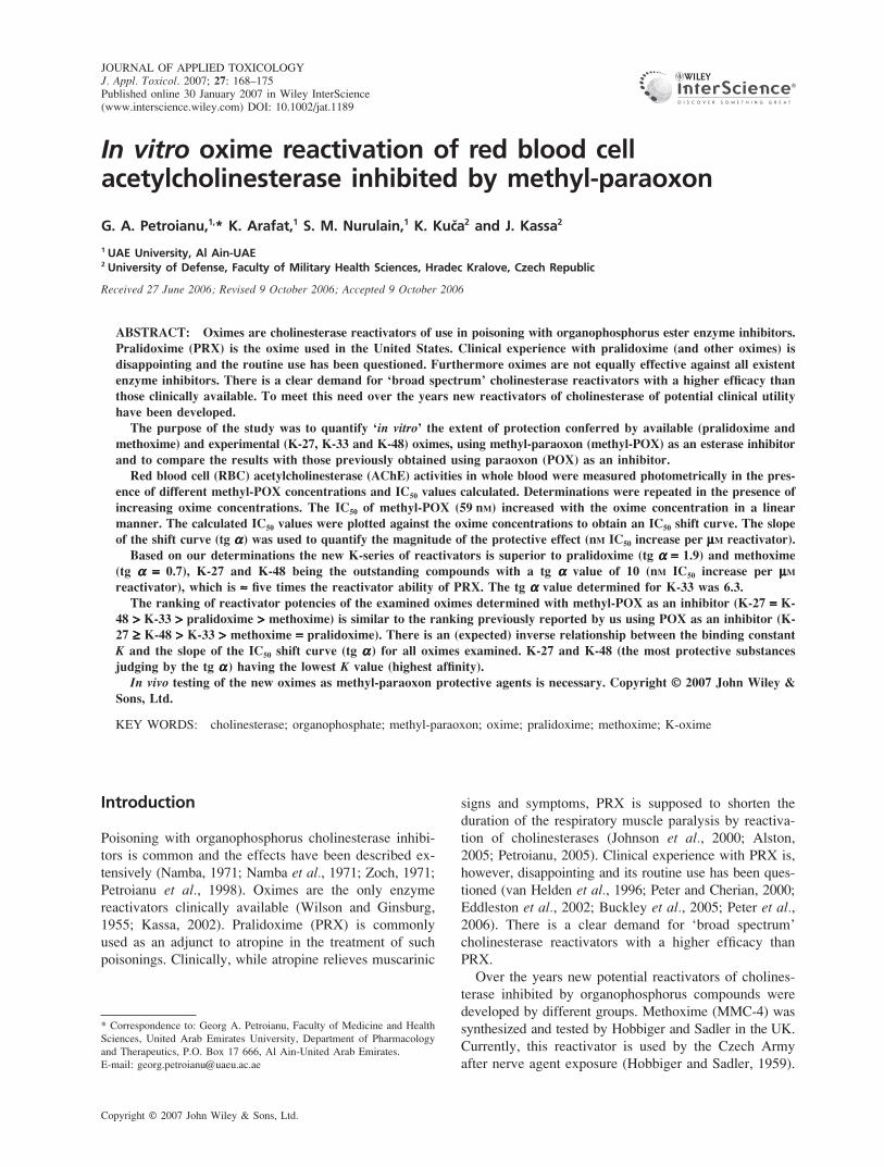
From a chemical point of view, the newly developed
oximes are bisquaternary symmetric (K-33 and
methoxime) or asymmetric (K-27 and K-48) pyridinium
aldoximes with the functional aldoxime group at position
two (K-33) or four (K-27, K-48 and methoxime) of the
pyridine (Fig. 1).
The newly synthesized AChE reactivators were pre-
viously examined with respect to their respective
abilities to reactivate paraoxon (POX) inhibited enzymes
Figure 1. Chemical structure of established and experimental oxime reactivators of organophosphorus inhibitedcholinesterase. From a chemical point of view, the newly developed oximes are bisquaternary symmetric (K-33 andmethoxime) or asymmetric (K-27 and K-48) pyridinium aldoximes with the functional aldoxime group at positiontwo (K-33) or four (K-27, K-48 and methoxime) at the pyridine rings
170 G. A. PETROIANU ET AL.
Copyright © 2007 John Wiley & Sons, Ltd. J. Appl. Toxicol. 2007; 27: 168–175
DOI: 10.1002/jat
(Petroianu et al., 2006). In this paper, in an attempt to
ellucidate the effect of the substituent (methyl vs ethyl)
on the ability of the oximes to reactivate an inhibited
enzyme, the efficacy of the compounds in reactivating
methyl-paraoxon (methyl-POX) inhibited RBC AChE
was examined.
Purpose of the Study
1. To determine in vitro in human blood the IC50 values
of methyl-POX
2. To quantify in vitro the protective effect of increasing
oxime concentrations on RBC-AChE activity when
exposed to methyl-POX, as assessed by the IC50 shift
3. To quantify in vitro the binding constant K of the new
oximes for RBC-AChE using methyl-POX inhibition
data (Schild plot)
Material and Methods
RBC-AChE Activity
The RBC-AChE activity was measured in diluted whole
blood samples in the presence of the selective butyryl-
cholinesterase inhibitor, ethopropazine, as previously
described (Worek et al., 1999). The assay which is
based on Ellman’s method, measures the reduction of
dithiobis-nitrobenzoic acid (DTNB) to nitrobenzoate
(TNB−) by thiocholine, the product of acetylthiocholine
hydrolysis (Ellman et al., 1961). Freshly drawn venous
blood samples were diluted in 0.1 M phosphate buffer
(pH 7.4) and incubated with DTNB (10 mM) and
ethopropazine (6 mM) for 20 min at 37 °C prior to addi-
tion of acetylthiocholine. The change in the absorbance
of DTNB was measured at 436 nm. The AChE activity
was calculated using an absorption coefficient of TNB−
at 436 nm (ε = 10.6 mM−1 cm1). The values were normal-
ized to the hemoglobin (Hb) content (determined as
cyanmethemoglobin) and expressed as mU µmol−1 Hb
(van Kampen and Zijlstra, 1961).
Determination in vitro in Human Blood of theIC50 value of Methyl-POX for RBC-AChE
Blood from human volunteers was used (n = 5, 2 males
and 3 females). None of the volunteers was on any drugs.
Enzyme activities were determined in the absence of and
then after the addition of methyl-POX. The inhibitor was
added before the incubation period. For the graphical
representation and IC50 calculation the SlideWrite™
(Advanced Graphics Software Inc, Encinitas, CA-USA)
software was used (user defined equation y = a0/[1 +(x/a1)exp a2]), where a1 corresponds to the IC50 value.
Enzyme activities were corrected for oxime induced
thiocholine-esteratic activity (Petroianu et al., 2004).
Quantification of the Protective Effect ofIncreasing Oxime Concentrations on RBC-AChEagainst Methyl-POX Inhibition, as assessed bythe IC50 Shift in vitro in Human Blood
IC50 determinations (POX for RBC-AChE) as described
above were repeated in the absence of and then in the
presence of increasing oxime concentrations. Enzyme
activities were corrected for oxime induced thiocholine-
esterase activity (Petroianu et al., 2004). The calculated
IC50 values were plotted against the oxime concentrations
to obtain an IC50 shift curve. For the graphical represen-
tation and calculations the SlideWrite™ (Advanced
Graphics Software Inc, Encinitas, CA-USA) software
was used (equation y = a0 + a1x) where a1 represents
the slope (tangent; tg α) of the IC50 shift graph. The slope
of the shift curve (tg α) was used to quantify the magnitude
of the protective effect (nM IC50 increase per µM reacti-
vator). The IC50 shift [tg α (nM/µM)] has no units.
Calculation of the Binding Constant K of Oximefor RBC-AChE
The performed measurements (IC50 shift) allow the calcu-
lation of the binding constant K of oxime for RBC-AChE.
K (the estimated amount of free substance required to
half saturate the maximal binding capacity of RBC-AChE)
was calculated using the Schild plot. The graphical method
requires plotting of log (dose ratio − 1) vs − log concen-
tration, where the dose ratio is defined as IC50 of methyl-
POX determined in the presence of oxime divided by the
IC50 of the methyl-POX determined in the absence of
oxime (Cheng, 2001; Arunlakshana and Schild, 1959).
For the graphical representation and calculations the
SlideWrite™ (Advanced Graphics Software Inc, Encinitas,
CA-USA) software was used (equation y = a0 + a1x).
Results
IC50 Value of Methyl-POX for RBC-AChE
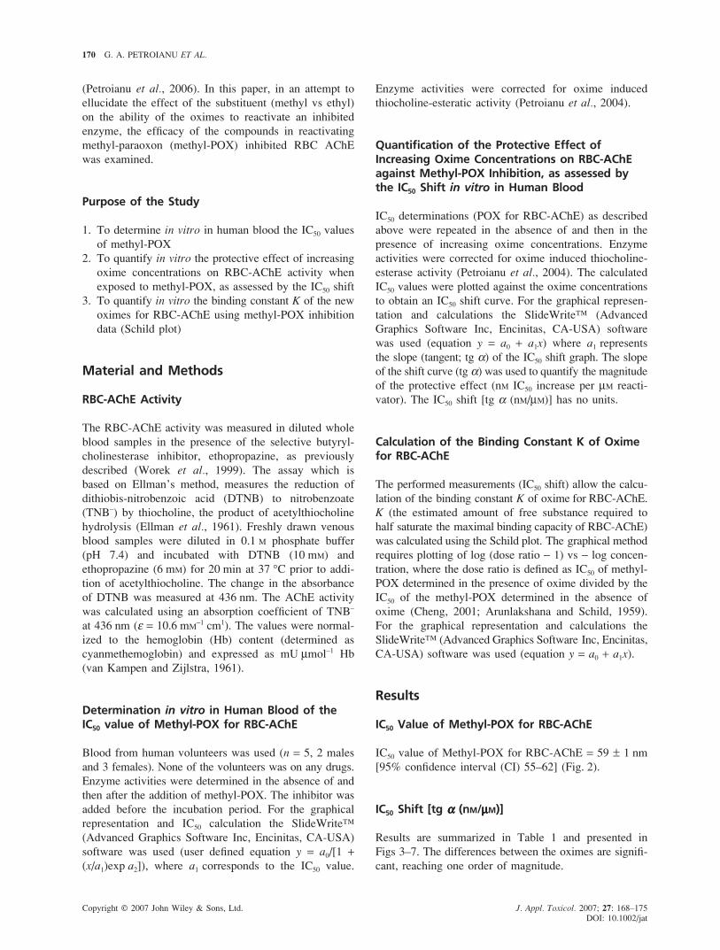
IC50 value of Methyl-POX for RBC-AChE = 59 ± 1 nm
[95% confidence interval (CI) 55–62] (Fig. 2).
IC50 Shift [tg ααααα (nM/µµµµµM)]
Results are summarized in Table 1 and presented in
Figs 3–7. The differences between the oximes are signifi-
cant, reaching one order of magnitude.
OXIME REACTIVATION OF ACETYLCHOLINESTERASE 171
Copyright © 2007 John Wiley & Sons, Ltd. J. Appl. Toxicol. 2007; 27: 168–175
DOI: 10.1002/jat
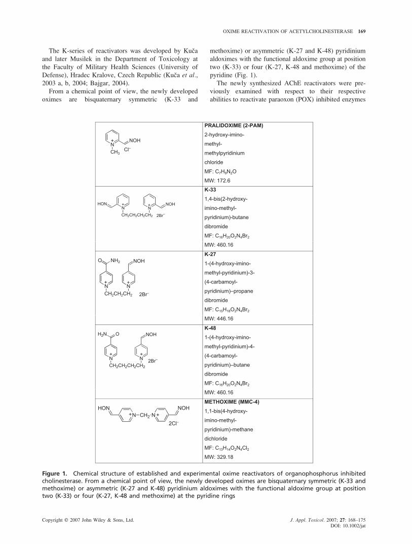
Figure 3. IC50 shift of methyl-POX for RBC-AChE in thepresence of increasing concentrations of PRX: increas-ing the PRX concentration from 0 to 50 µM increasesthe IC50 of methyl-POX for RBC-AChE from 59 to165 nM (∆ = 107 nM); tg α = 1.9 ± 0.1 (95% CI = 1.6–2.2). When using POX as an inhibitor the protectiveeffect of PRX was shown to be tg α = 0.3 ± 0.01 (95%CI = 0.29–0.33) (Petroianu et al., 2006). This figure isavailable in colour online at www.interscience.wiley.com/journal/jat
with PRX (and other available oximes) is mixed at best,
giving the impression of a therapeutic equivalent to ‘the
emperor has no clothes’ story.
Not only is the efficacy of the available reactivators
low, certainly none of them can be viewed as ‘broad
spectrum’, i.e. efficacious against organophosphates and
organophosphonates (Bajgar, 2004; Kassa, 2002). In view
of the real or perceived threat of organophosphorus
agent exposure the need for a highly efficacious ‘broad
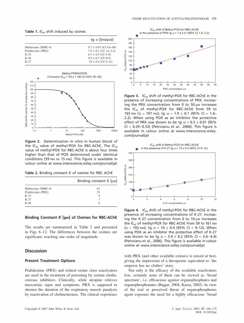
Figure 4. IC50 shift of methyl-POX for RBC-AChE in thepresence of increasing concentrations of K-27: increas-ing the K-27 concentration from 0 to 10 µM increasesthe IC50 of methyl-POX for RBC-AChE from 59 to 161 nM
(∆ ≈ 102 nM); tg α = 10 ± 0.4 (95% CI = 9–12). Whenusing POX as an inhibitor the protective effect of K-27was shown to be tg α = 3.9 ± 0.2 (95% CI = 3.4–4.4)(Petroianu et al., 2006). This figure is available in colouronline at www.interscience.wiley.com/journal/jat
Table 1. IC50 shift induced by oximes
tg α [(nM/µM)]
Methoxime (MMC-4) 0.7 ± 0.03 (CI 0.6–08)
Pralidoxime (PRX) 1.9 ± 0.1 (CI 1.6–2.2)
K-33 6.3 ± 0.5 (CI 5–8)
K-48 9.9 ± 0.7 (CI 812)
K-27 10 ± 0.4 (CI 9–12)
Binding Constant K [µµµµµM] of Oximes for RBC-AChE
The results are summarized in Table 2 and presented
in Figs 8–12. The differences between the oximes are
significant, reaching one order of magnitude.
Discussion
Present Treatment Options
Pralidoxime (PRX) and related oxime class reactivators
are used in the treatment of poisoning by certain cholin-
esterase inhibitors. Clinically, while atropine relieves
muscarinic signs and symptoms, PRX is supposed to
shorten the duration of the respiratory muscle paralysis
by reactivation of cholinesterases. The clinical experience
Figure 2. Determination in vitro in human blood ofthe IC50 value of methyl-POX for RBC-AChE. The IC50
value of methyl-POX for RBC-AChE is about four timeshigher than that of POX determined under identicalconditions (59 nM vs 15 nM). This figure is available incolour online at www.interscience.wiley.com/journal/jat
Table 2. Binding constant K of oximes for RBC AChE
Binding constant K [µM]
Methoxime (MMC-4) 63
Pralidoxime (PRX) 35
K-33 9
K-27 7
K-48 6
172 G. A. PETROIANU ET AL.
Copyright © 2007 John Wiley & Sons, Ltd. J. Appl. Toxicol. 2007; 27: 168–175
DOI: 10.1002/jat
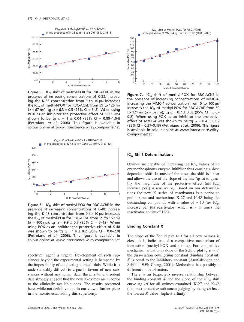
Figure 5. IC50 shift of methyl-POX for RBC-AChE in thepresence of increasing concentrations of K-33: increas-ing the K-33 concentration from 0 to 10 µM increasesthe IC50 of methyl-POX for RBC-AChE from 59 to 126 nM
(∆ ≈ 67 nM); tg α = 6.3 ± 0.5 (95% CI = 5–8). When usingPOX as an inhibitor the protective effect of K-33 wasshown to be tg α = 1 ± 0.04 (95% CI = 0.89–1.04)(Petroianu et al., 2006). This figure is available incolour online at www.interscience.wiley.com/journal/jat
Figure 7. IC50 shift of methyl-POX for RBC-AChE inthe presence of increasing concentrations of MMC-4:increasing the MMC-4 concentration from 0 to 100 µM
increases the IC50 of methyl-POX for RBC-AChE from 59to 121 nM (∆ ≈ 62 nM); tg α = 0.7 ± 0.03 (95% CI = 0.6–0.8). When using POX as an inhibitor the protectiveeffect of MMC-4 was shown to be tg α = 0.4 ± 0.02(95% CI = 0.37–0.48) (Petroianu et al., 2006). This figureis available in colour online at www.interscience.wiley.com/journal/jat
IC50 Shift Determinations
Oximes are capable of increasing the IC50 values of an
organophosphorus enzyme inhibitor thus causing a dose
dependent shift. In most of the cases the shift is linear
and allows the use of the slope of the line (tg α) to quan-
tify the magnitude of the protective effect (nM IC50
increase per µM reactivator). Based on our determina-
tions the new K series of reactivators is superior to
pralidoxime and methoxime, K-27 and K-48 being the
outstanding compounds with a value of ≈ 10 (nM IC50
increase per µM reactivator) which is ≈ 5 times the
reactivator ability of PRX.
Binding Constant K
The slope of the Schild plot (a1) for all new oximes is
close to 1, indicative of a competitive mechanism of
interaction (methyl-POX and oxime). For competitive
mechanism situations (slope of the Schild plot tg α ≈ 1)
the dissociation equilibrium constant (binding constant)
K is equal to the inhibitory constant (Arunlakshana and
Schild, 1959; Cheng, 2001). Methoxime has possibly a
different mode of action.
There is an (expected) inverse relationship between
the binding constant K and the slope of the IC50 shift
curve (tg α) for all oximes examined. K-27 and K-48
(the most protective substances judging by the tg α) have
the lowest K value (highest affinity).
spectrum’ agent is urgent. Development of such sub-
stances beyond the experimental setting is hampered by
the impossibility of conducting clinical trials. While it is
understandably difficult to argue in favour of new sub-
stances without any human data, the in vitro and rodent
data strongly suggest that the new K-oximes are superior
to the clinically available ones. The results presented
here, while not definitive, are in our view a further piece
in the mosaic establishing this superiority.
Figure 6. IC50 shift of methyl-POX for RBC-AChE in thepresence of increasing concentrations of K-48: increas-ing the K-48 concentration from 0 to 10 µM increasesthe IC50 of methyl-POX for RBC-AChE from 59 to 159 nM
(∆ ≈ 100 nM); tg α = 9.9 ± 0.7 (95% CI = 8–12). Whenusing POX as an inhibitor the protective effect of K-48was shown to be tg α = 1.4 ± 0.2 (95% CI = 0.8–2.0)(Petroianu et al., 2006). This figure is available incolour online at www.interscience.wiley.com/journal/jat
OXIME REACTIVATION OF ACETYLCHOLINESTERASE 173
Copyright © 2007 John Wiley & Sons, Ltd. J. Appl. Toxicol. 2007; 27: 168–175
DOI: 10.1002/jat
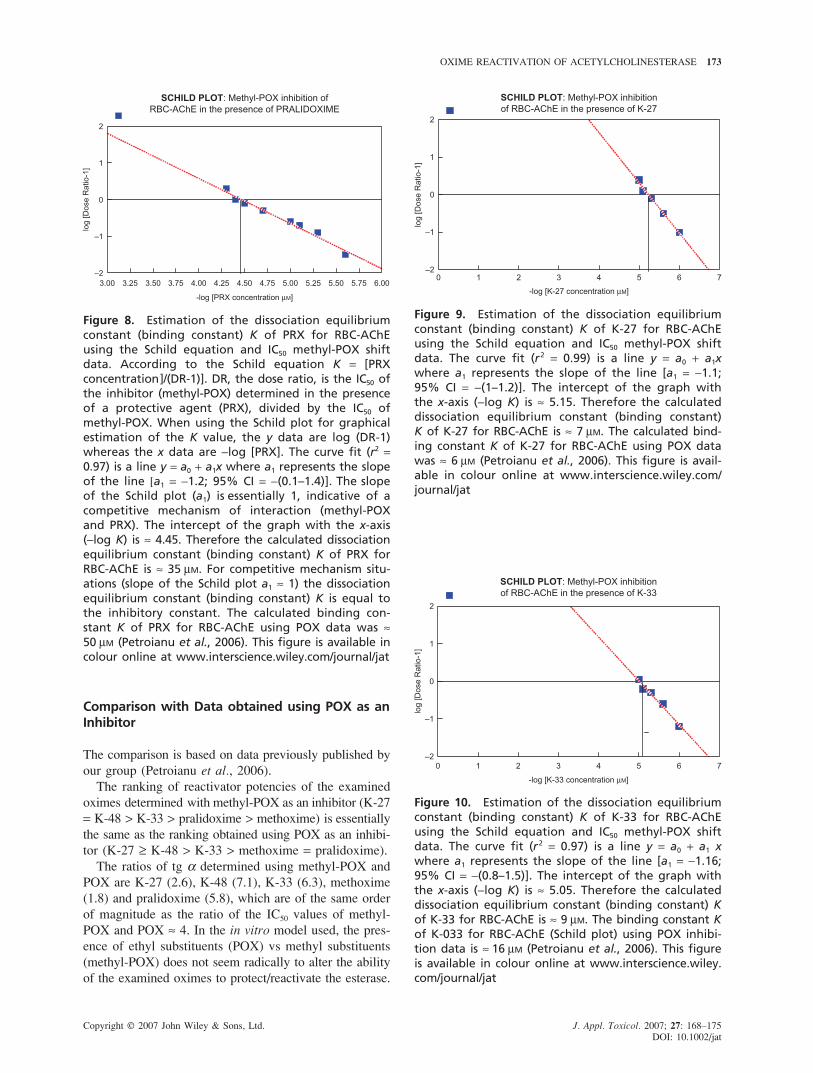
Figure 8. Estimation of the dissociation equilibriumconstant (binding constant) K of PRX for RBC-AChEusing the Schild equation and IC50 methyl-POX shiftdata. According to the Schild equation K = [PRXconcentration]/(DR-1)]. DR, the dose ratio, is the IC50 ofthe inhibitor (methyl-POX) determined in the presenceof a protective agent (PRX), divided by the IC50 ofmethyl-POX. When using the Schild plot for graphicalestimation of the K value, the y data are log (DR-1)whereas the x data are −log [PRX]. The curve fit (r2 =0.97) is a line y = a0 + a1x where a1 represents the slopeof the line [a1 = −1.2; 95% CI = −(0.1–1.4)]. The slopeof the Schild plot (a1) is essentially 1, indicative of acompetitive mechanism of interaction (methyl-POXand PRX). The intercept of the graph with the x-axis(−log K) is ≈ 4.45. Therefore the calculated dissociationequilibrium constant (binding constant) K of PRX forRBC-AChE is ≈ 35 µM. For competitive mechanism situ-ations (slope of the Schild plot a1 ≈ 1) the dissociationequilibrium constant (binding constant) K is equal tothe inhibitory constant. The calculated binding con-stant K of PRX for RBC-AChE using POX data was ≈50 µM (Petroianu et al., 2006). This figure is available incolour online at www.interscience.wiley.com/journal/jat
Figure 9. Estimation of the dissociation equilibriumconstant (binding constant) K of K-27 for RBC-AChEusing the Schild equation and IC50 methyl-POX shiftdata. The curve fit (r2 = 0.99) is a line y = a0 + a1xwhere a1 represents the slope of the line [a1 = −1.1;95% CI = −(1–1.2)]. The intercept of the graph withthe x-axis (−log K) is ≈ 5.15. Therefore the calculateddissociation equilibrium constant (binding constant)K of K-27 for RBC-AChE is ≈ 7 µM. The calculated bind-ing constant K of K-27 for RBC-AChE using POX datawas ≈ 6 µM (Petroianu et al., 2006). This figure is avail-able in colour online at www.interscience.wiley.com/journal/jat
Comparison with Data obtained using POX as anInhibitor
The comparison is based on data previously published by
our group (Petroianu et al., 2006).
The ranking of reactivator potencies of the examined
oximes determined with methyl-POX as an inhibitor (K-27
= K-48 > K-33 > pralidoxime > methoxime) is essentially
the same as the ranking obtained using POX as an inhibi-
tor (K-27 ≥ K-48 > K-33 > methoxime = pralidoxime).
The ratios of tg α determined using methyl-POX and
POX are K-27 (2.6), K-48 (7.1), K-33 (6.3), methoxime
(1.8) and pralidoxime (5.8), which are of the same order
of magnitude as the ratio of the IC50 values of methyl-
POX and POX ≈ 4. In the in vitro model used, the pres-
ence of ethyl substituents (POX) vs methyl substituents
(methyl-POX) does not seem radically to alter the ability
of the examined oximes to protect/reactivate the esterase.
Figure 10. Estimation of the dissociation equilibriumconstant (binding constant) K of K-33 for RBC-AChEusing the Schild equation and IC50 methyl-POX shiftdata. The curve fit (r2 = 0.97) is a line y = a0 + a1 xwhere a1 represents the slope of the line [a1 = −1.16;95% CI = −(0.8–1.5)]. The intercept of the graph withthe x-axis (−log K) is ≈ 5.05. Therefore the calculateddissociation equilibrium constant (binding constant) Kof K-33 for RBC-AChE is ≈ 9 µM. The binding constant Kof K-033 for RBC-AChE (Schild plot) using POX inhibi-tion data is ≈ 16 µM (Petroianu et al., 2006). This figureis available in colour online at www.interscience.wiley.com/journal/jat
174 G. A. PETROIANU ET AL.
Copyright © 2007 John Wiley & Sons, Ltd. J. Appl. Toxicol. 2007; 27: 168–175
DOI: 10.1002/jat
Interestingly enough the mathematical product of bind-
ing constant K and tg α is equal to the IC50 value of the
organophosphorus enzyme inhibitor used to generate the
data, in this case methyl-POX. This observation holds
true also for the previously published data using POX as
an inhibitor.
Conclusion
Newer K-oximes have, according to our in vitro results,
superior enzyme protective properties than the presently
used substances pralidoxime and methoxime when inhi-
bition is due to the organophosphate methyl-paraoxon.
This was previously shown to be true also for inhibition
due to paraoxon.
Further in vitro work using structurally different
organophosphorus enzyme inhibitors is needed in order to
consolidate the conclusions. In vivo testing of the new
oximes as organophosphate protective agents is also
necessary and unavoidable in order to validate the
in vitro results.
References
Alston TA. 2005. Pralidoxime rescues both muscarinic and nicotinicsystems. Anesth. Analg. 101: 926–927.
Arunlakshana O, Schild HO. 1959. Some quantitative uses of drugantagonists. Br. J. Pharmacol. 14: 48–58.
Bajgar J. 2004. Organophosphates/nerve agent poisoning: mechanism ofaction, diagnosis, prophylaxis, and treatment. Adv. Clin. Chem. 38:151–216.
Buckley N, Eddleston M, Szinicz L. 2005. Oximes for acute organo-phosphate pesticide poisoning. Cochrane Database Syst Rev. 1:CD005085.
Cheng HC. 2001. The power issue: determination of KB or Ki fromIC50. A closer look at the Cheng-Prusoff equation, the Schild plot andrelated power equations. J. Pharmacol. Toxicol. Methods 46: 61–71.
Eddleston M, Szinicz L, Eyer P, Buckley N. 2002. Oximes inacute organophosphorus pesticide poisoning: a systematic review ofclinical trials. Q J Med. 95: 275–283.
Ellman GL, Courtney KD, Feather-Stone RM, Andres V Jr. 1961. Anew and rapid colorimetric determination of acetyl-cholinesteraseactivity. Biochem. Pharmacol. 7: 88–95.
Hobbiger F, Sadler PW. 1959. Protection against lethal organophosphatepoisoning by quaternary pyridine aldoximes. Br. J. Pharmacol. 14:192–201.
Johnson MK, Jacobsen D, Meredith TJ, Eyer P, Heath AJ, LigtensteinDA, Marrs TC, Szinicz L, Vale JA, Haines JA. 2000. Evaluation ofantidotes for poisoning by organophosphorus pesticides. Emerg. Med.
12: 22–37.Kassa J. 2002. Review of oximes in the antidotal treatment of poison-
ing by organophosphorus nerve agents. J. Toxicol. Clin. Toxicol. 6:803–816.
Kuca K, Bielavsky J, Cabal J, Bielavska M. 2003a. Synthesis of apotential reactivator of acetylcholinesterase — 1-(4-hydroxyimin-omethylpyridinium)-3-(carbamoylpyridinium)-propane dibromide.Tetrahedron Lett. 44: 3123–3125.
Kuca K, Bielavsky J, Cabal J, Kassa J. 2003b. Synthesis of a newreactivator of tabun-inhibited acetylcholinesterase. Bioorg. Med.
Chem. Lett. 13: 3545–3547.Kuca K, Cabal J, Patocka J, Kassa J. 2004. Synthesis of bisquaternary
symmetric bis (2-hydroxyiminomethylpyridinium) alkane dibromides
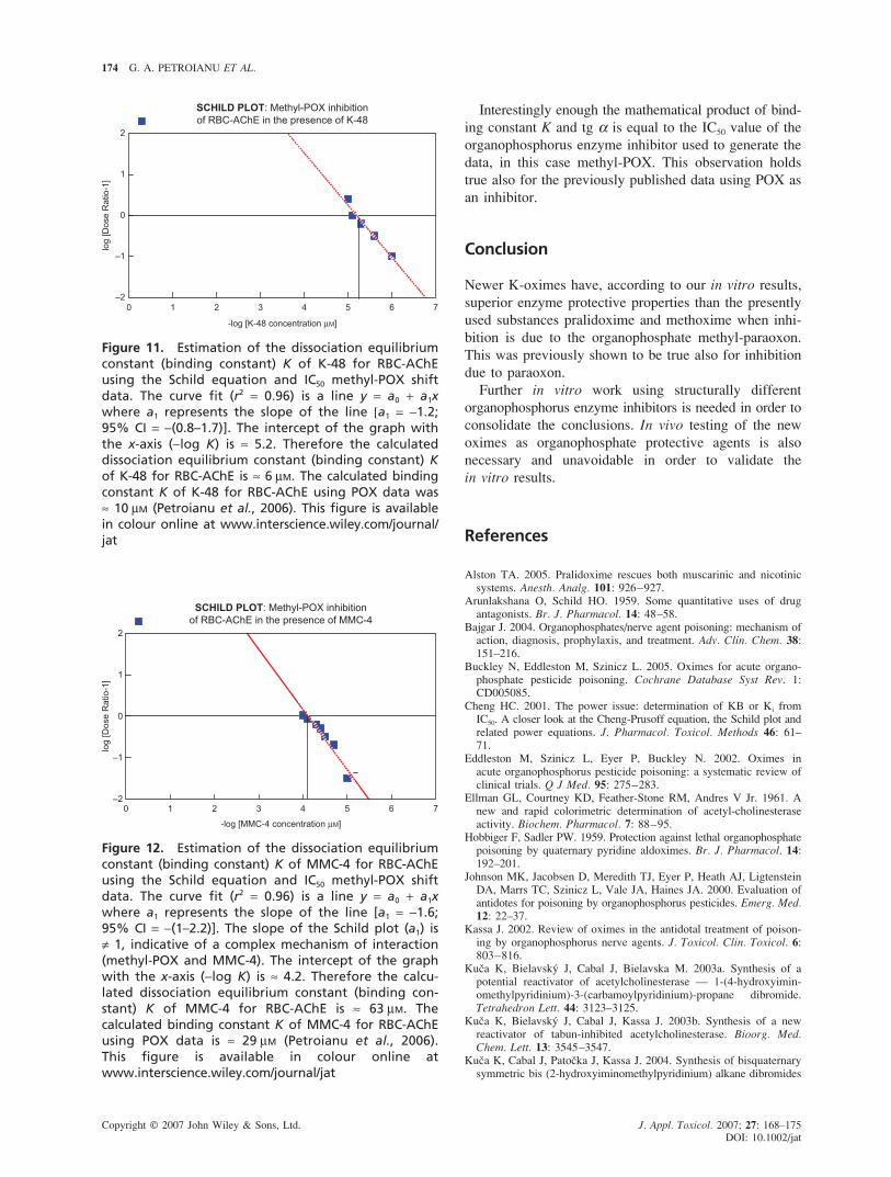
Figure 11. Estimation of the dissociation equilibriumconstant (binding constant) K of K-48 for RBC-AChEusing the Schild equation and IC50 methyl-POX shiftdata. The curve fit (r2 = 0.96) is a line y = a0 + a1xwhere a1 represents the slope of the line [a1 = −1.2;95% CI = −(0.8–1.7)]. The intercept of the graph withthe x-axis (−log K) is ≈ 5.2. Therefore the calculateddissociation equilibrium constant (binding constant) Kof K-48 for RBC-AChE is ≈ 6 µM. The calculated bindingconstant K of K-48 for RBC-AChE using POX data was≈ 10 µM (Petroianu et al., 2006). This figure is availablein colour online at www.interscience.wiley.com/journal/jat
Figure 12. Estimation of the dissociation equilibriumconstant (binding constant) K of MMC-4 for RBC-AChEusing the Schild equation and IC50 methyl-POX shiftdata. The curve fit (r2 = 0.96) is a line y = a0 + a1xwhere a1 represents the slope of the line [a1 = −1.6;95% CI = −(1–2.2)]. The slope of the Schild plot (a1) is≠ 1, indicative of a complex mechanism of interaction(methyl-POX and MMC-4). The intercept of the graphwith the x-axis (−log K) is ≈ 4.2. Therefore the calcu-lated dissociation equilibrium constant (binding con-stant) K of MMC-4 for RBC-AChE is ≈ 63 µM. Thecalculated binding constant K of MMC-4 for RBC-AChEusing POX data is ≈ 29 µM (Petroianu et al., 2006).This figure is available in colour online atwww.interscience.wiley.com/journal/jat
OXIME REACTIVATION OF ACETYLCHOLINESTERASE 175
Copyright © 2007 John Wiley & Sons, Ltd. J. Appl. Toxicol. 2007; 27: 168–175
DOI: 10.1002/jat
and their reactivation of cyclosarin-inhibited acetylcholinesterase.Lett. Org. Chem. 1: 84–86.
Namba T. 1971. Cholinesterase inhibition by organophosphoruscompounds, and its clinical effects. Bull WHO 44: 289–307.
Namba T, Nolte CT, Jackrel J, Grob D. 1971. Poisoning due toorganophosphate insecticides: acute and chronic manifestations. Am.
J. Med. 50: 457–492.Peter JV, Cherian AM. 2000. Organic insecticides. Anaesth. Intensive
Care 28: 11–21.Peter JV, Moran JL, Graham P. 2006. Oxime therapy and outcomes in
human organophosphate poisoning: an evaluation using meta-analytictechniques. Crit. Care Med. 34: 502–510.
Petroianu GA. 2005. Pralidoxime rescues both muscarinic and nicotinicsystems: In response. (letter) Anesth. Analg. 101: 926–927.
Petroianu GA, Arafat K, Kuca K, Kassa J. 2006. Five oximes(K-27, K-33, K-48, BI-6 and methoxime) in comparison withpralidoxime: in vitro reactivation of red blood cell acetylcholines-terase inhibited by paraoxon. J. Appl. Toxicol. 26: 64–71.
Petroianu GA, Missler A, Zuleger K, Thyes C, Ewald V, Maleck WH.2004. Enzyme reactivator treatment in organophosphate exposure:
the clinical relevance of thiocholine-esteratic activity of pralidoxime.J. Appl. Toxicol. 24: 429–435.
Petroianu G, Toomes LM, Petroianu A, Bergeler W, Rüfer R. 1998.Control of blood pressure, heart rate and haematocrit duringhigh-dose intravenous paraoxon exposure in mini pigs. J. Appl.
Toxicol. 18: 293–298.van Helden HP, Busker RW, Melchers BPC, Bruijnzeel PLB. 1996.
Pharmacological effects of oximes: how relevant are they? Arch.
Toxicol. 70: 779–786.van Kampen EJ, Zijlstra WG. 1961. Standardization of hemo-
globinometry II. The hemoglobincyanide method. Clin. Chim. Acta 6:538–544.
Wilson IB, Ginsburg S. 1955. A powerful reactivator of alkylphosphate.Biochim. Biophys. Acta 18: 168–170.
Worek F, Mast U, Kiderlen D, Diepold C, Eyer P. 1999. Improveddetermination of acetylcholinesterase activity in human whole blood.Clin. Chim. Acta 288: 73–90.
Zoch E. 1971. Die Wirkung organischer Phosphorsäureester bzw.Phosphorsäureester auf versch. Enzyme. Arzneimittelforschung. 21:181–187.