in vitro integration of human skin dermis with porous cationic hydrogels
TRANSCRIPT
Available online at www.sciencedirect.com
Acta Biomaterialia 5 (2009) 3337–3345
www.elsevier.com/locate/actabiomat
In vitro integration of human skin dermis with porouscationic hydrogels
Antonio Peramo a,c, Joong Hwan Bahng b, Cynthia L. Marcelo c, Nicholas Kotov b,David C. Martin a,d,e,*
a Department of Materials Science and Engineering, University of Michigan, Ann Arbor, MI 48109, USAb Department of Chemical Engineering, University of Michigan, Ann Arbor, MI 48109, USA
c Department of Surgery, University of Michigan, Ann Arbor, MI 48109, USAd Macromolecular Science and Engineering Center, University of Michigan, Ann Arbor, MI 48109, USA
e Department of Biomedical Engineering, University of Michigan, Ann Arbor, MI 48109, USA
Received 8 January 2009; received in revised form 6 April 2009; accepted 14 May 2009Available online 27 May 2009
Abstract
Porous poly(DMAA-co-AMTAC) hydrogels, fabricated using the inverted colloid crystal method, were used to observe their integra-tion with human skin. Full thickness human breast skin explants discarded from surgeries were cultured for up to 10 days at the air–liquid interface using a Transwell culture system. Cylindrical, disk- or other shaped hydrogels were placed inside the skin explants fittingpunctures produced by punch biopsies or scalpels and full section histological analysis of the skin explants with the inserted hydrogel wasthen performed. In addition, separated hydrogels were cultured up to 7 days with human fibroblasts. The results indicate that poly-(DMAA-co-AMTAC) hydrogels induce substantial extracellular matrix material deposition, maintain dermal integrity in the contactareas with the skin and permit dermal fibers to integrate into the hydrogel pores. Different types of cells remaining in the explantsmigrated into the hydrogels pores, including red blood cells. Fibroblasts adhered to and colonized separately cultured hydrogels. Weplan to use this type of soft material as an interface to permit skin integration with percutaneous devices in contact with skin.� 2009 Acta Materialia Inc. Published by Elsevier Ltd. All rights reserved.
Keywords: Wound healing; Skin biomaterial; Cationic hydrogel; Dermal integration; Porous scaffolds
1. Introduction
There has been a steady increase in the number of med-ical procedures with permanently or temporarily implantedpercutaneous devices. While totally implanted devices havebeen successfully used for several years [1], percutaneousdevices, defined as those that permanently cross the skinto perform their function, have been less successfully devel-oped. Following in part from the ideas of Branemark [2],recent research has led to the development of a perma-
1742-7061/$ - see front matter � 2009 Acta Materialia Inc. Published by Else
doi:10.1016/j.actbio.2009.05.031
* Corresponding author. Address: Department of Materials Science andEngineering, University of Michigan, 2644 CSE Building, Ann Arbor, MI48109, USA. Tel.: +1 734 936 3161.
E-mail address: [email protected] (D.C. Martin).
nently implanted prosthetic, attached directly to the bone.While some of the challenges have been addressed, onesubstantial problem limiting the development of suchdevices is the presence of chronic irritation, inflammationand infection [3–5], which is associated with implanting adevice through the skin and is derived, in part, from theimperfect seal between an organic tissue and the inorganicframe of the device [6]. Infection rates depend on the typeof device [7], but the costs, in economic and human terms,are very high [8,9]. While skin inflammation and infectionconstitutes a major hurdle in the development of perma-nent biointegrated prosthetic devices, the same problemsexist with smaller or temporary devices that are eitheralready in clinical use, like catheters and fixator pins [4],or are being actively developed, like glucose sensors [10].
vier Ltd. All rights reserved.
3338 A. Peramo et al. / Acta Biomaterialia 5 (2009) 3337–3345
These devices show high variability in their intrinsic prop-erties, with a disparate number of material composition,surface structure, porosities and topologies [11]. This vari-ability hampers the investigation of a general solution forthis unresolved problem.
Normally, percutaneous devices are made of hard materi-als where the soft tissue need to attach and integrate, thusthere is a transition between two elements of very differentchemical and mechanical properties. We propose that theuse of soft materials, for instance in the form of hydrogels,forming a preliminary area in contact with skin would allowbetter initial skin integration. For this reason, we sought touse a soft, biointegrative material allowing dermal integra-tion that could be produced as hydrogels. We have beeninvestigating the use of N,N-dimethylacrylamide (DMAA)copolymerized with (3-acrylamidopropyl)-trimethylammo-nium chloride (AMTAC) (poly(DMAA-co-AMTAC)) forits possible use as an interface in the ulterior biointegrationof human skin with percutaneous devices. This type of hydro-gel has been employed in gene therapies as vectors for DNAand oligonucleotide delivery [12], and was shown to promotecell adhesion [13,14], partially due to its cationic nature.
Given that cellular response is different in two- andthree-dimensional scaffoldings [15], the poly(DMAA-co-AMTAC) hydrogels were constructed as three-dimensionaltissue culture scaffolds. Although three-dimensional tissueculture matrices have been used, for instance by rapid pro-totyping [16], they have certain limitations, particularly intheir physical dimensions. The introduction of three-dimen-sional (3-D) hydrogel scaffolds of inverted colloidal crystal(ICC) topology made from poly(acrylamide) hydrogels [17]overcame these limitations, providing an easy control oftheir porous geometry. In contrast to poly(acrylamide) scaf-folds, the poly(DMAA-co-AMTAC) hydrogels we haveused have the advantage that they do not require surfacemodification to allow better cell adhesion.
To evaluate this soft material as an inductor of wholeskin integration, the poly(DMAA-co-AMTAC) hydrogelswere tested in different ways. First, whole human skinwas cultured with the hydrogels and the reaction when inte-grated to the hydrogels was studied. In this case, the hydro-gels were cultured using human organotypic breast skinbreast explants discarded from surgeries at the air–liquidinterface using a Transwell culture system. Secondly, iso-lated human fibroblasts were cultured inside the hydrogelsand their viability was assessed. Finally, the poly(DMAA-co-AMTAC) polymer was deposited as thin films on cover-slips and the adhesion of human fibroblasts was analyzed.These results are presented in this paper.
2. Materials and methods
2.1. Skin preparation
Full thickness human breast skin explants from dis-carded material from surgeries performed at the Universityof Michigan Health System were used. The specimens were
received from healthy human subjects after informed con-sent and immediately prepared for culture. After removalof subcutaneous fat, the tissue was rinsed abundantly with1� PBS containing 125 lg ml�1 of gentamicin (Invitro-gen/GIBCO, Carlsbad, CA) and 1.87 lg ml�1 of amphothe-ricin B (Sigma–Aldrich, Milwaukee, WI) and placed inaliquots of the same medium in an incubator at 37 �C for2 h, with change of medium after 1 h. The culture mediumused was EpiLife (Cascade Biologics, Portland, OR), sup-plemented with EpiLife defined Growth Supplement EDGS(Cascade Biologics, Portland, OR). In addition, the med-ium was supplemented with 75 lg ml�1 of gentamicin and1.125 lg ml�1 of amphothericin B. The final concentrationof calcium used in the culture medium was 1.2 mM. Afterpreparation, skin specimens of approximately 1.5 cm2 werecut using a scalpel and cultured for 5 or 10 days at 37 �C in a5% CO2 atmosphere, epidermal side up at the air–liquidinterface, in a Transwell system consisting of 6-well Trans-well carriers (Organogenesis, Canton, MA) and six CorningCostar supports (Fisher Scientific, Pittsburgh, PA). Culturemedium was changed every 24 h and the stratum corneumremained constantly exposed to the air.
2.2. Preparation of 3-D ICC poly(DMMA-co-AMTAC)
hydrogel scaffolds
The following materials were used for the hydrogelsynthesis: the neutral monomer N,N-dimethylacrylamide(CH2@CHCON(CH3)2 (DMAA), Aldrich), the cationicmonomer (3-acrylamidopropyl)-trimethylammonium chlo-ride (H2C@CHCONH(CH2)3N(CH3)3Cl (AMTAC),Aldrich), the cross-linker N,N0-methylenebisacrylamide((CH2@CHCONH)2CH2 (NMBA), Sigma) and the freeradical initiator potassium persulfate (K2SO4 (KPS),Sigma). Deionized distilled (DDI) water (E-pure, Barn-stead) was used to make the pre-polymer solution and thefree radical solution. An aqueous suspension of polystyrene(PS) microspheres (Duke Scientific, 3 � 104 particles per mlwith 1.4% size distribution) was used for the construction ofthe colloidal crystal.
The poly(DMAA-co-AMTAC) was prepared fromco-polymerization between DMAA and AMTAC in anaqueous environment. The monomer chains were chemi-cally cross-linked by NMBA, and the polymerization wastriggered by the free radical initiator KPS. In detail, aseries of DMAA and AMTAC ratios (variation in %Awhile keeping %T constant) dissolved in DDI water weremixed with a series of %C in a 20 ml glass vial. Eithernitrogen or argon gas was continuously purged so asto maintain an oxygen-free environment. The mixturewas stirred vigorously for 30 min and then partitionedinto 4 ml glass vials (1 ml in each vial). Aqueous KPSsolution (3 wt.%) was then added at a ratio of 1:10 byvolume into the partitioned mixtures to set off the poly-merization. The glass vials of precursors were kept in anoven at 75 �C for 3 h and at 60 �C overnight to aid theformation of the gels.
A. Peramo et al. / Acta Biomaterialia 5 (2009) 3337–3345 3339
2.3. Colloid crystal
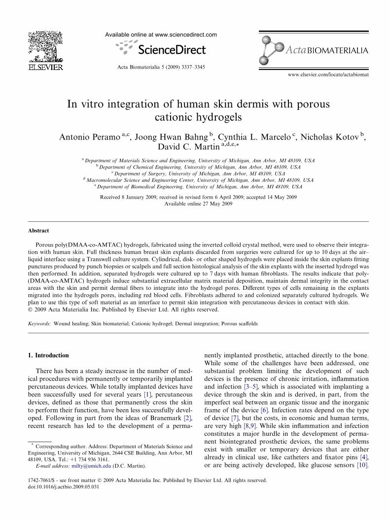
Colloidal crystals (CC) were used as an invertible moldfor the ICC poly(DMAA-co-AMTAC) hydrogel scaffolds,and the technique is illustrated in Fig. 1A and B. The con-struction of CC followed Lee et al.’s reported method [18]with only minor changes. The only component comprisingits structure is non-cross-linked PS microspheres. Plasticcentrifuge tubes were used as molds for the CC construc-tion. First, the PS microspheres were exchanged into iso-propanol. The plastic centrifuge tubes were fused withPasteur glass pipettes, filled with isopropanol and thenimmobilized on the top of an inverted glass beaker. Thewhole system was then placed on an ultrasonic bath(VWR) whose sonication supplied enough agitation to cre-ate well-ordered arrays. One drop of PS microsphere–iso-propanol solution was discharged at the top of thecomplex every 15–20 min. After the CC had grown to thedesired thickness, the CC was dried at 60 �C overnightand heat treated for 4 h at 120 �C while still in the tubemold. The heat treatment caused partial melting at the sur-faces, which resulted in the PS microspheres annealing withtheir adjacent neighbors. The resulting CC was then easilyextracted from the mold.
2.4. Hydrogel synthesis
The resulting CC was immersed in the oxygen-free gelprecursor solution. The gel precursor was infiltrated inbetween the microspheres via centrifugation at 3800 rpmfor 15 min. Again in an oxygen-free environment, aqueousKPS solution (3% w/v) was added at a ratio of 1:10 by vol-ume and then recentrifuged for a further 5 min to allowthorough penetration into the crystal. Subsequent heattreatment at 75 �C for 3 h and 60 �C overnight completedthe polymerization into a poly(DMAA-co-AMTAC)
Fig. 1. Composite image of the culture system used to analyze skin integratiinverted colloid crystal method, showing the highly reproducible porous structuwhich is shown in (B). The monomer dimethylacrylamide is cross-linked by Nmicrospheres was 190 lm, while the interconnecting hydrogel pore size was 50explants (D). The experimental setup uses the Organogenesis Transwell carrier sand (B) were provided by Meghan Cuddihy, Department of Chemical Engine
hydrogel. After polymerization, excess hydrogel pieceswere removed until the surface of the microspheres wasexposed. The hydrogel encapsulated CC was thenimmersed in a THF bath for 48 h to dissolve away theinternal PS microspheres, resulting in a 3-D ICC polyelec-trolyte hydrogel tissue culture scaffold. The THF bath wasrenewed after 24 h. The average diameter of the micro-spheres was 190 lm, while the pore size of the hydrogelswas 50 lm.
2.5. Culture of poly(DMMA-co-AMTAC) hydrogels with
skin specimens
Fig. 1 shows the preparation of the cultured skin withthe poly(DMAA-co-AMTAC) hydrogels. Experimentswere performed three times, with skin from three differentindividuals. The specimens were punctured with a 3 mmdiameter sterile biopsy punch and the hydrogels wereplaced inside the puncture, as shown in Fig. 1D. Afterinsertion, and during the experiments, the top part of thehydrogels remained exposed to air. To test a variety ofhydrogel shapes and type of contact with the skin, a specialtype of experiment was performed with skin specimenswhere the hydrogel was placed under the dermal area –in the dermal epibolic junction – as shown in Fig. 5Aand B. The hydrogels were washed first with PBS and thenwith culture medium before insertion in the punctures orplacement under the dermis. After placement, the hydro-gels remained tightly in contact with the specimens.Fig. 1E shows the skin and hydrogel in the Transwell cul-ture system used. Some experiments were performed wherethe hydrogels were placed under the dermis. This wasdevised to discard the possibility that the protrusion ofthe dermal parts inside the hydrogels could be solely dueto the pressure of the tissue when placed inside skin holesin the form of cylinders or disks.
ve response to poly(DMAA-co-AMTAC) hydrogels, fabricated using theres. (A) The CC molds used to prepare the final hydrogels, the structure ofMBA. Polymerization is triggered by KPS. The nominal diameter of thelm. Cylindrical or disk-shaped hydrogels (C) were placed inside the skinystem to culture the skin at the air–liquid interface (E). The pictures in (A)ering, University of Michigan.
3340 A. Peramo et al. / Acta Biomaterialia 5 (2009) 3337–3345
2.6. Extraction of fibroblasts from skin
Human fibroblasts were extracted and cultured from oneskin specimen as previously described [19], by sequentialtrypsin and collagenase digestion. After extraction, fibro-blasts were cultured in Dulbecco’s modified Eagle’s medium(Gibco) with 10% fetal calf serum, with the medium changedevery other day. Subconfluent primary cultures were pas-saged using trypsin–EDTA. For experiments, fibroblastswere used in passages 3–5.
2.7. Tissue histology of cultured skin specimens with
poly(DMMA-co-AMTAC) hydrogels
Whole skin specimens cultured with poly(DMAA-co-AMTAC) hydrogels were collected at 5 days for differenttypes of analysis. In particular, to test different configura-tions, we experimented with two types of specimen: a regularskin specimen with inserted 4 mm hydrogel disks (as inFig. 1D) and specimens where the hydrogels were placedpartially under the dermis of the skin, as seen in Fig. 5Aand B. For histological analysis, the specimens with hydro-gels were dehydrated in a graded series of ethanol, infiltratedand embedded in JB-4 embedding resin (Electron Micros-copy Sciences, Hatfield, PA). The blocks were sectioned ata thickness of 3 lm and the sections were stained with eithertoluidine blue or a mixture of toluidine blue and basic fuch-sin. While toluidine blue provides the background for extra-cellular matrix and collagen staining, basic fuchsin providescontrast to visualize the hydrogel. For light microscopyanalysis images were taken using a Nikon E800 microscope.
2.8. Scanning electron microscopy (SEM) of poly(DMAA-
co-AMTAC) hydrogels cultured with whole skin specimens
or with isolated fibroblasts
Poly(DMAA-co-AMTAC) hydrogels were SEM imagedafter being cultured with either skin explants or with iso-
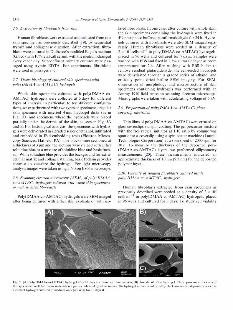
Fig. 2. (A) Poly(DMAA-co-AMTAC) hydrogel after 10 days in culture with hthe layer of extracellular matrix materials is 2 lm, as indicated by white arrows.a control hydrogel cultured in medium only (no skin) for 10 days (C).
lated fibroblasts. In one case, after culture with whole skin,the skin specimens containing the hydrogels were fixed in4% phosphate-buffered paraformaldehyde for 24 h. Hydro-gels cultured with fibroblasts were also SEM imaged sepa-rately. Human fibroblasts were seeded at a density of2 � 105 cells ml�1 in poly(DMAA-co-AMTAC) hydrogels,placed in 96 wells and cultured for 7 days. Samples werewashed with PBS and fixed in 2.5% glutaraldehyde at roomtemperature for 2 h. After washing with PBS buffer toremove residual glutaraldehyde, the cell-seeded hydrogelswere dehydrated through a graded series of ethanol andcritically point dried before SEM imaging. For SEM,observation of morphology and microstructure of skinspecimens containing hydrogels was performed with anAmray 1910 field emission scanning electron microscope.Micrographs were taken with accelerating voltage of 5 kV.
2.9. Preparation of poly(DMAA-co-AMTAC) glass
coverslip substrates
Thin films of poly(DMAA-co-AMTAC) were created onglass coverslips via spin-coating. The gel precursor mixturewith the free radical initiator at 1:10 ratio by volume wasspun onto a coverslip using a spin coater machine (LaurellTechnologies Corporation) at a spin speed of 2000 rpm for30 s. To measure the thickness of the deposited poly-(DMAA-co-AMTAC) layers, we performed ellipsometrymeasurements [20]. These measurements indicated anapproximate thickness of 10 nm (9.3 nm) for the depositedpolymer layer.
2.10. Viability of isolated fibroblasts cultured inside
poly(DMAA-co-AMTAC) hydrogels
Human fibroblasts extracted from skin specimens aspreviously described were seeded at a density of 2 � 105
cells ml�1 in poly(DMAA-co-AMTAC) hydrogels, placedin 96 wells and cultured for 3 days. To study cell viability
uman skin; (B) close detail of the hydrogel. The approximate thickness ofThe hydrogel surface is indicated by black arrows. No deposition is seen in
A. Peramo et al. / Acta Biomaterialia 5 (2009) 3337–3345 3341
on the hydrogels, viable cells were determined by using thecolorimetric MTS assay (CellTiter 96_ AQueous Assay,Madison, WI, USA). The assay measures the absorbanceat 492 nm of the soluble formazan produced by metaboli-cally active cells after reaction with the tetrazolium salt inthe MTS reagent. Afterwards, 20% MTS reagent wasadded to the culture medium and incubated for 1 h forfibroblasts and the absorbance at 492 nm was measured.Background absorbance contributed by the medium (nocells) only was subtracted. Viability data are presented asabsorbance value (mean ± SD).
2.11. Adhesion and viability of isolated fibroblasts to glass
substrates coated with poly(DMAA-co-AMTAC) thin films
For adhesion experiments, glass control, collagenIV-coated and poly(DMAA-co-AMTAC)-coated cover-slips were prepared in triplicate, as described earlier. Forcollagen IV, glass coverslips were prepared by submersingthem for 1 min in a solution of collagen IV (Fluka) at aconcentration of 100 lg ml�1 in 5% acetic acid. Before cellseeding, coverslips were washed with PBS and incubatedwith 1% BSA for 1 h. Afterwards, cells were seeded at adensity of 1 � 105 cells ml�1 and cultured overnight [21].After the experiments, coverslips were washed with PBSand remaining adhered cells were fixed with 2% glutaralde-hyde for 30 min, washed with deionized water and stainedwith Coomassie blue. Adherent cells were pictured andcounted in five different areas for each coverslip. To studycell viability on the substrates, viable cells were determinedby using the colorimetric MTS assay, as previouslydescribed. Triplicate glass control and poly(DMAA-co-AMTAC)-coated coverslips were used and 2.5 � 105 fibro-blasts were seeded and cultured overnight. Afterwards, thesubstrates were treated by adding 20% MTS reagent to theculture medium and incubated for 1 h. Thereafter, tripli-cate aliquots of the medium were pipetted into 96-wellplates and the absorbance at 492 nm was measured andbackground absorbance from the medium (no cells) wassubtracted. Viability data are presented as absorbancevalue (mean ± SD) and adhesion data are presented asnumber of cells (mean ± SD).
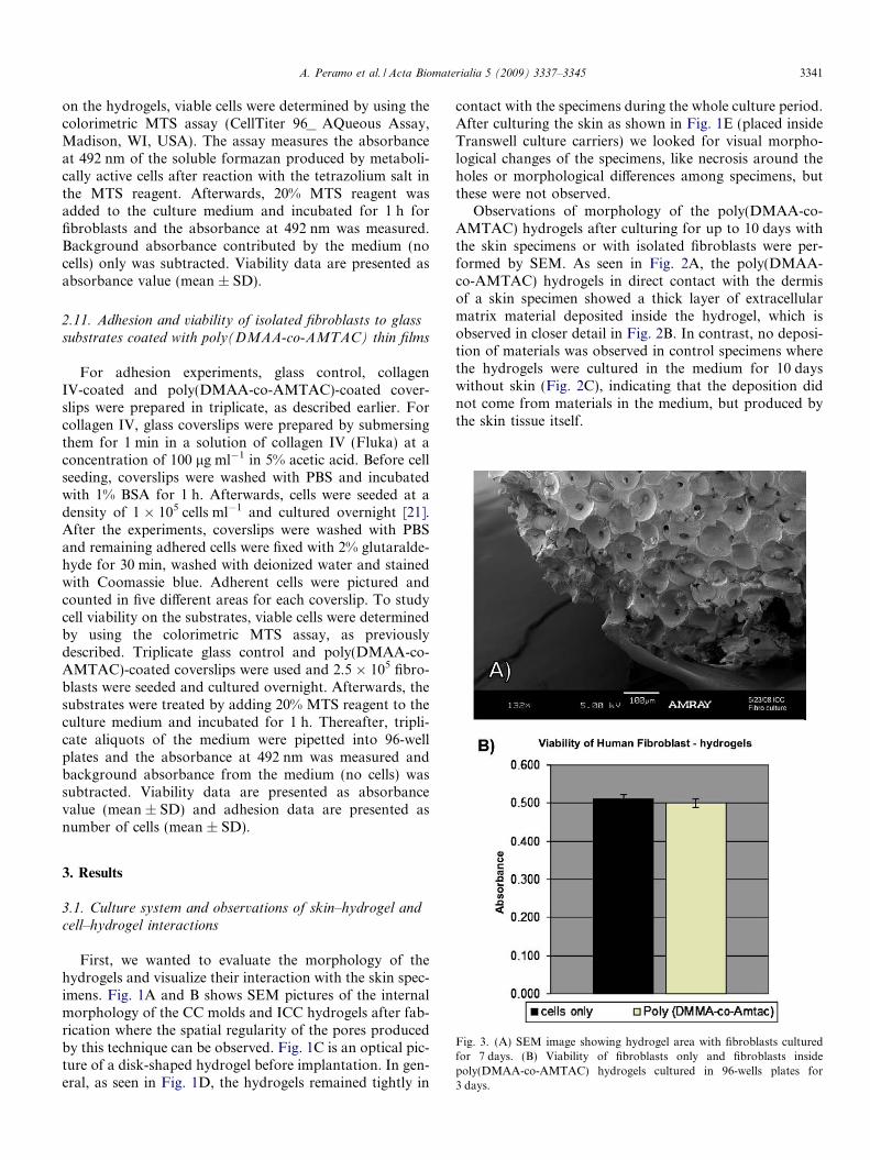
Fig. 3. (A) SEM image showing hydrogel area with fibroblasts culturedfor 7 days. (B) Viability of fibroblasts only and fibroblasts insidepoly(DMAA-co-AMTAC) hydrogels cultured in 96-wells plates for3 days.
3. Results
3.1. Culture system and observations of skin–hydrogel andcell–hydrogel interactions
First, we wanted to evaluate the morphology of thehydrogels and visualize their interaction with the skin spec-imens. Fig. 1A and B shows SEM pictures of the internalmorphology of the CC molds and ICC hydrogels after fab-rication where the spatial regularity of the pores producedby this technique can be observed. Fig. 1C is an optical pic-ture of a disk-shaped hydrogel before implantation. In gen-eral, as seen in Fig. 1D, the hydrogels remained tightly in
contact with the specimens during the whole culture period.After culturing the skin as shown in Fig. 1E (placed insideTranswell culture carriers) we looked for visual morpho-logical changes of the specimens, like necrosis around theholes or morphological differences among specimens, butthese were not observed.
Observations of morphology of the poly(DMAA-co-AMTAC) hydrogels after culturing for up to 10 days withthe skin specimens or with isolated fibroblasts were per-formed by SEM. As seen in Fig. 2A, the poly(DMAA-co-AMTAC) hydrogels in direct contact with the dermisof a skin specimen showed a thick layer of extracellularmatrix material deposited inside the hydrogel, which isobserved in closer detail in Fig. 2B. In contrast, no deposi-tion of materials was observed in control specimens wherethe hydrogels were cultured in the medium for 10 dayswithout skin (Fig. 2C), indicating that the deposition didnot come from materials in the medium, but produced bythe skin tissue itself.
3342 A. Peramo et al. / Acta Biomaterialia 5 (2009) 3337–3345
3.2. Viability of fibroblasts cells cultured inside
poly(DMMA-co-AMTAC) hydrogels and adhesion and
viability on thin film poly(DMMA-co-AMTAC) substrates
Given that skin in vivo contains fibroblasts that couldeventually migrate to the hydrogels, we devised experi-ments to analyze whether isolated fibroblasts would be via-ble inside hydrogels in vitro and whether they wouldadhere and be viable when seeded on poly(DMMA-co-AMTAC) substrates. Fibroblasts adhered to and colonizedthe cultured hydrogel scaffolds, as seen in Fig. 3A, suggest-ing that the hydrogels are a permissive substrate for cellsurvival. The fibroblast viability assays validated this initialassessment, indicating that the cells proliferated inside thehydrogel pores, as well as in the poly(DMAA-co-AMTAC)thin film substrates. Fig. 3B shows that there was nochange in the viability of the fibroblasts inside the hydro-gels compared with fibroblasts cultured in the 96-wellculture plates without hydrogels.
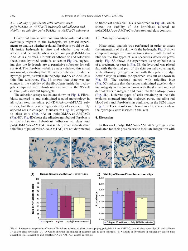
The adhesion assays results are shown in Fig. 4. Fibro-blasts adhered to and maintained a good morphology inall substrates, including poly(DMAA-co-AMTAC) sub-strates, but there was a higher density of extended, fullyadhered cells in collagen IV substrates (Fig. 4B) comparedto glass only (Fig. 4A) or poly(DMAA-co-AMTAC)(Fig. 4C). Fig. 4D shows the adhesion numbers of fibroblaststo the substrates. Fibroblast adhesion to glass andpoly(DMAA-co-AMTAC) was similar, which indicates thatthin films of poly(DMAA-co-AMTAC) are not detrimental
Fig. 4. Representative pictures of human fibroblasts adhered to glass coverslipIV-coated glass coverslips (C). (D) Graph showing the number of adherent celcoverslips, glass coverslips and poly(DMAA-co-AMTAC)-coated coverslips.
to fibroblast adhesion. This is confirmed in Fig. 4E, whichshows the viability of the fibroblasts adhered topoly(DMAA-co-AMTAC) substrates and glass controls.
3.3. Histological analysis
Histological analysis was performed in order to assessthe integration of the skin with the hydrogels. Fig. 5 showscomposite images of tissue sections stained with toluidineblue for the two types of skin specimens described previ-ously. Fig. 5A shows the experiment using epibolic cutsof a specimen. As seen in Fig. 5B, the hydrogel was placedflat with the dermal part of the skin partially covering it,while allowing hydrogel contact with the epidermis edge.After 5 days in culture the specimen was cut as shown inFig. 5B. The sections stained with toluidine blue(Fig. 5C) indicate that the tissues maintained excellent der-mal integrity in the contact areas with the skin and induceddermal fibers to integrate and move into the hydrogel pores(Fig. 5D). Different types of cells remaining in the skinexplants migrated into the hydrogel pores, including redblood cells and fibroblasts, as confirmed in the SEM image(Fig. 5E). These results were found in all specimens wherethe hydrogels were inserted in the skin.
4. Discussion
In this work, poly(DMAA-co-AMTAC) hydrogels wereevaluated for their possible use to facilitate integration with
s (A), poly(DMAA-co-AMTAC)-coated glass coverslips (B) and collagenls to each substrate. (E) Viability of fibroblasts in collagen IV-coated glass
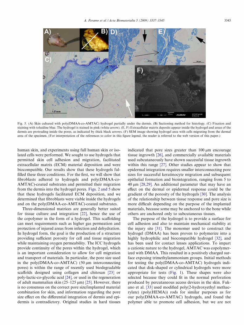
Fig. 5. (A) Skin cultured with poly(DMAA-co-AMTAC) hydrogel partially under the dermis. (B) Sectioning method for histology. (C) Fixation andstaining with toluidine blue. The hydrogel is stained in pink (white arrow). (E, F) Extracellular matrix deposits appear inside the hydrogel and areas of thedermis are protruding inside the pores, as indicated by thick black arrows. (F) SEM image showing hydrogel area with cells migrating from the dermalarea of the specimen. (For interpretation of the references to color in this figure legend, the reader is referred to the web version of this paper.)
A. Peramo et al. / Acta Biomaterialia 5 (2009) 3337–3345 3343
human skin, and experiments using full human skin or iso-lated cells were performed. We sought to use hydrogels thatpermitted skin cell adhesion and migration, facilitatedextracellular matrix (ECM) material deposition and werebiocompatible. Our results show that these hydrogels ful-filled these three conditions. For the first, we will show thatfibroblasts adhered to hydrogels and poly(DMAA-co-AMTAC)-coated substrates and permitted their migrationfrom the dermis into the hydrogel pores. Figs. 2 and 5 showthat these hydrogels facilitated ECM deposition, and wedetermined that fibroblasts were viable inside the hydrogelsand on the poly(DMAA-co-AMTAC)-coated substrates.
Three-dimensional matrices are generally better suitedfor tissue culture and integration [22], hence the use ofthe copolymer in the form of a hydrogel. This scaffoldingcan meet requirements such as higher gas permeation andprotection of injured areas from infection and dehydration.In hydrogel form, the goal is the production of a structureproviding sufficient porosity for cell and tissue migrationwhile maintaining oxygen permeability. The ICC hydrogelsprovide continuity of the pores within the hydrogel, whichis an important consideration to allow for cell migrationand transport of materials. In particular, the pore size usedin the poly(DMAA-co-AMTAC) (50 lm interconnectingpores) is within the range of recently used biodegradablescaffolds designed using collagen and chitosan [23] orpoly-lactic-co-glycolic acid [24], or used in the regenerationof adult mammalian skin (25–125 lm) [25]. However, thereis no consensus on the correct pore size/implanted materialcombination for skin, and information regarding the poresize effect on the differential integration of dermis and epi-dermis is contradictory. Original studies in hard tissues
indicated that pore sizes greater than 100 lm encouragetissue ingrowth [26], and commercially available materialsused subcutaneously have shown successful tissue ingrowthwithin this range [27]. Other studies appear to show thatepidermal integration requires smaller interconnecting poresizes for successful keratinocyte migration and subsequentepithelial formation and biointegration, ranging from 5 to40 lm [28,29]. An additional parameter that may have aneffect on the dermal or epidermal response could be thegradient of the porosity of the hydrogels [30]. The analysisof the relationship between tissue response and pore size ismore difficult depending on the purpose of the implantedmaterials, because some may be anchored to the bone whileothers are anchored only to subcutaneous tissues.
The purpose of the hydrogel is to provide a surface forcell adhesion and also to maintain mechanical stability atthe injury site [31]. The monomer used to construct thehydrogel (DMAA) has been proven to polymerize into ahighly hydrophilic and biocompatible hydrogel [32], andhas been used for contact lenses applications. To imparta cationic nature to the hydrogel, AMTAC was copolymer-ized with DMAA. This resulted in a positively charged sur-face exposing trimethylammonium groups. Initial methodsfor testing the poly(DMAA-co-AMTAC) hydrogels indi-cated that disk-shaped or cylindrical hydrogels were moreappropriate for tests (Fig. 1). These shapes were alsoselected because they could fit in the normal perforationproduced by percutaneous access devices in the skin. Fuk-ano et al. [33] used modified poly(2-hydroxyethyl methac-rylate) (poly(HEMA)) rods for similar purposes as forour poly(DMAA-co-AMTAC) hydrogels, and found thepolymer able to promote cell adhesion, but we are not
3344 A. Peramo et al. / Acta Biomaterialia 5 (2009) 3337–3345
aware of any other similar studies. The main differencewith the use of (1,10-carbonyldiimidazole)-modifiedpoly(HEMA) is that poly(DMAA-co-AMTAC) hydrogeldoes not require any further modification step of thesurface to increase cell adhesion, in part because it is con-structed as a cationic hydrogel.
In addition to the most common techniques for studyingthe cellular response to the copolymer, i.e. viability andadhesion (discussed later), we used organotypic cultures[34] of human skin explants because they are an attractivemodel for studying the biomaterial–skin interface [35,36].We note that organotypic cultures of skin cannot replicatethe time span required for clinical treatments of percutane-ous devices in vivo because these treatments last formonths or years. In accordance with previous studies oforganotypic cultures of human skin explants [37], no sub-stantial morphological changes were observed during theinitial 5 days for control specimens. To determine the effec-tiveness of the copolymer in inducing skin integration, weobserved histological features of the specimens culturedwith the hydrogels. The analysis of the SEM images com-paring control hydrogels and hydrogels cultured with skinfor 10 days indicated that the thick layer of materialsdeposited on the hydrogels came from the skin itself, as evi-denced by comparing Fig. 2A and C. We did not analyzethe precise composition of the materials, but the intenseblue stain by toluidine blue indicates they contain typicaldermal proteins, like collagen and fibrin. This depositionappeared to increase over time, as evidenced comparingSEM images of a sample cultured for 5 days (Fig. 5) and10 days (Fig. 2).
Given that most of the hydrogels were placed inside thepunctures of the punch biopsies or the cuts of scalpels, theprotrusion of the dermal parts inside the hydrogels may beattributed to the pressure of the tissue alone. For this rea-son, 5 day experiments were devised where the hydrogelswere placed partially underneath the dermis. The results,shown in Fig. 5, indicated that the skin protruded almosthorizontally into the pores (Fig. 5C) and that the depositedmaterials were accumulating all around the pores, not juston the bottom (Fig. 5D). This easy observation of depos-ited materials in these 3-D hydrogels is in contrast with2-D cultures. In 2-D cultures newly formed ECM is diffi-cult to observe [38], but when fibroblasts are seeded on3-D structures they integrate into the scaffold and secretecollagen [39]. It has been suggested that their function inwound beds is to synthesize collagen and other ECM mol-ecules [40]. Fibroblasts invade the wound by early synthesisof new skin tissue.
As well as analyzing dermal integration by observingcellular migration into the hydrogel and deposition ofextracellular materials far away from the dermal contactareas, separate experiments were performed seeding cellswith hydrogels or glass coverslips coated with a thin filmof the copolymer, to independently observe the levels of cellviability and adhesion permitted by the cationic copolymercompared to controls. In addition to glass, collagen IV was
also used as a control because of the high affinity of cellsfor this substrate [41]. We found small morphologicaldifferences between the cells in the different substrates(Fig. 4A–C), suggesting that cells on the glass andpoly(DMAA-co-AMTAC) substrates adhered in slightlylower numbers than on collagen IV (Fig. 4D). This sametrend was observed in the viability of the fibroblasts tothe substrates (Fig. 4E). An extended time point analysisof the viability of the fibroblasts in contact with thepoly(DMAA-co-AMTAC) copolymer is shown inFig. 3B. As mentioned earlier, fibroblasts adhered to andcolonized the cultured hydrogel scaffolds in experimentsof up to 7 days, as seen in Fig. 3A. The fibroblast viabilityassays validated this initial assessment, indicating that thecells proliferated inside the hydrogel pores. Fig. 3B showsthat there was no change in the viability of the fibroblastsinside the hydrogels compared with fibroblasts cultured inthe 96-well culture plates without hydrogels. Overall, weconclude that the copolymer is not detrimental to fibro-blast adhesion while the hydrogels are an environment thatclearly permits fibroblast viability.
5. Conclusions
Our results suggest that this type of soft, biodegradablematerial can be used as a general interface permitting skinintegration. This study proves that poly(DMAA-co-AMTAC) hydrogel scaffolds will support in vitro skin cellmigration and viability, producing and depositing extracel-lular matrix molecules within the structure of the hydrogels.In general, variations in scaffolding materials, structure andculture time affect extracellular matrix synthesis and theirsuitability for skin integration. Among the proposed possi-ble uses of these biodegradable hydrogels would be to allowskin cell transplantation into injured areas surrounding per-cutaneous devices. With this initial evidence, we are cur-rently adapting the mechanical properties of the hydrogelsto those of the skin. We will next work in animal modelswhere poly(DMAA-co-AMTAC) hydrogels will beimplanted in skin wounds to analyze skin integration andwhere cells will be seeded inside the scaffolds to see if thisspeeds up the wound healing and integration process.
Acknowledgements
We thank the Microscopy & Image-Analysis Labora-tory of the University of Michigan School of Medicinefor their work on specimen preparation; Ming Qin, fromthe Department of Chemical Engineering, University ofMichigan, Ann Arbor, for his help with the spin coater;and Meghan Cuddihy, Department of Chemical Engineer-ing, University of Michigan for the SEM hydrogel imagingof Fig. 1A and B. This report is presented as part of theresearch efforts within the Army Research Office Multidis-ciplinary University Research Initiative award on Bio-Inte-grating Structural and Neural Prosthetic Materials, and wegratefully acknowledge the funding provided.
A. Peramo et al. / Acta Biomaterialia 5 (2009) 3337–3345 3345
References
[1] Branemark R, Branemark PI, Rydevik B, Myers RR. Osseointegra-tion in skeletal reconstruction and rehabilitation: a review. J RehabilRes Dev 2001;38:175–81.
[2] Branemark PI, Albrektsson T. Titanium implants permanentlypenetrating human skin. Scand J Plast Reconstr Surg 1982;16:17–21.
[3] Winter GD. Transcutaneous implants: reactions of the skin–implantinterface. J Biomed Mater Res Symp No 5 (Part I) 1974;13:99–101.
[4] Mahan J, Seligson D, Henry SL, Hynes P, Dobbins J. Factors in pintract infections. Orthopedics 1991;14:305–8.
[5] Von Recum AF. Applications and failure modes of percutaneousdevices: a review. J Biomed Mater Res 1984;18:323–36.
[6] Albrektsson T, Branemark PI, Jacobsson M, Tjellstrom A. Presentclinical applications of osseointegrated percutaneous implants. PlastReconstr Surg 1987;79:721–30.
[7] Masse A, Bruno A, Bosetti M, Biasibetti A, Cannas M, Gallinaro P.Prevention of pin track infection in external fixation with silver coatedpins: clinical and microbiological results. J Biomed Mater Res (ApplBiomater) 2000;53:600–4.
[8] Mermel LA. Catheter-related blood stream infections. Ann InternMed 2000;133:395.
[9] NNIS System. National Nosocomial Infections Surveillance (NNIS)System Report, data summary from January 1992 through June 2003,issued August 2003. Am J Infect Control 2003;31:481–98.
[10] Pickup JC, Hussain F, Evans ND, Sachedina N. In vivo glucosemonitoring: the clinical reality and the promise. Biosens Bioelectron2005;20:1897–902.
[11] Pendergrass CJ, Goodship AE, Blunn GW. Development of a softtissue seal around bone-anchored transcutaneous amputation pros-theses. Biomaterials 2006;27:4183–91.
[12] De Smedt SC, Demeester J, Hennink WE. Cationic polymer basedgene delivery systems. Pharm Res 2000;17(2):113–26.
[13] Rosso F, Barbarisi A, Barbarisi M, Petillo O, Margarucci S, CalarcoA, et al. New polyelectrolite hydrogels for biomedical applications.Mater Sci Eng C 2003;23(3):371–6.
[14] Kang HS, Park S-H, Lee Y-G, Son T-I. Polyelectrolyte complexhydrogel composed of chitosan and poly(gamma-glutamic acid) forbiological application: preparation, physical properties, and cyto-compatibility. J Appl Polym Sci 2007;103(1):386–94.
[15] Cukierman E, Pankov R, Stevens DR, Yamada KM. Takingcell–matrix adhesions to the third dimension. Science 2001;294(5547):1708–12.
[16] Wang XH, Yan Y, Pan Y, Xiong Z, Liu H, Cheng J, et al. Generationof three-dimensional hepatocyte/gelatin structures with rapid proto-typing system. Tissue Eng 2006;12(1):83–90.
[17] Zhang YJ, Wang S, Eghtedari M, Motamedi M, Kotov NA.Inverted-colloidal-crystal hydrogel matrices as three-dimensional cellscaffolds. Adv Funct Mater 2005;15(5):725–31.
[18] Lee J, Shanbhag S, Kotov NA. Inverted colloidal crystals as three-dimensional microenvironments for cellular co-cultures. J MaterChem 2006;16(35):3558–64.
[19] Deveci M, Gilmont RR, Dunham WR, Mudge BP, Smith DJ,Marcelo CL. Glutathione enhances fibroblast collagen contractionand protects keratinocytes from apoptosis in hyperglycaemic culture.Br J Dermatol 2005;152(2):217–24.
[20] Tengvall P, Lundstrom I, Liedberg B. Protein adsorption studies onmodel organic surfaces: an ellipsometric and infrared spectroscopicapproach. Biomaterials 1998;19:407–22 [Review].
[21] Drumheller PD, Elbert DL, Hubbell JA. Multifunctional poly(ethylene glycol) semi-interpenetrating polymer networks as highly
selective adhesive substrates for bioadhesive peptide grafting. Bio-technol Bioeng 1994;43:772–80.
[22] Lee J, Cuddihy MJ, Kotov NA. Three-dimensional cell culturematrices: state of the art. Tissue Eng B Rev 2008;14(1):61–86[Review].
[23] Ma L, Gao C, Mao Z, Zhou J, Shen J, Hu X, et al. Collagen/chitosanporous scaffolds with improved biostability for skin tissue engineer-ing. Biomaterials 2003;24:4833.
[24] Lee JJ, Lee S-G, Park JC, Yang YI, Kim JK. Investigation onbiodegradable PLGA scaffold with various pore size structure for skintissue engineering. Curr Appl Phys 2007;7S(1):e37–40.
[25] Yang SF, Leong KF, Du Z, Chua CK. The design of scaffolds for usein tissue engineering. Part 1. Traditional factors. Tissue Eng 2001;7:679.
[26] Spector M, Flemming WR, Kreutner A. Bone growth into poroushigh-density polyethylene. J Biomed Mater Res 1976;10:595–603.
[27] Sclafani AP, Romo III T, Silver L. Clinical and histologic behavior ofexposed porous high-density polyethylene implants. Plast ReconstrSurg 1997;99:41–50.
[28] Knowles NG, Miyashita Y, Usui ML, Marshall AJ, Pirrone A,Hauch KD, et al. A model for studying epithelial attachment andmorphology at the interface between skin and percutaneous devices. JBiomed Mater Res A 2005;74:482–8.
[29] Squier CA, Collins P. The relationship between soft tissue attach-ment, epithelial downgrowth and surface porosity. J Periodontal Res1981;16:434–40.
[30] Honga H, Weia J, Liu C. Development of asymmetric gradational-changed porous chitosan membrane for guided periodontal tissueregeneration. Compos B Eng 2007;38:311–6.
[31] Hutmacher DW. Scaffold in tissue engineering bone and cartilage.Biomaterials 2000;20:2529.
[32] Mullarney MP, Seery TAP, Weiss RA. Drug diffusion in hydropho-bically modified N,N-dimethylacrylamide hydrogels. Polymer2006;47(11):3845–55.
[33] Fukano Y, Knowles NG, Usui ML, Underwood RA, Hauch KD,Marshall AJ, et al. Characterization of an in vitro model forevaluating the interface between skin and percutaneous biomaterials.Wound Repair Regen 2006;14:484–91.
[34] Resau JH, Sakamoto K, Cottrell JR, Hudson EA, Meltzer SJ.Explant organ culture: a review. Cytotechnology 1991;7:137–49.
[35] Le Poole IC, Van der Wijngaard RM, Westeerhof W, Dormans JA,Van der Berg FM, Verkruisen RP, et al. Organotypic culture ofhuman skin to study melanocyte migration. Pigment Cell Res1994;7:33–43.
[36] Tavakkol A, Varani J, Elder JT, Zouboulis CC. Maintenance ofhuman skin in organ culture: role for insulin-like growth factor-1receptor and epidermal growth factor receptor. Arch Dermatol Res1999;291:643–51.
[37] Tammi R, Jansen CT, Santti R. Histometric analysis of human skinin organ culture. J Invest Dermatol 1979;73:138–40.
[38] Doillon CJ, Silver FH, Berg RA. Fibroblast growth on a porouscollagen sponge containing hyaluronic acid and fibronectin. Bioma-terials 1987;8:195.
[39] Lee SB, Jeon HW, Lee YW, Lee YM, Song KW, Park MH, et al. Bio-artificial skin composed of gelatin and (1 ? 3), (1 ? 6)-b-glucan.Biomaterials 2003;24:2503.
[40] Yamada N, Uchinuma E, Kuroyanagi Y. Clinical evaluation of anallogenic cultured dermal substitute composed of fibroblasts within aspongy collagen matrix. Scand J Plast Reconstr 1999;33:147–54.
[41] Aumailley M, Timpl R. Attachment of cells to basement membranecollagen type IV. J Cell Biol 1986;103:1569–75.