in vitro inhibition of helicobacter pylori growth by...
TRANSCRIPT

Research ArticleIn Vitro Inhibition of Helicobacter pylori Growth by RedoxCycling Phenylaminojuglones
Julio Benites ,1,2 Héctor Toledo ,3 Felipe Salas,1 Angélica Guerrero,1 David Rios,1
Jaime A. Valderrama,2 and Pedro Buc Calderon 1,4
1Facultad de Ciencias de la Salud, Universidad Arturo Prat, Casilla 121, 1100000 Iquique, Chile2Instituto de Ciencias Exactas y Naturales, Universidad Arturo Prat, Casilla 121, 1100000 Iquique, Chile3Instituto de Ciencias Biomédicas (ICBM), Facultad de Medicina, Universidad de Chile, 8380453 Santiago, Chile4Research Group in Metabolism and Nutrition, Louvain Drug Research Institute, Université catholique de Louvain,Louvain-la-Neuve, Belgium
Correspondence should be addressed to Julio Benites; [email protected] and Héctor Toledo; [email protected]
Received 27 November 2017; Revised 1 February 2018; Accepted 21 February 2018; Published 24 April 2018
Academic Editor: Kota V. Ramana
Copyright © 2018 Julio Benites et al. This is an open access article distributed under the Creative Commons Attribution License,which permits unrestricted use, distribution, and reproduction in any medium, provided the original work is properly cited.
Infection by Helicobacter pylori increases 10 times the risk of developing gastric cancer. Juglone, a natural occurring 1,4-naphthoquinone, prevents H. pylori growth by interfering with some of its critical metabolic pathways. Here, we report thedesign, synthesis, and in vitro evaluation of a series of juglone derivatives, namely, 2/3-phenylaminojuglones, as potential H.pylori growth inhibitors. Results show that 5 out of 12 phenylaminojuglones (at 1.5 μg/mL) were 1.5–2.2-fold more active thanjuglone. Interestingly, most of the phenylaminojuglones (10 out of 12) were 1.1–2.8 fold more active than metronidazole,a known H. pylori growth inhibitor. The most active compound, namely, 2-((3,4,5-trimethoxyphenyl)amino)-5-hydroxynaphthalene-1,4-dione 7, showed significant higher halo of growth inhibitions (HGI = 32.25mm) to that of juglone andmetronidazole (HGI = 14.50 and 11.67mm). Structural activity relationships of the series suggest that the nature and location ofthe nitrogen substituents in the juglone scaffold, likely due in part to their redox potential, may influence the antibacterialactivity of the series.
1. Introduction
Helicobacter pylori (H. pylori) is a Gram-negative bacillaryspiral-shaped bacterium that colonizes the human stomach[1] and is associated with a number of human diseases,including gastritis, peptic ulceration, and gastric cancer[2, 3]. Due to its direct incidence in human cancer, H.pylori belongs to the group 1 of carcinogens according tothe International Agency for Research on Cancer (IARC)[4, 5]. In the stomach, as part of its mechanism of survivaladaptation, H. pylori express high levels of urease, convert-ing urea into ammonium and carbonic anhydride. Thiscreates an alkaline local medium that allows the survivalof H. pylori in the acidic environment of the stomach,facilitating the colonization of the gastric mucosa [6–8].Frequently, H. pylori infection is acquired during
childhood, and if it is not treated, it may remain through-out the entire patient life [9]. Approximately 50% of theworld population is chronically infected with H. pylori[1, 10–12] but most of the patients are asymptomatic[13, 14]. In spite of the fact that only a fraction of theinfected population develops a severe pathology, it hasbeen estimated that the risk of developing gastric canceris increased 10 times upon H. pylori infection [5].
Currently, the eradication treatment of H. pylori includesa double antibiotic therapy plus a proton pump inhibitor.This high-cost treatment regimen is often problematic(failure rates between 20 and 40%), with undesirable sideeffects that limit patient compliance and lead to the selectionof antibiotic-resistant bacteria [15–17]. Lower incidence ofinfection with H. pylori has been associated with the con-sumption of many food of vegetal origin, including wine
HindawiOxidative Medicine and Cellular LongevityVolume 2018, Article ID 1618051, 8 pageshttps://doi.org/10.1155/2018/1618051
and green tea which are rich in phytochemicals such as fla-vones, isoflavones, flavo- and flavanols, anthocyanidins, tan-nins, and stilbene derivatives [18–21]. Taken together, it isnecessary to find new therapies that would help to eradicateH. pylori infection and prevent gastric cancer [22].
Quinones represent an important class of naturallyoccurring compounds that are widely found in animals,plants, and microorganisms [23]. These compounds act asinhibitors of electron transport and uncouplers of oxidativephosphorylation and give rise to a wide range of cytostaticand antiproliferative activities [24]. 1,4-Naphthoquinones(i.e., juglone, plumbagin, and lawsone) are an interestingsubgroup of quinones that displays remarkable biologicalproperties [25–27]. The biological activity shown by 1,4-naphthoquinones relies upon their ability to accept oneand/or two electrons to form radical anion or hydroquinone[28], which leads to the generation of reactive oxygen species(ROS) such as hydrogen peroxide and superoxide that causecell damage [29]. It has been reported that hydrophobic andsteric factors may be determinants on the biological activityin 1,4-naphthoquinones [30].
Phytochemicals that display antimicrobial activity mayinhibit H. pylori growth by different mechanisms to thosereported for standard antibiotic drugs and could be used asan alternative approach to avoid the development of bacterialresistance. Regarding the antimicrobial effects mediated byquinones, they act on cell surface-exposed molecules, cell wallpolypeptides, and membrane-bound enzymes of H. pylori.For instance, juglone is a promising inhibitor of H. pylorigrowth because of its capacity to interfere with essential pro-cesses such as inhibition of 3 key H. pylori enzyme activities:cystathionine γ-synthase (HpCGS),malonyl-CoA acyl carrierprotein transacylase (HpFabD), and β-hydroxyacyl-ACPde-hydratase (HpFabZ) [31]. The anti-H. pylori activity of severalquinones, including juglone, menadione, and plumbagin, hasbeen shown by MIC90 values around 0.8–25 μg/mL [18].Meanwhile, lawsone analogs have shown inhibitory activityagainst the membrane-embedded protein quinol/fumaratereductase (QFR) fromWolinella succinogenes, a target closelyrelated to QFRs from H. pylori [32]. Moreover, other 1,4-napthoquinone derivatives, such as 2-methoxy-1,4-naphtho-quinone, also display a strong anti-H. pylori activity [18, 33].Finally, a series of 2-hydroxy-1,4-naphthoquinones showedactivity against H. pylori by acting on bacterial thymidylatesynthase [34].
The aim of the study was to design new anti-H. pyloriagents. To this end, a series of 2- and 3-phenylaminojuglone-based substances was prepared from juglone to assess theiranti-H. pylori activity. In addition, we evaluated the influenceof stereoelectronic and hydrophobic parameters of these com-pounds on the anti-H. pylori activity.
2. Materials and Methods
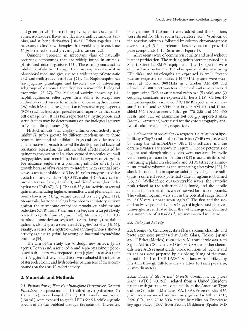
2.1. Preparation of Phenylaminojuglone Derivatives: GeneralProcedure. Suspensions of 1,5-dihydroxynaphthalene (1;1.25mmol), rose bengal (20mg; 0.02mmol), and water(150mL) were exposed to green LEDs for 5 h while a gentlestream of air was bubbled through the solution. Thereafter,
phenylamines 3 (1.5mmol) were added and the solutionswere stirred for 4 h at room temperature (RT). Work-up ofthe reaction mixtures followed by column chromatographyover silica gel (3 : 1 petroleum ether/ethyl acetate) providedpure compounds 4–15 (Scheme 1; Figure 1).
All reagents were of commercial quality and used withoutfurther purification. The melting points were measured in aStuart Scientific SMP3 equipment. The IR spectra wereobtained in a vector 22-FT Bruker spectrophotometer usingKBr disks, and wavelengths are expressed in cm−1. Protonnuclear magnetic resonance (1H NMR) spectra were mea-sured at 400 and 300MHz in a Bruker AM-400 andUltrashield-300 spectrometers. Chemical shifts are expressedin ppm using TMS as an internal reference (δ scale), and (J)coupling constants are expressed in hertz (Hz). Carbon-13nuclear magnetic resonance (13C NMR) spectra were mea-sured at 100 and 75MHz in a Bruker AM-400 and Ultra-shield-300, spectrometers. Silica gel (70–230 and 230–400mesh) and TLC on aluminum foil 60 F254-supported silica(Merck, Darmstadt) were used for the chromatography ana-lytical columns and TLC, respectively.
2.2. Calculation of Molecular Descriptors. Calculation of lipo-philicity (ClogP) and molar refractivity (CMR) was assessedby using the ChemBioDraw Ultra 11.0 software and theobtained values are shown in Figure 1. Redox potentials ofjuglone and phenylaminojuglones were measured by cyclicvoltammetry at room temperature (RT) in acetonitrile as sol-vent using a platinum electrode and 0.1M tetraethylammo-nium tetrafluoroborate as the supporting electrolyte [35]. Itshould be noted that in aqueous solution by using pulse radi-olysis, a different redox potential value of juglone is obtained[36, 37]. Well-defined quasi-reversible waves, the cathodicpeak related to the reduction of quinone, and the anodicone due to its reoxidation, were observed for the compounds.The voltammograms were run in the potential range from 0to −2.0V versus nonaqueous Ag/Ag+. The first and the sec-ond halfwave potential values (EI1/2) of juglone and phenyla-minojuglones, evaluated from the voltammograms obtainedat a sweep rate of 100mVs−1, are summarized in Figure 1.
2.3. Biological Activity
2.3.1. Reagents. Cellulose acetate filters, sodium chloride, andbacto agar were purchased at Asahi Glass, (Tokyo, Japan)and JT Baker (Mexico), respectively. Metronidazole was fromSigma Aldrich (St. Louis, MO 63103, USA). All other chemi-cals were ACS reagent grade. Stock solutions of juglone andits analogs were prepared by dissolving 50mg of the com-pound in 1mL of 100% DMSO. Solutions were sterilized byfiltration through cellulose acetate filters (0.2mm pore size;25mm diameter).
2.3.2. Bacterial Strain and Growth Conditions. H. pylori26695 (ATCC 700392), isolated from a United Kingdompatient with gastritis, was obtained from the American TypeCulture Collection (Manassas, VA, USA). Frozen stocks ofH.pylori were recovered and routinely grown for 48h at 37°C,5.5% CO2, and 70 to 80% relative humidity on Trypticasesoy agar plates (TSA) from Becton Dickinson (Sparks, MD
2 Oxidative Medicine and Cellular Longevity
21152, USA) supplemented with 0.4%H. pylori selective sup-plement Dent (Oxoid Basingstoke, Hampshire, England),0.3% IsoVitalex (Oxoid), and 5% horse serum from ThermoFisher Scientific HyClone (Utah 84321, USA) [38, 39]. Forliquid growth experiments, cells were grown in Trypticasesoy broth (TSB) (Becton Dickinson) with 5% horse serum,
supplemented with IsoVitalex and Dent (Oxoid). Bacteriawere first grown to an optical density of 0.6 to 1.0 at600 nm (OD600) at pH7.0 and subsequently diluted to astarting OD600 of 0.05. To measure the growth of H. pyloriin liquid medium, a serial dilution was prepared, aliquots ofthe various dilutions were plated on Trypticase soy agar
and/orNHAr
O
OOH
O
2
O
OH
OH
1
hv, O2 H2OSensitiser
ArNH2 (3)
EtOH
OH
4-7 8-15
5
O
OOH5 3 NHAr
2
Amines 3: 2-MePhNH2; 3-MeOPhNH2; 4-MeOPhNH2; 4-HOPhNH2; 3,4,5-(MeO)3PhNH2;4′-H2NPhSO2-Ph-4-NH2, 4′-H2NPh-Ph-4-NH2
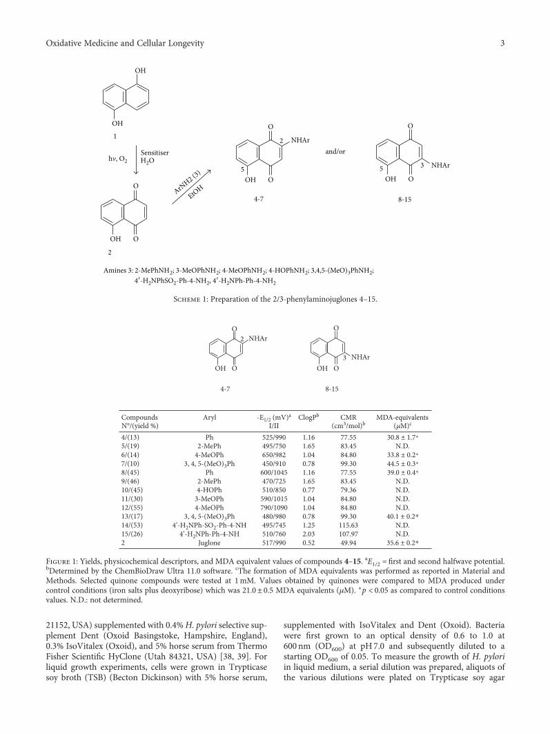
Scheme 1: Preparation of the 2/3-phenylaminojuglones 4–15.
CompoundsN°/(yield %)
Aryl -E1/2 (mV)a
I/IIClogPb CMR
(cm3/mol)bMDA-equivalents
(�휇M)c
4/(13) Ph 525/990 1.16 77.55 30.8 ± 1.7⁎5/(19) 2-MePh 495/750 1.65 83.45 N.D.6/(14) 4-MeOPh 650/982 1.04 84.80 33.8 ± 0.2⁎7/(10) 3, 4, 5-(MeO)3Ph 450/910 0.78 99.30 44.5 ± 0.3⁎8/(45) Ph 600/1045 1.16 77.55 39.0 ± 0.4⁎9/(46) 2-MePh 470/725 1.65 83.45 N.D.10/(45) 4-HOPh 510/850 0.77 79.36 N.D.11/(30) 3-MeOPh 590/1015 1.04 84.80 N.D.12/(55) 4-MeOPh 790/1090 1.04 84.80 N.D.13/(17) 3, 4, 5-(MeO)3Ph 480/980 0.78 99.30 40.1 ± 0.2⁎14/(53) 4’-H2NPh-SO2-Ph-4-NH 495/745 1.25 115.63 N.D.15/(26) 4’-H2NPh-Ph-4-NH 510/760 2.03 107.97 N.D.2 Juglone 517/990 0.52 49.94 35.6 ± 0.2⁎
O
NHAr
NHAr
O
4-7 8-15
2
3OH O
O
OH
Figure 1: Yields, physicochemical descriptors, and MDA equivalent values of compounds 4–15. aE1/2 = first and second halfwave potential.bDetermined by the ChemBioDraw Ultra 11.0 software. cThe formation of MDA equivalents was performed as reported in Material andMethods. Selected quinone compounds were tested at 1mM. Values obtained by quinones were compared to MDA produced undercontrol conditions (iron salts plus deoxyribose) which was 21.0± 0.5 MDA equivalents (μM). ∗p < 0.05 as compared to control conditionsvalues. N.D.: not determined.
3Oxidative Medicine and Cellular Longevity
plates, and the number colony-forming units (CFU) wasdetermined [40].
2.3.3. H. pylori Growth Assay in Liquid Medium. H. pylori(3× 10⁷ cells/mL) were inoculated in 5mL of TSB and sup-plemented with a range of concentrations (0.0 to 1.0 μg/mL) of juglone or a derivative compound. After incubationat 37°C for 48h with constant shaking at 250 rpm in a con-trolled atmosphere (5.5% CO2 and 70% relative humidity),bacterial growth was determined by turbidimetry at 600 nmor by counting colony-forming units on TSA plates [41, 42].
2.3.4. H. pylori Viability Assay. From each of the experimen-tal culture tubes described in the previous section, 100 μL ali-quots were taken at the end of the incubation period toprepare serial dilutions in PBS. Aliquots of 10 μL from eachof these dilutions were plated on TSA and incubated for48 h at 37°C [43]. The number of colony-forming units permL (CFU/mL) corresponding to each experimental condi-tion was determined.
2.3.5. Inhibition Halo Test on Agar Plates. The procedure wasperformed as described by Rodríguez et al. [44]. One hun-dred μL of H. pylori suspension containing 3× 10⁷ cells/mLwas evenly spread over the TSA plates with a metal handleloop. Then, three-millimeter diameter wells were made inthe plates and 30 μL of a series of compound solutions wasdeposited in the wells (corresponding to 0 to 1mg/well).After 48 h of incubation at 37°C, the diameter of the growthinhibition halos was determined.
2.3.6. Determination of Prooxidant Activity. The assay wasbased on TBARS method according to Halliwell et al. [45].Briefly, a mixture containing iron salts, phosphate buffer,and deoxyribose was incubated for 60min at RT in theabsence or presence of quinones. Then, the amount of mal-ondialdehyde (MDA) equivalent produced was determinedby reaction with thiobarbituric acid and further reading at532nm. Results are expressed as μM of MDA equivalents.The prooxidant activity of some selected quinones is shownin Figure 1.
2.3.7. Statistical Analysis. All experiments were performed atleast 3 times and groupswere compared byANOVA test usingGraphPad Prism software (SanDiego, CA 92037, USA). Two-way ANOVA test was used to analyze the dose-responsecurves. A p value< 0.05 was set as statistically significant.
3. Results
3.1. Synthesis of Phenylaminojuglones. The preparation of thephenylaminojuglone derivatives was achieved via a two syn-thetic step sequence from 1,5-dihydroxynaphthalene 1 andthe selected phenylamines 3 according to 1 and Figure 1. Inthe first step, sensitized photooxygenation of compound 1on water gave 5-hydroxy-1,4-naphthoquinone (2, juglone)in 64% yield [46]. Further reaction of juglone 2 with the phe-nylamines in ethanol [47, 48] at room temperature providedthe respective phenylaminojuglones 4–15. In all cases, thereaction gave a mixture of the respective regioisomers as
was observed by thin layer chromatography and proton mag-netic resonance. Pure samples of the regioisomers 4–7 (C-2)and 8–15 (C-3) were isolated by column chromatography(Figure 1). Efforts to isolate minor regioisomers were unsuc-cessful. The formation of regioisomers in these reactionsreveals that they proceed under regiochemical control. Thestructures of the phenylaminojuglones were established bynuclear magnetic resonance (1H-NMR and 13C-NMR) andhigh-resolution mass spectrometry (HRMS). The locationof the phenylamino substituents at the quinone nucleusin compounds 4–7 and 8–15 was determined by bidimen-sional nuclear magnetic resonance (2D-NMR) (data in theSupplementary Material available here).
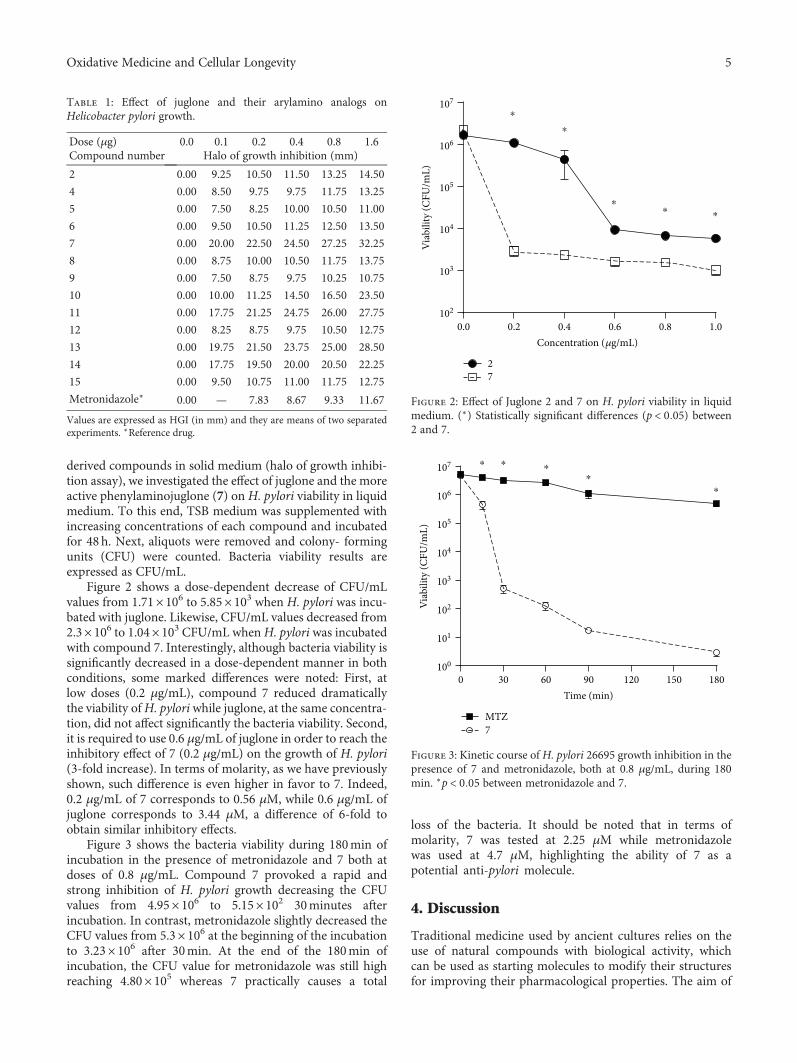
3.2. Inhibition of H. pylori Growth by Juglone,Phenylaminojuglones, andMetronidazole. To assess the effectof juglone and its analogs on H. pylori growth, increasingdoses of compounds were added into TSA well-plates previ-ously seeded with bacteria, which were further incubated for48 h. Table 1 shows the halo of growth inhibition (HGI) inmillimeter obtained for each compound as a function of theirconcentration by using the Diffusion Test assay.
Juglone andmost of its analogs (except 5 and 9)weremoreactive on H. pylori than metronidazole (HGI: 11.67mm).Compared to the antibacterial effect mediated by juglone(HGI: 14.50mm), 5 out of 12phenylaminojuglonesweremoreefficient than juglone with HGI values ranging from 22.25 to32.25mm. A clear representation of this inhibitory effect isunveiled when the antimicrobial activity of compounds basedon molar amounts was compared. For instance, the HGI of33.25mmof 7wasobtained at 4.5μMwhile theHGIof juglone(14.50mm) and metronidazole (11.67mm) were obtained at9.2 and 9.35μM, respectively. In other words, 7 reached a highinhibitory effect on H. pylori growth at half of the dosesrequired by juglone and metronidazole whose effects were byfar lower than 7.
The C-H functionalization in the 1,4-naphthoquinonescaffold at either C-2 or C-3, like in the pairs 4/8, 5/9, 6/12,and 7/13, resulted in similar antibacterial activities as shownby their halo of inhibition. For instance, 4 and 8 have an HGIof 13.25 and 13.75mm, respectively, and 7 and 13 have anHGI of 32.25 and 28.50mm, respectively.
Compound 14, obtained by oxidative amination of 2 withdapsone (4-H2NPhSO2Ph-4′-NH2), displayed higher inhibi-tory activity on H. pylori growth than juglone at all the testeddoses. Since dapsone may act against bacteria by inhibitingthe synthesis of dihydrofolic acid [49] it is likely that such anti-microbial ability mediated by dapsone is contributing to theoverall anti-pylori activity of 14. Finally, arylaminojuglone 15derived from 2 and benzidine (4-H2NPh-Ph-4′-NH2) showeda lower range of activity than juglone. It should be noted thatamines 3 phenylamine, 2-methylphenylamine, 3-methoxy-phenylamine, 4-methoxyphenylamine, 4-hydroxyphenyla-mine, 3,4,5-trimethoxyphenylamine, and benzidine weredevoid of anti-pylori activity when added in the absence ofjuglone (data not shown).
3.3. H. pylori Viability in the Presence of Juglone, 7, andMetronidazole. Once determining the effect of quinone-
4 Oxidative Medicine and Cellular Longevity
derived compounds in solid medium (halo of growth inhibi-tion assay), we investigated the effect of juglone and the moreactive phenylaminojuglone (7) on H. pylori viability in liquidmedium. To this end, TSB medium was supplemented withincreasing concentrations of each compound and incubatedfor 48h. Next, aliquots were removed and colony- formingunits (CFU) were counted. Bacteria viability results areexpressed as CFU/mL.
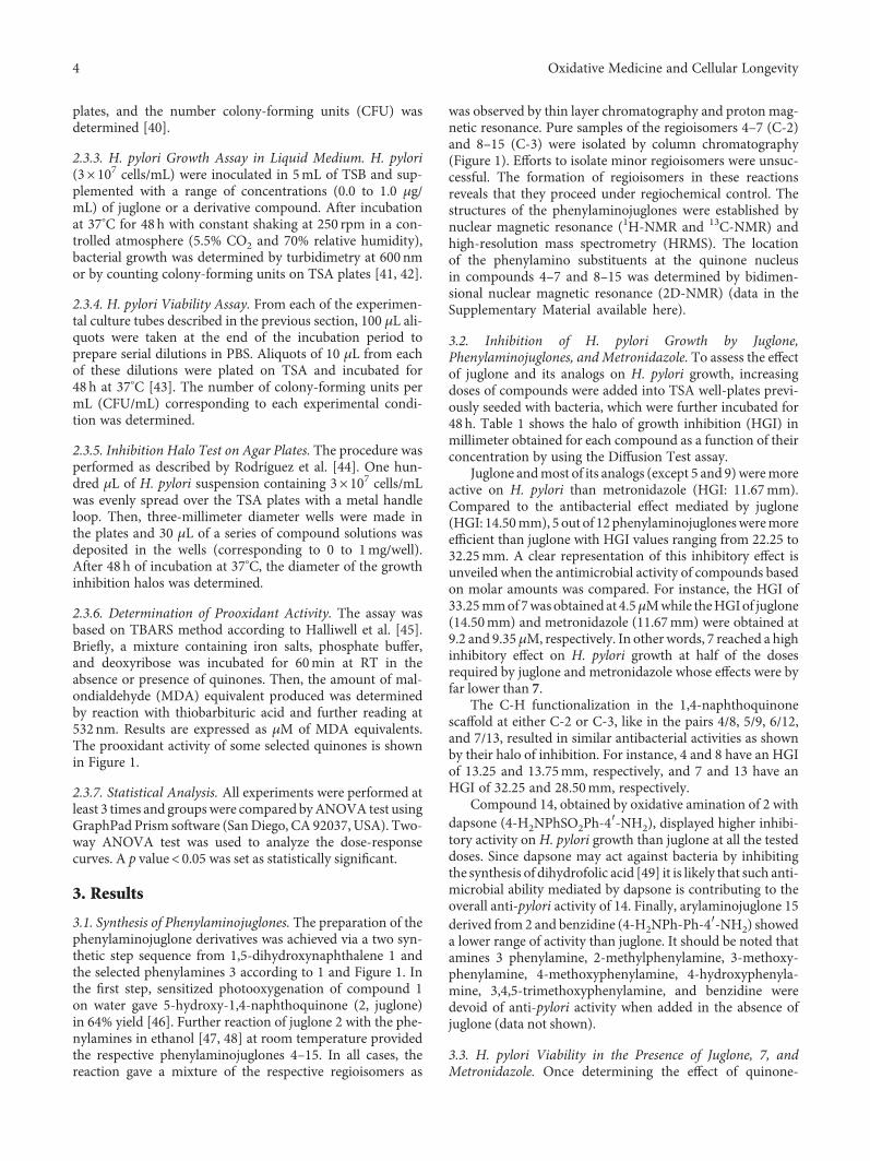
Figure 2 shows a dose-dependent decrease of CFU/mLvalues from 1.71× 10⁶ to 5.85× 10³ when H. pylori was incu-bated with juglone. Likewise, CFU/mL values decreased from2.3× 106 to 1.04× 10³ CFU/mL whenH. pylori was incubatedwith compound 7. Interestingly, although bacteria viability issignificantly decreased in a dose-dependent manner in bothconditions, some marked differences were noted: First, atlow doses (0.2 μg/mL), compound 7 reduced dramaticallythe viability ofH. pylori while juglone, at the same concentra-tion, did not affect significantly the bacteria viability. Second,it is required to use 0.6 μg/mL of juglone in order to reach theinhibitory effect of 7 (0.2 μg/mL) on the growth of H. pylori(3-fold increase). In terms of molarity, as we have previouslyshown, such difference is even higher in favor to 7. Indeed,0.2 μg/mL of 7 corresponds to 0.56 μM, while 0.6 μg/mL ofjuglone corresponds to 3.44 μM, a difference of 6-fold toobtain similar inhibitory effects.
Figure 3 shows the bacteria viability during 180min ofincubation in the presence of metronidazole and 7 both atdoses of 0.8 μg/mL. Compound 7 provoked a rapid andstrong inhibition of H. pylori growth decreasing the CFUvalues from 4.95× 10⁶ to 5.15× 10² 30minutes afterincubation. In contrast, metronidazole slightly decreased theCFU values from 5.3× 10⁶ at the beginning of the incubationto 3.23× 10⁶ after 30min. At the end of the 180min ofincubation, the CFU value for metronidazole was still highreaching 4.80× 10⁵ whereas 7 practically causes a total
loss of the bacteria. It should be noted that in terms ofmolarity, 7 was tested at 2.25 μM while metronidazolewas used at 4.7 μM, highlighting the ability of 7 as apotential anti-pylori molecule.
4. Discussion
Traditional medicine used by ancient cultures relies on theuse of natural compounds with biological activity, whichcan be used as starting molecules to modify their structuresfor improving their pharmacological properties. The aim of
Concentration (�휇g/mL)
Viab
ility
(CFU
/mL)
0.0 0.2 0.4 0.6 0.8 1.0102
103
104
105
106
107
27
⁎
⁎
⁎⁎ ⁎
Figure 2: Effect of Juglone 2 and 7 on H. pylori viability in liquidmedium. (∗) Statistically significant differences (p < 0 05) between2 and 7.
Time (min)
Viab
ility
(CFU
/mL)
1209060300 150 180100
101
102
103
104
105
106
107
MTZ7
⁎ ⁎ ⁎⁎
⁎
Figure 3: Kinetic course of H. pylori 26695 growth inhibition in thepresence of 7 and metronidazole, both at 0.8 μg/mL, during 180min. ∗p < 0 05 between metronidazole and 7.
Table 1: Effect of juglone and their arylamino analogs onHelicobacter pylori growth.
Dose (μg) 0.0 0.1 0.2 0.4 0.8 1.6Compound number Halo of growth inhibition (mm)
2 0.00 9.25 10.50 11.50 13.25 14.50
4 0.00 8.50 9.75 9.75 11.75 13.25
5 0.00 7.50 8.25 10.00 10.50 11.00
6 0.00 9.50 10.50 11.25 12.50 13.50
7 0.00 20.00 22.50 24.50 27.25 32.25
8 0.00 8.75 10.00 10.50 11.75 13.75
9 0.00 7.50 8.75 9.75 10.25 10.75
10 0.00 10.00 11.25 14.50 16.50 23.50
11 0.00 17.75 21.25 24.75 26.00 27.75
12 0.00 8.25 8.75 9.75 10.50 12.75
13 0.00 19.75 21.50 23.75 25.00 28.50
14 0.00 17.75 19.50 20.00 20.50 22.25
15 0.00 9.50 10.75 11.00 11.75 12.75
Metronidazole∗ 0.00 — 7.83 8.67 9.33 11.67
Values are expressed as HGI (in mm) and they are means of two separatedexperiments. ∗Reference drug.
5Oxidative Medicine and Cellular Longevity
this work was to synthesize a series of phenylaminojugloneswith anti-H. pylori biological activity. Among the membersof the series, five congeners were found 1.9- to 2.8-timesmore active than one standard therapeutic drug (i.e., metro-nidazole), a currently standard anti-pylori drug [40, 50–52].
Even though the discovery of molecular mechanismsunderlying the antibacterial effects of the phenylaminoju-glones was beyond our objectives, we noted that their anti-pylori activity depends on the nature and location of thenitrogen substituents at the quinone nucleus of the juglonescaffold. Thus, insertion of the 3,4,5-trimethoxyphenylaminogroup at the 2 position in juglone, as in compound 7 (HGI:32.25mm), induced a strong effect on the antibacterial activ-ity of the juglone scaffold (HGI: 14.50mm). Conversely, theinsertion of the phenylamino, 2-phenylamino, and 4-methoxyphenylamino groups, as in compounds 4 (HGI:13.25mm), 5 (HGI: 11.00mm) and 6 (13.50mm), causesdecreasing effects on the antibacterial activity of the juglonescaffold. Inspection of Table 1 reveals that, in general, theinsertion of the nitrogen substituents in the 3 position inducehigher effects on the antibacterial activity compared to theinsertion of nitrogen substituents in the 2 position of thejuglone scaffold. Among the members of the 3-arylaminojuglone derivatives 8–15, compounds 10, 12, and13 display remarkable antibacterial activities. Once again, interms of molarity, such high doses (1.6 μg/mL) correspondto 6 μM of compound 4, 4.5 μM of compound 7, and 9.2μM of juglone, strengthening the assumption about the effi-cacy of compound 7.
By comparing data from Figure 1 and Table 1 (HGI ofjuglone and its analogs as well as molecular descriptors), itcan be inferred that compounds 7, 10, and 13 (three of themost active phenylaminojuglones) have lower ClogP valuesthan other molecules of the series (around 0.78), showing amarked hydrophilic character. Moreover, when comparedwith compounds 9 and 13 that share similar values of redoxpotential and polarizability but different lipophilia, they havestrong differences in terms of anti-pylori activity: HGI: 10.75and 28.50mm, respectively. It appears then that compoundswith a significant hydrophilic degree will have a more pro-nounced antibacterial activity. Interestingly, it has beenreported that the membrane surface of H. pylori is ratherhydrophilic and it is negatively charged [53]. This propertywould facilitate the entry of these molecules inside the bacte-ria, facilitating their biological activity. This temptinghypothesis is however unlike because juglone has a ClogPvalue (0.52) even lower than the three former molecules butits HGI was only of 14.5mm.
It is to be expected that the redox status of the cellularsystem would be modulated by ROS. Since the ease of ROSgeneration through reduction of a quinonoid would dependon its electrochemical parameters, the redox potential of aquinone would influence its overall biological profile, whichencompasses the functional, toxicological, mutagenic, andantitumor activities. With this view, the redox potential ofthese naphthoquinones was determined by cyclic voltamme-try using acetonitrile, an aprotic solvent, which mimics theenvironment of the cell membrane [54]. Figure 1 shows thatE1/2 values for the first one electron transfer, corresponding
to the formation of the radical-anions of compounds 4–15,are spread into a broad potential range from −790 to−450mV. In addition, we noted that 7 out of 12 phenylami-nojuglones have higher redox potential values (from −450 to−510mV) than juglone (−517mV). It is tempting to assumethat the effect on redox potential by the insertion of nitrogengroups, such as PhNH-, in the 5-hydroxy-1,4-naphthoqui-none (juglone) scaffold may be clearly predicted, but the sit-uation is a little bit more complex. Indeed, it is reasonable toassume that the electron acceptor ability of these phenylami-nojuglone will depend, in part, on the location of the nitrogendonor in the quinone core and on the extent of the conjuga-tive effect of this group to the intramolecular hydrogen bondof the molecule. A similar situation by taking the 1,4-naphthoquinone scaffold has been discussed by Aguilar-Martinez et al. [55].
Regarding the influence of other molecular descriptorssuch as molar refractivity, it seems that high polarizabilityvalues enhance the anti-pylori activity. Indeed, when com-pared compounds 5 and 14, they have the same redox poten-tial values (−495mV) and similar lipophilia (1.65 versus1.25) but different polarizability values. Accordingly, com-pound 14 with a high molar refractivity (115.63), it has a highanti-pylori activity (HGI: 22.25mm). However, compound 7has less lipophilicity, high polarizability and low redoxpotential compared to 12, and their HGI values were mark-edly different: 32.25 and 12.75mm, respectively. All theseresults illustrate how difficult is to attribute a biologicalresponse to a given molecular descriptor. Interestingly,Figure 1 shows that phenylaminojuglones displaying highHGI (i.e., 7, 13) have the highest prooxidant activities asshown by the TBARS production, suggesting a potential linkbetween oxidative stress and antibacterial activity. Support-ing the role of oxidative stress during chronic gastritis associ-ated with H. pylori infection, it should be noted that theadministration of coenzyme q10 decreases mucosal inflam-mation in such patients [56].
In conclusion, compound 7 is a promissory anti-pyloricompound already active as soon as 30min of incubation ata very low concentration (0.56 μM). When using at 4.5 μM(1.6 μg/mL), the calculated halo of growth inhibition for 7was 32.25mm. These preliminary results make 7 an interest-ing lead molecule modulated by other substituting groupsand to conduct further assays.
Conflicts of Interest
The authors declare that they have no conflicts of interest.
Acknowledgments
The authors express an immense gratitude to the studentsConstanza González, Gabriel Vilches, and Cynthia Estela, fortheir significant contribution in some experiments. They alsothank Nicanor Villaroel for his excellent technical assistanceand to Fondo Nacional de Ciencia y Tecnología (Grant nos.1120050 to Julio Benites and 1120126 and 1150384 to HéctorToledo) for the financial support given to this study.
6 Oxidative Medicine and Cellular Longevity
Abbreviations
CFU: Colony-forming unitsHGI: Halo of growth inhibitionMDA: MalondialdehydeTSA: Trypticase soy agarTSB: Trypticase soy broth.
Supplementary Materials
Description of the general procedure for the preparationof phenylaminojuglone 4–7 and 8–15 and spectral dataof nuclear magnetic resonance (1H NMR and 13CNMR) and high-resolution mass spectrometry (HRMS).(Supplementary Materials)
References
[1] B. Dunn, H. Cohen, and M. Blaser, “Helicobacter pylori,” Clin-ical Microbiology Reviews, vol. 10, no. 4, pp. 720–741, 1997.
[2] J. D. Dubreuil, G. D. Giudice, and R. Rappuoli, “Helicobacterpylori interactions with host serum and extracellular matrixproteins: potential role in the infectious process,”Microbiologyand Molecular Biology Reviews, vol. 66, no. 4, pp. 617–629,2002.
[3] L. D. Butcher, G. den Hartog, P. B. Ernst, and S. E. Crowe,“Oxidative stress resulting from Helicobacter pylori infectioncontributes to gastric carcinogenesis,” Cellular and MolecularGastroenterology and Hepatology, vol. 3, no. 3, pp. 316–322,2017.
[4] International Agency for Research on Cancer (IARC), “Schis-tosomes, liver flukes, and Helicobacter pylori,” in IARC Work-ing group on the evaluation of carcinogenic risk to humans, vol.61, pp. 177–240, Lyon, France, 1994.
[5] Y. Yamaoka, Helicobacter pylori: Molecular Genetics and Cel-lular Biology, Horizon Scientific Press, Poole, UK, 2008.
[6] H. L. Mobley, M. J. Cortesia, L. E. Rosenthal, and B. D. Jones,“Characterization of urease from campylobacter pylori,” Jour-nal of Clinical Microbiology, vol. 26, no. 5, pp. 831–836, 1988.
[7] C. Montecucco and R. Rappuoli, “Living dangerously: howHelicobacter pylori survives in the human stomach,” NatureReviewsMolecular Cell Biology, vol. 2, no. 6, pp. 457–466, 2001.
[8] M. J. Blaser and J. C. Atherton, “Helicobacter pylori persis-tence: biology and disease,” The Journal of Clinical Investiga-tion, vol. 113, no. 3, pp. 321–333, 2004.
[9] C. T. Baldari, A. Lanzavecchia, and J. L. Telford, “Immune sub-version byHelicobacter pylori,” Trends in Immunology, vol. 26,no. 4, pp. 199–207, 2005.
[10] H. M. Mitchell, “The epidemiology of Helicobacter pylori,”Current Topics in Microbiology and Immunology, vol. 241,pp. 11–30, 1999.
[11] A. Covacci, J. L. Telford, G. Del Giudice, J. Parsonnet, andR. Rappuoli, “Helicobacter pylori virulence and genetic geogra-phy,” Science, vol. 284, no. 5418, pp. 1328–1333, 1999.
[12] J. Torres, G. Pérez-Pérez, K. J. Goodman et al., “A comprehen-sive review of the natural history of Helicobacter pylori infec-tion in children,” Archives of Medical Research, vol. 31, no. 5,pp. 431–469, 2000.
[13] J. Parsonnet, “Helicobacter pylori: the size of the problem,”Gut, vol. 43, Supplement 1, pp. S6–S9, 1998.
[14] P. Correa, “Helicobacter pylori as a pathogen and carcinogen,”Journal of Physiology and Pharmacology, vol. 48, Supplement4, pp. 19–24, 1997.
[15] B. C. Delaney, “Who benefits fromHelicobacter pylori eradica-tion?,” BMJ, vol. 332, no. 7535, pp. 187-188, 2006.
[16] J. A. Lane, L. J. Murray, S. Noble et al., “Impact of Helicobacterpylori eradication on dyspepsia, health resource use, and qual-ity of life in the Bristol helicobacter project: randomised con-trolled trial,” BMJ, vol. 332, no. 7535, pp. 199–204, 2006.
[17] Y. Nakayama and D. Y. Graham, “Helicobacter pylori infec-tion: diagnosis and treatment,” Expert Review of Anti-infective Therapy, vol. 2, no. 4, pp. 599–610, 2014.
[18] Y. C. Wang, “Medicinal plant activity on Helicobacter pylorirelated diseases,” World Journal of Gastroenterology, vol. 20,no. 30, pp. 10368–10382, 2014.
[19] P. Ruggiero, F. Tombola, G. Rossi et al., “Polyphenols reducegastritis induced by Helicobacter pylori infection or VacAtoxin administration in mice,” Antimicrobial Agents and Che-motherapy, vol. 50, no. 7, pp. 2550–2552, 2006.
[20] P. Ruggiero, G. Rossi, F. Tombola et al., “Red wine and greentea reduce H pylori- or VacA-induced gastritis in a mousemodel,” World Journal of Gastroenterology, vol. 13, no. 3,pp. 349–354, 2007.
[21] F. Tombola, S. Campello, L. De Luca et al., “Plant polyphenolsinhibit VacA, a toxin secreted by the gastric pathogen Helico-bacter pylori,” FEBS Letters, vol. 543, no. 1-3, pp. 184–189,2003.
[22] P. Ruggiero, S. Peppoloni, D. Berti, R. Rappuoli, and G. D. Giu-dice, “New strategies for the prevention and treatment of Heli-cobacter pylori infection,” Expert Opinion on InvestigationalDrugs, vol. 11, no. 8, pp. 1127–1138, 2002.
[23] R. H. Thomson, “Distribution of naturally occurring qui-nones,” Pharmaceutisch Weekblad, vol. 13, no. 2, pp. 70–73,1991.
[24] G. Powis, “Free radical formation by antitumor quinones,”Free Radical Biology and Medicine, vol. 6, no. 1, pp. 63–101,1989.
[25] H. Haraguchi, K. Yokoyama, S. Oike, M. Ito, and H. Nozaki,“Respiratory stimulation and generation of superoxide radicalsin pseudomonas aeruginosa by fungal naphthoquinones,”Archives of Microbiology, vol. 167, no. 1, pp. 6–10, 1997.
[26] P. F. Carneiro, S. B. do Nascimento, A. V. Pinto et al., “Newoxirane derivatives of 1,4-naphthoquinones and their evalua-tion against T. cruzi epimastigote forms,” Bioorganic &Medic-inal Chemistry, vol. 20, no. 16, pp. 4995–5000, 2012.
[27] Y. Kumagai, Y. Shinkai, T. Miura, and A. K. Cho, “The chem-ical biology of naphthoquinones and its environmental impli-cations,” Annual Review of Pharmacology and Toxicology,vol. 52, no. 1, pp. 221–247, 2012.
[28] L. Salmon-Chemin, E. Buisine, V. Yardley et al., “2- and 3-substituted 1,4-naphthoquinone derivatives as subversive sub-strates of trypanothione reductase and lipoamide dehydroge-nase from Trypanosoma cruzi: synthesis and correlationbetween redox cycling activities and in vitro cytotoxicity,”Journal of Medicinal Chemistry, vol. 44, no. 4, pp. 548–565,2001.
[29] T. Tran, E. Saheba, A. V. Arcerio et al., “Quinones as antimy-cobacterial agents,” Bioorganic &Medicinal Chemistry, vol. 12,no. 18, pp. 4809–4813, 2004.
[30] R. P. Verma and C. A. Hansch, “A comparison between twopolarizability parameters in chemical–biological interactions,”
7Oxidative Medicine and Cellular Longevity
Bioorganic & Medicinal Chemistry, vol. 13, no. 7, pp. 2355–2372, 2005.
[31] Y.-H. Kong, L. Zhang, Z.-Y. Yang et al., “Natural productjuglone targets three key enzymes from Helicobacter pylori:inhibition assay with crystal structure characterization,” ActaPharmacologica Sinica, vol. 29, no. 7, pp. 870–876, 2008.
[32] H. R. Nasiri, M. G. Madej, R. Panisch et al., “Design, synthesis,and biological testing of novel naphthoquinones as substrate-based inhibitors of the quinol/fumarate reductase from Woli-nella succinogenes,” Journal of Medicinal Chemistry, vol. 56,no. 23, pp. 9530–9541, 2013.
[33] Y. C. Wang and Y. H. Lin, “Anti-gastric adenocarcinomaactivity of 2-methoxy-1,4-naphthoquinone, an anti-Helicobac-ter pylori compound from Impatiens balsamina L,” Fitoterapia,vol. 83, no. 8, pp. 1336–1344, 2012.
[34] S. Skouloubris, K. Djaout, I. Lamarre et al., “Targeting of Heli-cobacter pylori thymidylate synthase ThyX by non-mitotoxichydroxy-naphthoquinones,” Open Biology, vol. 5, no. 6, article150015, 2015.
[35] Y. Prieto, M. Muñoz, V. Arancibia, M. Valderrama, F. J. Lahoz,and M. Luisa Martín, “Synthesis, structure and properties ofruthenium(II) complexes with quinolinedione derivatives aschelate ligands: crystal structure of [Ru(CO)2Cl2(6-methoxy-benzo[g]quinoline-5,10-dione)],” Polyhedron, vol. 26, no. 18,pp. 5527–5532, 2007.
[36] T. Mukherjee, “One-electron reduction of juglone (5-hydroxy-1,4-naphthoquinone): a pulse radiolysis study,” InternationalJournal of Radiation Applications and Instrumentation. PartC. Radiation Physics and Chemistry, vol. 29, no. 6, pp. 455–462, 1987.
[37] P. Wardman, “Reduction potentials of one-electron couplesinvolving free radicals in aqueous solution,” Journal of Physicaland Chemical Reference Data, vol. 18, no. 4, pp. 1637–1755,1989.
[38] H. Toledo, M. Valenzuela, A. Rivas, and C. A. Jerez, “Acidstress response in Helicobacter pylori,” FEMS MicrobiologyLetters, vol. 213, no. 1, pp. 67–72, 2002.
[39] O. Cerda, A. Rivas, and H. Toledo, “Helicobacter pylori strainATCC700392 encodes a methyl-accepting chemotaxis recep-tor protein (MCP) for arginine and sodium bicarbonate,”FEMS Microbiology Letters, vol. 224, no. 2, pp. 175–181, 2003.
[40] H. Toledo and R. López-Solís, “Tetracycline resistance in Chil-ean clinical isolates of Helicobacter pylori,” Journal of Antimi-crobial Chemotherapy, vol. 65, no. 3, pp. 470–473, 2010.
[41] K. A. Stevens, B. W. Sheldon, N. A. Klapes, and T. R. Klaen-hammer, “Nisin treatment for inactivation of Salmonella spe-cies and other gram-negative bacteria,” Applied andEnvironmental Microbiology, vol. 57, no. 12, pp. 3613–3615,1991.
[42] R. Díaz-Gómez, R. López-Solís, E. Obreque-Slier, andH. Toledo-Araya, “Comparative antibacterial effect of gallicacid and catechin againstHelicobacter pylori,” LWT - Food Sci-ence and Technology, vol. 54, no. 2, pp. 331–335, 2013.
[43] I. A. Eydelnant and N. Tufenkji, “Cranberry derived proantho-cyanidins reduce bacterial adhesion to selected biomaterials,”Langmuir, vol. 24, no. 18, pp. 10273–10281, 2008.
[44] M. J. Rodríguez Vaquero, M. R. Alberto, and M. C. Manca deNadra, “Antibacterial effect of phenolic compounds from dif-ferent wines,” Food Control, vol. 18, no. 2, pp. 93–101, 2007.
[45] B. Halliwell, J. M. C. Gutteridge, and O. I. Aruoma, “Thedeoxyribose method: a simple “test-tube” assay for
determination of rate constants for reactions of hydroxyl rad-icals,” Analytical Biochemistry, vol. 165, no. 1, pp. 215–219,1987.
[46] B. Julio, C. Michael, M. Luis et al., “Green synthetic approachesto furoylnaphthohydroquinone and juglone,” Journal of theChilean Chemical Society, vol. 59, no. 2, pp. 2455–2457, 2014.
[47] D. Bhasin, S. N. Chettiar, J. P. Etter, M. Mok, and P. K. Li,“Anticancer activity and SAR studies of substituted 1,4-naphthoquinones,” Bioorganic & Medicinal Chemistry,vol. 21, no. 15, pp. 4662–4669, 2013.
[48] L. I. Lopez-Lopez, J. J. Vaquera Garcia, A. Saenz-Galindo, andS. Y. Silva-Belmares, “Ultrasonic and microwave assisted syn-thesis of nitrogen-containing derivatives of juglone as poten-tial antibacterial agents,” Letters in Organic Chemistry,vol. 11, no. 8, pp. 573–582, 2014.
[49] M. D. Coleman, “Dapsone: modes of action, toxicity and pos-sible strategies for increasing patient tolerance,” British Journalof Dermatology, vol. 129, no. 5, pp. 507–513, 1993.
[50] C. Bonacorsi, M. S. G. Raddi, I. Z. Carlos, M. Sannomiya, andW. Vilegas, “Anti-Helicobacter pylori activity and immunosti-mulatory effect of extracts from Byrsonima crassa Nied. (Mal-pighiaceae),” BMC Complementary and Alternative Medicine,vol. 9, no. 1, pp. 1–7, 2009.
[51] S. Chaves, M. Gadanho, R. Tenreiro, and J. Cabrita, “Assess-ment of metronidazole susceptibility in Helicobacter pylori:statistical validation and error rate analysis of breakpointsdetermined by the disk diffusion test,” Journal of ClinicalMicrobiology, vol. 37, no. 5, pp. 1628–1631, 1999.
[52] L. Lang and F. Garcia, “Comparison of E-test and disk diffu-sion assay to evaluate resistance of Helicobacter pylori isolatesto amoxicillin, clarithromycin, metronidazole and tetracyclinein Costa Rica,” International Journal of Antimicrobial Agents,vol. 24, no. 6, pp. 572–577, 2004.
[53] J. I. Smith, B. Drumm, A. W. Neumann, Z. Policova, and P. M.Sherman, “In vitro surface properties of the newly recognizedgastric pathogen Helicobacter pylori,” Infection and Immunity,vol. 58, no. 9, pp. 3056–3060, 1990.
[54] F. C. Abreu, P. A. L. Ferraz, andM. O. F. Goulart, “Some appli-cations of electrochemistry in biomedical chemistry. emphasison the correlation of electrochemical and bioactive proper-ties,” Journal of the Brazilian Chemical Society, vol. 13, no. 1,pp. 19–35, 2002.
[55] M. Aguilar-Martinez, G. Cuevas, M. Jimenez-Estrada,I. Gonzalez, B. Lotina Hennsen, and M. Macias-Ruvalcaba,“An experimental and theoretical study of the substituenteffects on the redox properties of 2-[(R-phenyl)amine]-1,4-naphthalenediones in acetonitrile,” The Journal of OrganicChemistry, vol. 64, no. 10, pp. 3684–3694, 1999.
[56] A. Rahmani, G. Abangah, A. Moradkhani, M. R. HafeziAhmadi, and K. Asadollahi, “Coenzyme Q10 in combinationwith triple therapy regimens ameliorates oxidative stress andlipid peroxidation in chronic gastritis associated withH. Pyloriinfection,” Journal of Clinical Pharmacology, vol. 55, no. 8,pp. 842–847, 2015.
8 Oxidative Medicine and Cellular Longevity

Stem Cells International
Hindawiwww.hindawi.com Volume 2018
Hindawiwww.hindawi.com Volume 2018
MEDIATORSINFLAMMATION
of
EndocrinologyInternational Journal of
Hindawiwww.hindawi.com Volume 2018
Hindawiwww.hindawi.com Volume 2018
Disease Markers
Hindawiwww.hindawi.com Volume 2018
BioMed Research International
OncologyJournal of
Hindawiwww.hindawi.com Volume 2013
Hindawiwww.hindawi.com Volume 2018
Oxidative Medicine and Cellular Longevity
Hindawiwww.hindawi.com Volume 2018
PPAR Research
Hindawi Publishing Corporation http://www.hindawi.com Volume 2013Hindawiwww.hindawi.com
The Scientific World Journal
Volume 2018
Immunology ResearchHindawiwww.hindawi.com Volume 2018
Journal of
ObesityJournal of
Hindawiwww.hindawi.com Volume 2018
Hindawiwww.hindawi.com Volume 2018
Computational and Mathematical Methods in Medicine
Hindawiwww.hindawi.com Volume 2018
Behavioural Neurology
OphthalmologyJournal of
Hindawiwww.hindawi.com Volume 2018
Diabetes ResearchJournal of
Hindawiwww.hindawi.com Volume 2018
Hindawiwww.hindawi.com Volume 2018
Research and TreatmentAIDS
Hindawiwww.hindawi.com Volume 2018
Gastroenterology Research and Practice
Hindawiwww.hindawi.com Volume 2018
Parkinson’s Disease
Evidence-Based Complementary andAlternative Medicine
Volume 2018Hindawiwww.hindawi.com
Submit your manuscripts atwww.hindawi.com