in vitro inactivation of endodontic pathogens with nd:yag and er:yag lasers
TRANSCRIPT

ORIGINAL ARTICLE
In vitro inactivation of endodontic pathogens with Nd:YAGand Er:YAG lasers
Maarten A. Meire & Tom Coenye & Hans J. Nelis &
Roeland J. G. De Moor
Received: 4 March 2011 /Accepted: 1 June 2011 /Published online: 21 June 2011# Springer-Verlag London Ltd 2011
Abstract Both Nd:YAG and Er:YAG lasers have beensuggested as root canal disinfection aids. The aim of this invitro study is to compare both wavelengths in terms ofirradiation dose required for microbial inactivation, toquantify these irradiation doses and to investigate theinfluence of certain (laser) parameters on the antimicrobialefficacy. Agar plates containing a uniform layer ofEnterococcus faecalis, Candida albicans or Propionibacte-rium acnes were mounted perpendicularly underneath thelaser handpieces (5 mm spot). The Er:YAG laser wasoperated in single-pulse mode. Pulse energies of 40–400 mJand pulse lengths of 100, 300, 600, and 1,000 μs weretested. After incubation at 37°C for 48 h, growth on theplates was scored. The pulse energy yielding completeabsence of growth over the entire spot area was taken as thetotal inhibition threshold (TIT). TITs were determined forevery species and pulse length. The Nd:YAG laser wasoperated with pulse trains because single pulses wereineffective. Output power was 15 W and frequency was100 Hz. Spots were irradiated for 5–120 s. After incubation,the diameters of the inhibition zones were measured. Forthe Er:YAG laser, TITs varied between 100 and 210 mJ, anddiffered significantly between species and pulse lengths.Using Nd:YAG irradiation, TITs were around 5,300 J/cm2
for C. albicans and 7,100 J/cm2 for P. acnes. No inhibitionwas observed for E. faecalis. Er:YAG irradiation wassuperior to Nd:YAG in inactivating microorganisms on agarsurfaces.
Keywords Laser . Disinfection . Endodontics . Nd:YAG . Er:YAG .Enterococcus .Propionibacterium .Candida
Introduction
The Nd:YAG laser was among the first lasers to bescientifically evaluated for its use in endodontics. Thedisinfecting capabilities of this laser have been investigatedin a number of in vitro studies, mostly on experimentallyinfected root canals [1–10]. In these studies, the laser-treated teeth usually yield fewer bacteria than the untreatedcontrols. However, sterility was never observed, and theantibacterial effect of a laser treatment never supersedesthat of conventional NaOCl treatment. Therefore, the Nd:YAG laser has been regarded as an adjunct rather than analternative to existing root canal disinfection protocols [1].Another observation is the obvious variation in lasingparameters (e.g., output power, irradiation time, diameter ofthe fiber, use of a dye) used for root canal disinfection,proving the lack of scientific basis.
The ability of the Nd:YAG laser to kill microorganismshas been demonstrated in laboratory studies [11–13] but itsexact killing mechanism is still controversial. Ward et al.[12] concluded that the bactericidal action of Nd:YAG laserlight was due partly to heating and partly to an additional,as yet undefined, mechanism. Grönqvist et al. [11] statedthat the antimicrobial effect of the Nd:YAG laser light iscaused by a photothermal effect rather than a photochem-ical effect. Hibst et al. [14] showed that, at least for
M. A. Meire (*) : R. J. G. De MoorDepartment of Operative Dentistry and Endodontology,Dental School, Ghent University Hospital, Ghent University,De Pintelaan 185/P8,9000 Gent, Belgiume-mail: [email protected]
T. Coenye :H. J. NelisLaboratory for Pharmaceutical Microbiology, Ghent University,Harelbekestraat 72,9000 Gent, Belgium
Lasers Med Sci (2012) 27:695–701DOI 10.1007/s10103-011-0940-z

Escherichia coli, inactivation by high-power NIR laserirradiation is based solely on a thermal process. Pirnat et al.have recently demonstrated that most bacteria are virtuallytransparent to 1,064-nm wavelength light and that theinteraction between NIR laser light and the bacterialmicroenvironment, most likely in the form of heating, isthe primary mediator of cell death [15]. In both in vitrostudies of Ward et al. and Grönqvist et al., high energydensities were required for bacterial inactivation, i.e.,several thousand J/cm2.
The Er:YAG laser has been the subject of research indentistry since the late 1980s. The focus has beenprimarily on dental hard tissue removal, since itswavelength correlates closely with the absorption max-imum of hydroxyapatite. In addition, the Er:YAG laserhas a very high absorption in water and this increases itsdisinfection potential, since water is a main constituentof most microorganisms. Ando et al. [16] examined thebactericidal effect of the Er:YAG laser on periopathogenicbacteria in vitro and found that the Er:YAG laser has ahigh bactericidal potential at a low energy level. Theapplication of the Er:YAG laser for root canal disinfectionhas gained little attention in the literature. A fewinvestigators have reported the antibacterial effect of theEr:YAG laser in experimentally infected root canals [8, 17,18], or in bacterial biofilm models [19, 20]. The latterstudies concluded that Er:YAG lasers had an anti-biofilmeffect at a low energy and that their use could bebeneficial in endodontic treatment. Again, no scientificbackup exists for the antibacterial lasing parameters withthis wavelength.
A direct and systematic comparison between the anti-bacterial effect of Nd:YAG and Er:YAG lasers has not beenmade. In addition, the influence of laser parameters such aspulse length, fluence, and irradiance on the antibacterialaction of both wavelengths deserves further investigation.
Materials and methods
Strains and culture conditions
The microorganisms and the culture conditions that wereused in this study are listed in Table 1. Cultures weremaintained on solid growth media at 4°C and subculturedregularly. Prior to each experiment, cultures were grown inthe respective liquid medium (Table 1) and incubatedovernight at 37°C. P. acnes was cultured under anaerobicconditions using the Anaerocult A Mini system (Merck).
Inoculation of agar plates
Overnight cultures were diluted 1/10 in their respectiveliquid medium and 1.5-ml aliquots of this suspension werepipetted onto fresh agar plates. These plates had beenprepared with a standard volume of 20 ml, in order to havea similar level of agar in each plate. The liquid was spreadevenly over the agar surface, excess liquid was removed,and the plates were dried for 30 min in a laminar air flowcabinet. In this way, agar plates with a uniform layer ofmicroorganisms were obtained.
In addition, E. faecalis agar plates were prepared withthe bacteria inside the agar instead of at the surface. To thisend, an E. faecalis suspension was mixed with molten TSAand 20-ml volumes were poured into Petri dishes. In thisway, agar plates with E. faecalis cells homogeneouslydistributed inside the agar were obtained.
Laser devices and irradiation of agar plates
The Er:YAG laser (AT Fidelis, Fotona, Ljubljana, Slovenia)operating at 2,940 nm was used with a dermatological handpiece (R11, Fotona) mounted on the articulated arm. Thespot size was set at 5 mm. Each Petri dish containing theinoculated agar was placed perpendicularly underneath thelaser hand piece and the distance between both wasadjusted by means of a height-adjustable table so as toobtain a spot size of 5 mm. The Er:YAG laser was operatedwith a single pulse. To this end, the frequency was set at2 Hz. Pulse energies of 40 to 400 mJ were applied in stepsof 20 mJ. Four pulse lengths were tested: very short pulse(VSP, 100 μs), short pulse (SP, 300 μs), long pulse (LP,
Table 1 Strains and culture conditions
Species Strain Liquid growth medium Solid growth medium Culture condition
Enterococcus faecalis ATCC 10541 Tryptic soy broth (TSB) Tryptic soy agar (TSA) Aerobic
Propionibacterium acnes LMG 16711 Reinforced clostridial medium (RCM) RCM agar Anaerobic
Candida albicans ATCC 10231 Sabouraud broth Sabouraud dextrose agar Aerobic
696 Lasers Med Sci (2012) 27:695–701
The aim of the present study is to compare the in vitroantimicrobial action of Er:YAG and Nd:YAG laser irradia-tion on different microorganisms associated with endodon-tic infections (Enterococcus faecalis, Candida albicans,Propionibacterium acnes), to quantify the irradiation dosesrequired for microbial inactivation and to investigate theinfluence of certain laser parameters on the antimicrobialefficacy.
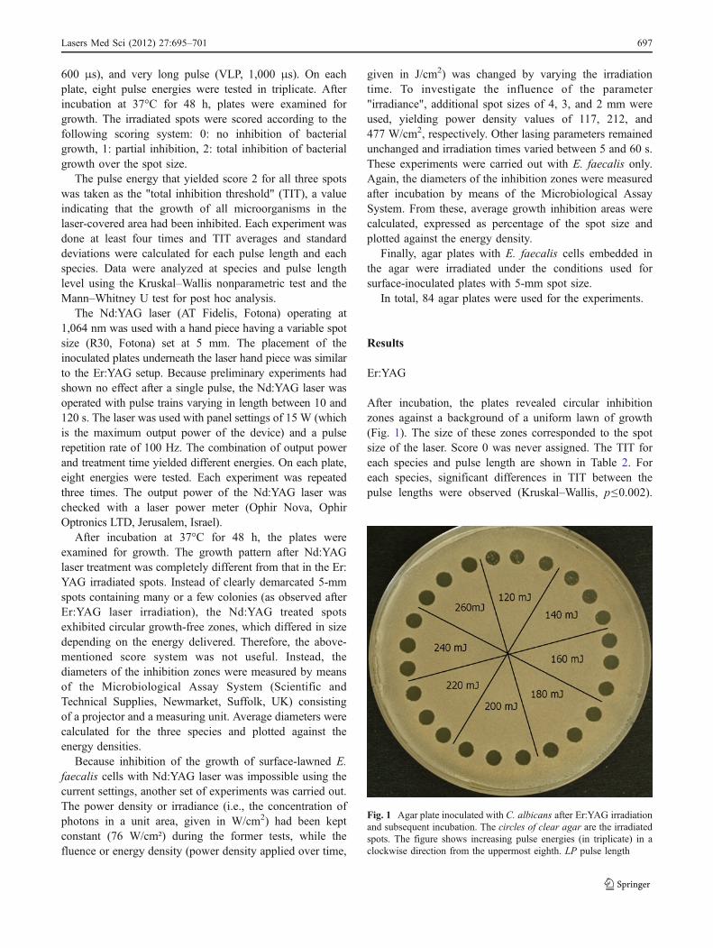
600 μs), and very long pulse (VLP, 1,000 μs). On eachplate, eight pulse energies were tested in triplicate. Afterincubation at 37°C for 48 h, plates were examined forgrowth. The irradiated spots were scored according to thefollowing scoring system: 0: no inhibition of bacterialgrowth, 1: partial inhibition, 2: total inhibition of bacterialgrowth over the spot size.
The pulse energy that yielded score 2 for all three spotswas taken as the "total inhibition threshold" (TIT), a valueindicating that the growth of all microorganisms in thelaser-covered area had been inhibited. Each experiment wasdone at least four times and TIT averages and standarddeviations were calculated for each pulse length and eachspecies. Data were analyzed at species and pulse lengthlevel using the Kruskal–Wallis nonparametric test and theMann–Whitney U test for post hoc analysis.
The Nd:YAG laser (AT Fidelis, Fotona) operating at1,064 nm was used with a hand piece having a variable spotsize (R30, Fotona) set at 5 mm. The placement of theinoculated plates underneath the laser hand piece was similarto the Er:YAG setup. Because preliminary experiments hadshown no effect after a single pulse, the Nd:YAG laser wasoperated with pulse trains varying in length between 10 and120 s. The laser was used with panel settings of 15 W (whichis the maximum output power of the device) and a pulserepetition rate of 100 Hz. The combination of output powerand treatment time yielded different energies. On each plate,eight energies were tested. Each experiment was repeatedthree times. The output power of the Nd:YAG laser waschecked with a laser power meter (Ophir Nova, OphirOptronics LTD, Jerusalem, Israel).
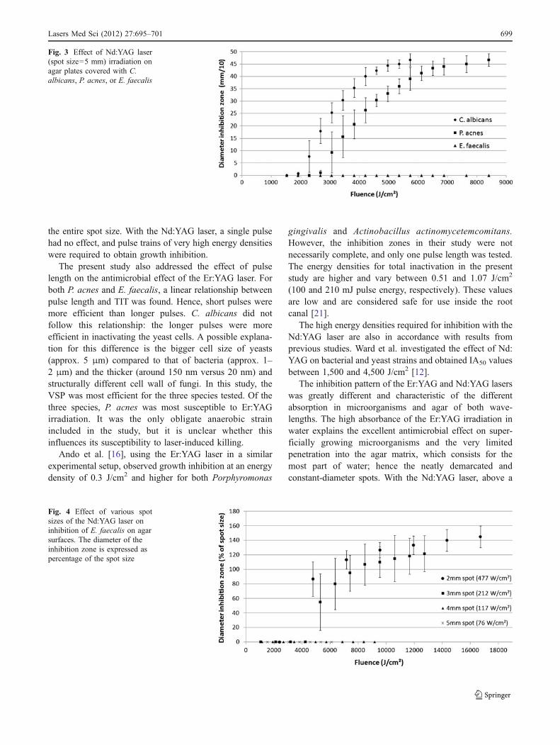
After incubation at 37°C for 48 h, the plates wereexamined for growth. The growth pattern after Nd:YAGlaser treatment was completely different from that in the Er:YAG irradiated spots. Instead of clearly demarcated 5-mmspots containing many or a few colonies (as observed afterEr:YAG laser irradiation), the Nd:YAG treated spotsexhibited circular growth-free zones, which differed in sizedepending on the energy delivered. Therefore, the above-mentioned score system was not useful. Instead, thediameters of the inhibition zones were measured by meansof the Microbiological Assay System (Scientific andTechnical Supplies, Newmarket, Suffolk, UK) consistingof a projector and a measuring unit. Average diameters werecalculated for the three species and plotted against theenergy densities.
Because inhibition of the growth of surface-lawned E.faecalis cells with Nd:YAG laser was impossible using thecurrent settings, another set of experiments was carried out.The power density or irradiance (i.e., the concentration ofphotons in a unit area, given in W/cm2) had been keptconstant (76 W/cm²) during the former tests, while thefluence or energy density (power density applied over time,
given in J/cm2) was changed by varying the irradiationtime. To investigate the influence of the parameter"irradiance", additional spot sizes of 4, 3, and 2 mm wereused, yielding power density values of 117, 212, and477 W/cm2, respectively. Other lasing parameters remainedunchanged and irradiation times varied between 5 and 60 s.These experiments were carried out with E. faecalis only.Again, the diameters of the inhibition zones were measuredafter incubation by means of the Microbiological AssaySystem. From these, average growth inhibition areas werecalculated, expressed as percentage of the spot size andplotted against the energy density.
Finally, agar plates with E. faecalis cells embedded inthe agar were irradiated under the conditions used forsurface-inoculated plates with 5-mm spot size.
In total, 84 agar plates were used for the experiments.
Results
Er:YAG
After incubation, the plates revealed circular inhibitionzones against a background of a uniform lawn of growth(Fig. 1). The size of these zones corresponded to the spotsize of the laser. Score 0 was never assigned. The TIT foreach species and pulse length are shown in Table 2. Foreach species, significant differences in TIT between thepulse lengths were observed (Kruskal–Wallis, p≤0.002).
Fig. 1 Agar plate inoculated with C. albicans after Er:YAG irradiationand subsequent incubation. The circles of clear agar are the irradiatedspots. The figure shows increasing pulse energies (in triplicate) in aclockwise direction from the uppermost eighth. LP pulse length
Lasers Med Sci (2012) 27:695–701 697

For every pulse length, significant differences in TITbetween the species were found (Kruskal–Wallis, p≤0.016).
P. acnes required the least energy for growth inhibition.For shorter pulse lengths (100 and 300 μs), C. albicansrequired the highest energies, while for longer pulse lengths(600 and 1,000 μs), E. faecalis required the highestenergies for inhibition. For both bacterial species (P. acnesand E. faecalis), a proportional relationship between pulselength and TIT was observed, while for C. albicans, nosuch relationship was found (Table 2). Irradiation of plateswith embedded bacteria did not result in inhibition ofbacterial growth inside the agar.
Nd:YAG
After incubation, most plates revealed circular growth-freezones where the microorganisms had been inactivated,against a background of a uniform lawn of growth (Fig. 2).The diameter of these zones varied between 0 and
4.7 mm depending on the energy that had been releasedon the spot. In Fig. 3, the inhibition diameter is plottedagainst the delivered energy for the three species. Nogrowth-free zones were observed after Nd:YAG irradiationof plates inoculated with E. faecalis, even after delivery of8,400 J/cm2. For C. albicans and P. acnes, the diameter ofthe inhibition zone increased with increasing fluence. Dueto peripheral growth of the microorganisms in theirradiated spot the during incubation, the fluencecorresponding to a diameter of 90% of the spot size wastaken as TIT. This was around 5,300 J/cm2 for C. albicansand 7,100 J/cm2 for P. acnes.
The influence of irradiance on bacterial growth inhibi-tion is shown in Fig. 4. Four and 5-mm spot sizes (117 and76 W/cm2) did not result in inhibition; 2- and 3-mm spotsizes (477 and 212 W/cm2) resulted in inhibition above2,400 and 4,200 J/cm2, respectively. It can be seen that forthe same fluence, higher irradiance values result in largerinhibition areas.
Plates with E. faecalis cells embedded in the agarshowed circular inhibition zones against a background ofuniform growth (similar to the pattern observed forsuperficially inoculated C. albicans and P. acnes plates)after Nd:YAG irradiation. This in contrast to superficiallyinoculated E. faecalis plates after Nd:YAG treatment forwhich no growth inhibition was observed. In Fig. 5, theinhibition area (IA) is plotted against the delivered energyfor both inoculation methods. IA50-values (the energydensity that gave an IA equal to 50% of the beam area)for E. faecalis cells embedded into the agar were around5,500 J/cm2.
In all experiments, the output power of the Nd:YAG laseras measured with the power meter at the target rangedbetween 96.7 and 99.3% of the panel setting.
Discussion
In this study, the Er:YAG laser was superior to the Nd:YAGlaser for inhibiting growth of microorganisms on agarsurfaces. A single pulse of relatively low energy (less than220 mJ) was sufficient to inhibit all microorganisms over
Table 2 Average total inhibition thresholds (mJ) per species and per pulse length±standard deviation (n=4)
Pulse length (µs) P. acnes E. faecalis C. albicans
100 100 ± 0.00 130 ± 11.5 175 ± 10.0
300 115 ± 10.0 150 ± 11.5 210 ± 11.5
600 130 ± 11.5 190 ± 11.5 a 170 ± 11.5 a
1000 155 ± 10.0 a 200 ± 16.3 b 155 ± 19.1 a,b
Connection bars indicate differences between pulse lengths that are NOT significant at the 0.05 level. Similar superscript letters indicatedifferences between species that are NOT significant at the 0.05 level (Kruskal–Wallis/Mann–Whitney U test)
Fig. 2 Agar plate inoculated with C. albicans after Nd:YAGirradiation and subsequent incubation. The circles of clear agar arethe inactivated areas. The figure shows increasing energy densities in aclockwise direction from the uppermost spot
698 Lasers Med Sci (2012) 27:695–701
the entire spot size. With the Nd:YAG laser, a single pulsehad no effect, and pulse trains of very high energy densitieswere required to obtain growth inhibition.
The present study also addressed the effect of pulselength on the antimicrobial effect of the Er:YAG laser. Forboth P. acnes and E. faecalis, a linear relationship betweenpulse length and TIT was found. Hence, short pulses weremore efficient than longer pulses. C. albicans did notfollow this relationship: the longer pulses were moreefficient in inactivating the yeast cells. A possible explana-tion for this difference is the bigger cell size of yeasts(approx. 5 μm) compared to that of bacteria (approx. 1–2 μm) and the thicker (around 150 nm versus 20 nm) andstructurally different cell wall of fungi. In this study, theVSP was most efficient for the three species tested. Of thethree species, P. acnes was most susceptible to Er:YAGirradiation. It was the only obligate anaerobic strainincluded in the study, but it is unclear whether thisinfluences its susceptibility to laser-induced killing.
Ando et al. [16], using the Er:YAG laser in a similarexperimental setup, observed growth inhibition at an energydensity of 0.3 J/cm2 and higher for both Porphyromonas
gingivalis and Actinobacillus actinomycetemcomitans.However, the inhibition zones in their study were notnecessarily complete, and only one pulse length was tested.The energy densities for total inactivation in the presentstudy are higher and vary between 0.51 and 1.07 J/cm2
(100 and 210 mJ pulse energy, respectively). These valuesare low and are considered safe for use inside the rootcanal [21].
The high energy densities required for inhibition with theNd:YAG laser are also in accordance with results fromprevious studies. Ward et al. investigated the effect of Nd:YAG on bacterial and yeast strains and obtained IA50 valuesbetween 1,500 and 4,500 J/cm2 [12].
The inhibition pattern of the Er:YAG and Nd:YAG laserswas greatly different and characteristic of the differentabsorption in microorganisms and agar of both wave-lengths. The high absorbance of the Er:YAG irradiation inwater explains the excellent antimicrobial effect on super-ficially growing microorganisms and the very limitedpenetration into the agar matrix, which consists for themost part of water; hence the neatly demarcated andconstant-diameter spots. With the Nd:YAG laser, above a
Fig. 3 Effect of Nd:YAG laser(spot size=5 mm) irradiation onagar plates covered with C.albicans, P. acnes, or E. faecalis
Fig. 4 Effect of various spotsizes of the Nd:YAG laser oninhibition of E. faecalis on agarsurfaces. The diameter of theinhibition zone is expressed aspercentage of the spot size
Lasers Med Sci (2012) 27:695–701 699

certain threshold, a linear relationship between exposureand growth inhibition area was observed, similar to theresults described by Grönqvist et al. [11]. They noted anagar temperature of 70°C immediately after exposure toapproximately 1,000 J/cm2, indicating killing due to aphotothermal effect, i.e., due to heating of the agar. Indeedduring our experiments with Nd:YAG laser, a local meltingof the agar surface was observed.
Pipetting a microbial suspension and immediatelyremoving the excess fluid resulted in a thin and uniformlayer of bacteria at the surface of the agar plates. Placing E.faecalis cells into the agar had a great influence on theoutcome of the experiments. Firstly, the Er:YAG laser wasunable to affect the cells in the agar. The Er:YAG light isreadily absorbed on the surface and does not penetrate theagar. Er:YAG pulse trains resulted in disk-shaped depres-sions in the agar due to ablation (data not shown). It is verylikely that thicker layers of microorganisms than those usedin this study would require higher Er:YAG pulse energy ormore pulses to obtain total inhibition. Second, the IA50
values for Nd:YAG laser inactivation decreased, which canmost likely be explained by the fact that the temperaturesare highest in the middle of the agar and less at the surfacewhere a cooling effect due to contact with air is present.This, together with the fact that E. faecalis is known to berelatively heat-resistant [22], is a possible reason why wewere not able to inactivate E. faecalis cells on the agarsurfaces.
Evidence suggests that the antimicrobial action of theNd:YAG laser is due to absorption of laser light in thesubstrate onto which the microorganisms are located,resulting in local temperature rise causing microbial death[15]. The substrate in the present study is agar, whichabsorbs the Nd:YAG wavelength very little, hence the highenergies required for microbial inactivation. In the case ofendodontic therapy, the substrate is dentine, which is also apoor absorber of Nd:YAG irradiation. In contrast, the Er:YAG laser light is highly absorbed in both microorganismsand their substrate (agar or dentine). This represents a
different but more efficient mode of action, requiring lessirradiation and reducing the likelihood of adverse thermalside-effects in the tooth. Thus, for disinfection of rootcanals, the Er:YAG laser seems to have better potential,provided the irradiation can be efficiently delivered to theroot canal system. Although the forward emission of mostfiber optic tips has been a major problem in delivery oflaser irradiation to the root canal walls, fiber modificationtechniques have recently been developed that result in acone-shaped tip with significant lateral emission. Theseallow a much better exposure of the root canal walls formost infrared lasers [23].
The proposed killing mechanism of the Nd:YAG laserbased on heating of the micro-environment of bacteria isbased on the in vitro observation that microorganisms arevirtually transparent to the Nd:YAG wavelength [11, 15](this can also be observed from the results in the presentstudy). In these studies, suspensions of bacteria were used.However, planktonic bacteria do not generally represent thein vivo growth state found in an infected tooth wherebacteria grow as a biofilm on the dentinal wall [24, 25]. Thehigh cell density and the presence of an extracellular matrixcharacteristic of a biofilm potentially reflects a differentsituation in terms of the levels of target chromophores andhence of the antimicrobial susceptibility to Nd:YAG laserirradiation. This is an area that remains to be investigatedfurther. Likewise, the presence of blood or blood productsin the clinical setting may increase levels of porphyrins andmelanins in bacteria and improve killing with Nd:YAG.
The different absorption into dentine of both lasers hasconsequences for their penetration depth. Bacteria locateddeep inside the dentinal tubules are most likely not affectedby the Er:YAG laser, while Nd:YAG has greater penetrationdepth in dentine and has more potential to reach these areas.
Comparing studies that investigate the antimicrobial effectof lasers is often difficult because the description of energydensity or irradiation conditions for calculating energydensity is often lacking. The present study shows thatirradiance also influences the antimicrobial effect and should
Fig. 5 Effect of Nd:YAG laserirradiation on agar plates withembedded or superficialE. faecalis cells
700 Lasers Med Sci (2012) 27:695–701

be taken into account. Increasing the irradiance resulted inlower IA50 values, indicating a stronger antimicrobial effect.
Conclusions
Within the limitations of an in vitro study, it can be concludedthat Er:YAG laser irradiation was far superior to irradiationwith the Nd:YAG laser for inactivatingmicroorganisms on agarsurfaces. Single Er:YAG laser pulses of relatively low energy(less than 220 mJ) were sufficient to inactivate all micro-organisms. The antimicrobial effect of this laser was species-specific and was influenced by pulse energy and pulse length.
With the Nd:YAG laser, pulse trains with very highenergy densities (several thousand mJ) were required toobtain microbial inactivation. Higher irradiance valuesincreased the antimicrobial effect. The pattern of inactiva-tion with Nd:YAG laser supports the hypothesis of aphotothermal antimicrobial effect.
Acknowledgements We would like to thank High Tech Laser(Herzele, Belgium) for providing the laser handpieces that have beenused in this study. We are also grateful to G. Eelsing and P. Rigole fortheir technical assistance.
References
1. Bergmans L, Moisiadis P, Teughels W, Van Meerbeek B, QuirynenM, Lambrechts P (2006) Bactericidal effect of Nd:YAG laserirradiation on some endodontic pathogens ex vivo. Int Endod J 39(7):547–557
2. Berkiten M, Berkiten R, Okar I (2000) Comparative evaluation ofantibacterial effects of Nd:YAG laser irradiation in root canals anddentinal tubules. J Endod 26(5):268–270
3. Fegan SE, Steiman HR (1995) Comparative evaluation of theantibacterial effects of intracanal Nd:YAG laser irradiation: an invitro study. J Endod 21(8):415–417
4. Folwaczny M, Mehl A, Jordan C, Hickel R (2002) Antibacterialeffects of pulsed Nd:YAG laser radiation at different energysettings in root canals. J Endod 28(1):24–29
5. Gutknecht N, Moritz A, Conrads G, Sievert T, Lampert F (1996)Bactericidal effect of the Nd:YAG laser in in vitro root canals. JClin Laser Med Surg 14(2):77–80
6. Hardee MW, Miserendino LJ, Kos W, Walia H (1994) Evaluationof the antibacterial effects of intracanal Nd:YAG laser irradiation. JEndod 20(8):377–380
7. Meire MA, De Prijck K, Coenye T, Nelis HJ, De Moor RJ (2009)Effectiveness of different laser systems to kill Enterococcusfaecalis in aqueous suspension and in an infected tooth model.Int Endod J 42(4):351–359
8. Moritz A, Schoop U, Goharkhay K, Jakolitsch S, Kluger W,Wernisch J, Sperr W (1999) The bactericidal effect of Nd:YAG,
Ho:YAG, and Er:YAG laser irradiation in the root canal: an in vitrocomparison. J Clin Laser Med Surg 17(4):161–164
9. Moshonov J, Orstavik D, Yamauchi S, Pettiette M, Trope M(1995) Nd:YAG laser irradiation in root canal disinfection. EndodDent Traumatol 11(5):220–224
10. Piccolomini R, D'Arcangelo C, D'Ercole S, Catamo G, SchiaffinoG, De Fazio P (2002) Bacteriologic evaluation of the effect of Nd:YAG laser irradiation in experimental infected root canals. JEndod 28(4):276–278
11. Gronqvist A, Wistrom J, Axner O, Monsen TJ (2000) Bactericidaleffect of pulsed 1,064 nm Nd:YAG laser light on Staphylococcusepidermidis is of photothermal origin: an in vitro study. LasersSurg Med 27(4):336–340
12. Ward GD, Watson IA, Stewart-Tull DE, Wardlaw AC, Chatwin CR(1996) Inactivation of bacteria and yeasts on agar surfaces withhigh power Nd:YAG laser light. Lett Appl Microbiol 23(3):136–140
13. Ward GD, Watson IA, Stewart-Tull DE, Wardlaw AC, Wang RK,Nutley MA, Cooper A (2000) Bactericidal action of high-powerNd:YAG laser light on Escherichia coli in saline suspension. JAppl Microbiol 89(3):517–525
14. Hibst R, Graser R, Udart M, Stock K (2010) Mechanism ofhigh-power NIR laser bacteria inactivation. J Biophotonics3(5–6):296–303
15. Pirnat S, Lukac M, Ihan A (2010) Study of the direct bactericidaleffect of Nd:YAG and diode laser parameters used in endodonticson pigmented and nonpigmented bacteria. Lasers Med Sci
16. Ando Y, Aoki A, Watanabe H, Ishikawa I (1996) Bactericidaleffect of erbium YAG laser on periodontopathic bacteria. LasersSurg Med 19(2):190–200
17. Schoop U, Moritz A, Kluger W, Patruta S, Goharkhay K, Sperr W,Wernisch J, Gattringer R, Mrass P, Georgopoulos A (2002) The Er:YAG laser in endodontics: results of an in vitro study. Lasers SurgMed 30(5):360–364
18. Mehl A, Folwaczny M, Haffner C, Hickel R (1999) Bactericidaleffects of 2.94 microns Er:YAG-laser radiation in dental rootcanals. J Endod 25(7):490–493
19. Noiri Y, Katsumoto T, Azakami H, Ebisu S (2008) Effects of Er:YAG laser irradiation on biofilm-forming bacteria associated withendodontic pathogens in vitro. J Endod 34(7):826–829
20. Sennhenn-Kirchner S, Schwarz P, Schliephake H, Konietschke F,Brunner E, Borg-von Zepelin M (2009) Decontamination efficacyof erbium:yttrium-aluminium-garnet and diode laser light on oralCandida albicans isolates of a 5-day in vitro biofilm model.Lasers Med Sci 24(3):313–320
21. Kimura Y, Yonaga K, Yokoyama K, Kinoshita J, Ogata Y,Matsumoto K (2002) Root surface temperature increase duringEr:YAG laser irradiation of root canals. J Endod 28(2):76–78
22. Kearns AM, Freeman R, Lightfoot NF (1995) Nosocomialenterococci: resistance to heat and sodium hypochlorite. J HospInfect 30(3):193–199
23. George R, Walsh LJ (2009) Performance assessment of novel sidefiring flexible optical fibers for dental applications. Lasers SurgMed 41(3):214–221
24. Chavez de Paz LE (2007) Redefining the persistent infection inroot canals: possible role of biofilm communities. J Endod 33(6):652–662
25. Nair PN (1987) Light and electron microscopic studies of rootcanal flora and periapical lesions. J Endod 13(1):29–39
Lasers Med Sci (2012) 27:695–701 701