in vitro α-glucosidase inhibition and antioxidative potential of an endophyte species (streptomyces...
TRANSCRIPT
In Vitro a-Glucosidase Inhibition and Antioxidative Potentialof an Endophyte Species (Streptomyces sp. Loyola UGC) Isolatedfrom Datura stramonium L
I. V. S. Nimal Christhudas • P. Praveen Kumar •
P. Agastian
Received: 3 August 2012 / Accepted: 18 January 2013 / Published online: 17 February 2013
� Springer Science+Business Media New York 2013
Abstract Endophytic actinomycetes isolated from Datura
stramonium L. was evaluated for its effects against in vitro
a-glucosidase inhibition, antioxidant, and free radical scav-
enging activities. Based on microbial cultural characteristic
and 16S rRNA sequencing, it was identified as Streptomyces
sp. loyola UGC. The methanolic extract of endophytic acti-
nomycetes (MeEA) shows remarkable inhibition of a-glu-
cosidase (IC50 730.21 ± 1.33 lg/ml), scavenging activity
on 2,2-diphenyl-picrylhydrazyl (DPPH) (IC50 435.31 ±
1.79 lg/ml), hydroxyl radical (IC50 350.21 ± 1.02 lg/ml),
nitric oxide scavenging (IC50 800.12 ± 1.05 lg/ml),
superoxide anion radical (IC50 220.31 ± 1.47 lg/ml), as
well as a high and dose-dependent reducing power. The
MeEA also showed a strong suppressive effect on rat liver
lipid peroxidation. Antioxidants of b-carotene linoleate
model system revels significantly lower than BHA. The total
phenolic content of the extract was 176 mg of catechol
equivalents/gram extract. Perusal of this study indicates
MeEA can be used as natural resource of a-glucosidase
inhibitor and antioxidants.
Introduction
Diabetes mellitus (DM) is a metabolic disorder character-
ized by hyperglycemia resulting from defects in insulin
secretion, insulin action, or both. Along with hyperglyce-
mia and abnormalities in serum lipids, diabetes is associ-
ated with micro- and macrovascular complications, which
are the major causes of morbidity and death in diabetic
subjects [15]. The currently available antidiabetic agents
including sulfonylureas, biguanide, and thiazolidinedione
inhibitors are widely used to control the hyperglycemia and
hyperlipidemia, but these drugs fail to significantly alter
the course of diabetic complications and have limited use
because of undesirable side effects and high rates of sec-
ondary failure [24]. Thus, it is essential to look for more
effective antidiabetic agents with lesser side effects. There
are many articles related to antidiabetic compounds from
plants and also microbial resources have been proven to be
a rewarding source of antidiabetic compounds reported
from Streptomyces hygroscopicus-limoneus [6, 19], Strep-
tomyces calvus [17]. However, normalizing blood glucose
level is a formidable challenge in clinical practice. The
pharmacological agents with greatest effect on postprandial
hyperglycemia include insulin, lispro, amylin analogs, and
a-glucosidase (acarbose and voglibose) inhibitors [10]. It
has been well acknowledged that plant-derived extracts,
phytochemicals, and microbial sources are potential alter-
natives to synthetic inhibitors against a-glucosidase.
Datura stramonium L. is a wild-growing herb known as
Jimson weed belongs to the family Solanaceae. The plant
distributed throughout most parts of temperate regions of the
world [1]. Whole plant is used as anti-inflammatory, central
nervous system stimulant [28], for the treatment of respira-
tory decongestion [35] dental and skin infections, toothache,
and alopecia [5]. It is a hallucinogenic plant that causes
serious poisoning. Consumption of any part of the plant may
result in a severe anticholinergic reaction that may lead to
toxicity and occasionally cause diagnostic difficulties [7]. It
is used recreationally for its anticholinergic effects, resulting
I. V. S. Nimal Christhudas � P. Praveen Kumar
Department of Plant Biology and Biotechnology,
Loyola College, Chennai 600 034, India
P. Agastian (&)
Research Department of Plant Biology and Biotechnology,
School of Life Science, Loyola College, Chennai 600 034, India
e-mail: [email protected];
123
Curr Microbiol (2013) 67:69–76
DOI 10.1007/s00284-013-0329-2
in hallucinations. The entire plant has anticholinergic com-
pounds, but the seeds contain the highest concentration. An
extract made by boiling crushed seeds retains the anticho-
linergic activity, has a rapid onset of action [3], and thus may
be potentially useful as an alternative to atropine for the
treatment of the muscarinic symptoms of organophosphate
toxicity and some of the central anticholinergic effects [29].
There were overexploitations of this plant by the pharma-
ceutical industries without making any alternative methods
to conserve this medicinally important plant. Attempt
is made to isolate the endophytic actinomycetes from
D. stramonium L. to evaluate the in vitro a-glucosidase and
antioxidant potential of MeEA (Methanolic extract of
endophytic actinomycetes).
Materials and Methods
Plant Materials
Datura stramonium L. was collected from Irula Tribal
Women’s Welfare Society (ITWWS), Chengalpattu, Kan-
chipuram district, Tamil Nadu, South India, in the month
of January. The species was identified and authenticated by
Dr. D. Narashiman, Department of Botany, Madras Christian
College, Chennai, South India.
Isolation of Actinomycetes
The roots and transition zones of D. stramonium L. were
surface sterilized by the methodology of Johannes et al.,
[14] with some modifications. Samples were thoroughly
washed with running tap water and all the visibly damaged
materials were excluded. Plant parts were rinsed in 0.1 %
Tween 20 for 30 s and followed by bevastin for 2–3 min to
inhibit the fungal growth, sequentially immersed in 0.1 %
sodium hypochlorite (NaClO) for 30 s and in 75 % ethanol
for 3–5 min. After each treatment, samples were rinsed
three times in sterile distilled water. Finally the surface-
sterilized samples were thoroughly dried in a laminar flow
chamber. The samples was aseptically dissected to expose
cortex region and plated onto actinomycetes isolation
medium, incubated for 12–15 days at 28 �C. The isolation
medium was supplemented with nalidixic acid and actidion
both to a final concentration of 50 lg/ml to inhibit the
growth of non actinomycetes organisms.
Identification of Actinomycetes
The isolated actinomycetes were dereplicated for cultural
and morphological characteristics, including morphology
and color of aerial mycelium: characteristics of colonies on
the plate, mycelium and spore color, color of diffusible
pigments and growth. Visual observation by light micros-
copy and Gram-staining were performed for further iden-
tification [31]. The total genomic DNA was extracted by
CTAB-Method. The actinomycetes DNA fragments were
amplified using Universal primers 16S rRNA and PCR
reaction were standardized as initial denaturation at 94 �C
for 3 min, followed by 35 cycle of 1 min at 94 �C,
54 �C for 1 min, 72 �C for 2 min and a final extension at
72 �C for 8–10 min, stop at 4 �C for 1 h. The PCR prod-
ucts were stored at 4 �C and visualized by electrophoresis.
The gel was photographed in gel documentation. The
amplified product was purified and sequenced with two
fragments of the 27F (5‘ AGT TTG ATC CTG GCT CAG 3‘)
and 1492R (5‘ ACG GCT ACC TTG TTA CGA CTT 3‘)
region in both the directions and the sequences obtained were
submitted to Genbank for homology search with Blast.
Culture Media and Extraction
The spore suspensions of the culture were inoculated on the
Modified Nutrient Glucose Broth (MNGB) medium with
pH 7.0 and incubated at 28 �C for 7 days in a shaker. Then,
the mycelium filtered and the culture fluid was extracted
with methanol (1:1). The organic phase was evaporated to
dryness under reduced pressure at 35 �C using rotor vac-
uum evaporator.
Determination of In Vitro a-Glucosidase Inhibition
and Antioxidant Assay
a-Glucosidase Inhibition Activity
In order to investigate the inhibition activity of MeEA, an
in vitro a-glucosidase inhibition test was performed. a-glu-
cosidase from yeast is used extensively as a screening
material for a-glucosidase inhibitors, but the results do not
always agree with those obtained in mammals. Therefore, we
used the mouse small intestine homogenate as a-glucosidase
solution because we speculated that it would better reflect the
in vivo state. The inhibitory effect was measured by the
method slightly modified method of Dahlqvist [4]. After
fasting for 20 h, the small intestine between the part imme-
diately below duodenum and the part immediately above the
cecum was cut, rinsed with ice-cold saline, and homogenized
with 12 ml of maleate buffer (100 mM, pH 6.0). The
homogenate was used as the a-glucosidase solution. The
assay mixture consisted of 100 mM maleate buffer (pH 6.0),
2 % (w/v) each sugar substrate solution (100 ll), and the
sample extract (200–1,000 lg/mL). It was preincubated for
5 min at 37 �C and the reaction was initiated by adding the
crude a-glucosidase solution (50 ll) to it, followed by
incubation for 10 min at 37 �C. Acarbose was used as a
70 I. V. S. Nimal Christhudas et al.: In Vitro a-Glucosidase Inhibition and Antioxidative Potential
123
standard. The percentage of inhibition was calculated by the
formula.
Inhibition (%) = [(amount of glucose produced by the
positive control) – (amount of glucose produced by the addition
of sample)/(amount of glucose produced by the positive
control)] 9 100.
Total Phenolic Content (TPC)
Total phenolic content was assessed according to the Folin–
Ciocalteau method [27] with some modifications. Briefly,
0.1 ml of sample (200–1,000 lg/ml), 1.9 ml distilled water,
and 1 ml of Folin–Ciocalteau’s reagent were added in a tube
and then 1 ml of 100 g/l Na2CO3 was added. The reaction
mixture was incubated at 25 �C for 2 h and the absorbance of
the mixture was read at 765 nm. The sample was tested in
triplicate and a calibration curve with six data points for
catechol was obtained. The results were compared to a cat-
echol calibration curve and the total phenolic content of
MeEA was expressed as mg of catechol equivalents per gram
of extract.
Reducing Power Activity
The reducing power of MeEA was determined according to
Yen and Duh [32]. Different concentrations of MeEA
(200–1,000 lg/ml) were mixed with 2.5 ml of phosphate
buffer (200 mM, pH 6.6) and 2.5 ml of 1 % potassium fer-
ricyanide. The mixtures were incubated for 20 min at 50 �C.
After incubation, 2.5 ml of 10 % trichloroacetic acid was
added to each mixture followed by centrifugation at
3,000 rpm for 10 min. The upper layer (5 ml) was mixed
with 5 ml of distilled water and 1 ml of 0.1 % ferric chloride.
The absorbance of the resultant solution was measured at
700 nm and was compared with standard BHT absorbance.
DPPH Radical Scavenging Assay
DPPH quenching ability of MeEA was measured according
to Hanato et al. [11]. A methanol DPPH solution (0.15 %)
was mixed with serial dilutions (200–1,000 lg/ml) of the
MeEA and after 10 min, the absorbance was read at
515 nm. The antiradical activity was expressed as IC50 (lg/
ml), (the antiradical dose required to cause a 50 % inhi-
bition). Vitamin C was used as standard. The ability to
scavenge the DPPH radical was calculated by the following
formula:
DPPH radical scavenging activity %ð Þ¼ A0 � A1=A0ð Þ � 100½ � ð1Þ
where A0 is the absorbance of the control at 30 min and
A1 is the absorbance of the sample at 30 min. All samples
were analyzed in triplicate.
Hydroxyl Radical Scavenging Activity
The hydroxyl scavenging assay was performed as described
by the method of Elizabeth and Rao [8] with minor changes.
All solutions were prepared freshly. One milliliter of the
reaction mixture contained 100 ll of 28 mM 2-deoxy-2-
ribose (dissolved in phosphate buffer, pH 7.4), 500 ll solu-
tion of various concentrations of MeEA (200–1,000 lg/ml),
200 ll of 200 lM FeCl3 and 1.04 mM EDTA (1:1 v/v),
100 ll H2O2 (1 mM), and 100 ll ascorbic acid (1 mM).
After an incubation period of 1 h at 37 �C, the extent of
deoxyribose degradation was measured by the TBA reaction.
The absorbance was read at 532 nm against the blank solu-
tion. Vitamin C was used as a positive control. The scav-
enging activity was calculated by the formula (1).
Nitric Oxide Scavenging Activity
Sodium nitroprusside in aqueous solution at physiologic pH
spontaneously generates nitric oxide; it interacts with oxy-
gen to produce nitrite ions, which can be estimated by the use
of Griess Illosvoy reaction [9]. In the present investigation,
Griess Illosvoy reagent was modified using naphthyl ethy-
lenediamine dihydrochloride (0.1 % w/v) instead of
1-naphthylamine (5 %). The reaction mixture (3 ml) con-
taining sodium nitroprusside (10 mM, 2 ml), phosphate
buffer saline (0.5 ml), and different concentrations of MeEA
(200–1,000 lg/ml) or standard solution (0.5 ml) was incu-
bated at 25 �C for 150 min. After incubation, 0.5 ml of the
reaction mixture containing nitrite was pipetted and mixed
with 1 ml of sulphanilic acid reagent (0.33 % in 20 % glacial
acetic acid) and allowed to stand for 5 min for completing
diazotization. Then, 1 ml of naphthyl ethylenediamine
dihydrochloride (1 %) was added, mixed, and allowed to
stand for 30 min. A pink colored chromophore was formed in
diffused light. The absorbance of these solutions was mea-
sured at 540 nm against the corresponding blank. Ascorbic
acid was used as standard. The scavenging activity was
calculated by Eq. (1).
Superoxide Scavenging Activity
Superoxide scavenging activity of MeEA was determined by
monitoring the competition of those with NBT for the
superoxide anion generated by the PMS–NADH system
[16]. Superoxide radicals were generated in 1 ml of 20 mM
Tris–HCl buffer pH 8.0 containing 0.05 mM nitroblue tet-
razolium (NBT), 0.01 mM phenazine methosulphate (PMS),
and different concentrations (200–1,000 lg/ml) of MeEA
were preincubated for 2 min. The reaction was initiated by
the addition of 0.078 mM NADH. Blue chromogen, formed
due to NBT reduction, was read at 560 nm. Results were
expressed as percentage of inhibition of superoxide radicals.
I. V. S. Nimal Christhudas et al.: In Vitro a-Glucosidase Inhibition and Antioxidative Potential 71
123
Vitamin C was used as positive control. The scavenging
activity was calculated by the formula (1).
Lipid Peroxidation Assay
The inhibition effect of MeEA on lipid peroxidation was
determined according to the thiobarbituric acid method.
FeCl2–H2O2 was used to induce the liver homogenate
peroxidation [33]. In this method, 0.2 ml of MeEA
(200–1,000 lg/ml) was mixed with 1.0 ml of 1 % liver
homogenate (each 100 ml homogenate solution contains
1.0 g rat liver), then 50 ll of FeCl2 (0.5 mM) and H2O2
(0.5 mM) was added. The mixture was incubated at 37 �C
for 60 min, 1.0 ml of trichloroacetic acid (15 %), and
thiobarbituric acid (0.67 %) was added and the mixture
was heated up in boiled water for 15 min. The absorbance
was recorded at 532 nm. Ascorbic acid was used as the
positive control. The percentage of inhibition effect was
calculated according to the formula (1).
Antioxidant Assay Using b-Carotene Linoleate Model
System
The antioxidant activity of MeEA was evaluated by the b-
carotene linoleate model system [21]. A solution of b-
carotene was prepared by dissolving 2 mg of b-carotene in
10 ml of chloroform. Two milliliters of this solution was
pipetted into a 100-ml round-bottom flask. After chloro-
form was removed under vacuum, 40 mg of purified lino-
leic acid, 400 mg of Tween 40 emulsifier, and 100 ml of
aerated distilled water were added to the flask with vigor-
ous shaking. Aliquot (4.8 ml) of this emulsion was trans-
ferred into different test tubes containing different
concentrations of MeEA (200–1,000 lg/ml). BHA was
used for comparative purposes. As soon as the emulsion
was added to each tube, the zero time absorbance was
measured at 470 nm. The tubes were then placed at 50 �C
in a water bath. Measurement of absorbance was continued
until the color of b-carotene disappeared; a blank, devoid
of b-carotene, was prepared for background subtraction.
Antioxidant activity (AA) was calculated by the following
equation:
AA ¼ b� carotene content after 2h of assayðinitial b� carotene content= Þ � 100
Statistical Analysis
The data for biochemical and physiologic parameters were
analyzed and expressed as mean ± SD. The IC50 values
were calculated from linear regression analysis. Results
were processed by computer program, Microsoft Excel
(2007).
Results
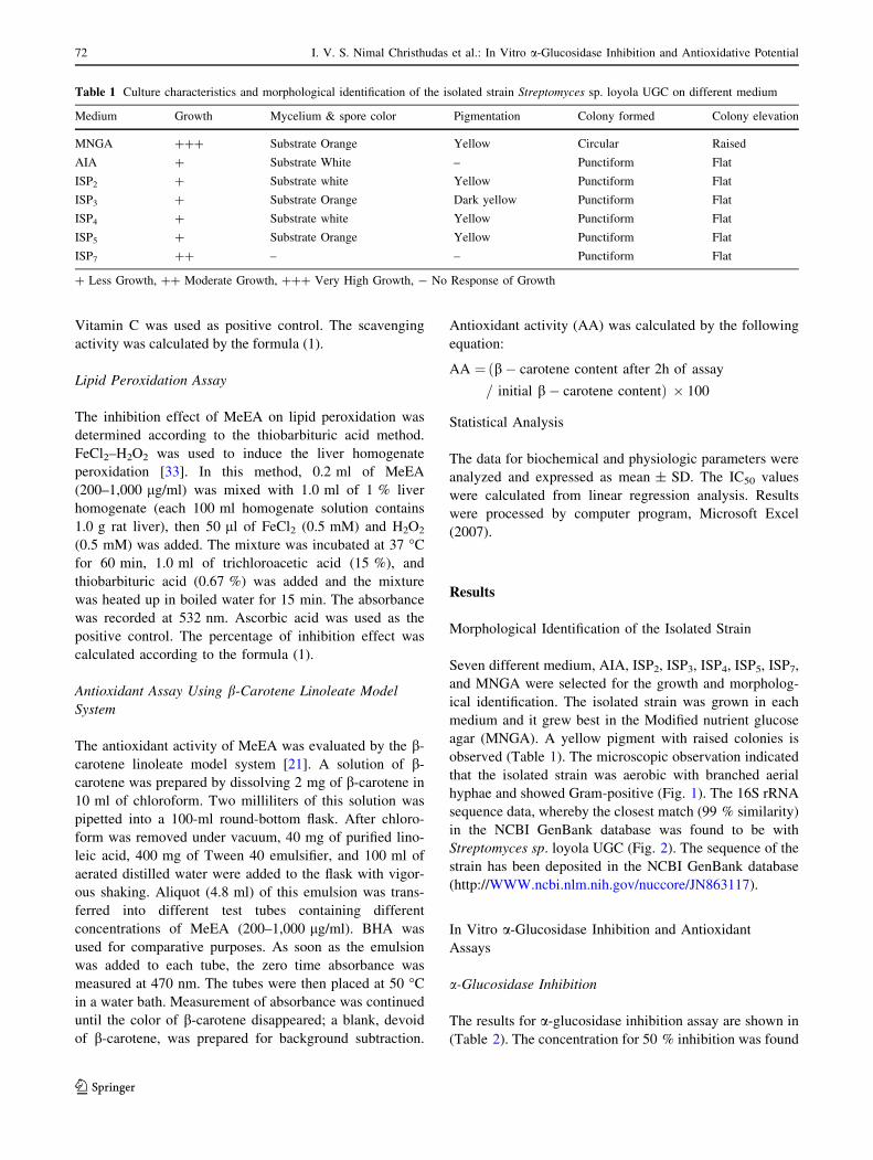
Morphological Identification of the Isolated Strain
Seven different medium, AIA, ISP2, ISP3, ISP4, ISP5, ISP7,
and MNGA were selected for the growth and morpholog-
ical identification. The isolated strain was grown in each
medium and it grew best in the Modified nutrient glucose
agar (MNGA). A yellow pigment with raised colonies is
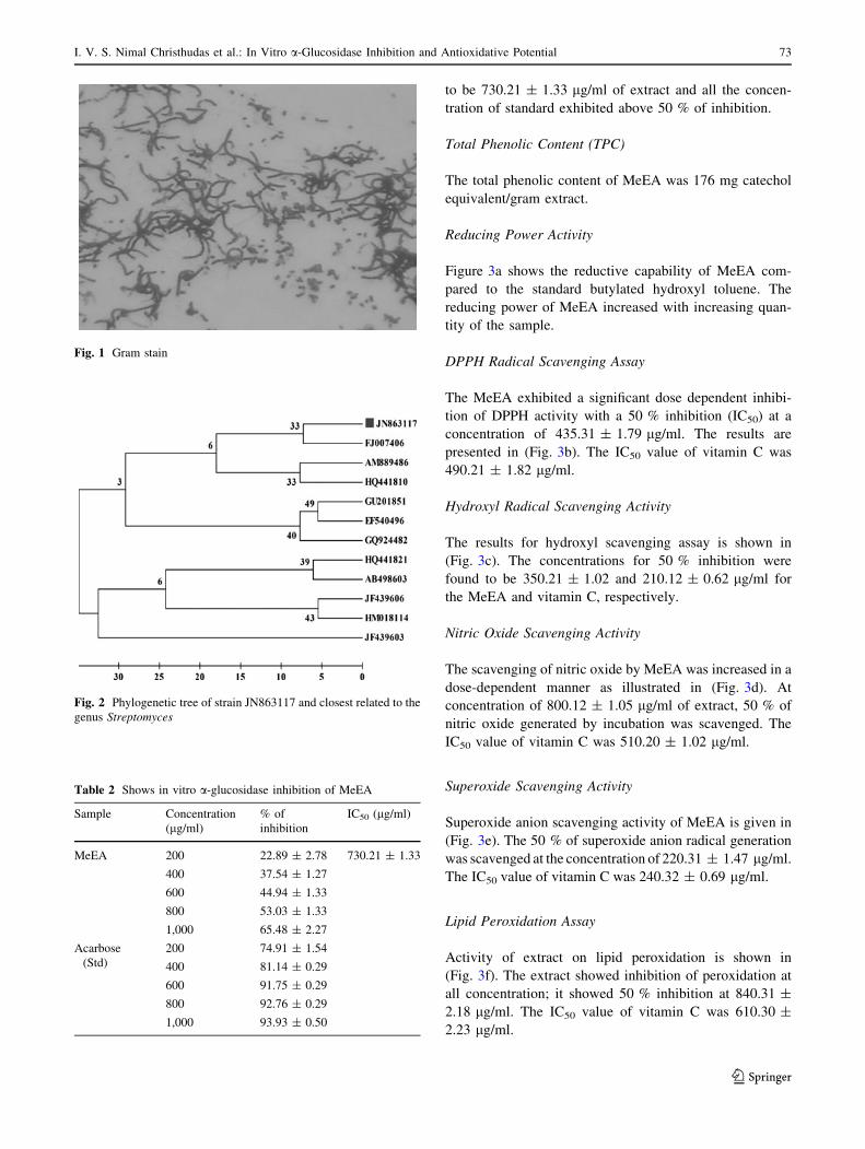
observed (Table 1). The microscopic observation indicated
that the isolated strain was aerobic with branched aerial
hyphae and showed Gram-positive (Fig. 1). The 16S rRNA
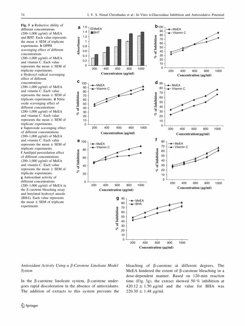
sequence data, whereby the closest match (99 % similarity)
in the NCBI GenBank database was found to be with
Streptomyces sp. loyola UGC (Fig. 2). The sequence of the
strain has been deposited in the NCBI GenBank database
(http://WWW.ncbi.nlm.nih.gov/nuccore/JN863117).
In Vitro a-Glucosidase Inhibition and Antioxidant
Assays
a-Glucosidase Inhibition
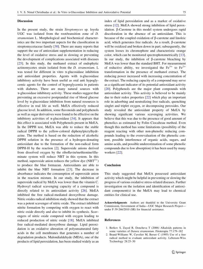
The results for a-glucosidase inhibition assay are shown in
(Table 2). The concentration for 50 % inhibition was found
Table 1 Culture characteristics and morphological identification of the isolated strain Streptomyces sp. loyola UGC on different medium
Medium Growth Mycelium & spore color Pigmentation Colony formed Colony elevation
MNGA ??? Substrate Orange Yellow Circular Raised
AIA ? Substrate White – Punctiform Flat
ISP2 ? Substrate white Yellow Punctiform Flat
ISP3 ? Substrate Orange Dark yellow Punctiform Flat
ISP4 ? Substrate white Yellow Punctiform Flat
ISP5 ? Substrate Orange Yellow Punctiform Flat
ISP7 ?? – – Punctiform Flat
? Less Growth, ?? Moderate Growth, ??? Very High Growth, - No Response of Growth
72 I. V. S. Nimal Christhudas et al.: In Vitro a-Glucosidase Inhibition and Antioxidative Potential
123
to be 730.21 ± 1.33 lg/ml of extract and all the concen-
tration of standard exhibited above 50 % of inhibition.
Total Phenolic Content (TPC)
The total phenolic content of MeEA was 176 mg catechol
equivalent/gram extract.
Reducing Power Activity
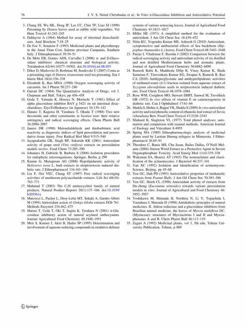
Figure 3a shows the reductive capability of MeEA com-
pared to the standard butylated hydroxyl toluene. The
reducing power of MeEA increased with increasing quan-
tity of the sample.
DPPH Radical Scavenging Assay
The MeEA exhibited a significant dose dependent inhibi-
tion of DPPH activity with a 50 % inhibition (IC50) at a
concentration of 435.31 ± 1.79 lg/ml. The results are
presented in (Fig. 3b). The IC50 value of vitamin C was
490.21 ± 1.82 lg/ml.
Hydroxyl Radical Scavenging Activity
The results for hydroxyl scavenging assay is shown in
(Fig. 3c). The concentrations for 50 % inhibition were
found to be 350.21 ± 1.02 and 210.12 ± 0.62 lg/ml for
the MeEA and vitamin C, respectively.
Nitric Oxide Scavenging Activity
The scavenging of nitric oxide by MeEA was increased in a
dose-dependent manner as illustrated in (Fig. 3d). At
concentration of 800.12 ± 1.05 lg/ml of extract, 50 % of
nitric oxide generated by incubation was scavenged. The
IC50 value of vitamin C was 510.20 ± 1.02 lg/ml.
Superoxide Scavenging Activity
Superoxide anion scavenging activity of MeEA is given in
(Fig. 3e). The 50 % of superoxide anion radical generation
was scavenged at the concentration of 220.31 ± 1.47 lg/ml.
The IC50 value of vitamin C was 240.32 ± 0.69 lg/ml.
Lipid Peroxidation Assay
Activity of extract on lipid peroxidation is shown in
(Fig. 3f). The extract showed inhibition of peroxidation at
all concentration; it showed 50 % inhibition at 840.31 ±
2.18 lg/ml. The IC50 value of vitamin C was 610.30 ±
2.23 lg/ml.
Fig. 1 Gram stain
Fig. 2 Phylogenetic tree of strain JN863117 and closest related to the
genus Streptomyces
Table 2 Shows in vitro a-glucosidase inhibition of MeEA
Sample Concentration
(lg/ml)
% of
inhibition
IC50 (lg/ml)
MeEA 200 22.89 ± 2.78 730.21 ± 1.33
400 37.54 ± 1.27
600 44.94 ± 1.33
800 53.03 ± 1.33
1,000 65.48 ± 2.27
Acarbose
(Std)
200 74.91 ± 1.54
400 81.14 ± 0.29
600 91.75 ± 0.29
800 92.76 ± 0.29
1,000 93.93 ± 0.50
I. V. S. Nimal Christhudas et al.: In Vitro a-Glucosidase Inhibition and Antioxidative Potential 73
123
Antioxidant Activity Using a b-Carotene Linoleate Model
System
In the b-carotene linoleate system, b-carotene under-
goes rapid discoloration in the absence of antioxidants.
The addition of extracts to this system prevents the
bleaching of b-carotene at different degrees. The
MeEA hindered the extent of b-carotene bleaching in a
dose-dependent manner. Based on 120-min reaction
time (Fig. 3g), the extract showed 50 % inhibition at
420.12 ± 1.50 lg/ml and the value for BHA was
220.30 ± 1.48 lg/ml.
0
0.2
0.4
0.6
0.8
1
1.2
1.4
1.6
Abs
orba
nce
Concentraton (µg/ml)
MeEABHT
a
0102030405060708090
100
% o
f In
hibi
tion
Concentration (µg/ml)
MeEAVitamin C
b
0102030405060708090
100
% o
f In
hibi
tion
Concentration (µg/ml)
MeEAVitamin C
c
0
10
20
30
40
50
60
70
80
90
% o
f In
hibi
tion
Concentration(µg/ml)
MeEAVitamin C
d
0
20
40
60
80
100
% o
f In
hibi
tion
Concentration (µg/ml)
MeEAVitamin C
e
0
10
20
30
40
50
60
70
80
% o
f In
hibi
tion
Concentration (µg/ml)
MeEAVitamin C
f
0
10
20
30
40
50
60
70
80
90
200 400 600 800 1000200 400 600 800 1000
200 400 600 800 1000 200 400 600 800 1000
200 400 600 800 1000200 400 600 800 1000
200 400 600 800 1000
% o
f In
hibi
tion
Concentration (µg/ml)
MeEABHA
g
Fig. 3 a Reductive ability of
different concentrations
(200–1,000 lg/ml) of MeEA
and BHT. Each value represents
the mean ± SEM of triplicate
experiments. b DPPH
scavenging effect of different
concentrations
(200–1,000 lg/ml) of MeEA
and vitamin C. Each value
represents the mean ± SEM of
triplicate experiments.
c Hydroxyl radical scavenging
effect of different
concentrations
(200–1,000 lg/ml) of MeEA
and vitamin C. Each value
represents the mean ± SEM of
triplicate experiments. d Nitric
oxide scavenging effect of
different concentrations
(200–1,000 lg/ml) of MeEA
and vitamin C. Each value
represents the mean ± SEM of
triplicate experiments.
e Superoxide scavenging effect
of different concentrations
(200–1,000 lg/ml) of MeEA
and vitamin C. Each value
represents the mean ± SEM of
triplicate experiments.
f Antilipid peroxidation effect
of different concentrations
(200–1,000 lg/ml) of MeEA
and vitamin C. Each value
represents the mean ± SEM of
triplicate experiments.
g Antioxidant activity of
different concentrations
(200–1,000 lg/ml) of MeEA in
the b carotene bleaching assay
and butylated hydroxyl anisole
(BHA). Each value represents
the mean ± SEM of triplicate
experiments
74 I. V. S. Nimal Christhudas et al.: In Vitro a-Glucosidase Inhibition and Antioxidative Potential
123
Discussion
In the present study, the strain Streptomyces sp. loyola
UGC was isolated from the root/transition zone of D.
stramonium L. Morphological and biochemical character-
istics are the two important aspects for the classification in
streptomycetaceae family [30]. There are many reports that
support the use of antioxidant supplementation in reducing
the level of oxidative stress and in slowing or preventing
the development of complications associated with diseases
[25]. In this study, the methanol extract of endophytic
actinomycetes strain isolated from the D. stramonium. L
was tested for different in vitro a-glucosidase inhibition
and antioxidant properties. Agents with a-glucosidase
inhibitory activity have been useful as oral anti hypogly-
cemic agents for the control of hyperglycemia in patients
with diabetes. There are many natural sources with
a-glucosidase inhibitory activity. These studies suggest that
preventing an excessive postprandial rise of blood glucose
level by a-glucosidase inhibition from natural resources is
effective in real life as well. MeEA effectively reduced
glucose level. In addition, some flavonoids and polyphenols
as well as sugar derivatives were found to be effective on the
inhibitory activities of a-glucosidase [34]. It appears that
this effect is associated with Polyphenols present in MeEA.
In the DPPH test, MeEA was able to reduce the stable
radical DPPH to the yellow-colored diphenylpicrylhydr-
azine. The method is based on the reduction of alcoholic
DPPH solution in the presence of a hydrogen-donating
antioxidant due to the formation of the non-radical form
DPPH-H by the reaction [2]. Superoxide anions derived
from dissolved oxygen by the riboflavin/methionine/illu-
minate system will reduce NBT in this system. In this
method, superoxide anion reduces the yellow dye (NBT2?)
to produce the blue formazan. Antioxidants are able to
inhibit the blue NBT formation [23]. The decrease in
absorbance indicates the consumption of superoxide anion
in the reaction mixture. In our study, the inhibition of
superoxide radical by MeEA was lower than the vitamin C.
Hydroxyl radical scavenging capacity of a compound is
directly related to its antioxidant activity [26]. MeEA
inhibited the free radical-mediated deoxyribose damage.
Nitric oxides radical inhibition study showed that the extract
was a potent scavenger of nitric oxide. The extract inhibited
nitrite formation by competing with oxygen to react with
nitric oxide directly and also to inhibit its synthesis. Scav-
engers of nitric oxide competed with oxygen leading to
reduced production of nitric oxide [18]. MeEA inhibited
free radical-mediated deoxyribose damage. Lipid peroxi-
dation is an oxidative alteration of polyunsaturated fatty
acids in the cell membranes that generates a number of
degradation products. Malondialdehyde (MDA), one of the
products of lipid peroxidation, has been studied widely as an
index of lipid peroxidation and as a marker of oxidative
stress [12]. MeEA showed strong inhibition of lipid perox-
idation. b-Carotene in this model system undergoes rapid
discoloration in the absence of an antioxidant. This is
because of the coupled oxidation of b-carotene and linoleic
acid, which generates free radicals. As a result, b-carotene
will be oxidized and broken down in part; subsequently, the
system looses its chromophore and characteristic orange
color, which can be monitored spectrophotometrically [13].
In our study, the inhibition of b-carotene bleaching by
MeEA was lower than the standard BHT. For measurement
of reductive ability, we investigated the Fe3? to Fe2?
transformation in the presence of methanol extract. The
reducing power increased with increasing concentration of
the extract. The reducing capacity of a compound may serve
as a significant indicator of its potential antioxidant activity
[20]. Polyphenols are the major plant compounds with
antioxidant activity. This activity is believed to be mainly
due to their redox properties [22] which play an important
role in adsorbing and neutralizing free radicals, quenching
singlet and triplet oxygen, or decomposing peroxides. Our
study revealed the antioxidant property of MeEA by
showing significant various scavenging activities. We
believe that this was due to the presence of good amount of
phenolics as estimated by Folin–Ciocalteau method. Even
though this method has some limitations (possibility of the
reagent reacting with other non-phenolic reducing com-
pounds leading to the overevaluation of the phenolic con-
tent, possible interference of organic acids, sugars, and
amino acids, and possible underestimation of some phenolic
compounds due to low absorption) it has been used by many
workers [22].
Conclusion
This study suggested that MeEA possessed antioxidant
activity which might be helpful in preventing or slowing the
progress of various oxidative stress-related diseases. Further
investigation on the isolation and identification of antioxi-
dant component(s) in the MeEA may lead to chemical
entities for clinical use.
Acknowledgments Authors are thankful to the University Grant
Commission, Government of India—UGC Major Research Project—
under F-39-266/2010 (SR) for financial assistance.
References
1. Berkov S, Zayed R, Doncheva T (2006) Alkaloids patterns in
some varieties of Datura stramonium. Fitoterapia 77:179–182
2. Brand-Williams W, Cuvelier M, Berset C (1995) Use of a free
radical method to evaluate antioxidant activity. Lebensm-Wiss
Technology 28:25–30
I. V. S. Nimal Christhudas et al.: In Vitro a-Glucosidase Inhibition and Antioxidative Potential 75
123
3. Chang SS, Wu ML, Deng JF, Lee CC, Chin TF, Liao SJ (1999)
Poisoning by Datura leaves used as edible wild vegetables. Vet
Hum Toxicol 41:242–245
4. Dahlqvist A (1964) Method for assay of intestinal disaccharid-
ases. Anal Biochem 7:18–25
5. De Foe V, Senatore F (1993) Medicinal plants and phytotherapy
in the Amal Fitan Cost, Salerno province Campania, Southern
Italy. J Ethnopharmacol 39:39–51
6. De Melo EB, Gomes ADS, Carvalho I (2006) a- and b-Gluco-
sidase inhibitors: chemical structure and biological activity.
Tetrahedron 62(44):10277–10302. doi:10.1016/j.tet.08.055
7. Diker D, Markovitz D, Rothman M, Sendovski U (2007) Coma as
a presenting sign of Datura stramonium seed tea poisoning. Eur J
Intern Med 18(4):336–338
8. Elizabeth K, Rao MNA (1990) Oxygen scavenging activity of
curcumin. Int J Pharm 58:237–240
9. Garratt DC (1964) The Quantitative Analysis of Drugs, vol 3.
Chapman and Hall, Tokyo, pp 456–458
10. Goda T, Yamada K, Hosoya N, Moriuchi Y (1981) Effect of
alpha glucosidase inhibitor BAY g 5421 on rat intestinal disac-
charidases. EiyoToShokuryo (in Japanese) 34:139–143
11. Hanato T, Kagawa H, Yasuhara T, Okuda T (1988) Two new
flavonoids and other constituents in licorice root: their relative
astringency and radical scavenging effects. Chem Pharm Bull
36:2090–2097
12. Janero DR (1990) Malondialdehyde and thiobarbituric acid
reactivity as diagnostic indices of lipid peroxidation and peroxi-
dative tissue injury. Free Radical Biol Med 9:515–540
13. Jayaprakasha GK, Singh RP, Sakariah KK (2001) Antioxidant
activity of grape seed (Vitis vinifera) extracts on peroxidation
models invitro. Food Chem 73:285–290
14. Johannes H, Gabriele B, Barbara S (2006) Isolation procedures
for endophytic microorganisms. Springer, Berlin, p 299
15. Kumar G, Murugesan AG (2008) Hypolipidaemic activity of
Helicteres isora L. bark extracts in streptozotocin induced dia-
betic rats. J Ethnopharmacol 116:161–166
16. Liu F, Ooi VEC, Chang ST (1997) Free radical scavenging
activities of mushroom polysaccharide extracts. Life Sci 60(10):
763–771
17. Mahmud T (2003) The C7N aminocyclitol family of natural
products. Natural Product Reports 20(1):137–166. doi:10.1039/
b205561a
18. Marcocci L, Packer L, Droy-Lefai MT, Sekaki A, Gardes-Albert
M (1994) Antioxidant action of Ginkgo biloba extracts EGb 761.
Methods Enzymol 234:462–475
19. Matsui T, Ueda T, Oki T, Sugita K, Terahara N (2001) a-Glu-
cosidase inhibitory action of natural acylated anthocyanins.
Journal Agricultural Food Chemistry 49:1948–1951
20. Meir S, Kanner J, Akiri B, Hadas SP (1995) Determination and
involvement of aqueous reducing compounds in oxidative defense
systems of various senescing leaves. Journal of Agricultural Food
Chemistry 43:1813–1817
21. Miller HE (1971) A simplified method for the evaluation of
antioxidant. J Am Oil Chem Soc 18:439–452
22. Nitin KU, Yogendra Kumar MS, Asheesh G (2010) Antioxidant,
cytoprotective and antibacterial effects of Sea buckthorn (Hip-pophae rhamnoides L.) leaves. Food Chem Toxicol 48:3443–3448
23. Parejo I, Viladomat F, Bastida J (2002) Comparison between the
radical scavenging activity and antioxidant activity of six distilled
and non distilled Mediterranean herbs and aromatic plants.
Journal of Agricultural Food Chemistry 50:6882–6890
24. Ramesh Babu K, Maddirala Dilip R, Vinay Kumar K, Shaik
Sameena F, Tiruvenkata Kumar EG, Swapna S, Ramesh B, Rao
CA (2010) Antihyperglycemic and antihyperlipidemic activities
of methanol:water (4:1) fraction isolated from aqueous extract of
Syzygium alternifolium seeds in streptozotocin induced diabetic
rats. Food Chem Toxicol 48:1078–1084
25. Rose WM, Creighton MO, Stewart DHPJ, Sanwal M, Trevithick
GR (1972) In vivo effects of vitamin E on cataractogenesis in
diabetic rats. Can J Ophthalmol 17:61–66
26. Shukla S, Mehta A, Bajpai VK, Shukla S (2009) In vitro antioxidant
activity and total phenolic content of ethanolic leaf extract of Steviarebaudiana Bert. Food Chem Toxicol 47:2338–2343
27. Slinkard K, Singleton VL (1977) Total phenol analyses: auto-
mation and comparison with manual methods. American Journal
of Enology and Viticulture 8:4955
28. Spring MA (1989) Ethnopharmacologic analysis of medicinal
plants used by Laotian Hmong refugees in Minnesota. J Ethno-
pharmacol 26:65–91
29. Theodore C, Bania MS, Chu Jasan, Bailes Dallas, O’Neill Mel-
anie (2004) Jimson Weed Extract as a Protective Agent in Severe
Organophosphate Toxicity. Acad Emerg Med 11(4):335–338
30. Waksman SA, Henrici AT (1943) The nomenclature and classi-
fication of the actinomycetes. J Bacteriol 46:337–341
31. Yan XC (1992) Isolation and identification of actinomycete.
Science, Beijing, pp 45–68
32. Yen GC, Duh PD (1993) Antioxidative properties of methanolic
extracts from Peanut Hulls. J Am Oil Chem Soc 70:383–386
33. Yen GC, Hsieh CL (1998) Antioxidant activity of extracts from
Du-zhong (Eucommia urmoides) towards various peroxidation
models in vitro. Journal of Agricultural and Food Chemistry 46:
3952–3957
34. Yoshikawa M, Shimada H, Norihisa N, Li Y, Toguchida I,
Yamahara J, Matsuda H (1998) Antidiabetic principles of natural
medicines. II. Aldose reductase and a-glucosidase inhibitors from
Brazilian natural medicine, the leaves of Myrcia multiflora DC.
(Myrtaceae): structures of Myrciacitrins I and II and Myrcia-
phenones A and B. Chem Pharm Bull 46:113–119
35. Zagari A (1992) Medicinal plants, vol 3, 5th edn. Tehran Uni-
versity Publication, Tehran, p 889
76 I. V. S. Nimal Christhudas et al.: In Vitro a-Glucosidase Inhibition and Antioxidative Potential
123