in vitro evaluation of crystalline silicon nanoparticles cytotoxicity
TRANSCRIPT
ISSN 0006�3509, Biophysics, 2014, Vol. 59, No. 1, pp. 105–109. © Pleiades Publishing, Inc., 2014.Original Russian Text © A.N. Shubenkov, S.B. Korovin, E.R. Andreeva, L.B. Buravkova, V.I. Pustovoy, 2014, published in Biofizika, 2014, Vol. 59, No. 1, pp. 134–139.
105
INTRODUCTION
Silicon�based nanoparticles attract attention ofinvestigators owing to a high degree of biocompatibil�ity [1], possibility of modification of their surface withvarious functional groups, greater stability and ele�vated intensity of glow as compared with organic poly�meric matrices and organic fluorescent probes [2]. Inthe case of hollow nanoparticles their internal contentis protected from external impacts, in particular oxy�gen [3]. Silicon nanoparticles may be used in medicineand biology for directed transport of drugs [1], in thequality of optical tags in diagnostics and possible ther�apy of oncological diseases. Owing to low cytotoxicity,silicon nanoparticles may be ideal candidates for bio�logical fluorescent mapping [4].
The merits of nanoparticles investigated in thegiven work consist in that nanoparticles of pure crys�talline silicon are capable of fluorescence and, in thisway, can be used in the quality of biological probes.Nanoparticles of SiB are promising for boron�neu�tron�capture therapy [5, 6]. Boron in form of nano�particles will allow realizing addressed delivery andelevating the effectiveness of impact on target cells.For palladium especially important is that it is used asa catalyst of various chemical reactions [7].
Actively elaborated are methods of synthesis of sil�icon nanoparticles with covalently attached variousmolecules on surface. At that it is worth noting thatvarious types of surface modification are potentiallycapable of conferring one or another degree of toxicity
to the given nanomaterials [4]. In the long run we aredealing with a practically infinite number of varietiesof “interfaces” between nanoparticles and biologicalobjects, inasmuch as nanoparticles differ not only ingeometric parameters but also in physicochemical andcrystalline properties [8]. In this connection specialsignificance is acquired by investigation of the mecha�nisms of these interactions, which is necessary for suc�cessful design of nanomaterials in the future.
At the present time a large quantity of works isavailable devoted to the study of nanoparticles basedon silicon oxide (silica) [9–11] and porous silicon[12]. However the articles devoted to studying nano�particles of pure crystalline silicon describe their phys�icochemical properties necessary for biotechnologybut do not touch on problems of biocompatibility withliving objects [13, 14].
For testing silicon nanoparticles, chosen weremononuclears (MNC) of human peripheral blood,inasmuch as on the organismic level regardless of theway of getting into the organism, be it contact withskin or injection into tissue or blood, one of the maincellular systems with which nanoparticles will interactare blood cells. In vitro evaluation of the influence ofnanoparticles on these cells allows characterizing notonly cellular effect but also the possible biomedicalrisk of using nanoparticles on the level of the organism.
The aim of the given investigation consisted inevaluation in vitro of the damaging effects of silicon�
CELL BIOPHYSICS
In vitro Evaluation of Crystalline Silicon Nanoparticles CytotoxicityA. N. Shubenkova, S. B. Korovinb, E. R. Andreevaa, L. B. Buravkovaa, and V. I. Pustovoyb
aInstitute of Biomedical Problems, Russian Academy of Sciences, Moscow, 123007 Russia
e�mail: [email protected] General Physics Institute, Russian Academy of Sciences, Moscow, 119991 Russia
e�mail: [email protected]
Received June 25, 2013; in final form, September 3, 2013
Abstract—The effects of silicon�based nanoparticles on viability and cellular organelle state were evaluatedin human lymphocytes in vitro. We did not find any changes in cell viability in experimental groups comparedto control. Cell death occurred mainly due to apoptosis and late apoptosis, and necrosis/apoptosis ratio inthe control and after exposure to nanoparticles remained unchanged. All silicon�based nanoparticles (Si,SiB, SiPd) caused an increase of reactive oxygen species in the cells. Evaluation of mitochondria and lysos�omes state after interaction with modified nanoparticles demonstrated slight decrease in its function. Thus,modification of silicon nanoparticles did not significantly reduce their biocompatibility.
Keywords: silicon�based nanoparticles, cytotoxicity, human peripheral blood
DOI: 10.1134/S0006350914010205
106
BIOPHYSICS Vol. 59 No. 1 2014
SHUBENKOV et al.
based nanoparticles (Si, SiB, SiPd) on human periph�eral blood MNC.
EXPERIMENTAL
For obtaining silicon nanoparticles use was madeof the method of laser pyrolysis, which differs from allthe other methods of obtaining nanoparticles, in themain, by its purity. In the reactor there are no extrinsicsubstances that might contaminate the obtained parti�cles. Besides that, the method allow in broad limitschanging the parameters of reaction and obtainingparticles with priorly set parameters, such as size,structure, relationship of components, state of surfaceetc.
Synthesis of nanosized silicon powder was actual�ized in a flow�through reactor in a stream of monosi�lane (SiH4) (Horst, Russia) surrounded by a cylindri�cal flow of buffer gas (argon or helium). A reaction ofpyrolysis was induced with continuous radiation of aCO2 laser ILGN�802 with wave length λ = 10.6 μm,output power 70 W and beam diameter 6 mm. Collec�tion of the powder turned out was actualized upon theend of reaction in a block with changeable filter cellsin argon atmosphere [15, 16]. Analysis of sizes of theobtained nanoparticles and their dispersion in sizewith the aid of transmission electron microscopy(TEM) has shown that a suspension of nanoparticles issufficiently uniform and their diameter constitutes onaverage 7 nm. The conducted analysis of crystallinestructure of samples with the use of electron diffrac�tion on the crystal lattice has allowed establishing thatnanoparticles have the crystalline structure of silicon.
For synthesis of nanoparticles containing boronand silicon, we conducted laser pyrolysis of a mixtureof monosilane and boron trichloride (BCl3) (Horst,Russia) [17]. With the aid of TEM was shown thatnanoparticles had spherical shape with mean effectivediameter about 10 nm. Investigation of the obtainednanoparticles by the method of X�ray photoelectronspectroscopy in three arbitrarily chosen points of asample showed the presence in the spectrum of linescorresponding to bonding energy of 2s�electrons ofsilicon (150–154 eV) and 1s�electrons of boron(187 eV).
For experiments we chose nanoparticles of SiBcontaining about 20% of boron, such particlesretained the crystalline structure of silicon (siliconlegated with boron).
Precipitation of metallic palladium on the surfaceof a nanoparticle was conducted in two steps. First weperformed passivation of the surface of a silicon nano�particle by adding into a water colloid a small amountof hydrofluoric acid, which removes the atmosphericoxide of silicon and creates on the surface a monolayerof hydrogen. We washed the obtained colloid withwater for removal of reaction products, added a solu�tion of palladium chloride (Horst, Russia) and placedinto an ultrasonic bath. Precipitation of metallic palla�dium on the surface of a nanoparticle proceeded at theexpense of a strong local field on the Si–H bond andintensive ultrasonic treatment. The percentage con�tent of nanosilicon and palladium salt was calculatedso that the layer thickness would constitute 5 nm. Thereally obtained layer thickness constituted 2.5–3.0 nm.
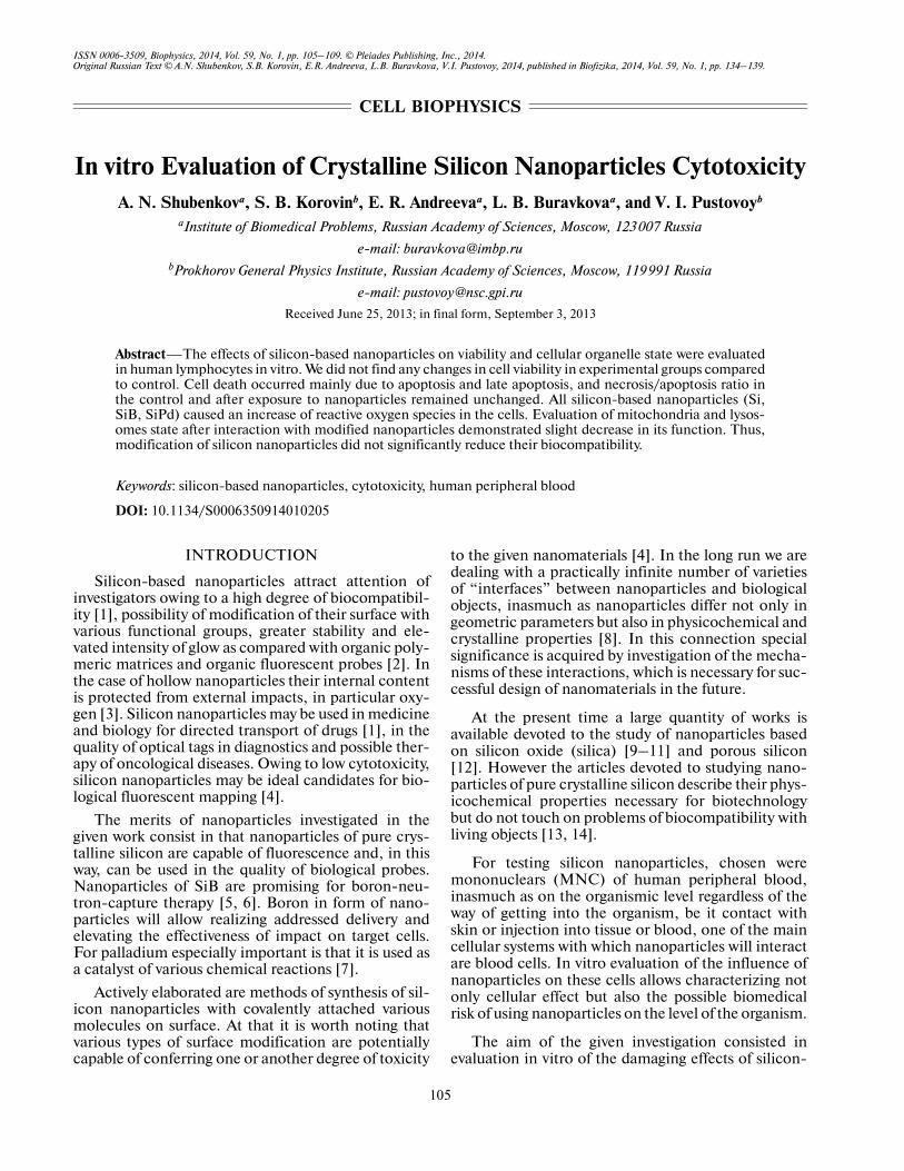
The characteristic of used nanoparticles is pre�sented in Table 1.
Isolation of MNC. MNC were obtained fromperipheral blood of healthy donors (n = 5) by themethod of centrifugation in a density gradient ofFicoll�Histopaque (Sigma�Aldrich, USA) asdescribed earlier [18].
Cultivation. MNC were cultivated for 24 h inmedium RPMI 1640 (Biolot, Russia) containing100 U/mL penicillin, 100 μg/mL streptomycin(PanEko, Russia), 2 mM glutamine (PanEko, Russia)and 5% FCS (HyClone, USA) in a concentration1 ⋅ 106 cell/mL in a Sanyo (Japan) CO2�incubator at37°C in an atmosphere of 5% CO2, 95% air and 100%humidity.
The concentration of initial suspension of Si andSiB nanoparticles constituted 10 mg/mL, the concen�tration of SiPd nanoparticles – 1 mg/mL. The nano�particles were added into the cultivation medium at aconcentration of 1, 10 and 100 μg/mL. The cells incu�bated in a medium not containing nanoparticles wereused in the quality of control for determination of ini�tial values of the studied indices.
Evaluation of MNC viability, characteristic oftransmembrane potential of mitochondria, state oflysosomal compartment and production of reactiveoxygen species. The cytotoxicity of nanoparticles wasdetermined in suspension with the aid of ANNEXINV – FITC Kit (Immunotech, France) according to theinstruction of the manufacturing company by a stan�dard method jointly with staining by propidium iodide
Table 1. Parameters of nanoparticles
Type of particles Diameter, nm Structure Solvent Initial concentration, mg/mL
Si 7 Crystal lattice of diamond type H2O 10
SiB 10 Crystal lattice of diamond type H2O 10
SiPd 15 Crystal lattice of diamond type H2O 1
BIOPHYSICS Vol. 59 No. 1 2014
IN VITRO CYTOTOXICITY OF CRYSTALLINE SILICON NANOPARTICLES 107
on a flow cytofluorimeter Epics XL (BeckmanCoulter, USA). We determined the share of live cells(Annexin V–/PI–), apoptotic cells (Annexin V+/PI–),necrotic cells (Annexin V–/PI+) and cells in a state ofpostapoptotic necrosis (Annexin V+/PI+).
The transmembrane potential of mitochondria wascharacterized with the aid of a fluorescent probe MitoTracker red FM (λexc = 581 nm, λem = 644 nm) (Invit�rogen, USA). The given substance passively penetratesthrough the cell membrane and accumulates in activemitochondria, while the intensity of its fluorescencereflects the state of the transmembrane potential.
The state of the lysosomal compartment was evalu�ated with the aid of a pH�sensitive fluorescent probeLyso Tracker Green DND 26 (λexc = 504 nm, λem =511 nm) (Invitrogen, USA).
Reactive oxygen species (ROS) in cells wererevealed using probe CM�H2DCFDA (λexc = 492–495 nm, λem = 517–527 nm) (Invitrogen, USA). Thegiven probe upon interaction with ROS transformsinto a fluorescing product, by the fluorescence inten�sity of which one can judge about the amount of ROSin cells.
All probes were used according to manufacturer’sinstructions, cells were analyzed on an Epics XL flowcytofluorimeter. Preliminarily the culture medium waswashed off with phosphate buffer by means of centrif�ugation at 1500 rpm in the course of 5 min on a centri�fuge (Eppendorf, Germany), after which we sus�pended the cell pellet in 1 mL of medium containing aprobe and incubated in a Sanyo (Japan) CO2�incuba�tor in the course of 30 min. We analyzed not less than10000 events per each sample.
Statistical analysis of obtained data was conductedwith the aid of program package MS Office Exel 2003and Statistica 7.0, using nonparametric Mann–Whit�ney test, differences were deemed significant at p < 0.05.
RESULTS AND DISCUSSION
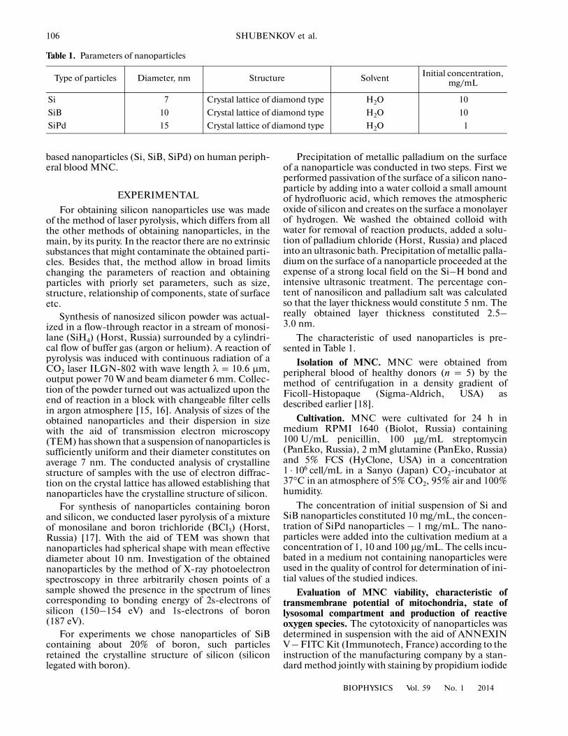
All the investigated nanoparticles did not exert pro�nounced influence on cell viability (Table 2).
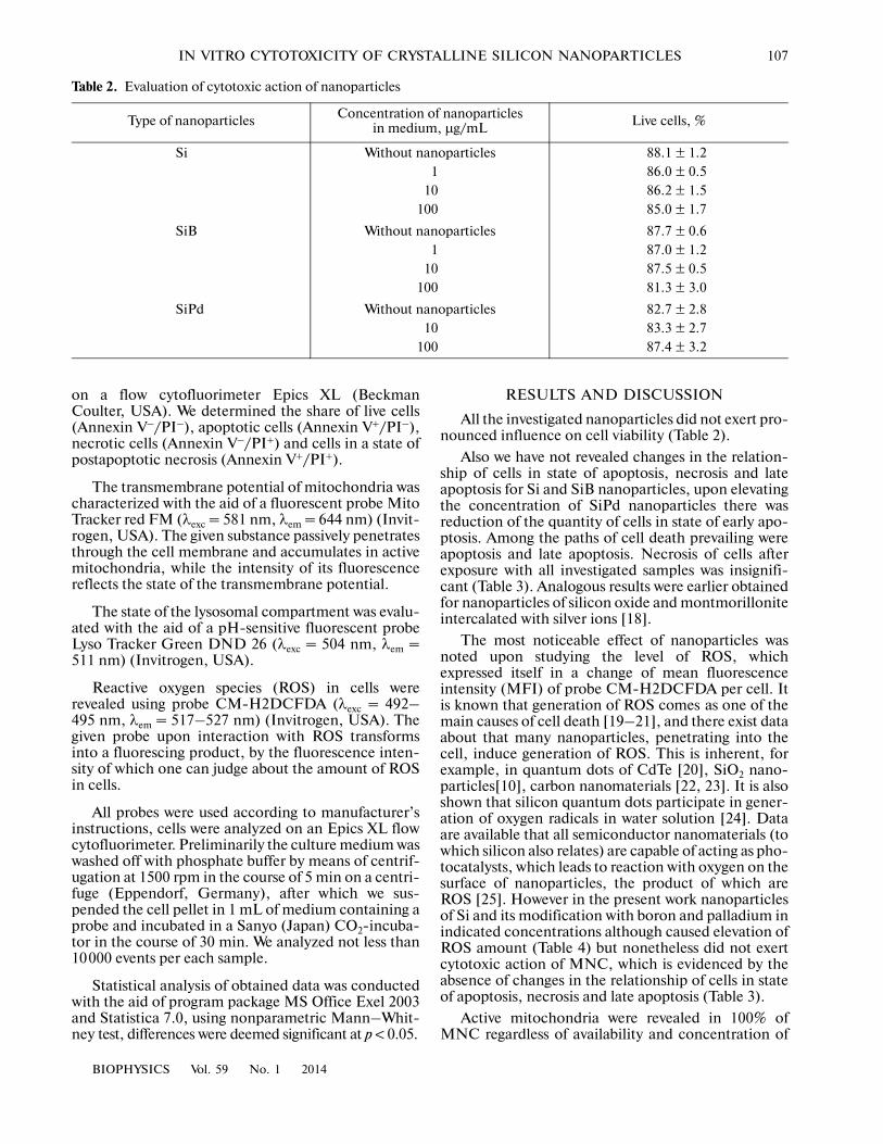
Also we have not revealed changes in the relation�ship of cells in state of apoptosis, necrosis and lateapoptosis for Si and SiB nanoparticles, upon elevatingthe concentration of SiPd nanoparticles there wasreduction of the quantity of cells in state of early apo�ptosis. Among the paths of cell death prevailing wereapoptosis and late apoptosis. Necrosis of cells afterexposure with all investigated samples was insignifi�cant (Table 3). Analogous results were earlier obtainedfor nanoparticles of silicon oxide and montmorilloniteintercalated with silver ions [18].
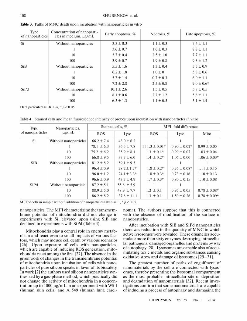
The most noticeable effect of nanoparticles wasnoted upon studying the level of ROS, whichexpressed itself in a change of mean fluorescenceintensity (MFI) of probe CM�H2DCFDA per cell. Itis known that generation of ROS comes as one of themain causes of cell death [19–21], and there exist dataabout that many nanoparticles, penetrating into thecell, induce generation of ROS. This is inherent, forexample, in quantum dots of CdTe [20], SiO2 nano�particles[10], carbon nanomaterials [22, 23]. It is alsoshown that silicon quantum dots participate in gener�ation of oxygen radicals in water solution [24]. Dataare available that all semiconductor nanomaterials (towhich silicon also relates) are capable of acting as pho�tocatalysts, which leads to reaction with oxygen on thesurface of nanoparticles, the product of which areROS [25]. However in the present work nanoparticlesof Si and its modification with boron and palladium inindicated concentrations although caused elevation ofROS amount (Table 4) but nonetheless did not exertcytotoxic action of MNC, which is evidenced by theabsence of changes in the relationship of cells in stateof apoptosis, necrosis and late apoptosis (Table 3).
Active mitochondria were revealed in 100% ofMNC regardless of availability and concentration of
Table 2. Evaluation of cytotoxic action of nanoparticles
Type of nanoparticles Concentration of nanoparticles in medium, µg/mL Live cells, %
Si Without nanoparticles 88.1 ± 1.21 86.0 ± 0.5
10 86.2 ± 1.5100 85.0 ± 1.7
SiB Without nanoparticles 87.7 ± 0.61 87.0 ± 1.2
10 87.5 ± 0.5100 81.3 ± 3.0
SiPd Without nanoparticles 82.7 ± 2.810 83.3 ± 2.7
100 87.4 ± 3.2
108
BIOPHYSICS Vol. 59 No. 1 2014
SHUBENKOV et al.
nanoparticles. The MFI characterizing the transmem�brane potential of mitochondria did not change inexperiments with Si, elevated upon using SiB anddeclined in experiments with SiPd (Table 4).
Mitochondria play a central role in energy metab�olism and react even to small impacts of various fac�tors, which may induce cell death by various scenarios[26]. Upon exposure of cells with nanoparticles,which are capable of inducing ROS generation, mito�chondria react among the first [27]. The absence in thegiven work of changes in the transmembrane potentialof mitochondria upon incubation of cells with nano�particles of pure silicon speaks in favor of its biosafety.In work [2] the authors used silicon nanoparticles syn�thesized by a gas�phase method, which practically didnot change the activity of mitochondria in a concen�tration up to 1000 μg/mL in an experiment with WS 1(human skin cells) and A 549 (human lung carci�
noma). The authors suppose that this is connectedwith the absence of modification of the surface ofnanoparticles.
After incubation with SiB and SiPd nanoparticlesthere was reduction in the quantity of MNC in whichactive lysosomes were revealed. These organelles accu�mulate more than sixty enzymes destroying intracellu�lar pathogens, damaged organelles and proteins by wayof autophagy [28]. Lysosomes are capable also of accu�mulating toxic metals and organic substances causingoxidative stress and damage of lysosomes [29–31].
The greatest number of paths of engulfment ofnanomaterials by the cell are connected with lysos�omes, thereby presenting the lysosomal compartmentas the most probable intracellular site of depositionand degradation of nanomaterials [32]. Recent inves�tigations confirm that some nanomaterials are capableof inducing a process of autophagy and damaging the
Table 3. Paths of MNC death upon incubation with nanoparticles in vitro
Typeof nanoparticles
Concentration of nanoparti�cles in medium, µg/mL Early apoptosis, % Necrosis, % Late apoptosis, %
Si Without nanoparticles 3.3 ± 0.3 1.1 ± 0.3 7.4 ± 1.1
1 3.6 ± 0.7 1.6 ± 0.3 8.8 ± 1.1
10 3.7 ± 0.4 2.5 ± 1.0 7.7 ± 1.1
100 3.9 ± 0.7 1.9 ± 0.8 9.3 ± 1.2
SiB Without nanoparticles 5.5 ± 1.6 1.3 ± 0.4 5.5 ± 0.9
1 6.2 ± 1.8 1.0 ± 0 5.8 ± 0.6
10 5.7 ± 1.4 0.7 ± 0.3 6.0 ± 1.1
100 7.2 ± 2.8 2.5 ± 0.8 9.0 ± 0.6*
SiPd Without nanoparticles 10.1 ± 2.6 1.5 ± 0.5 5.7 ± 0.5
10 8.1 ± 0.6 2.7 ± 1.2 5.8 ± 1.1
100 6.3 ± 1.3 1.1 ± 0.5 5.1 ± 1.4
Data presented as M ± m, * p < 0.05.
Table 4. Stained cells and mean fluorescence intensity of probes upon incubation with nanoparticles in vitro
Typeof nanoparticles
Nanoparticles, µg/mL
Stained cells, % MFI, fold difference
ROS Lyso ROS Lyso Mito
Si Without nanoparticles 66.2 ± 7.4 43.0 ± 6.2 1 1 1
1 78.1 ± 6.3 36.5 ± 7.8 11 1.3 ± 0.01* 0.90 ± 0.02* 0.99 ± 0.05
10 75.2 ± 6.2 35.9 ± 8.1 1.3 ± 0.1* 0.99 ± 0.07 1.03 ± 0.04
100 66.8 ± 9.5 37.7 ± 6.0 1.4 ± 0.2* 1.06 ± 0.00 1.06 ± 0.03*
SiB Without nanoparticles 81.2 ± 8.2 59.1 ± 9.5 1 1 1
1 96.4 ± 0.9 28.2 ± 1.7* 1.8 ± 0.2* 0.76 ± 0.08* 1.11 ± 0.15
10 96.0 ± 1.2 24.1 ± 3.3* 1.8 ± 0.3* 0.73 ± 0.16 1.10 ± 0.13
100 96.6 ± 0.9 43.7 ± 4.9 1.7 ± 0.3* 0.80 ± 0.15 1.10 ± 0.08
SiPd Without nanoparticle 87.2 ± 5.1 55.8 ± 5.9 1 1 1
10 88.9 ± 5.0 48.9 ± 7.7 1.2 ± 0.1 0.95 ± 0.05 0.78 ± 0.08*
100 86.2 ± 8.2 37.8 ± 11.1 1.3 ± 0.1 1.50 ± 0.26 0.78 ± 0.09*
MFI of cells in sample without addition of nanoparticles taken as 1, * p < 0.05.
BIOPHYSICS Vol. 59 No. 1 2014
IN VITRO CYTOTOXICITY OF CRYSTALLINE SILICON NANOPARTICLES 109
lysosomal membrane [32]. It was shown that activa�tion of lysosomes is capable of elevating oxidativestress [22]. In the given work a similar effect wasobserved in experiments with SiPd nanoparticles atmaximal concentration, when we revealed simulta�neous elevation of the level of ROS and MFI of spe�cific lysosomal probe. Although changes of lysosomalpermeability are often regarded as a factor leading tonecrosis, there are data about that loss of lysosomalintegrity is associated with loss of membrane potentialof mitochondria [11, 33] and is capable of leading toapoptosis [9, 34]. Thus nanoparticles of silicon oxidewere capable of causing apoptosis of line MH�S cells,one of the causes of activation of which comes to bethe activity of lysosomal enzymes [9]. Nonetheless inthe present work we did not reveal a connectionbetween the state of the lysosomal compartment andcell viability.
In this way, it is shown that silicon�based nanopar�ticles are nontoxic, which is evidenced by the absenceof changes in viability and proportion of apoptotic andnecrotic cells after 24�h incuation. However, beingintercalated by atoms of boron and palladium, thegiven nanomaterials are capable of causing changes ofmitochondria, lysosomal compartment and amount ofROS in cells.
All the examined silicon�based nanoparticles pro�voked the elevation of elevated the amount of ROS incells. The smallest changes were disclosed for nano�particles of unmodified silicon. Modification withboron led to reduction of lysosome activity and eleva�tion of mitochondrial transmembrane potential.Modification with palladium, conversely, causedreduction of mitochondrial transmembrane potential.The obtained results point to that modification of sili�con nanoparticles by atoms of other chemical ele�ments is capable of changing the degree of their cyto�toxicity in relation to various cellular parameters,which can be revealed in a in vitro system.
ACKNOWLEDGMENTS
The work was supported by the Russian Founda�tion for Basic Research (11�02�12210�ofi�m).
REFERENCES
1. J. Lu, M. Liong, Z. Li, et al., NIH Public Access 6 (16),1794 (2011).
2. K. Fujoka, S. Hanada, F. Kanaya, et al., J. Phys.: Conf.Series 304 (2011).
3. V. A. Livshits, I. V. Demisheva, and B. B. Meshkov,Ross. Nanotekhologii 4 (1–2), 99 (2009).
4. A. Shiohara, S. Hanada, S. Prabakar, et al., J. Am.Chem. Soc. 138 (1), 248 (2010).
5. Yu. A. Koldaeva, E. Yu. Grigor’eva, and V. N. Kulakov,Luchevaya Terapiya 2, (1) (2011).
6. V. N. Mitin, N. G. Kozlovskaya, and A. M. Arno�pol’skaya, Onkologiya, no. 1, (2006).
7. Y. J. Wang, Am. Cem. Sac. No. 116, 397 (1994).8. A. E. Nel, L. Mädler, D. Velegol, et al., Nature Materi�
als 8 (7), 543 (2009).9. M. S. Thibodeau, C. Giardina, D. A. Knecht, et al.,
Toxicological sciences: an official journal of the Societyof Toxicology 80 (1), 34 (2004).
10. W. Lin, Y�w Huang, X�D Zhou, et al., Toxicol. Appl.Pharmacol. 217 (3), 252 (2006).
11. S. K. Sohaebuddin, P. T. Thevenot, D. Baker, et al.,Particle and fibre toxicology 7, 22 (2010).
12. Ji�Ho Park., L. Gu, G. von Maltzahn, et al., NatureMaterials (8) (2009).
13. Z. F. Li and E. Ruckenstein, Nano Lett. 4 (8), 1463(2004).
14. J. H. Warner, A. Hoshino, and K. Yamamoto, ImagingAgents 117, 4626 (2005).
15. A. Vladimirov, S. Korovin, A. Surkov, et al., Laser Phys.21 (4), 830 (2011).
16. E. Kelm, S. Korovin, V. Pustovoy, et al., Appl. Phys. B:Lasers and optics 105 (3), 599 (2011).
17. V. Beklemishev, V. Pustovoy, S. Korovin, et al., Nanoin�dustriya (5), 44 (2011).
18. E. R. Andreeva, E. G. Rudimov, and A. N. Gornos�taeva, Byul. Eksperim. Biol. Med. 155 (3), 377 (2013).
19. M. Green and E. Hawman, Chem. Commun. (Cam�bridge, England), No. 1, 123 (2005).
20. B. I. Ipe, M. Lehnig, and C. M. Niemeyer, Small(Weinheim an der Bergstrasse, Germany) 1 (7), 706(2005).
21. J. Lovri , S. J. Cho, F. M. Winnik, et al., Chem. Biol.12 (11), 1227 (2005).
22. B. Halamoda Kenzaoui, C. Chapuis Bernasconi,S. Guney�ayra, et al., Biochem. J. 441 (3), 813 (2012).
23. M. N. Moore, J. A. J. Redman, J. W. Redman, et al.,Nanotoxicology 3 (1), 40 (2009).
24. K. Fujioka, M. Hiruoka, K. Sato, et al., Nanotechnol�ogy 19 (41), 415102 (2008).
25. N. O’Farrell, A. Houlton, and B. R. Horrocks, Int.J. Nanomedicine 1 (4), 451 (2006).
26. J. M. Hansen, Y�M. Go, and D. P. Jones, Ann. Rev.Pharmacol. Toxicol. 46, 215 (2006).
27. M. Dusica, B. Maik, and P. Ewa, NanoPharmaceuti�cals Online J. 1, 1 (2006).
28. D. J. Klionsky, Nature Rev. Mol. Cell Biol. 8 (11), 931(2007).
29. M. N. Moore, Histochemical 22,187 (1990).30. M. N. Moore, M. H. Depledge, J. W. Readman, et al.,
Mutation Res. 552, 247 (2004).31. M. N. Moore, D. Lowe, and A. Köhler, ICES Tech�
niques in Marine Environmental Sciences (36), 31(2004).
32. S. T. Stern, P. P. Adiseshaiah, and R. M. Crist, Part.Fibre Toxicol. 9, 20 (2012).
33. T. Xia, M. Kovochich, M. Liong, et al., ACS Nano 2(1), 85 (2008).
34. U. T. Brunk and I. Svensson, RedoxRep. 4 (1–2), 3(1999).
c�