in vitro cytotoxicity of monodispersed hematite nanoparticles on hek 293 cells
TRANSCRIPT
Materials Letters 65 (2011) 815–817
Contents lists available at ScienceDirect
Materials Letters
j ourna l homepage: www.e lsev ie r.com/ locate /mat le t
In vitro cytotoxicity of monodispersed hematite nanoparticles on Hek 293 cells
Haijuan Yan a,b, Benshu Zhang a,⁎a Tianjin Medical University, 22 Qixiangtai Road, HePing District, TianJin 300070, Chinab Goettingen University, Goettingen 37075, Germany
⁎ Corresponding author. Tel./fax: +86 354 4632268.E-mail address: [email protected] (B. Zhang)
0167-577X/$ – see front matter © 2010 Elsevier B.V. Adoi:10.1016/j.matlet.2010.12.004
a b s t r a c t
a r t i c l e i n f oArticle history:Received 1 September 2010Accepted 5 December 2010Available online 10 December 2010
Keywords:Hematite nanoparticlesHydrothermalMonodispersionCytotoxicityOxidative stress
A simple hydrothermal method was developed to prepare monodispersed and well-crystallized hematitenanoparticles with a diameter distribution of about 180 nm. In-vitro cytotoxicity of such nanoparticles wascarried out using Hek 293 cell culture system with different dosages. Assessment of cell viability reveals thathematite nanoparticles reduce cell viability in a dose- and time-dependent manner within a rather widedosage range. Such cytotoxicity is correlative to the decrease of the activity of antioxidative enzymes inducedby the oxidative stress in cells.
.
ll rights reserved.
© 2010 Elsevier B.V. All rights reserved.
1. Introduction
Hematite is an important multifunctional material and has greatpotential applications in fine ceramics, pigments, paints, catalysis,energy conversion, gas sensor andmedicine [1–7]. Controlling synthesisof hematite particles is considerably important as their physical andchemical properties drastically depend on their particle size, morphol-ogy and dispersion. Various methods have been introduced to prepareshape- and size-controlled hematite nanostructures, such as the sol–gelmethod, microwave, thermal decomposition, chemical precipitationand hydrothermal treatment [1–3,8,9]. Among these methods, hydro-thermal synthesis demonstrates its advantages over conventionaltechnologies in effective control of size and shape of the particles, andrelatively low reaction temperature as well as high homogeneity andwell-crystallized products [8,9].
Due to the rapid development of nanotechnology, considerableattention has been paid to investigate the health impact of occupa-tional and environmental “ultrafine”particles [10,11]. The reduction insize of particles frommicro to nanoscale not only provides benefits todiverse scientific fields but also introduces potential risks to humansand the environment [12]. Nanostructured iron oxides allow a widerange of potential applications in nanotechnology related fieldsincluding bio-medical imaging, magnetic target drug delivery, envi-ronmental catalysis, magnetic storage and so forth [13,14]. In view ofthese environmental and biomedical applications, it necessitates thefurther study of the nano–bio interaction behavior of nanostructurediron nanomaterials to be aware of the importance of analyzing the
positive aspects of nanomaterials while avoiding their potential toxiceffects [15]. In this paper,monodispersed hematite nanoparticlesweresuccessfully prepared via a simple hydrothermal method in aqueoussolution. And the in vitro cytotoxicity of nanoparticleswas tested usingKidney Hek 293 cell line.
2. Experimental
2.1. Synthesis of monodispersed hematite nanoparticles
Typically, 1.3 g Fe(NO3)3 and 0.46 g BaCl2 were dissolved in 30 mLDI water containing 0.40 g PVP (40,000 g/mol) under stirring. Then0.22 g NaOHwas added into this mixture. Finally, the resultant solutionwas transferred into an autoclave with a capacity of 50 mL, and kept at200 °C for 12 h. After the autoclave cools to room temperature, theprecipitants were collected via centrifuge and washed with DI waterand ethanol for several times.
2.2. Characterization
The crystal structure and phase identification of nanoparticleswere performed by X-ray diffraction (XRD Rigaku DMax-2200) with amonochromatized source of Cu Kα1 radiation (λ=0.15405 nm).Field-emission transmission electron microscopy (TEM JEM 2100F)was carried out to study the morphology, crystallinity and particlesize of the samples. Size distributions of nanoparticles wereperformed using DLS measurement in aqueous solution (pH 6–7).FTIR spectrum (SHIMADZU IRPrestige-21) was applied using the KBrpellet.
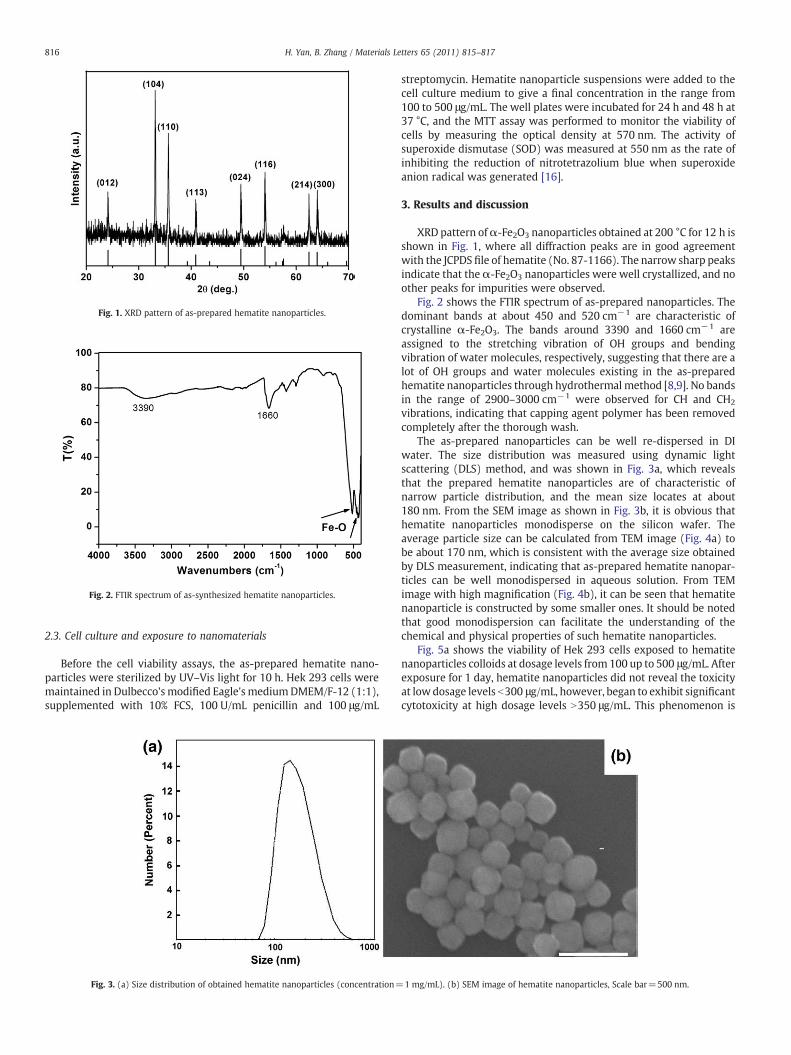
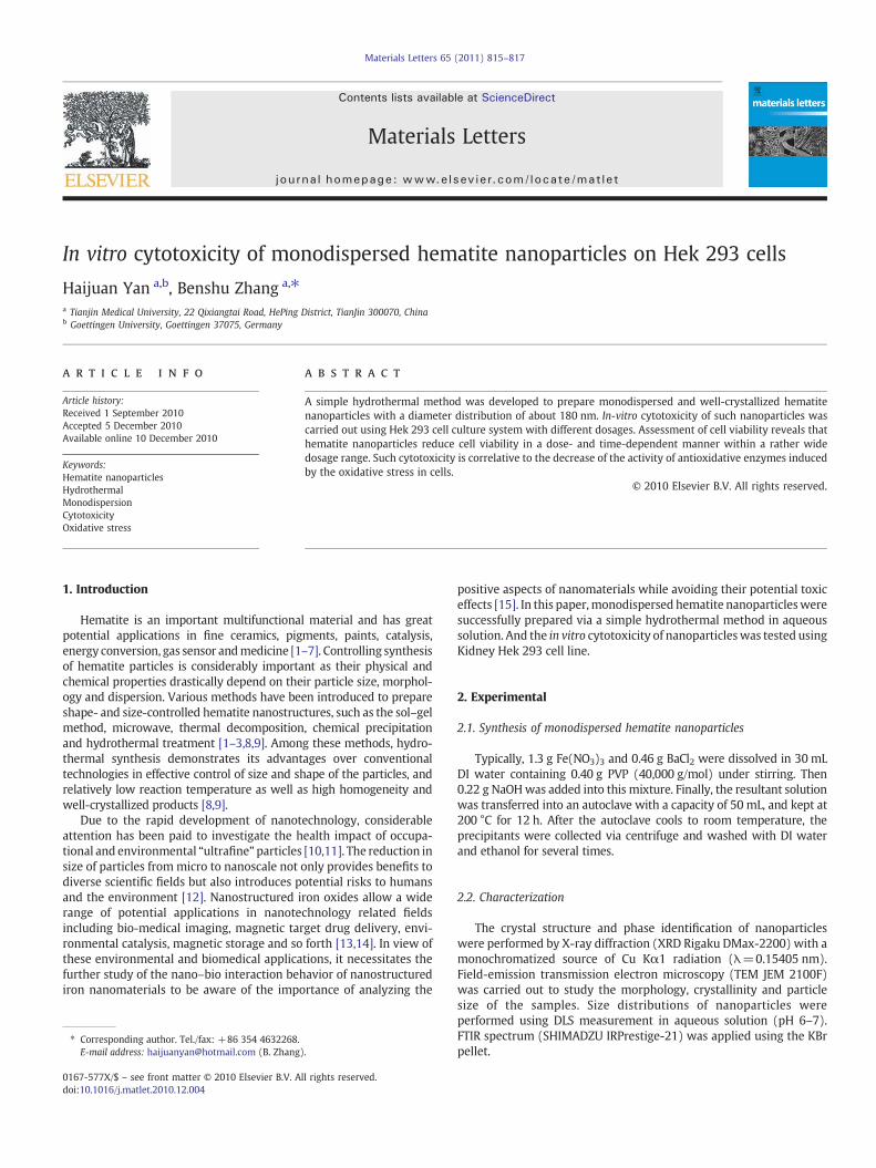
Fig. 1. XRD pattern of as-prepared hematite nanoparticles.
Fig. 2. FTIR spectrum of as-synthesized hematite nanoparticles.
816 H. Yan, B. Zhang / Materials Letters 65 (2011) 815–817
2.3. Cell culture and exposure to nanomaterials
Before the cell viability assays, the as-prepared hematite nano-particles were sterilized by UV–Vis light for 10 h. Hek 293 cells weremaintained in Dulbecco's modified Eagle's mediumDMEM/F-12 (1:1),supplemented with 10% FCS, 100 U/mL penicillin and 100 μg/mL
Fig. 3. (a) Size distribution of obtained hematite nanoparticles (concentration=
streptomycin. Hematite nanoparticle suspensions were added to thecell culture medium to give a final concentration in the range from100 to 500 μg/mL. The well plates were incubated for 24 h and 48 h at37 °C, and the MTT assay was performed to monitor the viability ofcells by measuring the optical density at 570 nm. The activity ofsuperoxide dismutase (SOD) was measured at 550 nm as the rate ofinhibiting the reduction of nitrotetrazolium blue when superoxideanion radical was generated [16].
3. Results and discussion
XRD pattern ofα-Fe2O3 nanoparticles obtained at 200 °C for 12 h isshown in Fig. 1, where all diffraction peaks are in good agreementwith the JCPDS file of hematite (No. 87-1166). The narrow sharp peaksindicate that the α-Fe2O3 nanoparticles were well crystallized, and noother peaks for impurities were observed.
Fig. 2 shows the FTIR spectrum of as-prepared nanoparticles. Thedominant bands at about 450 and 520 cm−1 are characteristic ofcrystalline α-Fe2O3. The bands around 3390 and 1660 cm−1 areassigned to the stretching vibration of OH groups and bendingvibration of water molecules, respectively, suggesting that there are alot of OH groups and water molecules existing in the as-preparedhematite nanoparticles through hydrothermal method [8,9]. No bandsin the range of 2900–3000 cm−1 were observed for CH and CH2
vibrations, indicating that capping agent polymer has been removedcompletely after the thorough wash.
The as-prepared nanoparticles can be well re-dispersed in DIwater. The size distribution was measured using dynamic lightscattering (DLS) method, and was shown in Fig. 3a, which revealsthat the prepared hematite nanoparticles are of characteristic ofnarrow particle distribution, and the mean size locates at about180 nm. From the SEM image as shown in Fig. 3b, it is obvious thathematite nanoparticles monodisperse on the silicon wafer. Theaverage particle size can be calculated from TEM image (Fig. 4a) tobe about 170 nm, which is consistent with the average size obtainedby DLS measurement, indicating that as-prepared hematite nanopar-ticles can be well monodispersed in aqueous solution. From TEMimage with high magnification (Fig. 4b), it can be seen that hematitenanoparticle is constructed by some smaller ones. It should be notedthat good monodispersion can facilitate the understanding of thechemical and physical properties of such hematite nanoparticles.
Fig. 5a shows the viability of Hek 293 cells exposed to hematitenanoparticles colloids at dosage levels from 100 up to 500 μg/mL. Afterexposure for 1 day, hematite nanoparticles did not reveal the toxicityat lowdosage levels b300 μg/mL, however, began to exhibit significantcytotoxicity at high dosage levels N350 μg/mL. This phenomenon is
1 mg/mL). (b) SEM image of hematite nanoparticles, Scale bar=500 nm.
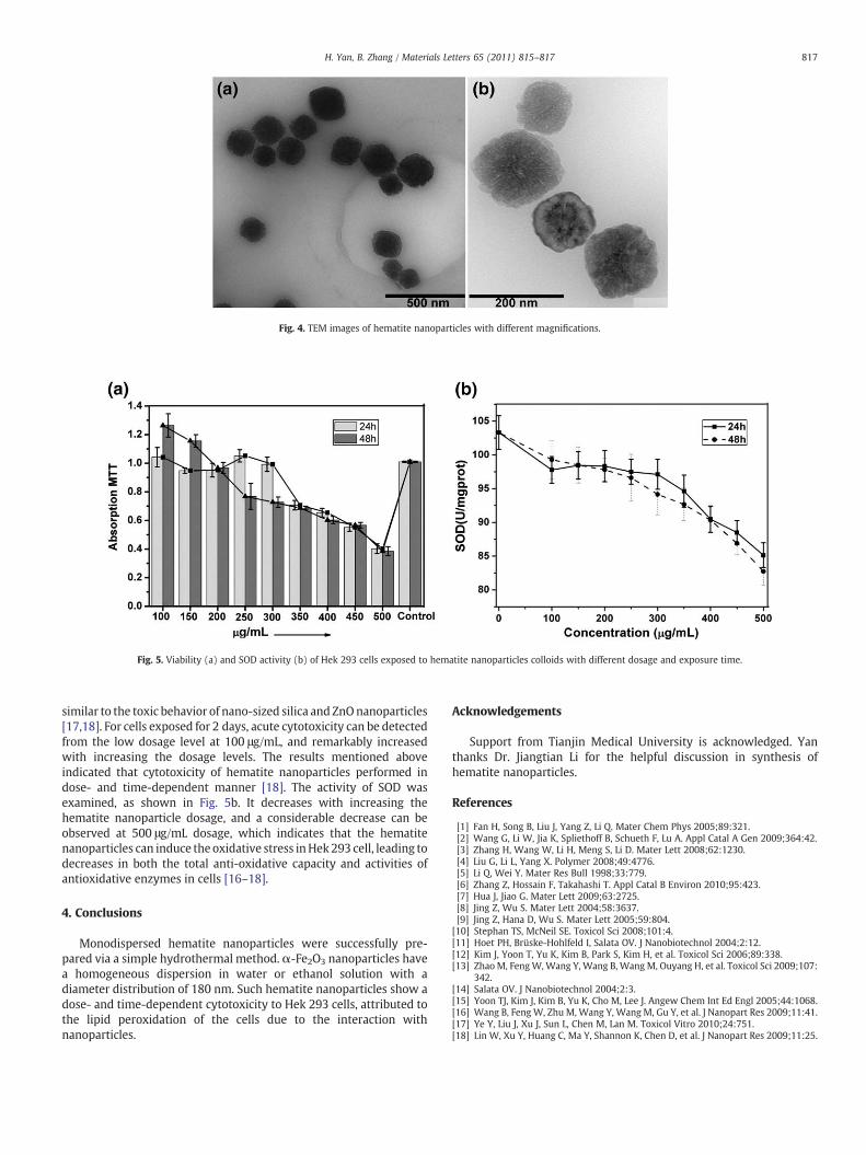
Fig. 4. TEM images of hematite nanoparticles with different magnifications.
Fig. 5. Viability (a) and SOD activity (b) of Hek 293 cells exposed to hematite nanoparticles colloids with different dosage and exposure time.
817H. Yan, B. Zhang / Materials Letters 65 (2011) 815–817
similar to the toxic behavior of nano-sized silica and ZnOnanoparticles[17,18]. For cells exposed for 2 days, acute cytotoxicity can be detectedfrom the low dosage level at 100 μg/mL, and remarkably increasedwith increasing the dosage levels. The results mentioned aboveindicated that cytotoxicity of hematite nanoparticles performed indose- and time-dependent manner [18]. The activity of SOD wasexamined, as shown in Fig. 5b. It decreases with increasing thehematite nanoparticle dosage, and a considerable decrease can beobserved at 500 μg/mL dosage, which indicates that the hematitenanoparticles can induce the oxidative stress inHek 293 cell, leading todecreases in both the total anti-oxidative capacity and activities ofantioxidative enzymes in cells [16–18].
4. Conclusions
Monodispersed hematite nanoparticles were successfully pre-pared via a simple hydrothermal method. α-Fe2O3 nanoparticles havea homogeneous dispersion in water or ethanol solution with adiameter distribution of 180 nm. Such hematite nanoparticles show adose- and time-dependent cytotoxicity to Hek 293 cells, attributed tothe lipid peroxidation of the cells due to the interaction withnanoparticles.
Acknowledgements
Support from Tianjin Medical University is acknowledged. Yanthanks Dr. Jiangtian Li for the helpful discussion in synthesis ofhematite nanoparticles.
References
[1] Fan H, Song B, Liu J, Yang Z, Li Q. Mater Chem Phys 2005;89:321.[2] Wang G, Li W, Jia K, Spliethoff B, Schueth F, Lu A. Appl Catal A Gen 2009;364:42.[3] Zhang H, Wang W, Li H, Meng S, Li D. Mater Lett 2008;62:1230.[4] Liu G, Li L, Yang X. Polymer 2008;49:4776.[5] Li Q, Wei Y. Mater Res Bull 1998;33:779.[6] Zhang Z, Hossain F, Takahashi T. Appl Catal B Environ 2010;95:423.[7] Hua J, Jiao G. Mater Lett 2009;63:2725.[8] Jing Z, Wu S. Mater Lett 2004;58:3637.[9] Jing Z, Hana D, Wu S. Mater Lett 2005;59:804.[10] Stephan TS, McNeil SE. Toxicol Sci 2008;101:4.[11] Hoet PH, Brüske-Hohlfeld I, Salata OV. J Nanobiotechnol 2004;2:12.[12] Kim J, Yoon T, Yu K, Kim B, Park S, Kim H, et al. Toxicol Sci 2006;89:338.[13] Zhao M, FengW,Wang Y, Wang B, Wang M, Ouyang H, et al. Toxicol Sci 2009;107:
342.[14] Salata OV. J Nanobiotechnol 2004;2:3.[15] Yoon TJ, Kim J, Kim B, Yu K, Cho M, Lee J. Angew Chem Int Ed Engl 2005;44:1068.[16] Wang B, Feng W, Zhu M, Wang Y, Wang M, Gu Y, et al. J Nanopart Res 2009;11:41.[17] Ye Y, Liu J, Xu J, Sun L, Chen M, Lan M. Toxicol Vitro 2010;24:751.[18] Lin W, Xu Y, Huang C, Ma Y, Shannon K, Chen D, et al. J Nanopart Res 2009;11:25.