in vitro biocompatibility of chitosan/hyaluronic acid-containing calcium phosphate bone cements
TRANSCRIPT
ORIGINAL PAPER
In vitro biocompatibility of chitosan/hyaluronic acid-containingcalcium phosphate bone cements
Saeed Hesaraki • Nader Nezafati
Received: 19 October 2013 / Accepted: 24 December 2013
� Springer-Verlag Berlin Heidelberg 2014
Abstract The need for bone repair has increased as the
population ages. In this research, calcium phosphate
cements, with and without chitosan (CS) and hyaluronic
acid (HA), were synthesized. The composition and mor-
phological properties of cements were evaluated by X-ray
diffraction and scanning electron microscopy. The acellular
in vitro bioactivity revealed that different apatite mor-
phologies were formed on the surfaces of cements after
soaking in simulated body fluid. The in vitro osteoblastic
cell biocompatibility of in situ forming cements was
evaluated and compared with those of conventional cal-
cium phosphate cements (CPCs). The viability and growth
rate of the cells were similar for all CPCs, but better
alkaline phosphatase activity was observed for CPC with
CS and HA. Calcium phosphate cements supported
attachment of osteoblastic cells on their surfaces. Spindle-
shaped osteoblasts with developed cytoplasmic membrane
were found on the surfaces of cement samples after 7 days
of culture. These results reveal the potential of the CPC–
CS/HA composites to be used in bone tissue engineering.
Keywords Calcium phosphate cement � Chitosan �Hyaluronic acid � Alkaline phosphatase � Osteoblast
Introduction
Calcium phosphates (Ca–P) constitute an important group
of compounds widely used as synthetic bone substitution
materials [1–3], in clinical applications due to their good
biocompatibility, osteoconduction, simulation of bone
minerals, and bone replacement capability [4, 5]. The Ca–P
minerals also provide a desirable substrate for cell attach-
ment and expression of osteoblast phenotype [6, 7]. Cal-
cium phosphate cement (CPC) can be molded or injected to
form a scaffold in situ that properly adapts to the shape of
complicated bone defects [8]. Injectable scaffolds are
appropriate for cell delivery because they can lessen the
time of surgical operation, decrease postoperative pain and
scar size, make rapid recovery and reduce cost [9]. A
typical CPC was first reported by Brown and Chow [10].
This CPC powder could be mixed with aqueous liquid to
form a paste that would set in situ and form hydroxyapatite
as the main constituent part of the mineral phase of bone
[11]. Since then, several other calcium phosphate cements
and injectable cements have been developed [12–15]. On
the other hand, it is necessary to develop CPC with os-
teopromotive or osteoinductive factors to improve its bio-
logical performance [16, 17].
Hyaluronic acid (HA) is an anionic glycosaminoglycan
distributed widely through connective, epithelial and neural
tissues. In the extracellular matrix, hyaluronate can par-
ticipate as a hydrated network and operate as an organizing
core to connect complex intercellular aggregates [18, 19]
that assists significantly in cell proliferation and migration
[20]. Several applications of HA in tissue engineering have
been reported [21–23].
Chitosan (CS) has different biological properties that
make it attractive to medical applications including: bio-
degradability, lack of toxicity, antifungal effects, acceler-
ation of wound healing and stimulation of immune system
[24].
It has been reported that its handling, rheological
properties and antibacterial effect of CPC based on trical-
cium phosphate, tetracalcium phosphate (TTCP) and
S. Hesaraki � N. Nezafati (&)
Department of Nanotechnology and Advanced Materials,
Materials and Energy Research Center, Alborz, Iran
e-mail: [email protected]
123
Bioprocess Biosyst Eng
DOI 10.1007/s00449-013-1122-0
dicalcium phosphate (DCP) can get better if HA and/or
sodium hyaluronate and CS, as gel-forming polymers are
used [14, 25–27]. For example, Xu and colleagues have
shown that CS increases the flexural strength of a CS–CPC
composite composed of tetracalcium phosphate–dicalcium
phosphate anhydrous considerably, and the highest value
was reached when 15–20 wt% CS was incorporated into
the CPC, although Zhang reported that optimum results
also occurred when CPC–CS was synergistically combined
with Vicryl fibers or alginate microbeads [28]. Kai et al.
have investigated that the injectability, setting time and
mechanical strength of CPC could be significantly
improved by using 0.6 %wt of sodium hyaluronate (NaHA)
when NaHA, as the liquid phase, was introduced into the
system [29]. Hence, proper injectability along with bio-
compatibility can have good prospects for medical
application.
Despite the above-mentioned studies, it is worth to
mentioning that there is little information available con-
cerning the biological properties and evaluation of cell
behavior of CS/HA-based calcium phosphate cements for
bone composite cement. Thus, in the present study, after
the composition and morphological properties of cements
were assessed, apatite formation, cytocompatibility and
cell responses of the injectable CS/HA–CPC composites to
act as a bone substitute and its potential for bone tissue
engineering were investigated in vitro.
Experimental procedures
Sample preparation
The following starting materials were purchased and uti-
lized without further purification in this study: calcium
carbonate (CaCO3, Merck 2076, Germany), DCP anhy-
drous (Merck 2144, Germany), DCP dehydrate (DCPD,
Merck 172/06, Germany), high molecular weight HA
(1750 kDa; Jinan Haohua Industry Co., China), chitosan
(Chitotech Company, Iran) and acetic acid (Merck 100062,
Germany). The TTCP powder was synthesized by solid-
state reaction between equimolar amounts of DCP anhy-
drous and calcium carbonate, as described elsewhere [30].
Formulation of cements
The cement powder phase of the experimental groups was
a mixture of ground TTCP and DCPD (Brushite) powder in
a molar ratio of 1:1, and their liquid phases were prepared
by adding HA (CPC 1750) and CS (CPC–CS) at concen-
trations of 3 g ml-1 to a 1 % (v/v) solution of acetic acid.
Additionally, the pure CPC was also prepared with the
same powder phase and distilled water as a liquid phase to
compare its properties with those of the other two cements.
In each paste, the powder/liquid ratio (P/L) was selected in
a way that the best consistent paste can be obtained. This
proportion was 3/5 for CPC, 3/1 for CPC 1750 and
2/8 g ml-1 for CPC–CS. The powder phase was mixed
with the liquid, and the obtained paste was molded.
Characterization and measurement
Particle size distributions of synthesized powders of TTCP
and DCPD as a main part of cements were examined by
laser particle size analyzer (LPSA) via a Fritsch Particle
Size ‘Analysette 22’ instrument in the acetone–alcohol
mode.
Phase analysis of cement powder was performed by a
Philips PW3710 diffractometer. This instrument worked
with voltage and current settings of 40 kV and 30 mA,
respectively, and utilized Cu–Ka radiation (1.54 A). For
qualitative analysis, X-ray diffraction (XRD) diagrams
were recorded in the interval 10o B 2h B 50o.
The microstructure of the gold-coated fractured surfaces
of set cements was observed using SEM (Stereoscan S-360
Cambridge).
Acellular in vitro bioactivity
The in vitro surface reactivity of calcium phosphate-based
composites was accomplished after the samples were
soaked in simulated body fluid (SBF) at solid (S) to liquid
(L) loading of 1 g/100 ml and then kept at 37 �C for
14 days. The SBF solution was prepared according to the
procedure described by Kokubo et al. [31] by dissolving
NaCl 8.035 g/l, KCl 0.225 g/l, K2HPO4�3H2O 0.231 g/l,
MgCl2�6H2O 0.311 g/l, CaCl2 0.292 g/l, NaHCO3 0.355 g/
l and Na2SO3 0.072 g/l into distilled water, and buffered at
pH = 7.25 with 6.118 g/l tris–hydroxymethyl amino-
methane and 1 N HCl solution at 37C. The SBF solution
was chosen because of its characteristic of being highly
supersaturated with respect to apatite. The SBF solution is
so far the best for in vitro measurement of apatite-forming
ability in implant materials [32].
The surface morphology and microstructure of the
samples were evaluated using SEM after soaking in the
SBF solution for 14 days.
In vitro assay
The cement specimens (CPC, CPC–CS and CPC 1750)
were selected for clinical applications because of their
ability in setting and also due to diverse usage of such
materials in reconstruction of bone injuries. In this part of
the study, polystyrene pieces with dimensions similar to
that of cement specimens were used as control. It is
Bioprocess Biosyst Eng
123
necessary to apply biological tests for studying biocom-
patibility of cement materials. In this study, to evaluate the
biological properties of pastes, osteoblastic cells were
derived from newborn rat calvaria. These cells were pur-
chased from Royan Institute Research Center.
Sample preparation
The cylindrical cement pastes were formed in Teflon mold
(10 mm in diameter and 3 mm in height) for measuring the
level of cytotoxicity and cell morphology. To obtain
specimens with smooth surfaces, the molds filled with
cement pastes were packed with a glass plate. After the
cements hardened, the samples were sterilized with 70 %
ethanol and seeded with the cells [33]. The cultured cells
were continuously checked by a microscope to change the
cell medium if it was necessary.
Cell culture procedure
The cells were cultured in Dulbecco modified Eagle med-
ium (DMEM;Gibco-BRL, LifeTechnologies, GrandIsland,
NY) supplemented with 15 % fetal bovine serum
(FBS;Dainippon Pharmaceutical, Osaka, Japan) in a 5 %
CO2 atmosphere at 37˚C. The size of the cell flask was
50 mm. To store and freeze the cells, a suspension of cells
(2 9 104 cells for each vial) was poured into a 0.5 ml
medium including 90 % fetal bovine serum and 10 %
dimethylsulfoxide (DMSO). This suspension was conveyed
to cryotubes and frozen at -20 �C for 2 h. The tubes were
frozen at -70 �C and then transferred to a liquid nitrogen
tank after about 4 h. To subculture the cells, the tubes were
heated at 37 �C in a Benmary bath and the cells were
washed with culture medium after the ingredients of the
tube were melted. Finally, the resultant cell suspension was
cultured. The confluent cells were dissociated with trypsin
and subcultured to three passages which were used for
tests.
Cells proliferation assay and alkaline phosphatase (ALP)
activity
The osteoblastic cells (cultured for 1 week) were cultured
on the surfaces of cements (cell density: 2 9 104 cell/
sample) and incubated for 4 h. The specimen/cell samples
were placed in 24-well culture plates and left undisturbed
in an incubator for 3 h to allow the cells to attach to them
and then an additional 30 ml of culture medium was added
into each well. The cell/specimen constructs were cultured
in a humidified incubator at 37 �C with 95 % air and 5 %
CO2 for different periods. Note that every 3 days, the
medium was exchanged.
The proliferation of the osteoblastic cells was deter-
mined using the MTT (3-{4,5-dimethylthiazol-2yl}-2,5-
diphenyl-2H-tetrazolium bromide) assay. For this purpose,
at the end of each evaluating period, the medium was
removed and 2 ml of MTT solution was added to each
well. Following incubation at 37 �C for 4 h in a fully
humidified atmosphere at 5 % CO2 in air, MTT was taken
up by active cells and reduced in the mitochondria to
insoluble purple formazon granules. Subsequently, the
medium was discarded and the precipitated formazon was
dissolved in dimethylsulfoxide, DMSO, and optical density
(OD) of the solution was read using a microplate spectro-
photometer (BIO-TEK Elx 800, Highland park, USA) at a
wavelength of 570 nm. The recorded OD values were
converted into cell number by preparing a calibration curve
using known cell numbers.
The osteoblast activity was determined by measuring the
level of alkaline phosphatase enzyme. The unit of ALP is
defined as the amount of enzyme that hydrolyzes 1 lmol of
p-nitrophenyl phosphate to p-nitrophenol in a total reaction
volume of 1 ml in 1 min at 37 �C [34]. The cells were seeded
on the samples under the same culturing condition described
elsewhere and the level of ALP was ascertained on different
days. The osteoblast lysates were frozen and thawed three
times to disrupt the cell membranes. ALP activity was
determined at 410 nm using p-nitrophenyl phosphate in
diethanolamide buffer as chromogenic substrate.
To observe the morphologies of the cells attached onto
the surfaces of the specimens, after 7 days the culture
medium was removed, the cell-cultured specimens were
rinsed with phosphate-buffered saline (PBS) twice and then
the cells were fixed with 500 ml/well of 2.5 % glutaral-
dehyde solution. After 30 min, they were rinsed again and
kept in PBS at 4 �C. The specimens were then fixed with
1 % osmium tetroxide. After cell fixation, the specimens
were dehydrated in ethanol solutions of varying concen-
trations (30, 50, 70, 90, and 100 %) for about 20 min at
each concentration. The specimens were then dried in air,
coated with gold and analyzed by SEM.
Results
Particle size distribution and specific surface area
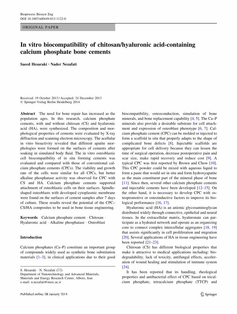
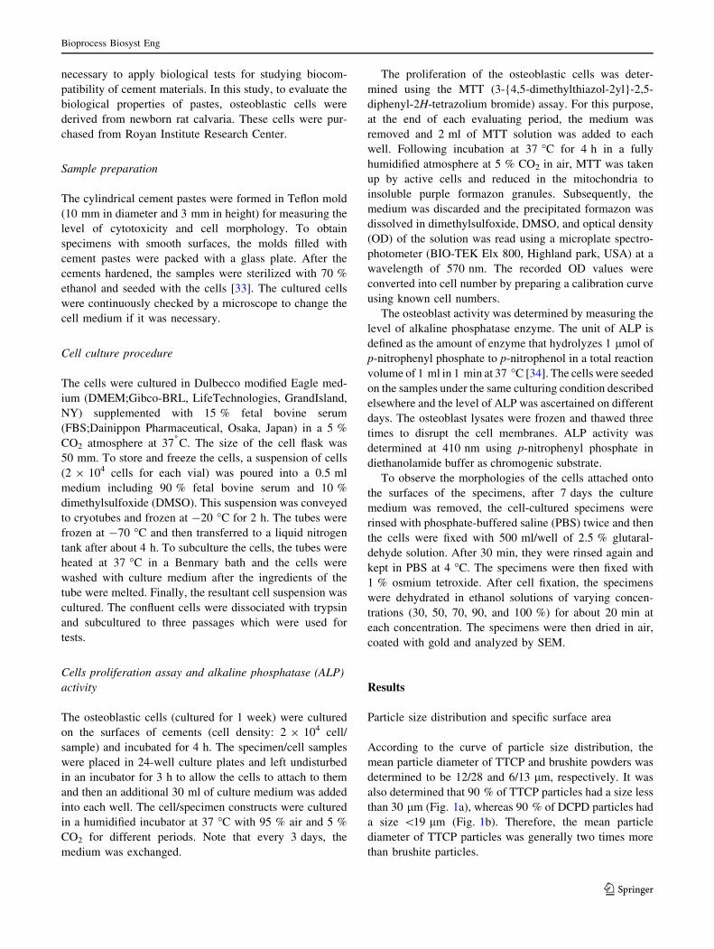
According to the curve of particle size distribution, the
mean particle diameter of TTCP and brushite powders was
determined to be 12/28 and 6/13 lm, respectively. It was
also determined that 90 % of TTCP particles had a size less
than 30 lm (Fig. 1a), whereas 90 % of DCPD particles had
a size \19 lm (Fig. 1b). Therefore, the mean particle
diameter of TTCP particles was generally two times more
than brushite particles.
Bioprocess Biosyst Eng
123
Phase analysis of the specimens
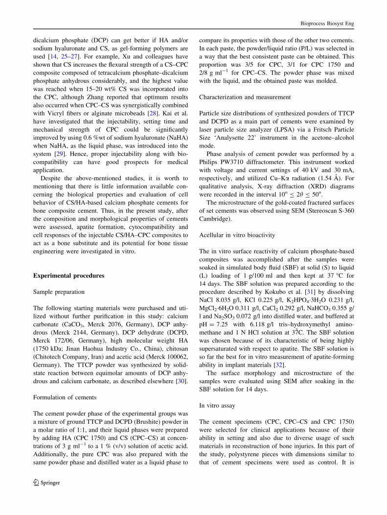
Figure 2 depicts the compositional variations in the CPC
samples after being set for 24 h in an incubator. In all three
groups of set cements, the formation of the apatite phase,
which is the main product of the setting reaction, could be
detected along with the TTCP and DCPD phases as
reactants.
SEM observation of samples before and after soaking
in SBF solution
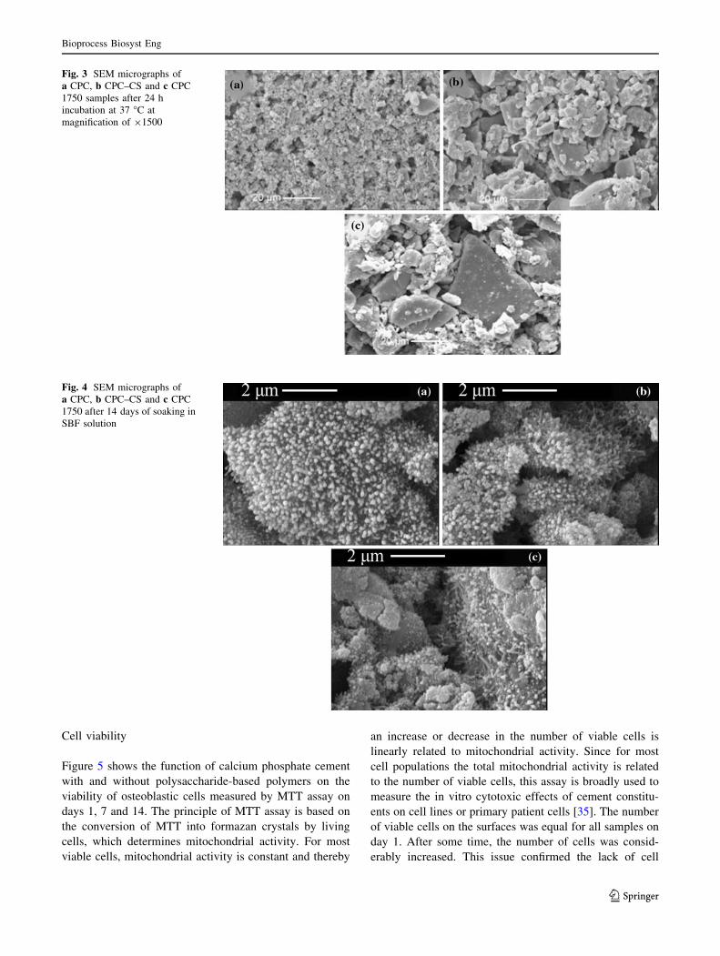
The microstructures of CPC, CPC–CS and CPC 1750
samples after 24 h incubation at 37 C are shown in Fig. 3.
According to Fig. 3a, two different morphologies of cal-
cium phosphate reactant particles were observed for the
CPC sample: first, coarse particles with dimension of
nearly 15 lm; second, finer particles (\5 lm) which were
situated on the surface of larger ones. In addition, the mi-
cropores (\3 lm) were produced by removing the liquid
phase from the cement structure. The microstructure of
calcium phosphate particles in CPC–CS (Fig. 3b) and CPC
1750 (Fig. 3c) was completely different from the CPC
sample. It seemed that reactive components in these
cements were covered with bindery phase, and the surface
of particles was smoother in these samples. The tiny holes
on the surface of CPC–CS and CPC 1750 were probably
generated by the air-trapped bubbles.
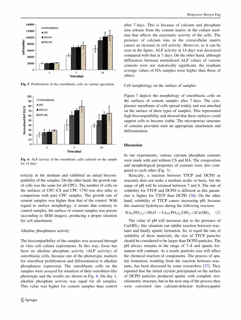
Scanning electron microscopy images of samples, after
14 days of soaking, showed that tiny spherical apatite
particles were formed on the surface of CPC (Fig. 4a),
while the morphology was changed for CPC–CS (Fig. 4b)
and CPC 1750 (Fig. 4c). For these samples, the particles
were connected by entangled needle-like apatite crystals. It
seems that this kind of morphology is nearly more
detectable for CPC 1750 than CPC–CS.
Fig. 1 Particle size distribution curve of TTCP and brushite powders by LPSA analyzer
Fig. 2 XRD pattern of 1 CPC,
2 CPC–CS and 3 CPC 1750
specimens after being set for
24 h in an incubator
Bioprocess Biosyst Eng
123
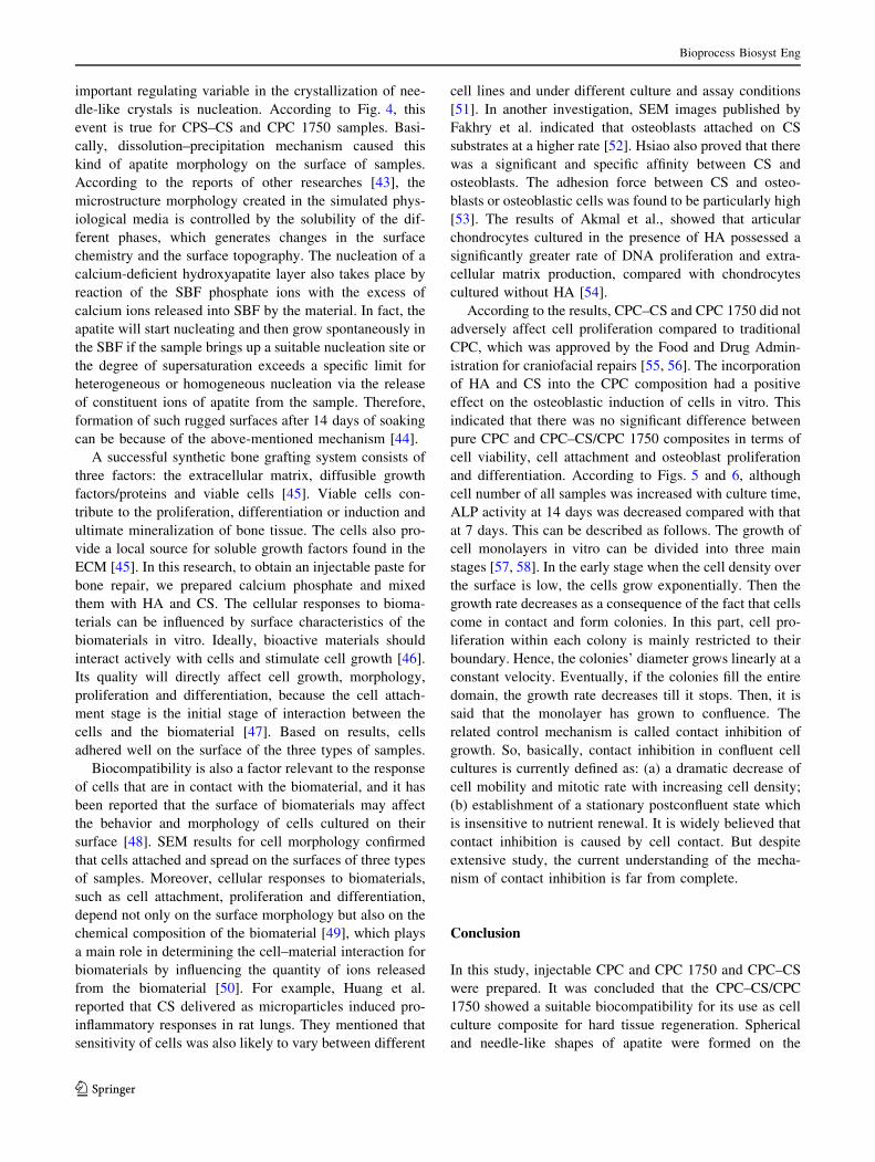
Cell viability
Figure 5 shows the function of calcium phosphate cement
with and without polysaccharide-based polymers on the
viability of osteoblastic cells measured by MTT assay on
days 1, 7 and 14. The principle of MTT assay is based on
the conversion of MTT into formazan crystals by living
cells, which determines mitochondrial activity. For most
viable cells, mitochondrial activity is constant and thereby
an increase or decrease in the number of viable cells is
linearly related to mitochondrial activity. Since for most
cell populations the total mitochondrial activity is related
to the number of viable cells, this assay is broadly used to
measure the in vitro cytotoxic effects of cement constitu-
ents on cell lines or primary patient cells [35]. The number
of viable cells on the surfaces was equal for all samples on
day 1. After some time, the number of cells was consid-
erably increased. This issue confirmed the lack of cell
Fig. 3 SEM micrographs of
a CPC, b CPC–CS and c CPC
1750 samples after 24 h
incubation at 37 �C at
magnification of 91500
Fig. 4 SEM micrographs of
a CPC, b CPC–CS and c CPC
1750 after 14 days of soaking in
SBF solution
Bioprocess Biosyst Eng
123
toxicity in the medium and exhibited an initial biocom-
patibility of the samples. On the other hand, the growth rate
of cells was the same for all CPCs. The number of cells on
the surfaces of CPC–CS and CPC 1750 was also alike in
comparison with pure CPC samples. The growth rate of
cement samples was higher than that of the control. With
regard to surface morphology, it means that contrary to
control samples, the surface of cement samples was porous
(according to SEM images), producing a proper situation
for cell attachment.
Alkaline phosphatase activity
The biocompatibility of the samples was assessed through
in vitro cell culture experiments. In this way, focus has
been on alkaline phosphate activity (ALP activity) of
osteoblastic cells, because one of the phenotypic markers
for osteoblast proliferation and differentiation is alkaline
phosphatase expression. The osteoblastic cells on the
samples were assayed for retention of their osteoblast-like
phenotype and the results are shown in Fig. 6. On day 1,
alkaline phosphate activity was equal for all samples.
This value was higher for cement samples than control
after 7 days. This is because of calcium and phosphate
ions release from the cement matrix in the culture med-
ium that affects the enzymatic activity of the cells. The
presence of calcium ions in the extracellular matrix
causes an increase in cell activity. However, as it can be
seen in the figure, ALP activity at 14 days was decreased
compared with that at 7 days. On the other hand, although
differences between normalized ALP values of various
cements were not statistically significant, the resultant
average values of HA samples were higher than those of
others.
Cell morphology on the surface of samples
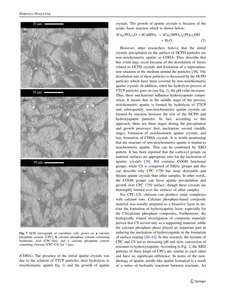
Figure 7 depicts the morphology of osteoblastic cells on
the surfaces of cement samples after 7 days. The cyto-
plasmic membrane of cells spread widely and was attached
on the surface of three types of samples. This represented
high biocompatibility and showed that these surfaces could
support cells to become viable. The microporous structure
of cements provided such an appropriate attachment and
differentiation.
Discussion
In our experiments, various calcium phosphate cements
were made with and without CS and HA. The composition
and morphological properties of cements were also com-
pared to each other (Fig. 3).
Basically, a reaction between TTCP and DCPD as
reactants does not make a medium acidic or basic, but the
range of pH will be retained between 7 and 8. The rate of
solubility for TTCP and DCPD is different as this param-
eter is higher for TTCP than DCPD [36]. On the other
hand, solubility of TTCP causes increasing pH, because
this material hydrolyzes during the following reaction:
3Ca4 PO4ð Þ2þ3H2O! Ca10 PO4ð Þ6 OHð Þ2þ2Ca OHð Þ2 ð1Þ
The value of pH will increases due to the presence of
Ca(OH)2; this situation can inhibit reaction between reac-
tants and finally apatite formation. So, to equal the rate of
solubility of these materials, the size of TTCP particles
should be considered to be larger than DCPD particles. The
pH always remains in the range of 7–8 and apatite for-
mation will continue. As a result, particles size will affect
the chemical reaction of components. The process of apa-
tite formation, resulting from the reaction between reac-
tants, has been discussed by some researchers [37]. They
reported that the initial crystals precipitated on the surface
of DCPD particles produced apatite with complete stoi-
chiometric structure, but in the next step of the process they
were converted into calcium-deficient hydroxyapatite
Fig. 5 Proliferation of the osteoblastic cells on various specimens
Fig. 6 ALP activity of the osteoblastic cells cultured on the sample
for 14 days
Bioprocess Biosyst Eng
123
(CDHA). The presence of the initial apatite crystals was
due to the solution of TTCP particles, their hydrolysis to
stoichiometric apatite Eq. 1) and the growth of apatite
crystals. The growth of apatite crystals is because of the
acidic–basic reaction which is shown below:
3Ca4 PO4ð Þ2Oþ 6CaHPO4 ! 2Ca2 HPO4ð Þ6 PO4ð Þ5OH
þ H2O: ð2Þ
However, other researchers believe that the initial
crystals precipitated on the surface of DCPD particles are
non-stoichiometric apatite or CDHA. They describe that
this event may occur because of the dissolution of layers
related to DCPD crystals and formation of a supersatura-
tion situation in the medium around the particles [38]. The
dissolution rate of these particles is decreased by the DCPD
particles which have been covered by non-stoichiometric
apatite crystals. In addition, when the hydrolysis process of
TTCP particles goes on (see Eq. 2), the pH value increases.
Thus, these mechanisms influence hydroxyapatite compo-
sition. It means that in the middle stage of the process,
stoichiometric apatite is formed by hydrolysis of TTCP
and, subsequently, non-stoichiometric apatite crystals are
formed by reaction between the rest of the DCPD and
hydroxyapatite particles. In fact, according to this
approach, there are three stages during the precipitation
and growth processes: first, nucleation; second (middle
stage), formation of stoichiometric apatite crystals; and
last, formation of CDHA crystals. It is worth mentioning
that the structure of non-stoichiometric apatite is similar to
stoichiometric apatite. This can be confirmed by XRD
pattern. It has been reported that the carboxyl groups on
material surfaces are appropriate sites for the nucleation of
apatitic crystals [39]. HA contains COOH functional
groups, while CS is composed of NHAc groups and this
can describe why CPC 1750 has more detectable and
thicker apatite crystals than other samples. In other words,
the COOH groups can favor apatite precipitation and
growth over CPC 1750 surface, though these crystals are
thoroughly formed over the surfaces of other samples.
For CPC–CS, chitosan can produce some complexes
with calcium ions. Calcium phosphate-based composite
material was usually prepared as a bioactive layer to ini-
tiate the formation of hydroxyapatite layer, especially for
the CS/calcium phosphate composites. Furthermore, the
biologically related investigation of composite materials
proved that CS served only as a supporting material, while
the calcium phosphate phase played an important part in
inducing the nucleation of hydroxyapatite in the formation
of surface coating [40–42]. In this research, the mixture of
CPC and CS led to increasing pH and slow conversion of
reactants to hydroxyxapatite. According to Fig. 2, the XRD
patterns of three kinds of CPCs are similar to each other
and have no significant difference. In terms of the mor-
phology of apatite, needle-like apatite formation is a result
of a series of hydraulic reactions between reactants. An
Fig. 7 SEM micrograph of osteoblast cells grown on a calcium
phosphate cement (CPC), b calcium phosphate cement containing
hyaluronic acid (CPC–HA) and c calcium phosphate cement
containing chitosan (CPC–CS) for 7 days
Bioprocess Biosyst Eng
123
important regulating variable in the crystallization of nee-
dle-like crystals is nucleation. According to Fig. 4, this
event is true for CPS–CS and CPC 1750 samples. Basi-
cally, dissolution–precipitation mechanism caused this
kind of apatite morphology on the surface of samples.
According to the reports of other researches [43], the
microstructure morphology created in the simulated phys-
iological media is controlled by the solubility of the dif-
ferent phases, which generates changes in the surface
chemistry and the surface topography. The nucleation of a
calcium-deficient hydroxyapatite layer also takes place by
reaction of the SBF phosphate ions with the excess of
calcium ions released into SBF by the material. In fact, the
apatite will start nucleating and then grow spontaneously in
the SBF if the sample brings up a suitable nucleation site or
the degree of supersaturation exceeds a specific limit for
heterogeneous or homogeneous nucleation via the release
of constituent ions of apatite from the sample. Therefore,
formation of such rugged surfaces after 14 days of soaking
can be because of the above-mentioned mechanism [44].
A successful synthetic bone grafting system consists of
three factors: the extracellular matrix, diffusible growth
factors/proteins and viable cells [45]. Viable cells con-
tribute to the proliferation, differentiation or induction and
ultimate mineralization of bone tissue. The cells also pro-
vide a local source for soluble growth factors found in the
ECM [45]. In this research, to obtain an injectable paste for
bone repair, we prepared calcium phosphate and mixed
them with HA and CS. The cellular responses to bioma-
terials can be influenced by surface characteristics of the
biomaterials in vitro. Ideally, bioactive materials should
interact actively with cells and stimulate cell growth [46].
Its quality will directly affect cell growth, morphology,
proliferation and differentiation, because the cell attach-
ment stage is the initial stage of interaction between the
cells and the biomaterial [47]. Based on results, cells
adhered well on the surface of the three types of samples.
Biocompatibility is also a factor relevant to the response
of cells that are in contact with the biomaterial, and it has
been reported that the surface of biomaterials may affect
the behavior and morphology of cells cultured on their
surface [48]. SEM results for cell morphology confirmed
that cells attached and spread on the surfaces of three types
of samples. Moreover, cellular responses to biomaterials,
such as cell attachment, proliferation and differentiation,
depend not only on the surface morphology but also on the
chemical composition of the biomaterial [49], which plays
a main role in determining the cell–material interaction for
biomaterials by influencing the quantity of ions released
from the biomaterial [50]. For example, Huang et al.
reported that CS delivered as microparticles induced pro-
inflammatory responses in rat lungs. They mentioned that
sensitivity of cells was also likely to vary between different
cell lines and under different culture and assay conditions
[51]. In another investigation, SEM images published by
Fakhry et al. indicated that osteoblasts attached on CS
substrates at a higher rate [52]. Hsiao also proved that there
was a significant and specific affinity between CS and
osteoblasts. The adhesion force between CS and osteo-
blasts or osteoblastic cells was found to be particularly high
[53]. The results of Akmal et al., showed that articular
chondrocytes cultured in the presence of HA possessed a
significantly greater rate of DNA proliferation and extra-
cellular matrix production, compared with chondrocytes
cultured without HA [54].
According to the results, CPC–CS and CPC 1750 did not
adversely affect cell proliferation compared to traditional
CPC, which was approved by the Food and Drug Admin-
istration for craniofacial repairs [55, 56]. The incorporation
of HA and CS into the CPC composition had a positive
effect on the osteoblastic induction of cells in vitro. This
indicated that there was no significant difference between
pure CPC and CPC–CS/CPC 1750 composites in terms of
cell viability, cell attachment and osteoblast proliferation
and differentiation. According to Figs. 5 and 6, although
cell number of all samples was increased with culture time,
ALP activity at 14 days was decreased compared with that
at 7 days. This can be described as follows. The growth of
cell monolayers in vitro can be divided into three main
stages [57, 58]. In the early stage when the cell density over
the surface is low, the cells grow exponentially. Then the
growth rate decreases as a consequence of the fact that cells
come in contact and form colonies. In this part, cell pro-
liferation within each colony is mainly restricted to their
boundary. Hence, the colonies’ diameter grows linearly at a
constant velocity. Eventually, if the colonies fill the entire
domain, the growth rate decreases till it stops. Then, it is
said that the monolayer has grown to confluence. The
related control mechanism is called contact inhibition of
growth. So, basically, contact inhibition in confluent cell
cultures is currently defined as: (a) a dramatic decrease of
cell mobility and mitotic rate with increasing cell density;
(b) establishment of a stationary postconfluent state which
is insensitive to nutrient renewal. It is widely believed that
contact inhibition is caused by cell contact. But despite
extensive study, the current understanding of the mecha-
nism of contact inhibition is far from complete.
Conclusion
In this study, injectable CPC and CPC 1750 and CPC–CS
were prepared. It was concluded that the CPC–CS/CPC
1750 showed a suitable biocompatibility for its use as cell
culture composite for hard tissue regeneration. Spherical
and needle-like shapes of apatite were formed on the
Bioprocess Biosyst Eng
123
surfaces of cements when they were soaked in SBF solution
for 14 days. In vitro experiments revealed that cell viability
and growth rate of cells for CPC–CS and CPC 1750 samples
was nearly similar. Collectively, these data indicated that
CPC 1750 may give greater results concerning average
value of ALP compared to the CPC–CS and pure CPC in this
in vitro study. Overall, we suggest an acceptable applica-
bility of the CPC–CS/CPC 1750 samples as bone
substitutes.
Acknowledgments The authors would like to acknowledge the Iran
National Science Foundation (INSF) for the financial support of this
work through Grant No. 89001740.
References
1. Navarro M, Michiardi A, Castano O, Planell JA, Soc JR (2008)
Biomaterials in orthopaedics. Interface 5:1137–1158
2. Bohner M (2010) Resorbable biomaterials as bone graft substi-
tutes. Mater Today 13:24–30
3. Heinemann S, Gelinsky M, Worch H, Hanke T (2011) Resorbable
bone substitution materials: an overview of commercially avail-
able materials and new approaches in the field of composites.
Orthopade 40:761–773
4. Wu F, Wei L, Guo H, Liu CS (2008) Self-setting bioactive cal-
cium–magnesium phosphate cement with high strength and de-
gradability for bone regeneration. Acta Biomater 4:1873–1884
5. Pilliar RM, Filiaggi MJ, Wells JD, Grynpas MD, Kandel RA
(2001) Porous calcium polyphosphate scaffolds for bone substitute
applications: in vitro characterization. Biomaterials 22:963–972
6. Reilly GC, Radin S, Chen AT, Ducheyne P (2007) Differential
alkaline phosphatase responses of rat and human bone marrow
derived mesenchymal stem cells to 45S5 bioactive glass. Bio-
materials 28:4091–4097
7. Deville S, Saiz E, Nalla RK, Tomsia AP (2006) Freezing as a
path to build complex composites. Science 311:515–518
8. Link DP, van den Dolder J, van den Beucken JJ, Wolke JG,
Mikos AG et al (2008) Bone response and mechanical strength of
rabbit femoral defects filled with injectable CaP cements con-
taining TGF-b1 loaded gelatin microspheres. Biomaterials 29:
675–682
9. Kretlow JD, Young S, Klouda L, Wong M, Mikos AG (2009)
Injectable biomaterials for regenerating complex craniofacial
tissues. Adv Mater 21:3368–3393
10. Brown WE, Chow LC (1986) A new calcium phosphate water
setting cement. In: Brown PW (ed) Cements research progress.
American Ceramic Society, Westerville, pp 352–379
11. Julien M, Khairoun I, LeGeros RZ, Delplace S, Pilet P et al
(2007) Physicochemical–mechanical and in vitro biological
properties of calcium phosphate cements with doped amorphous
calcium phosphates. Biomaterials 28:956–965
12. Moreau JL, Xu HHK (2009) Mesenchymal stem cell proliferation
and differentiation on an injectable calcium phosphate–chitosan
composite scaffold. Biomaterials 30:2675–2682
13. Xu HHK, Zhao L, Detamore MS, Takagi S, Chow LC (2010)
Umbilical cord stem cell seeding on fast-resorbable calcium
phosphate bone cement. Tissue Eng A 16:2743–2753
14. Ahmadzadeh-Asl S, Hesaraki S, Zamanian A (2011) Preparation
and characterisation of calcium phosphate–hyaluronic acid
nanocomposite bone cement. Adv Appl Ceram 110:340–345
15. Yokoyama A, Yamanoto S, Kawasaki T, Kohgo T, Nakasu M
(2002) Development of calcium phosphate cement using chitosan
and citric acid for bone substitute materials. Biomaterials
23:1091–1101
16. Bodde E, Boerman O, Russel F, Mikos A, Spauwen P et al (2008)
The kinetic and biological activity of different loaded rhBMP-2
calcium phosphate cement implants in rats. J Biomed Mater Res
A 87A:780–791
17. Wang X, Ma J, Wang Y, He B (2001) Structural characterization of
phosphorylated chitosan and their applications as effective addi-
tives of calcium phosphate cements. Biomaterials 22:2247–2255
18. Almond A, Deangelis PL, Blundell CD (2006) Hyaluronan: the
local solution conformation determined by NMR and computer
modeling is close to a contracted left-handed 4-fold helix. J Mol
Biol 358:1256–1269
19. Shin DY, Hwang E, ChoI H, Moon MH (2007) Molecular weight
and structure characterization of sodium hyaluronate and its
gamma radiation degradation products by flow field-flow frac-
tionation and on-line multiangle light scattering. J Chromatogr A
1160:270–275
20. Toole BP (2000) Hyaluronan is not just a goo. J Clin Invest
106:335–336
21. Sasaki T, Watanabe C (1995) Stimulation of osteoinduction in
bone wound healing by high-molecular hyaluronic acid. Bone
16:9–15
22. Huang L, Cheng YY, Koo PL, Lee KM, Qin L, Cheng JC, Kumta
SM (2003) The effect of hyaluronan on osteoblast proliferation
and differentiation in rat calvarial-derived cell cultures. J Biomed
Mater Res A 66A:880–884
23. Lisignoli G, Zini N, Remiddi G, Piacentini A, Puggioli A,
Trimarchi C, Fini M, Maraldi NM, Facchini A (2001) Basic
fibroblast growth factor enhances in vitro mineralization of rat
bone marrow stromal cells grown on non-woven hyaluronic acid
based polymer scaffold. Biomaterials 22:2095–2105
24. Di Martino A, Sittinger M, Risbud MV (2005) Chitosan: a ver-
satile biopolymer for orthopaedic tissue-engineering. Biomateri-
als 26:5983–5990
25. Alkhraisat MH, Rueda C, Marino FT, Torres J, Jerez LB, Gbu-
reck U, Cabarcos EL (2009) The effect of hyaluronic acid on
brushite cement cohesion. Acta Biomater 5:3150–3156
26. Cherng B, Takagi S, Chow LC (1997) Effects of hydroxypropyl
methyl cellulose and other gelling agents on the handling prop-
erties of calcium phosphate cement. J Biomed Mater Res
35:273–277
27. Wu T, Hua X, He Z, Wang X, Yu X, Ren W (2012) The bac-
tericidal and biocompatible characteristics o f reinforced calcium
phosphate cements. Biomed Mater 7:045003–045013
28. Xu HH, Quinn JB, Takagi S (2002) Processing and properties of
strong and non-rigid calcium phosphate cement. J Dent Res
81(3):219–224
29. Kai D, Li D, Zhu X, Zhang L, Fan H, Zhang X (2009) Addition of
sodium hyaluronate and the effect on performance of the inject-
able calcium phosphate cement. J Mater Sci Mater Med
20:1595–1602
30. Hesaraki S, Zamanian A, Nazarian H (2009) Physical and phys-
icochemical evaluation of calcium phosphate cement made using
human derived blood plasma. Adv Appl Ceram 108:253–260
31. Kokubo T, Kushitani H, Sakka S, Kitsugi T, Yamamuro T (1990)
Solutions able to reproduce in vivo surface-structure changes in
bioactive glass-ceramic A-W. J Biomed Mater Res 24:721–734
32. Oyane A, Kim HM, Furuya T, Kokubo T, Miyazaki T, Nakamura
T (2003) Preparation and assessment of revised simulated body
fluids. J Biomed Mater Res Part A 65:188–195
33. Washburn NR, Weir M, Anderson P, Potter K (2004) Bone for-
mation in polymeric scaffolds evaluated by proton magnetic
Bioprocess Biosyst Eng
123
resonance microscopy and X-ray microtomography. J Biomed
Mater Res 69A:738–747
34. Mossner E, Boll M, Pfleiderer G (1980) Purification of human
and bovine alkaline phosphatases by affinity chromatography.
Hoppe Seylers Z Physiol Chem 361(4):543–549
35. van Meerloo J, Kaspers GJL, Cloos J (2011) Cell sensitivity
assays: the MTT assay. Methods Mol Biol 731:237–245
36. Hench LL, Wilson J (1995) An introduction to bioceramics.
World Scientific, Singapore
37. Hench LL (1991) Bioceramics: from concept to clinic. J Am
Ceram Soc 74:1487–1510
38. ReD Rawlings (1993) Bioactive glasses and glass–ceramics. Clin
Mater 14:155–179
39. Tadashi K, Hyun-Min K, Masakazu K (2003) Novel bioactive
materials with different mechanical properties. Biomaterials
24:2161–2175
40. Xu HHK, Simon CG (2005) Fast setting calcium phosphate–
chitosan scaffold: mechanical properties and biocompatibility.
Biomaterials 26:1337–1348
41. Zhang Y, Zhang M (2002) Calcium phosphate/chitosan com-
posite scaffolds for controlled in vitro antibiotic drug release.
J Biomed Mater Res A 62:378–386
42. Wang J, van Apeldoorn A, de Groot K (2006) Electrolytic
deposition of calcium phosphate/chitosan coating on titanium
alloy: growth kinetics and influence of current density, acetic
acid, and chitosan. J Biomed Mater Res A 76:503–511
43. Sainz M, Pena P, Serena S, Caballero A (2010) Influence of
design on bioactivity of novel CaSiO3–CaMg(SiO3)2 bioce-
ramics: in vitro simulated body fluid test and thermodynamic
simulation. Acta Biomater 6:2797–2807
44. Rhee SH (2003) Effect of calcium salt content in the poly(epsi-
lon-caprolactone)/silica nanocomposite on the nucleation and
growth behavior of apatite layer. J Biomed Mater Res A
67:1131–1138
45. Reddi AH (1994) Symbiosis of biotechnology and biomaterials:
applications in tissue engineering of bone and cartilage. J Cell
Biochem 56:192–195
46. Chen QZ, Efthymiou A, Salih V, Boccaccini AR (2008) Bio-
galss�-derived glass ceramic scaffolds: study of cell proliferation
and scaffold degradation in vitro. J Biomed Mater Res A
84:1049–1060
47. Low SP, Williams KA, Canham LT, Voelcker NH (2006) Eval-
uation of mammalian cell adhesion on surface modified porous
silicon. Biomaterials 27:4538–4546
48. Lee JY, Kang BS, Hicks B, Chancellor TF, Chu BH et al (2008)
The control of cell adhesion and viability by zinc oxide nanorods.
Biomaterials 29:3743–3749
49. Chou SY, Cheng CM, LeDuc PR (2009) Composite polymer
systems with control of local substrate elasticity and their effect
on cytoskeletal and morphological characteristics of adherent
cells. Biomaterials 30:3136–3142
50. Wu C, Ramaswmy Y, Zhu YF, Zheng R, Appleyard R et al
(2009) The effect of mesoporous bioactive glass on the physio-
chemical, biological and drug-release properties of poly(DL-lac-
tide-co-glycolide) films. Biomaterials 30:2199–2208
51. Huang YC, Vieira A, Huang KL, Yeh MK, Chiang CH (2005)
Pulmonary inflammation caused by chitosan microparticles.
J Biomed Mater Res 75A:283–287
52. Fakhry A, Schneider GB, Zaharias R, Senel S (2004) Chitosan
supports the initial attachment and spreading of osteoblasts
preferentially over fibroblasts. Biomaterials 25:2075–2079
53. Hsiao SW, Thien DVH, Ho MH, Hsieh HJ, Li CH, Hung CH, Li
HH (2010) Interactions between chitosan and cells measured by
AFM. Biomed Mater 5:054117–054125
54. Akmal M, Singh A, Anand A, Kesani A, Aslam N, Goodship A,
Bentley G (2005) The effects of hyaluronic acid on articular
chondrocytes. J Bone Joint Surg Br 87B:1143–1149
55. Weiss DD, Sachs MA, Woodard CR (2003) Calcium phosphate
bone cements: a comprehensive review. J Long Term Eff Med
Implant 13:41–47
56. Schmitz JP, Hollinger JO, Milan SB (1999) Reconstruction of
bone using calcium phosphate bone cements: a critical review.
J Oral Maxillofac Surg 57:1122–1126
57. Takai Y, Miyoshi J, Ikeda W, Ogita H (2008) Nectins and nectin-
like molecules: roles in contact inhibition of cell movement and
proliferation. Nat Rev Mol Cell Biol 9:603–615
58. Heckman CA (2009) Contact inhibition revisited. J Cell Physiol
220:574–575
Bioprocess Biosyst Eng
123