impact of myocardial thickness on viability assessment on late gadolinium enhancement sequences...
TRANSCRIPT

Impact of Myocardial Thickness on Viability assessment on Late Gadolinium
Enhancement Sequences
Ismahen BEN YAACOUB-KZADRI, Sana HARGUEM, Rym BENNACEUR, Imen GANZOUI, Aicha BEN MILED, Najla MNIF
Radiology department, Charles Nicolle Hospital, Tunis - TUNISIA

INTRODUCTION
Late Gadolinium enhancement (LGE) in CMR detects
replacement of normal viable myocytes by necrosis or
fibrosis with high spatial resolution and excellent correlation
to histopathology.
The extension of LGE within the myocardial wall is crucial for
viability assessment.
Because myocardial necrosis is associated with myocardial
thinning, preserved end diastolic myocardial wall thickness
(MWT) may represent a reliable marker of viability.

OBJECTIVES
Our work aims to assess whether end-diastolic MWT is an
important marker of myocardial viability in patients with
suspected hibernating myocardium through an evaluation
of the relationship between the MWT and the extension of
LGE through the myocardial wall.

Retrospective study of 50 conscutive patients with documented CAD, enrolled during the last 2 years
All patients underwent a standrized CMR examination for myocardial viability on a 1.5 T scanner (GE HDXT)
CMR protocol:
Cine Bright Blood sequences: SSFP (Fiesta)
Perfusion (FGRET) with 0.2mmol/kg of Gadolinate contrast media
LGE: Double IR T1 sequences, 10 min after the injection
MATERIAL & METHODS
LGE MWT
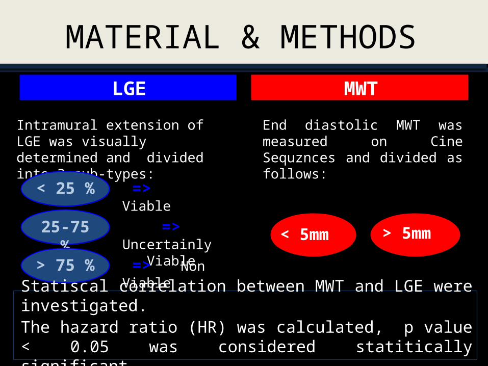
Intramural extension of LGE was visually determined and divided into 3 sub-types:
End diastolic MWT was measured on Cine Sequznces and divided as follows:
MATERIAL & METHODS
< 25 % 25-75 %
25-75 %
> 75 %
=> Viable
=> Uncertainly Viable
=> Non Viable
> 5mm < 5mm
Statiscal correlation between MWT and LGE were investigated.The hazard ratio (HR) was calculated, p value < 0.05 was considered statitically significant.

Cardio-vascular risk factors
hype
rten
sion
diab
etes
smok
ers
hype
rlipi
dem
ia
obes
ity
fam
ily h
isto
ry o
f CAD
05
101520253035
3022
29
11 7 11risk factors
mean age: 61 y (37-79y)Sex ratio: 44/6
RESULTSCharacteristics of the patient population
< 25 % 25 - 75 % > 75 %0
5
10
15
20
25
30
35
40
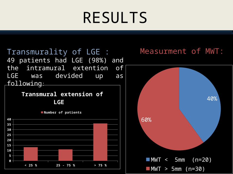
Transmural extension of LGE
Number of patients
Transmurality of LGE :49 patients had LGE (98%) and the intramural extention of LGE was devided up as following:
RESULTS
Measurment of MWT:
40%
60%
MWT < 5mm (n=20)MWT > 5mm (n=30)

RESULTS
Severe myocardial wall thining with transmural LGE
LGE of 25-75% transmurality with preserved MWT
Transmural LGE with preserved MWT Subendocardial LGE with preserved MWT

Analysis of statical correlation showed:
LGE transmural extension
< 25% Or < 25% and 25-75 %
> 75% Or < 25 % and > 75% Or 25 – 75 % and > 75%
No case of MWT < 5mm The risk of myocardial wall thining
increased
RESULTS
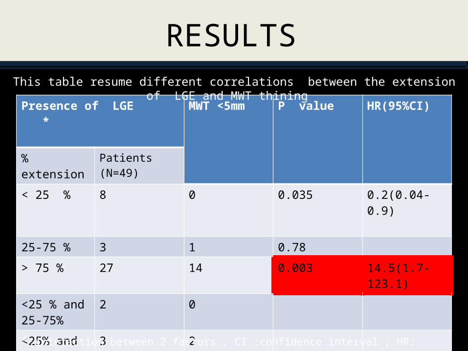
Presence of LGE *
MWT <5mm
P value HR(95%CI)
% extension
Patients (N=49)
< 25 % 8 0 0.035 0.2(0.04-0.9)
25-75 % 3 1 0.78
> 75 % 27 14 0.003 14.5(1.7-123.1)
<25 % and 25-75%
2 0
<25% and >75%
3 2
25-75 % and >75%
6 3*:interaction between 2 factors ; CI :confidence interval ; HR: hazard ratio
This table resume different correlations between the extension of LGE and MWT thining
RESULTS

Myocardial viability in patients with CAD is associated with improvement in contractile function after revascularisation.
Early detection of viable myocardium has become an increasingly important guide to prognosis and therapy.
CMR is considered as the new gold standard in the detection of irreversibly damaged myocardium.
In the present study, we focused on LGE extension through the myocardial wall and end diastolic MWT as markers of myocardial viability of proven value.
DISCUSSION
The use of an inversion-recovery prepared T1-weighted gradient-echo sequence after an intravenous administration of a gadolinium-chelate allows a direct imaging of myocardial necrosis
This CMR technique demonstrates non viable tissue as “hyperenhanced” or bright :
‘ White is dead ‘
DISCUSSION

LGE is due to persistance Gadolinium in necrosis or fibrosis because of : Increase of the volume of the interstitial space Slow down of wash in (arrival) and wash out (elimination)
DISCUSSION
Viable myocardium Chronic myocardial infarction
Acute myocardial infarction
GadoliniumNormal cell Destructed cell Fibroblast
Inte
rsti
tiu
m

LGE extent is critical determinant of contractile recovery.
Several studies evaluated the relationship between the transmural extent of hyperenhancement and the contractile recovery after myocardial infarction.
When the segmental transmural extent of LGE was:
< 25% : The majority of segments improved function > 75% : Functional recovery was unlikely 25 to 75 % : Intermediate likelihood of recovery
DISCUSSION
Prior studies demonstrated that a transmural myocardial necrosis is responsible for myocardial muscle loss due to cell death.
Fibrosis replacement secondary to ischemic injury in chronic transmural infarction leads to myocardial thining frequently measuring less than 5.5 mm.
In the present study, the statiscal analysis demonstrated that a MWT < 5 mm is highly associated with a transmural extension of LGE (> 75% )
It extends the earlier observations demonstrating that in patients with suspected myocardial hibernation, a simple measurement of MWT obtained with CMR is an important parameter of myocardial viability.
DISCUSSION
Wall thikeness is an important marker of myocardial viability in patients with myocardial infarction.
MWT < 5mm practically excludes relevant amount of viable myocardium.
A simple measurment of MWT is a valuable adjunct to LGE in the assessment of myocardial viability.
CONCLUSION

REFERENCES1. Elena Biagini, Tjebbe W Galema, Arend F.L Schinkel, Willem B Vletter, Jos R.T.C Roelandt,
Folkert J Ten Cate et al.Myocardial wall thickness predicts recovery of contractile function after primary coronary intervention for acute myocardial infarction. J Am Coll Cardiol. 2004;43(8):1489-1493
2. Cwajg JM, Cwajg E, Nagueh SF, He ZX, Qureshi U, Olmos LI et al .End-diastolic wall thickness as a predictor of recovery of function in myocardial hibernation: relation to rest-redistribution T1-201 tomography and dobutamine stress echocardiography. J Am Coll Cardiol. 2000 Apr;35(5):1152-61
3. De Waha S, Eitel I, Desch S, Fuernau G, Lurz P, Schuler G, et al. Diagnosis and therapy of chronic myocardial ischemia. Role of cardiac magnetic resonance imaging. Herz. 2013 Jun;38(4):350-8.
4. Shah DJ , Kim HW, James O, Parker M, Wu E, Bonow RO et al. Prevalence of regional myocardial thinning and relationship with myocardial scarring in patients with coronary artery disease.JAMA. 2013 Mar 6;309(9):909-18
5. El Aidi H, Adams A, Moons KG, Den Ruijter HM, Mali WP, Doevendans PA et al . Cardiac magnetic resonance imaging findings and the risk of cardiovascular events in patients with recent myocardial infarction or suspected or known coronary artery disease: a systematic review of prognostic studies. J Am Coll Cardiol. 2014 Mar 25;63(11):1031-45
6. Di Bella,Valeria Siciliano,Giovanni Donato Aquaro,Sabrina Molinaro,Massimo Lombardi,Scipione Carerj et al. Scar extent, left ventricular end-diastolic volume, and wall motion abnormalities identify high-risk patients with previous myocardial infarction: a multiparametric approach for prognostic stratification. Eur Heart J (2013) 34 (2): 104-111