immune checkpoint inhibitors in hodgkin and non-hodgkin...
TRANSCRIPT
Immune checkpoint inhibitors in Hodgkin and non-Hodgkin Lymphoma:
How do they work? Where will we use them?
Stephen M. Ansell, MD, PhD
Mayo Clinic
Conflicts of Interest
• Research Funding from –– Bristol Myers Squibb
– Celldex Therapeutics
– Seattle Genetics
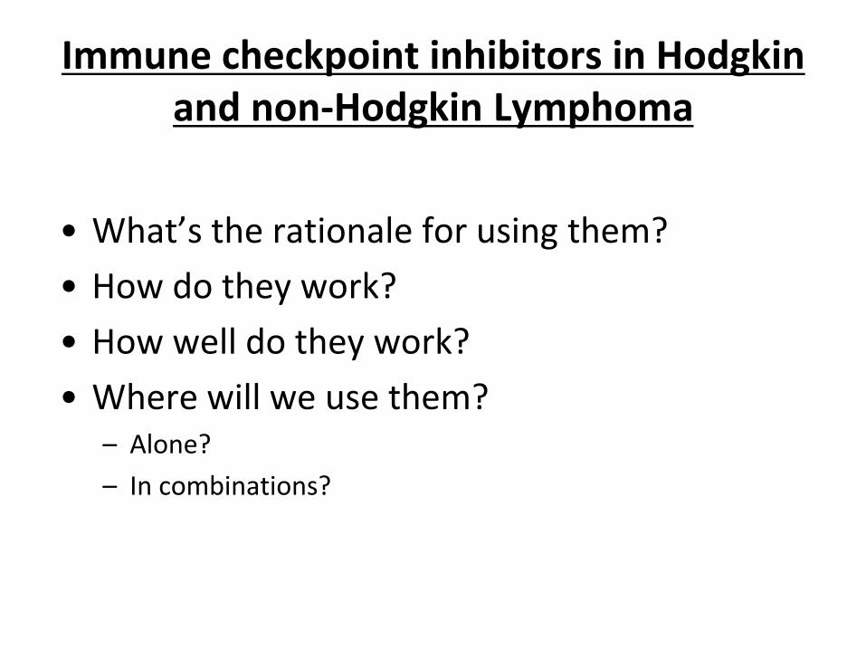
Immune checkpoint inhibitors in Hodgkin and non-Hodgkin Lymphoma
• What’s the rationale for using them?
• How do they work?
• How well do they work?
• Where will we use them?– Alone?
– In combinations?
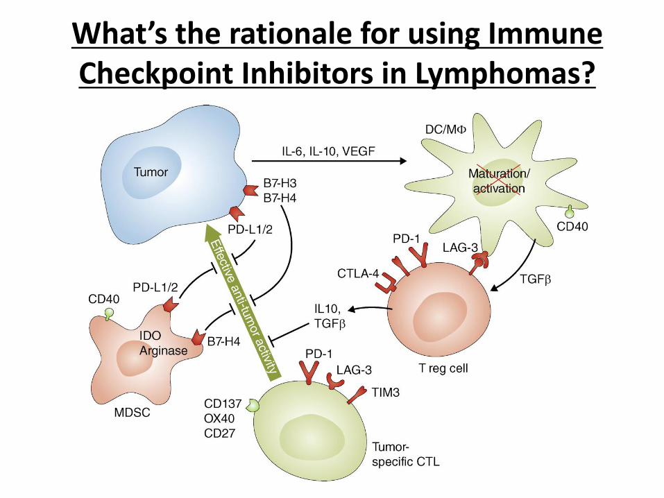
What’s the rationale for using Immune Checkpoint Inhibitors in Lymphomas?
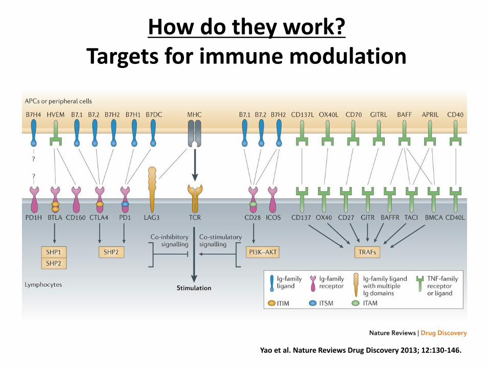
Yao et al. Nature Reviews Drug Discovery 2013; 12:130-146.
How do they work?Targets for immune modulation
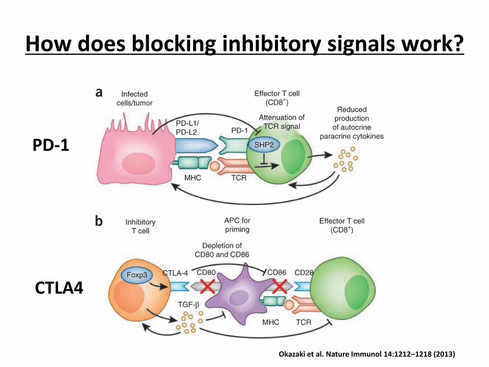
How does blocking inhibitory signals work?
Okazaki et al. Nature Immunol 14:1212–1218 (2013)
PD-1
CTLA4
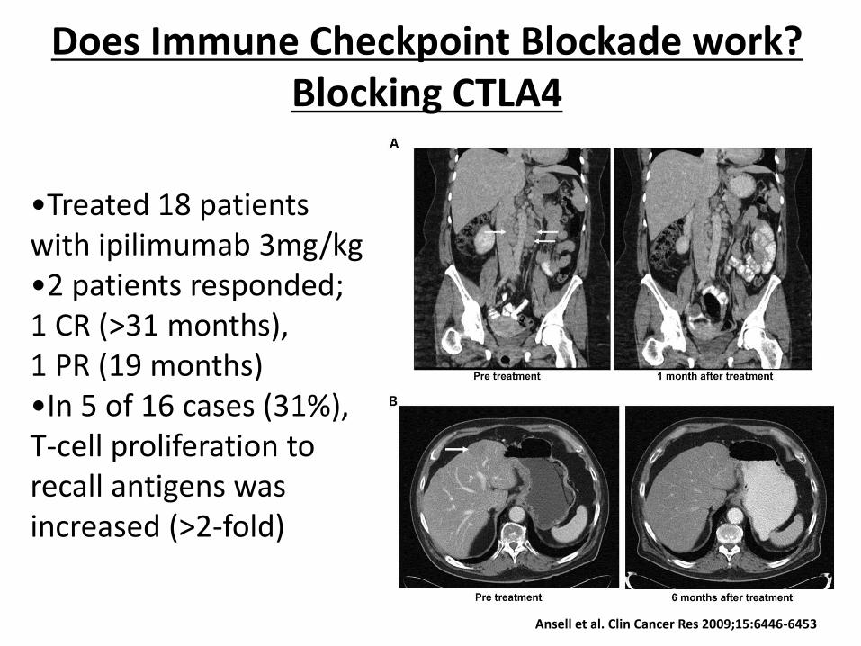
Does Immune Checkpoint Blockade work? Blocking CTLA4
Ansell et al. Clin Cancer Res 2009;15:6446-6453
•Treated 18 patients with ipilimumab 3mg/kg•2 patients responded; 1 CR (>31 months), 1 PR (19 months)•In 5 of 16 cases (31%), T-cell proliferation to recall antigens was increased (>2-fold)
Ipilimumab to treat relapse after allogeneic hematopoietic cell transplantation
• 29 patients with relapsed hematologic disease.
• Three patients with lymphoid malignancy developed objective disease responses following ipilimumab:
– CR in 2 patients with Hodgkin disease
– PR in a patient with refractory mantle cell lymphoma.
• Ipilimumab did not induce or exacerbate clinical GVHD
Bashey et al. Blood. 2009;113(7):1581-8.
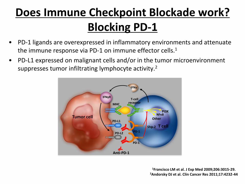
• PD-1 ligands are overexpressed in inflammatory environments and attenuate the immune response via PD-1 on immune effector cells.1
• PD-L1 expressed on malignant cells and/or in the tumor microenvironment suppresses tumor infiltrating lymphocyte activity.2
MHC
PD-L1
PD-1
PD-1
T-cellreceptor
PD-L2
T cell
NFκBOther
PI3K
IFNγ
IFNγR
Shp-2
Anti-PD-1
Tumor cell
1Francisco LM et al. J Exp Med 2009;206:3015-29.2Andorsky DJ et al. Clin Cancer Res 2011;17:4232-44
Does Immune Checkpoint Blockade work? Blocking PD-1
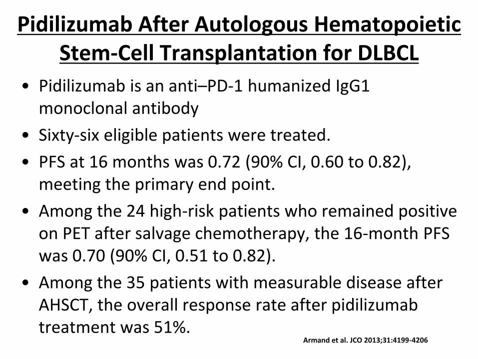
Pidilizumab After Autologous Hematopoietic Stem-Cell Transplantation for DLBCL
• Pidilizumab is an anti–PD-1 humanized IgG1 monoclonal antibody
• Sixty-six eligible patients were treated.
• PFS at 16 months was 0.72 (90% CI, 0.60 to 0.82), meeting the primary end point.
• Among the 24 high-risk patients who remained positive on PET after salvage chemotherapy, the 16-month PFS was 0.70 (90% CI, 0.51 to 0.82).
• Among the 35 patients with measurable disease after AHSCT, the overall response rate after pidilizumab treatment was 51%.
Armand et al. JCO 2013;31:4199-4206
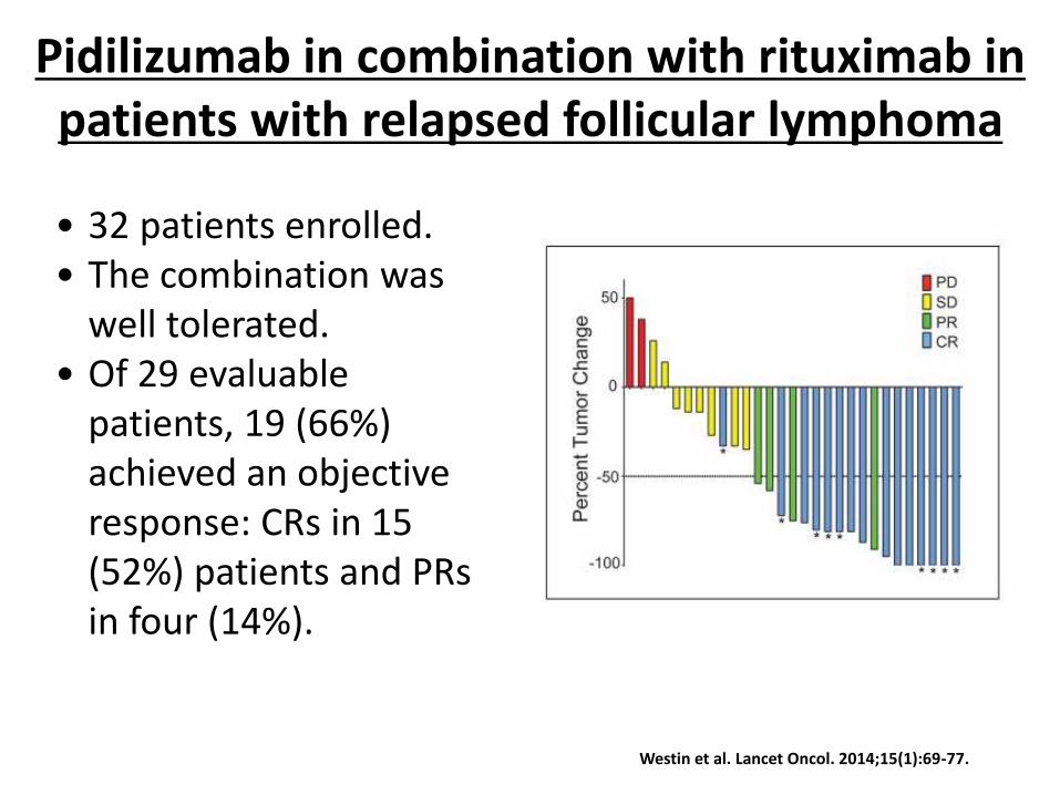
Pidilizumab in combination with rituximab in patients with relapsed follicular lymphoma
Westin et al. Lancet Oncol. 2014;15(1):69-77.
• 32 patients enrolled. • The combination was
well tolerated. • Of 29 evaluable
patients, 19 (66%) achieved an objective response: CRs in 15 (52%) patients and PRs in four (14%).
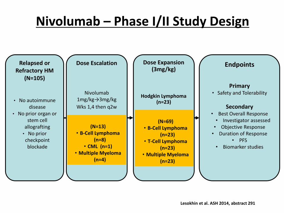
Relapsed or Refractory HM
(N=105)
• No autoimmune disease
• No prior organ or stem cell
allografting• No prior checkpoint blockade
Endpoints
Primary• Safety and Tolerability
Secondary• Best Overall Response• Investigator assessed• Objective Response• Duration of Response
• PFS• Biomarker studies
Nivolumab – Phase I/II Study Design
Dose Expansion (3mg/kg)
Hodgkin Lymphoma (n=23)
Dose Escalation
Nivolumab1mg/kg→3mg/kg
Wks 1,4 then q2w
(N=69)• B-Cell Lymphoma
(n=23)• T-Cell Lymphoma
(n=23)• Multiple Myeloma
(n=23)
(N=13)• B-Cell Lymphoma
(n=8)• CML (n=1)
• Multiple Myeloma (n=4)
Lesokhin et al. ASH 2014, abstract 291
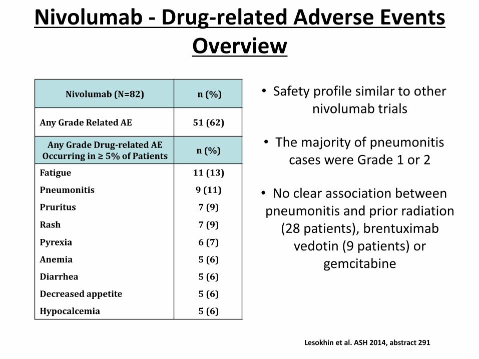
Nivolumab - Drug-related Adverse Events Overview
• Safety profile similar to other nivolumab trials
• The majority of pneumonitis cases were Grade 1 or 2
• No clear association between pneumonitis and prior radiation
(28 patients), brentuximabvedotin (9 patients) or
gemcitabine
Nivolumab (N=82) n (%)
Any Grade Related AE 51 (62)
Any Grade Drug-related AE Occurring in ≥ 5% of Patients
n (%)
Fatigue 11 (13)
Pneumonitis 9 (11)
Pruritus 7 (9)
Rash 7 (9)
Pyrexia 6 (7)
Anemia 5 (6)
Diarrhea 5 (6)
Decreased appetite 5 (6)
Hypocalcemia 5 (6)
Lesokhin et al. ASH 2014, abstract 291
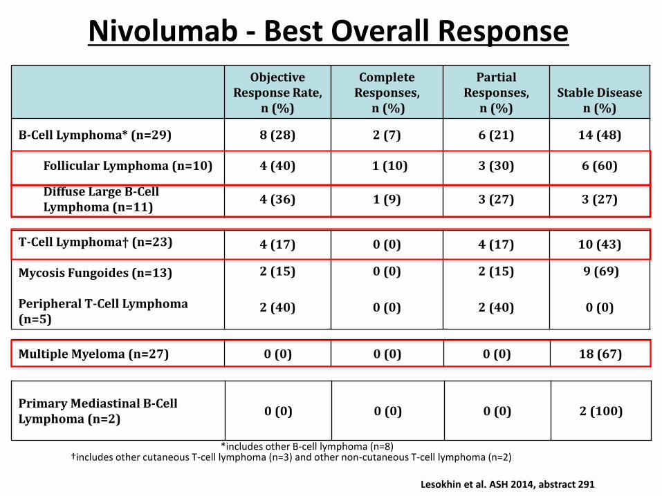
Multiple Myeloma (n=27) 0 (0) 0 (0) 0 (0) 18 (67)
Nivolumab - Best Overall ResponseObjective
Response Rate, n (%)
Complete Responses,
n (%)
Partial Responses,
n (%)Stable Disease
n (%)
B-Cell Lymphoma* (n=29) 8 (28) 2 (7) 6 (21) 14 (48)
Follicular Lymphoma (n=10) 4 (40) 1 (10) 3 (30) 6 (60)
Diffuse Large B-Cell Lymphoma (n=11)
4 (36) 1 (9) 3 (27) 3 (27)
†includes other cutaneous T-cell lymphoma (n=3) and other non-cutaneous T-cell lymphoma (n=2)
T-Cell Lymphoma† (n=23)
Mycosis Fungoides (n=13)
Peripheral T-Cell Lymphoma (n=5)
4 (17) 0 (0) 4 (17) 10 (43)
2 (15) 0 (0) 2 (15) 9 (69)
2 (40) 0 (0) 2 (40) 0 (0)
*includes other B-cell lymphoma (n=8)
Primary Mediastinal B-Cell Lymphoma (n=2)
0 (0) 0 (0) 0 (0) 2 (100)
Lesokhin et al. ASH 2014, abstract 291
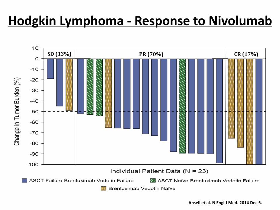
Hodgkin Lymphoma - Response to Nivolumab
PR (70%) CR (17%)SD (13%)
Ansell et al. N Engl J Med. 2014 Dec 6.
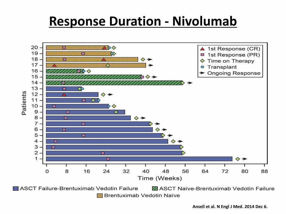
Response Duration - NivolumabP
ati
en
ts
Ansell et al. N Engl J Med. 2014 Dec 6.
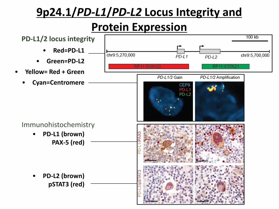
• Red=PD-L1
• Green=PD-L2
• Yellow= Red + Green
• Cyan=Centromere
• PD-L1 (brown)PAX-5 (red)
• PD-L2 (brown)pSTAT3 (red)
Immunohistochemistry
PD-L1/2 locus integrity
9p24.1/PD-L1/PD-L2 Locus Integrity and Protein Expression
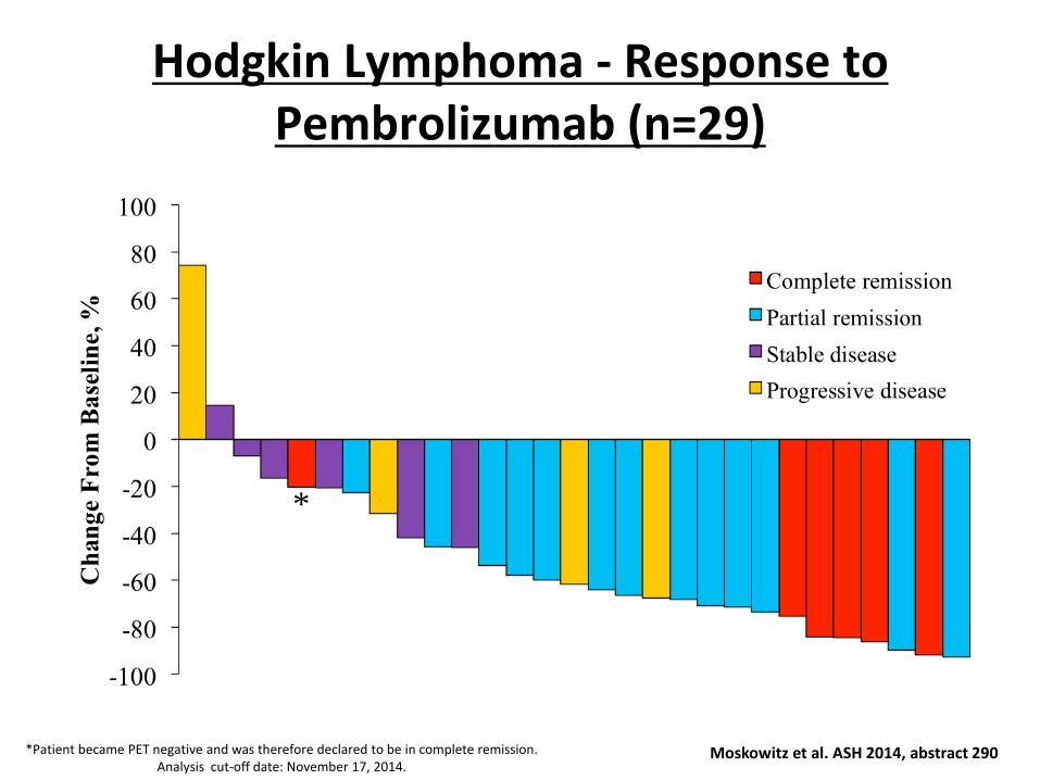
Hodgkin Lymphoma - Response to Pembrolizumab (n=29)
*Patient became PET negative and was therefore declared to be in complete remission.Analysis cut-off date: November 17, 2014.
*
Moskowitz et al. ASH 2014, abstract 290
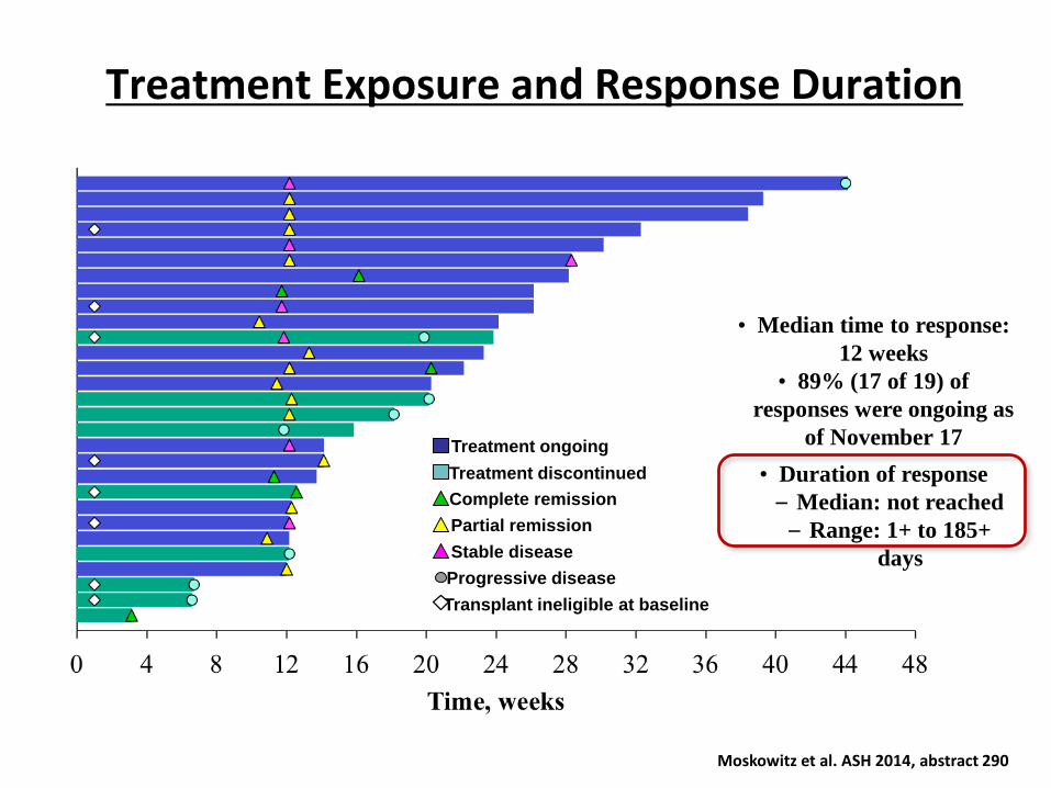
Treatment Exposure and Response Duration
Treatment ongoing
Treatment discontinued
Complete remission
Partial remission
Stable disease
Progressive disease
Transplant ineligible at baseline
• Median time to response:
12 weeks
• 89% (17 of 19) of
responses were ongoing as
of November 17
• Duration of response
– Median: not reached
– Range: 1+ to 185+
days
Moskowitz et al. ASH 2014, abstract 290
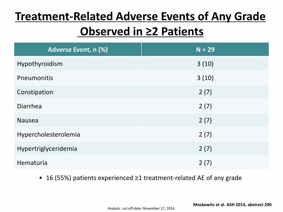
Treatment-Related Adverse Events of Any GradeObserved in ≥2 Patients
Analysis cut-off date: November 17, 2014.
• 16 (55%) patients experienced ≥1 treatment-related AE of any grade
Adverse Event, n (%) N = 29
Hypothyroidism 3 (10)
Pneumonitis 3 (10)
Constipation 2 (7)
Diarrhea 2 (7)
Nausea 2 (7)
Hypercholesterolemia 2 (7)
Hypertriglyceridemia 2 (7)
Hematuria 2 (7)
Moskowitz et al. ASH 2014, abstract 290
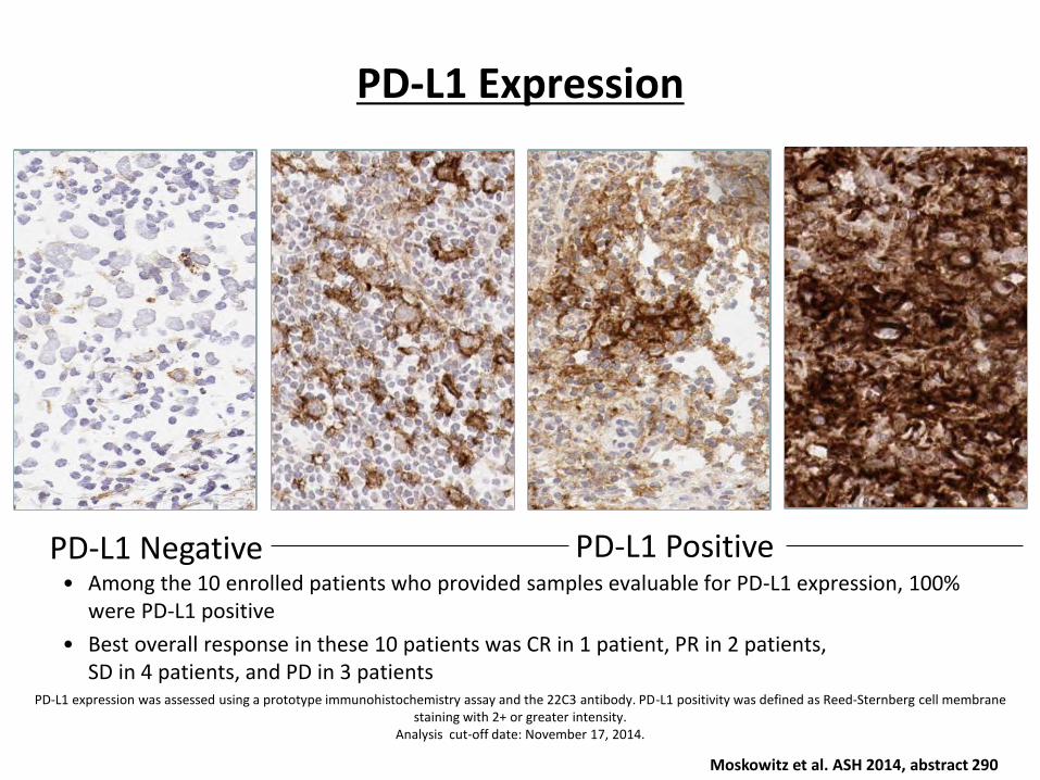
PD-L1 Expression
• Among the 10 enrolled patients who provided samples evaluable for PD-L1 expression, 100% were PD-L1 positive
• Best overall response in these 10 patients was CR in 1 patient, PR in 2 patients, SD in 4 patients, and PD in 3 patients
PD-L1 expression was assessed using a prototype immunohistochemistry assay and the 22C3 antibody. PD-L1 positivity was defined as Reed-Sternberg cell membrane staining with 2+ or greater intensity.
Analysis cut-off date: November 17, 2014.
PD-L1 Negative PD-L1 Positive
Moskowitz et al. ASH 2014, abstract 290
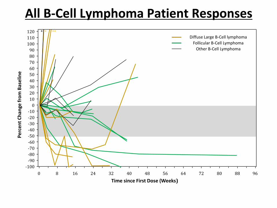
All B-Cell Lymphoma Patient ResponsesP
erc
en
t C
han
ge f
rom
Bas
elin
e
0 8 16 24 32 40 48 56 64 72 80 88 96
Time since First Dose (Weeks)
Diffuse Large B-Cell lymphoma
Follicular B-Cell Lymphoma
Other B-Cell Lymphoma
-30
-20
-10
0
10
20
30
40
50
60
70
80
90
100
110
120
-40
-50
-60
-70
-80
-90
-100
* ***
Time since First Dose (Weeks)
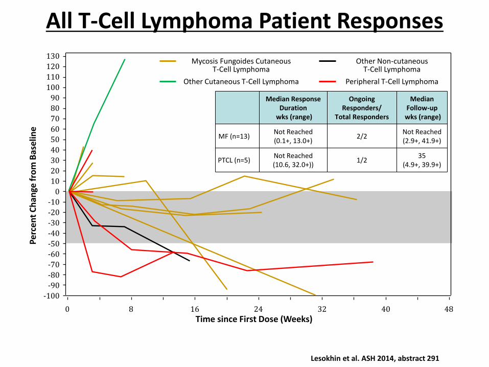
All T-Cell Lymphoma Patient Responses
-30
-20
-10
0
10
20
30
40
50
60
70
80
90
100
110
120
130
0 8 16 24 32 40 48
-40
-50
-60
-70
-80
-90
-100
Pe
rce
nt
Ch
ange
fro
m B
ase
line
Other Non-cutaneousT-Cell Lymphoma
Peripheral T-Cell Lymphoma
Mycosis Fungoides CutaneousT-Cell Lymphoma
Other Cutaneous T-Cell Lymphoma
Median ResponseDuration
wks (range)
Ongoing Responders/
Total Responders
MedianFollow-up
wks (range)
MF (n=13)Not Reached(0.1+, 13.0+)
2/2Not Reached(2.9+, 41.9+)
PTCL (n=5)Not Reached(10.6, 32.0+))
1/235
(4.9+, 39.9+)
Lesokhin et al. ASH 2014, abstract 291
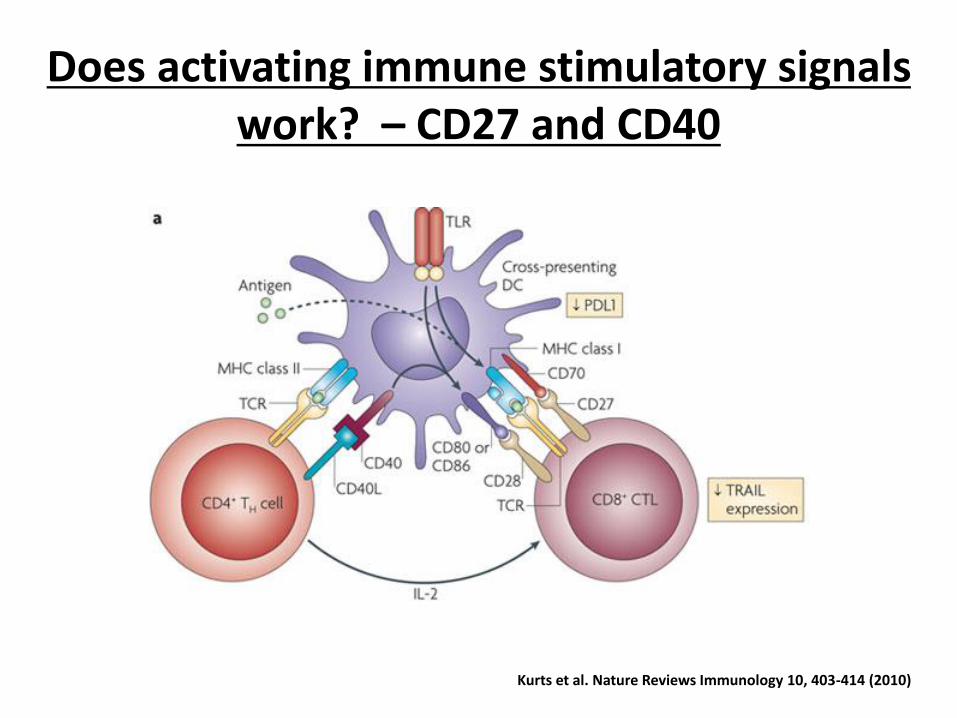
Does activating immune stimulatory signals work? – CD27 and CD40
Kurts et al. Nature Reviews Immunology 10, 403-414 (2010)
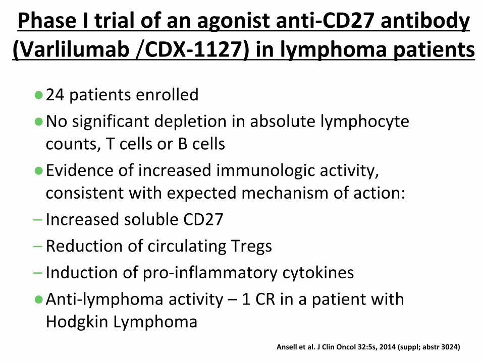
●24 patients enrolled
●No significant depletion in absolute lymphocyte counts, T cells or B cells
●Evidence of increased immunologic activity, consistent with expected mechanism of action:
‒ Increased soluble CD27
‒ Reduction of circulating Tregs
‒ Induction of pro-inflammatory cytokines
●Anti-lymphoma activity – 1 CR in a patient with Hodgkin Lymphoma
Phase I trial of an agonist anti-CD27 antibody (Varlilumab /CDX-1127) in lymphoma patients
Ansell et al. J Clin Oncol 32:5s, 2014 (suppl; abstr 3024)
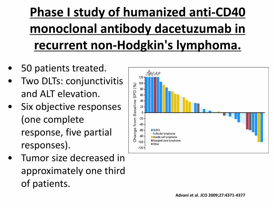
Phase I study of humanized anti-CD40 monoclonal antibody dacetuzumab in recurrent non-Hodgkin's lymphoma.
Advani et al. JCO 2009;27:4371-4377
• 50 patients treated.• Two DLTs: conjunctivitis
and ALT elevation. • Six objective responses
(one complete response, five partial responses).
• Tumor size decreased in approximately one third of patients.
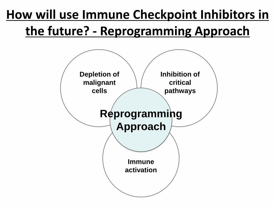
How will use Immune Checkpoint Inhibitors in the future? - Reprogramming Approach
Reprogramming
Approach
Depletion of
malignant
cells
Inhibition of
critical
pathways
Immune
activation
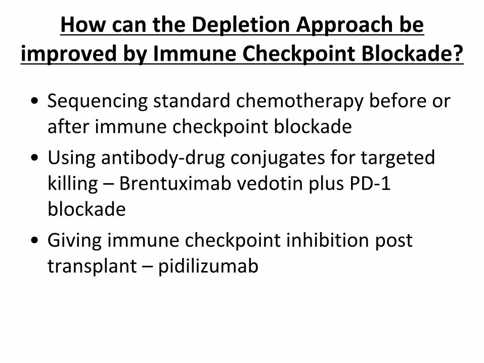
How can the Depletion Approach be improved by Immune Checkpoint Blockade?
• Sequencing standard chemotherapy before or after immune checkpoint blockade
• Using antibody-drug conjugates for targeted killing – Brentuximab vedotin plus PD-1 blockade
• Giving immune checkpoint inhibition post transplant – pidilizumab
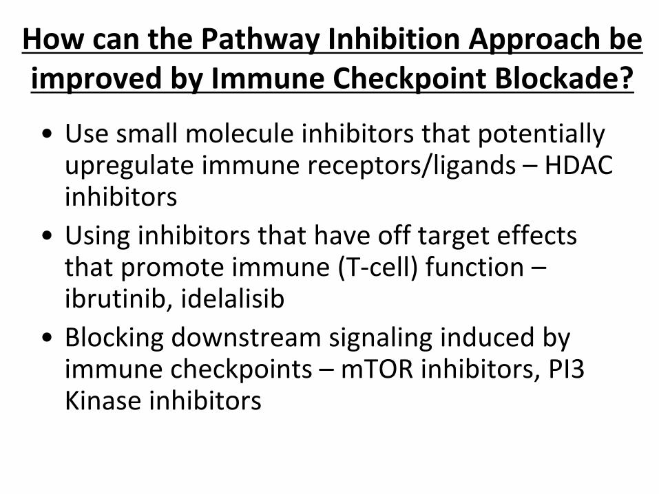
How can the Pathway Inhibition Approach be improved by Immune Checkpoint Blockade?
• Use small molecule inhibitors that potentially upregulate immune receptors/ligands – HDAC inhibitors
• Using inhibitors that have off target effects that promote immune (T-cell) function –ibrutinib, idelalisib
• Blocking downstream signaling induced by immune checkpoints – mTOR inhibitors, PI3 Kinase inhibitors
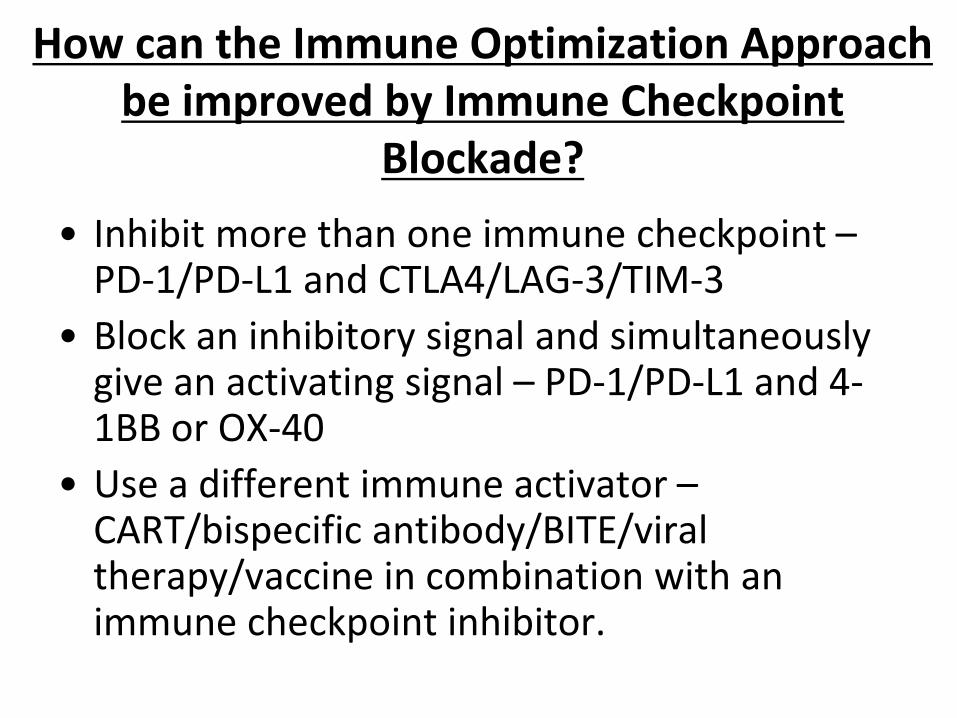
How can the Immune Optimization Approach be improved by Immune Checkpoint
Blockade?
• Inhibit more than one immune checkpoint –PD-1/PD-L1 and CTLA4/LAG-3/TIM-3
• Block an inhibitory signal and simultaneously give an activating signal – PD-1/PD-L1 and 4-1BB or OX-40
• Use a different immune activator –CART/bispecific antibody/BITE/viral therapy/vaccine in combination with an immune checkpoint inhibitor.
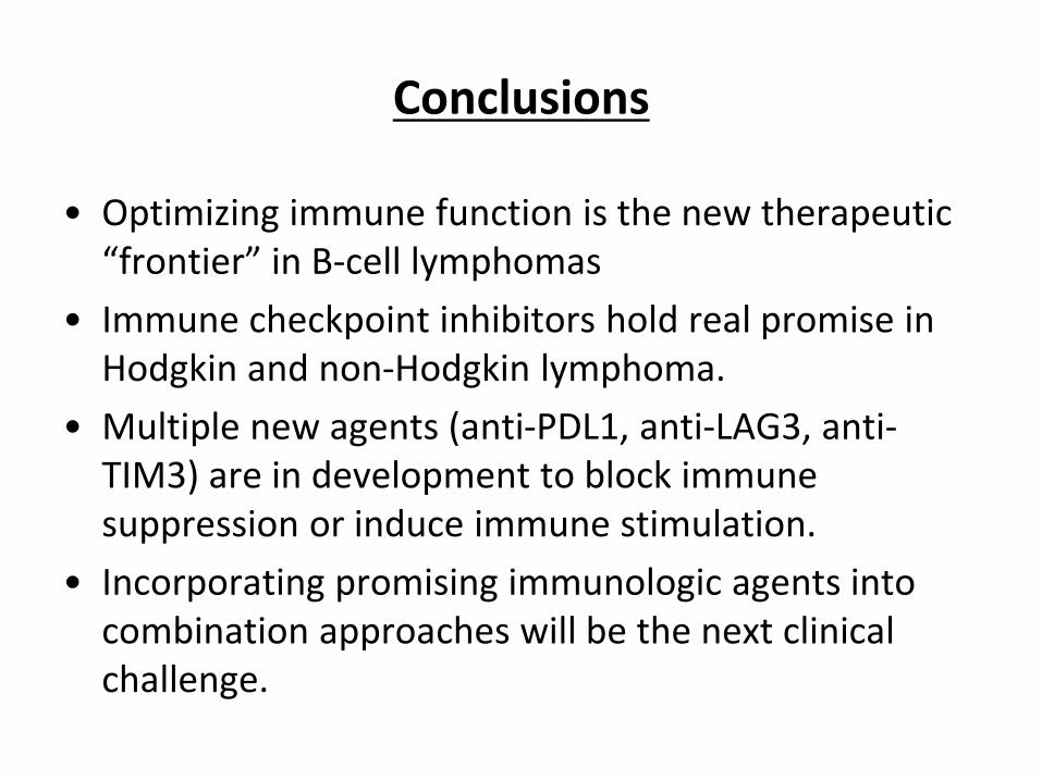
Conclusions
• Optimizing immune function is the new therapeutic “frontier” in B-cell lymphomas
• Immune checkpoint inhibitors hold real promise in Hodgkin and non-Hodgkin lymphoma.
• Multiple new agents (anti-PDL1, anti-LAG3, anti-TIM3) are in development to block immune suppression or induce immune stimulation.
• Incorporating promising immunologic agents into combination approaches will be the next clinical challenge.