identification ofgenesandgeneproducts necessaryfor ... · beenshownto...
TRANSCRIPT
Proc. Nati. Acad. Sci. USAVol. 81, pp. 4154-4158, July 1984Genetics
Identification of genes and gene products necessary forbacterial bioluminescence
(lux genes/recombinant DNA/complementation/minicells)
JOANNE ENGEBRECHT*t AND MICHAEL SILVERMAN**The Agouron Institute, 505 Coast Boulevard South, La Jolla, CA 92037; and tScripps Institution of Oceanography, Department of Marine Biology A-002,La Jolla, CA 92093
Communicated by Donald R. Helinski, March 23, 1984
ABSTRACT Expression of luminescence in Escherichiacoli was recently achieved by cloning genes from the marinebacterium Vibrio fischeri. One DNA fragment on a hybridplasmid encoded regulatory functions and enzymatic activitiesnecessary for light production. We report the results of a ge-netic analysis to identify the luminescence genes (lux) that re-side on this recombinant plasmid. lax gene mutations weregenerated by hydroxylamine treatment, and these mutationswere ordered on a linear map by complementation in transwith a series of polar transposon insertions on other plasmids.lux genes were defined by complementation of lax gene defectson pairs of plasmids in trans in E. coli. Hybrid plasmids werealso used to direct the synthesis of polypeptides in the E. coliminicell system. Seven lax genes and the corresponding geneproducts were identified from the complementation analysisand the minicell programing experiments. These genes, in theorder of their position on a linear map, and the apparent mo-lecular weights of the gene products are luxR (27,000), luax(25,000), luxC (53,000), luxD (33,000), luxA (40,000), luxB(38,000), and luxE (42,000). From the luminescence pheno-types of E. coli containing mutant plasmids, functions wereassigned to these genes: laxA, luxB, luxC, luxD, and luxE en-code enzymes for light production and luxR and lux! encoderegulatory functions.
Luminescent bacteria are common in the ocean and occupya variety of ecological niches (1, 2). Vibrio fischeri (strainMJ-1) colonizes the light organ of the fish Monocentris ja-ponicus, and we have initiated studies to determine the ge-netic and functional components of the luminescence systemfrom this bacterium. The emission of light by marine bacte-ria is catalyzed by the enzyme luciferase, a mixed functionoxidase that has two subunits, a and f3, with molecularweights of -40,000 each (3). In the generation of light, lucif-erase oxidizes a reduced flavin and a long-chain aldehyde,producing oxidized flavin and the corresponding long chainfatty acid:
luciferaseRCHO + FMNH2 + 02 -e RCOOH + FMN + H20 + hp.
Other components unique to the bioluminescence system in-clude enzymes involved in the synthesis or recycling of thealdehyde substrate. Light production occurs in dense bacte-rial cultures and is controlled by the synthesis of a sensorymolecule termed autoinducer. This molecule is secreted intothe extracellular environment where it accumulates and at acritical concentration signals expression of luminescence.This induction can result in a 10,000-fold increase in lightemission per cell (4). The autoinducer from V. fischeri hasbeen shown to be N-(/3-ketocaproyl)homoserine lactone (5).We previously isolated a 9-kilobase-pair (kbp) DNA frag-
ment from V. fischeri that encoded all of the functions neces-sary for light production and that also contained the regula-tory elements required for their expression in Escherichiacoli. By using transposon mutagenesis, the regions on thisDNA fragment that encoded aldehyde, luciferase, and regu-latory functions were defined. These functions were orga-nized into two transcriptional units, operon L and operon R.Furthermore, by using the lacZ gene fusions created by tran-sposon mini-Mu insertion, transcription of operon R wasfound to be induced by the presence of autoinducer, whosesynthesis was controlled by a gene product also encoded bythis same operon. Therefore, lux gene expression was con-trolled by a positive feedback circuit, and induction resultedin a logarithmic increase in the synthesis of enzymes for thelight reaction (6). Due to the polar nature of these transposoninsertions, we were unable to define the individual genesthat encoded these functions. We report here the identifica-tion of lux genes and gene products by performing comple-mentation tests with hydroxylamine-generated point muta-tions and by programing protein synthesis in the E. coli mini-cell system.
MATERIALS AND METHODS
Bacterial Strains, Cloning Vehicles, and Media. E. colistrain ED8654 (supE supF met hsdR- hsdM+) was used forpropagation of recombinant plasmids. Complementationtests were performed in HB101 (hsdS recA ara proA lacgalK rpsL). E. coli strain P678-54 (thr ara leu azi tonA lacYtsx minA minB gal malA thi xyl rpsL) was used for the pro-duction of minicells for the protein programing. DNA frag-ments were subcloned into various sites in vehicle pBR322(7) and pACYC184 (8). Plasmids were recovered by transfor-mation (9) and selection for transformants containing recom-binants in pBR322 was on Luria agar plates containing 80 pugof ampicillin per ml and recombinants in pACYC184 was onLuria agar containing 50 ,ug of chloramphenicol per ml. Hy-brid plasmids in complementation studies were propagatedon Luria agar plates containing 80 ,4g of ampicillin per ml and50 gg of chloramphenicol per ml. Restriction endonucleaseswere purchased from New England Biolabs and T4 DNA li-gase was purchased from Bethesda Research Laboratories.Antibiotics were purchased from Calbiochem.Hydroxylamine Mutagenesis. Mutagenesis was as de-
scribed for phage by Berman et al. (10). Five micrograms ofpJE202 (6) or pJE737 DNA was incubated at 370C in 0.5 MNH2OH, pH 6/5 mM Tris/0.5 mM EDTA in a final volumeof 50 Al. Five-microliter samples were removed at 60-minintervals, and the reaction was stopped by the addition of 95,41 of 100 mM CaC12. This DNA was then used to transformthe strain HB101. Since mutagenesis was in vitro and noplasmid replication could occur, transformants with a mu-tant luminescence phenotype were the result of independent
Abbreviation: kbp, kilobase pair(s).
4154
The publication costs of this article were defrayed in part by page chargepayment. This article must therefore be hereby marked "advertisement"in accordance with 18 U.S.C. §1734 solely to indicate this fact.
Dow
nloa
ded
by g
uest
on
Janu
ary
5, 2
020
Proc. NatL. Acad. Sci. USA 81 (1984) 4155
mutations. To insure that transformants arose from a singlemutant plasmid, plasmid DNA was prepared (11) from eachmutant strain and used to retransform strain HB101. Thesetransformants were subsequently reexamined for the mutantphenotype. With mutagenized pJE737 DNA, transformationwas into HB101 containing plasmid pJE502 (luxR). Minimalamounts of DNA were used to avoid the isolation of multipletransformed bacteria. Seventy-four independent dark (no de-tectable light production) or dim (ca. 1% of wild-type lightproduction) mutant plasmids were isolated from the pJE202mutagenesis. Twenty mutant plasmids-i.e., no comple-mentation of the luxR defect on the plasmid pJE502-wereisolated from the pJE737 mutagenesis. To insure that therecombinant plasmids contained single mutations, hydroxyl-amine treatment was adjusted to yield a frequency of Lux-mutants of about 5%. Approximately 240 min of exposure tohydroxylamine was necessary to achieve this level of muta-genesis.
Complementation Tests. Complementation of Tn5 muta-tions on hybrid plasmids (pJE300s) (6) with hydroxylamine-generated mutations on hybrid plasmids (pJE500s) was per-formed by cotransforming pairs of plasmids into a Rec- E.coli strain, HB101, as described (6). To perform additionalcomplementation tests, lux mutations had to be transferredby reciprocal recombination to another hybrid plasmid witha compatible replicon. lux mutations on a pACYC184 repli-con (pJE600s) were made by cotransforming pJE500 plas-mids (pBR322 replicon) and pJE212 (Sal I lux fragment inpACYC184) into a Rec+ E. coli strain, ED8654. PlasmidDNA was isolated from these strains and retransformed intostrain HB101 with selection for the pACYC184 replicon. Ap-proximately 1% of the transformants had the mutant pheno-type of the donor plasmid (pJE500s). Complementation intrans of lux gene mutations was then measured by cotrans-forming plasmids of the pJE500 and pJE600 series intoHB101. Light production was measured in these strains visu-ally, by autoradiography or with a LKB 1211 Minibeta scin-tillation counter in the chemiluminescence mode, and com-plementation was scored as positive if light production was>10% of wild-type light production (HB101 containing plas-mid pJE202 or pJE212) and negative if <10%. Hybrid plas-mids containing amber mutations in specific genes were gen-erated by hydroxylamine mutagenesis. Strains with mutantplasmids were selected on the basis of complementation (toidentify the gene defect) and suppression with strain ED8654(to recognize amber mutations).
Minicell Preparation and Electrophoresis of Gene Products.Minicells were made from strain P678-54 as described byMatsumura et al. (12). The hybrid plasmids were introducedby transformation. Two milliliters of a stationary culture wasused to inoculate 1 liter of Luria broth containing 80 ,ug ofampicillin per ml or 50 ,g of chloramphenicol per ml andincubated on a 37°C shaker overnight. The culture was cen-trifuged for 5 min at 5000 x g to remove whole bacteria, andthe supernatant was centrifuged for 10 min at 10,000 x g toconcentrate minicells. The resulting pellet was suspended in1.5 ml of buffered saline gelatin (0.85% NaCl/0.03%KH2PO4/0.06% Na2HPO4/100 ,ug of gelatin per ml). Thecells were then sedimented on a 5-20% sucrose gradient for20 min at 5000 x g. The top band was collected, pelleted,and resuspended in 1.5 ml of buffered saline gelatin. Thissuspension was rerun on a second 5-20% sucrose gradient,the upper band was collected, and the minicells were pellet-ed. The pellet was suspendedto -2 x 10⁹ minicells per ml inminimal salts medium with 0.5% glycerol and 2 ,ug of aminoacids (minus methionine) per ml. Minicells (0.5 ml) were in-cubated for 30 min at 30'C, and then 10 /iCi (1 Ci = 37 GBq)of [355]methionine with a specific activity of 970 Ci/mmol(New England Nuclear) or 10 ,uCi of 3H-labeled L-amino acidmixture with a specific activity of =50 Ci/mmol (New En-
gland Nuclear) was added. After 20 min, the minicells werepelleted and then frozen at -20'C. The details of Na-DodSO4/polyacrylamide gel electrophoresis have been de-scribed (13). The gel was incubated in Autofluor (NationalDiagnostics, Somerville, NJ) before drying and autoradiog-raphy.
RESULTSSeventy-four dark or dim mutants of strains carrying pJE202mutagenized with hydroxylamine were isolated (see Materi-als and Methods). As observed previously with transposonmutants (6), there were four classes of mutant phenotypes:(i) mutants that could not produce or respond to autoin-ducer; (ii) those that could respond to autoinducer but couldno longer produce autoinducer; (iii) those that were dark butproduced light with the addition of exogenous aldehyde; and(iv) those that were dark but could not respond to the addi-tion of aldehyde. Mutants in categories i and ii were defec-tive in functions required for regulation of lux gene expres-sion. Mutants in categories iii and iv produced and respond-ed to autoinducer but had defects in enzymes for lightproduction. Mutants in categories i-iii were dim (1% or lessof wild-type light production), whereas mutants in categoryiv were generally dark (no detectable light production). Togenerate a linear map of the hydroxylamine mutations, com-plementation tests were performed with lux plasmids con-taining TnS insertions that were previously mapped by re-striction analysis (6) (see Fig. 2A). Due to the polar nature ofthese transposon insertions, genes within an operon down-stream from the transposon insertion would not be tran-scribed, whereas any genes upstream would be expressed.Therefore, in complementation tests with hybrid plasmidscontaining lux point mutations in trans with hybrid plasmidscontaining transposon insertions, complementation wouldnot occur if the point mutation were in a gene identical to one(in trans) inactivated by transposon insertion (target gene ordistal gene), but complementation would occur if the pointmutation were in a gene identical to a gene (in trans) proxi-mal to transposon insertion. All 74 mutant plasmids werecharacterized by this test, and Table 1 shows the result ofthis type of analysis. Thus, lux mutations onhybrid plasmidswere mapped to six regions of the cloned DNA fragment.Nonsense mutations resulting from hydroxylamine muta-
genesis would be expected to show a degree of polarity ontranscription of downstream genes. However, none of thepoint mutations tested showed polarity of the extent exhibit-
Table 1. Complementation of polar mutations
Tn5 mutation (pACYC184 replicon)*331 337 307 309 301 320
501> 502C4 560a, 5611-
r 5250* 528= 546E 547.5 508E 510-> 5052 515>, 537
545
++++++++++++
+ + + + ++ + + + +- + + + +- + + + +- - + + +
- - + + +
~+ +
- - + ~+ +
+~~~~
- - +~~~~
- - +~~~~
+~~~~
Complementation of lux defects was measured in RecA- strainsharboring pairs of mutant plasmids (+, light producing; -, little orno light produced).*Location of transposon Tn5 insertions in lux operons is shown inFig. 2A.
Genetics: Engebrecht and Silverman
Dow
nloa
ded
by g
uest
on
Janu
ary
5, 2
020
4156 Genetics: Engebrecht and Silverman
ed by the TnS transposon mutations (see Table 1). Thus, hy-brid plasmids containing these point mutations could be usedin complementation tests to define individual genes. To per-
form these tests, it was first necessary to transfer point mu-
tations to a second compatible replicon (pJE212, derivativeof pACYC184) via recombination with the original plasmidmutations (pJE500s). These hybrid plasmids were designat-ed pJE600s and would replicate in the same cell as thepJE500s (see Materials and Methods). Fifty lux mutationsfrom the pJE500 series were transferred to the pACYC184replicon (pJE600 series). Complementation studies were
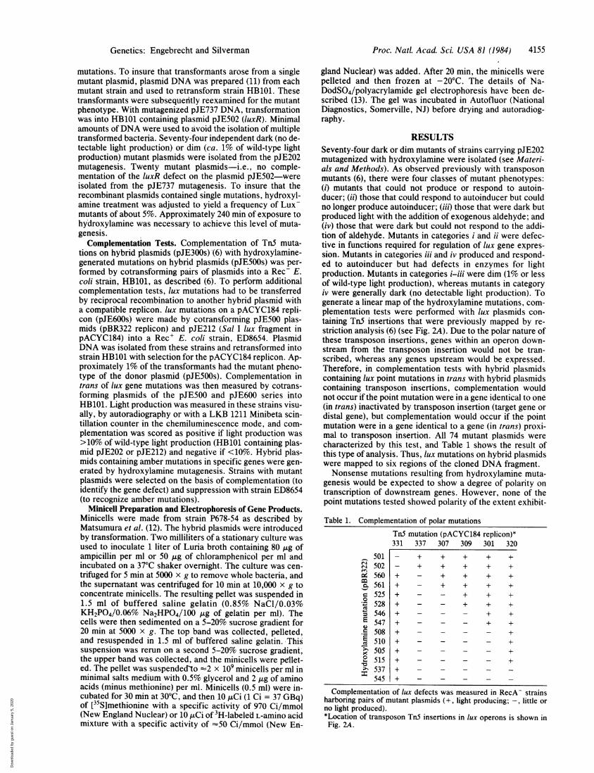
then undertaken by introducing pJE500 and pJE600 plasmidsinto the same E. coli strain. To reduce the number of com-plementation tests, the entire matrix of lux mutation comnbi-nations (50 pJE500 x 50 pJE600 series plasmids or 2500strains) was not constructed. Instead, subgroups of plasmidswith linked lux mutations (generally 6 pJE500 x 6 pJE600series plasmids or 36 strains) were used in complementationtests to define individual genes. Pairs of mutant plasmidsrepresenting each complementation group were then assem-
bled into the matrix shown in Fig. 1. Light production oc-curred when the mutations on the plasmids complemented.Those cells that produced little or no light harbored plasmidswith noncomplementing mutations. Since mutations in thesame gene did not complement and seven groups of noncom-plementing mutations were observed, we concluded thatthere were seven lux genes encoded by the fragment clonedfrom V. fischeri. These genes are designated luxR, luxI,luxC, luxD, luxA, luxB, and luxE. The locations of thesegenes in operon L and operon R, as determined by data suchas those in Table 1, are shown in Fig. 2B. The function ofeach gene was inferred from the phenotypes of mutants iso-lated in this study and from the properties of transposon mu-
tants analyzed in a previous report (6). The products of luxRand luxI regulate expression of luminescence; luxi encodes afunction required for the synthesis of autoinducer and luxRencodes a function necessary for response to autoinducer.The luxC, luxD, and luxE gene products function to providethe aldehyde substrate, and luxA and luxB encode the a andp8 subunits of luciferase.The E. coli minicell system has been used extensively to
identify plasmid-encoded gene products. Since only small re-
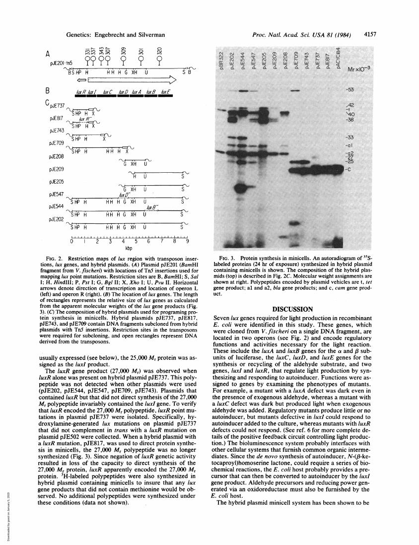
plicons such as the hybrid lux plasmids segregate with theminicells during their formation, protein synthesis in purifiedminicell preparations is directed exclusively by hybrid plas-mid genes. To use this system to program the synthesis oflux gene products, we constructed hybrid plasmids that con-tained subclones of the lux gene region and also generatedchain-terminating (amber) mutations in specific genes in hy-brid plasmids (see Fig. 2C). These hybrid plasmids were thentransferred into the minicell strain by transformation, andminicells were prepared for protein labeling experiments.Fig. 3 shows an autoradiogram of a polyacrylamide gel con-
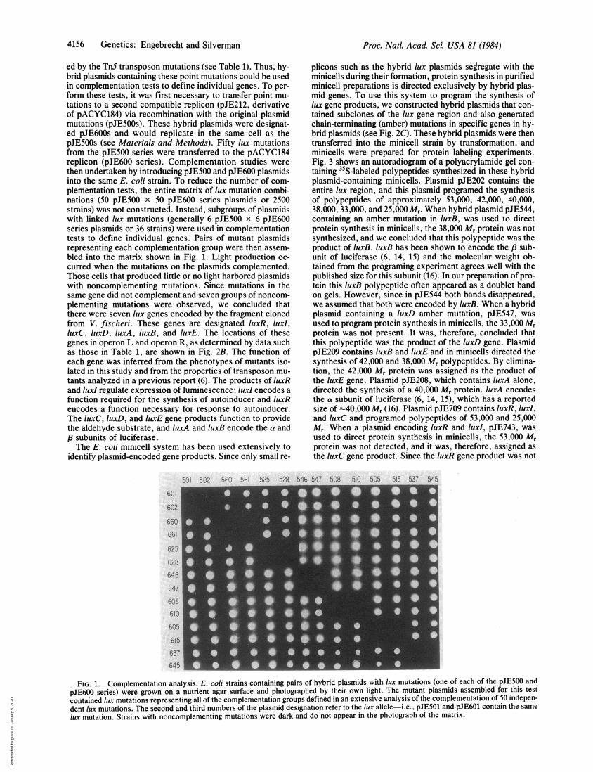
taining 35S-labeled polypeptides synthesized in these hybridplasmid-containing minicells. Plasmid pJE202 contains theentire lux region, and this plasmid programed the synthesisof polypeptides of approximately 53,000, 42,000, 40,000,38,000, 33,000, and 25,000 Mr. When hybrid plasmid pJE544,containing an amber mutation in luxB, was used to directprotein synthesis in minicells, the 38,000 Mr protein was notsynthesized, and we concluded that this polypeptide was theproduct of luxB. luxB has been shown to encode the P sub-unit of luciferase (6, 14, 15) and the molecular weight ob-tained from the programing experiment agrees well with thepublished size for this subunit (16). In our preparation of pro-tein this luxB polypeptide often appeared as a doublet bandon gels. However, since in pJE544 both bands disappeared,we assumed that both were encoded by luxB. When a hybridplasmid containing a luxD amber mutation, pJE547, was
used to program protein synthesis in minicells, the 33,000 Mr
protein was not present. It was, therefore, concluded thatthis polypeptide was the product of the luxD gene. PlasmidpJE209 contains luxB and luxE and in minicells directed thesynthesis of 42,000 and 38,000 Mr polypeptides. By elimina-tion, the 42,000 Mr protein was assigned as the product ofthe luxE gene. Plasmid pJE208, which contains luxA alone,directed the synthesis of a 40,000 Mr protein. luxA encodesthe a subunit of luciferase (6, 14, 15), which has a reportedsize of -40,000 Mr (16). Plasmid pJE709 contains luxR, luxI,and luxC and programed polypeptides of 53,000 and 25,000Mr. When a plasmid encoding luxR and luxI, pJE743, was
used to direct protein synthesis in minicells, the 53,000 Mrprotein was not detected, and it was, therefore, assigned as
the luxC gene product. Since the luxR gene product was not
501 502 560 561 525 528 546 547 508 510 505 515 537 545
601
602
660
661
625
628646
647
608610 -
605
615
637645
FIG. 1. Complementation analysis. E. coli strains containing pairs of hybrid plasmids with lux mutations (one of each of the pJE500 andpJE600 series) were grown on a nutrient agar surface and photographed by their own light. The mutant plasmids assembled for this testcontained lux mutations representing all of the complementation groups defined in an extensive analysis of the complementation of 50 indepen-dent lux mutations. The second and third numbers of the plasmid designation refer to the lux allele-i.e., pJE501 and pJE601 contain the samelux mutation. Strains with noncomplementing mutations were dark and do not appear in the photograph of the matrix.
Proc. NatL Acad Sci. USA 81 (1984)
Dow
nloa
ded
by g
uest
on
Janu
ary
5, 2
020
Proc. Natl. Acad. Sci. USA 81 (1984) 4157
A 4
pJE201:tn5 ?BS HP H
.r- ro) N-r n C
~) V4)e'
(7) c c
H H H G XH U S B
N o t o O o o rorirv, CN I)L N N r- 1- r-. OD >-
L Lw Lu w w Lu 0
m - -, -, -) -, --) ) -,n)-7 <( C CL 0. 0L CL CL C0
-53/uxR/uxl/IxC /UXP luxA /x8 /lUXE
C pJE737SHP H X
pJE 817 tuj R-SHP H X
pJE743
pJE709 SHP H X
SHP H
42-t`40-38
-33-al-a2-27-25-C
pJE208
pJE209
pJE205
pJE547
pJE544
pJE202
G XH U
H U S
G XH U S
HHHG XHHH H GXH
SHP H HH H G XH U S
SHP H 2 3 4 6 7 S
0' 3 4 5 6 7 8 9kbp
FIG. 2. Restriction maps of lux region with transposon inser-tions, lux genes, and hybrid plasmids. (A) Plasmid pJE201 (BamHIfragment from V. fischeri) with locations of TnS insertions used formapping lux point mutations. Restriction sites are B, BamHI; S, SalI; H, HindIlI; P, Pst I; G, Bgl II; X, Xho I; U, Pvu II. Horizontalarrows denote direction of transcription and location of operon L(left) and operon R (right). (B) The location of lux genes. The lengthof rectangles represents the relative size of lux genes as calculatedfrom the apparent molecular weights of the lux gene products (Fig.3). (C) The composition of hybrid plasmids used for programing pro-tein synthesis in minicells. Hybrid plasmids pJE737, pJE817,pJE743, and pJE709 contain DNA fragments subcloned from hybridplasmids with TnS insertions. Restriction sites in the transposonswere required for subcloning, and open rectangles represent DNAderived from the transposons.
usually expressed'(see below), the 25,000 Mr protein was as-signed as the luxI product.The luxR gene product (27,000 Mr) was observed when
luxR alone was present on hybrid plasmid pJE737. This poly-peptide was not detected when other plasmids were used(pJE202, pJE544, pJE547, pJE709, pJE743). Plasmids thatcontained luxR but that did not direct synthesis of the 27,000Mr polypeptide invariably contained the luxI gene. To verifythat luxR encoded the 27,000 Mr polypeptide, luxR point mu-tations in plasmid pJE737 were isolated. Specifically, hy-droxylamine-generated lux mutations on plasmid pJE737that did not complement in trans with a luxR mutation onplasmid pJE502 were collected. When a hybrid plasmid witha luxR mutation, pJE817, was used to direct protein synthe-sis in minicells,'the 27,000 Mr polypeptide was no longersynthesized (Fig. 3). Since negation of luxR genetic activityresulted in loss of the capacity to direct synthesis of the27,000 Mr protein, luxR apparently encoded the 27,000 Mrprotein. 3H-labeled polypeptides were also synthesized inhybrid plasmid containing minicells to insure that any luxgene products that did not contain methionine would be ob-served. No additional polypeptides were synthesized underthese conditions (data not shown).
w, ...
Wc,
.:U
FIG. 3. Protein synthesis in minicells. An autoradiogram of 35S-labeled proteins (24 hr of exposure) synthesized in hybrid plasmidcontaining minicells is shown. The composition of the hybrid plas-mids (top) is described in Fig. 2C. Molecular weight assignments areshown at right. Polypeptides encoded by plasmid vehicles are t, tetgene product; al and a2, bla gene products; and c, cam gene prod-uct.
DISCUSSIONSeven lux genes required for light production in recombinantE. coli were identified in this study. These genes, whichwere cloned from V. fischeri on a single DNA fragment, are
located in two operons (see Fig. 2) and encode regulatoryfunctions and activities necessary for the light reaction.These include the luxA and luxB genes for the a and sub-units of luciferase, the luxC, luxD, and luxE genes for thesynthesis or recycling of the aldehyde substrate, and twogenes, luxI and luxR, that regulate light production by syn-thesizing and responding to autoinducer. Functions were as-signed to genes by examining the phenotypes of mutants.For example, a mutant with a luxA defect was dark even inthe presence of exogenous aldehyde, whereas a mutant witha luxC defect was dark but produced light when exogenousaldehyde was added. Regulatory mutants produce little or noautoinducer, but mutants defective in luxI could respond toautoinducer added to the culture, whereas mutants with luxRdefects could not respond. (See ref. 6 for more complete de-tails of the positive feedback circuit controlling light produc-tion.) The bioluminescence system probably interfaces withother cellular systems that furnish common organic interme-diates. Since the de novo synthesis of autoinducer, N-(,B-ke-tocaproyl)homoserine lactone, could require a series of bio-chemical reactions, the E. coli host probably provides a pre-cursor that can then be converted to autoinducer by the luxIgene product. Aldehyde precursors and reducing power gen-erated via an oxidoreductase must also be furnished by theE. coli host.The hybrid plasmid minicell system has been shown to be
B
Mrx103
.Genetics: Engebrecht and Silverman
Amummak-f.qw :,
" :.:-,:,...mll-f..
40.
A-qm
w
Dow
nloa
ded
by g
uest
on
Janu
ary
5, 2
020
4158 Genetics: Engebrecht and Silverman
a useful method for selectively synthesizing gene products(12). We have used this system to identify the seven lux geneproducts. It is unlikely that we have failed to identify a geneand its gene product since 95% of the coding capacity of thecloned DNA fragment was used to direct the synthesis ofthose seven polypeptides. In certain cases the gene-productassignment could be compared with independent values, andthe molecular weights of the luxA and luxB gene productscorresponded well with the reported molecular weights ofthe V. fischeri luciferase subunits (16). There does not seemto be correspondence between polypeptides from V. fischeriand the fatty acid reductase isolated from the distantly relat-ed luminescent bacterium Photobacterium phosphoreum,which has subunit Mrs of 51,000 and 58,000 (17). Several hy-brid plasmids used in minice'll programing experiments didnot contain the lux operon promoter elements. Since luxgene products were detected, it is apparent that plasmid pro-moter elements must have been responsible for transcriptionof these' genes. However, other plasmids did contain lux pro-moter elements and protein synthesis could be stimulated bythe exogenous addition. of autoinducer. Thus, the lux pro-moter elements did function in minicells but the relative con-tributions of plasmid and lux promoters to protein synthesishave not been determined.A variety of plasmids containing luxR were used to direct
polypeptide synthesis in the minicell system, but the luxRproduct was detected only when'plasmids with a particulargenetic composition were used. The luxR gene product ap-peared only in the absence of luxI gene activity. With thesehybrid plasmids transcription of the luxR gene was probablydirected by the operon L promoter since synthesis of theluxR product' was independent of alignment with definedplasmid promoters. It is possible that expression of operon L(containing luxR) is repressed by the luxi gene product. Op-eron R, containing luxI and genes for light reaction enzymes,is positively regulated by the combined actions of the luxRand luxI gene products (6). The possibility that biolumines-cence is controlled by both negative and positive regulatorycircuits must be explored.The definition of gene functions resulting from this study
is in agreement with the general categories of function in-ferred from the earlier use of transposon-generated muta-tions (6). With the identification of lux genes and gene prod-ucts, we can now begin to dissect further this system. Thesubcellular location of these lux gene products can now be
determined by using labeled polypeptides synthesized in theE. coli minicell system. Knowing the identity of lux geneproducts should assist in the purification and biochemicalanalysis of poorly understood components of the biolumi-nescence system such as those for aldehyde cycling and forthe synthesis, excretion, and sensing of autoinducer.
We thank Melvin Simon, Robert Belas, Margo Haygood, KaarenJanssen, and Peter Johnson for helpful advice. Michele Platten ablyprepared this manuscript. This research was supported by a contract(N00014-81-K-0343) from the Office of Naval Research.
1. Nealson, K. H. & Hastings, J. W. (1979) Microbiol. Rev. 43,496-518.
2. Baumann, P. & Baumann, L. (1977) Annu. Rev. Microbiol. 31,39-61.
3. Ziegler, M. M. & Baldwin, T. 0. (1981) Curr. Top. Bioenerg.12, 65-113.
4. Nealson, K. H. (1977) Arch. Microbiol. 112, 73-79.5. Eberhard, A., Burlingame, A. L., Eberhard, C., Kenyon,
G. L., Nealson, K. H. & Oppenheimer, N. J. (1981) Biochem-istry 20, 2444-2449.
6. Engebrecht, J., Nealson, K. H. & Silverman, M. R. (1983)Cell 32, 773-781.
7. Bolivar, F., Rodriguez, R. L., Greene, P. J., Betlach, M. C.,Heyneker, H. L., Boyer, H. W., Crosa, J. H. & Falkow, S.(1977) Gene 2, 95-113.
8. Chang, A. C. Y. & Cohen, S. N. (1978) J. Bacteriol. 134,1141-1156.
9. Mandel, M. & Higa, A. (1970) J. Mol. Biol. 53, 159-162.10. Berman, M. L., Enquist, L. W. & Silhavy, T. J. (1982) Ad-
vanced Bacterial Genetics (Cold Spring Harbor Laboratory,Cold Spring Harbor, NY), p. 125.
11. Birnboim, H. C. & Doly, J. (1979) Nucleic Acids Res. 7, 1513-1523.
12. Matsumura, P., Silverman, M. & Simon, M. (1977) J. Bacteri-ol. 132, 996-1002.
13. Silverman, M., Matsumura, P., Draper, R., Edwards, S. & Si-mon, M. (1976) Nature (London) 261, 248-250.
14. Cohn, D. H., Ogden, R. C., Abelson, J. N., Baldwin, T. O.,Nealson, K. H., Simon, M. I. & Mileham, A. J. (1983) Proc.Natl. Acad. Sci. USA 80, 120-123.
15. Belas, R., Mileham, A., Cohn, D., Hilmen, M., Simon, M. &Silverman, M. (1982) Science 218, 791-793.
16. Gunsalus-Miguel, A., Meighen, E. A., Nicoli, M. Z., Nealson,K. H. & Hastings, J. W. (1972) J. Biol. Chem. 247, 398-404.
17. Rodriguez, A., Riendeau, D. & Meighen, E. (1983) J. Biol.Chem. 258, 5233-5237.
Proc. Natl. Acad Sci. USA 81 (1984)
Dow
nloa
ded
by g
uest
on
Janu
ary
5, 2
020