identification of phenprocoumon metabolites in human urine by high-performance liquid chromatography...
TRANSCRIPT
325
Journal of Chromatography, 338 (1985) 325-334 Biomedical Applications Elsevier Science Publishers B.V., Amsterdam - Printed in The Netherlands
CHROMBIO. 2438
IDENTIFICATION OF PHENPROCOUMON METABOLITES IN HUMAN URINE BY HIGH-PERFORMANCE LIQUID CHROMATOGRAPHY AND GAS CHROMATOGRAPHY-MASS SPECTROMETRY
JAN X. DE VRIES*, MICHAEL SIMON, RAINER ZIMMERMANN and JOB HARENBERG
Medizinische Klinik der Universitiit Heidelberg, Abteilung fiir Klinische Pharmakologie und Abteilung fiir Innere Medizin I, Bergheimerstrasse 58, O-6900 Heidelberg (F.R.G.)
(First received July 23rd, 1984;revised manuscript received October lOth, 1984)
SUMMARY
The oral anticoagulant phenprocoumon is eliminated in urine mainly as the glucuronide conjugate to an extent of 20% of the dose. The urine from patients undergoing phen- procoumon therapy was investigated and the following metabolites were isolated and
identified: 7-hydroxyphenprocoumon as the main component, and 4’-hydroxyphenpro- coumon and 6-hydroxyphenprocoumon as conjugates. They were characterized by high- performance liquid chromatography and, after methylation, by gas chromatography-mass
spectrometry.
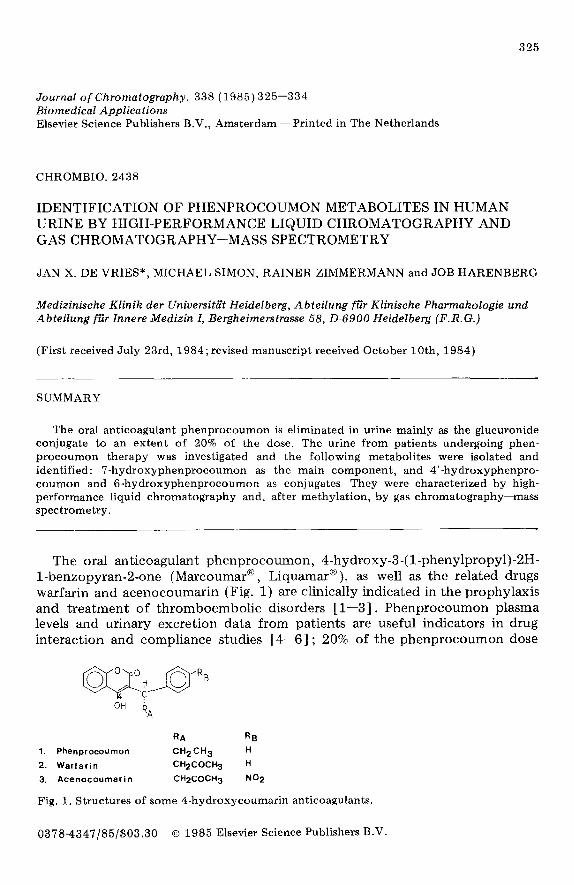
The oral anticoagulant phenprocoumon, 4-hydroxy-3-(l-phenylpropyl)-ZH- 1-benzopyran-2-one (Marcoumar@ , Liquamar@), as well as the related drugs warfarin and acenocoumarin (Fig. 1) are clinically indicated in the prophylaxis and treatment of thromboembolic disorders [l-3] . Phenprocoumon plasma levels and urinary excretion data from patients are useful indicators in drug interaction and compliance studies [4-f5] ; 20% of the phenprocoumon dose
gFjI.p$gjrRB OH ’
RA
RA R~
1. Phenprocoumon CH2CH3 H
2. Warfarin CH2COCH3 H
3. Acenocoumarin CH2COCH3 NO2
Fig. 1. Structures of some 4-hydroxycoumarin anticoagulants.
0378~4347/85/$03.30 0 1985 Elsevier Science Publishers B.V.
326
is recovered as conjugate in urine [4, 71 1 Rats and rat liver microsomes treated with phenprocoumon yield a series of hydroxylated derivatives [8-101 but little is known about the metabolism of the drug in man.
We describe the extraction, purification and identification of phenprocoumon metabolites from human urine, using for characterization high- performance liquid chromatography (HPLC) and gas chromatography-mass spectrometry (GC-MS) after derivatization.
MATERIALS AND METHODS
Apparatus Liquid chromatography was run with the following system: an automatic
injector (WISP 710B), a gradient programmer (M660), two pumps (M6000A), an ultraviolet detector (M440) (all from Waters, Eschborn, F.R.G.) and a fluorimetric detector (Model SFM 23; Kontron, Eching, F.R.G.); the chroma- tograms were recorded with the printer-plotter R-1B (Shimadzu, Dusseldorf, F.R.G.). For the GC-MS analysis a Model 5986A quadrupole system (Hewlett- Packard, Frankfurt, F.R.G.) was used. Centrifugations were performed at 2000 g at room temperature.
Reagents All test substances were analytical grade; diethyl ether and isooctane were
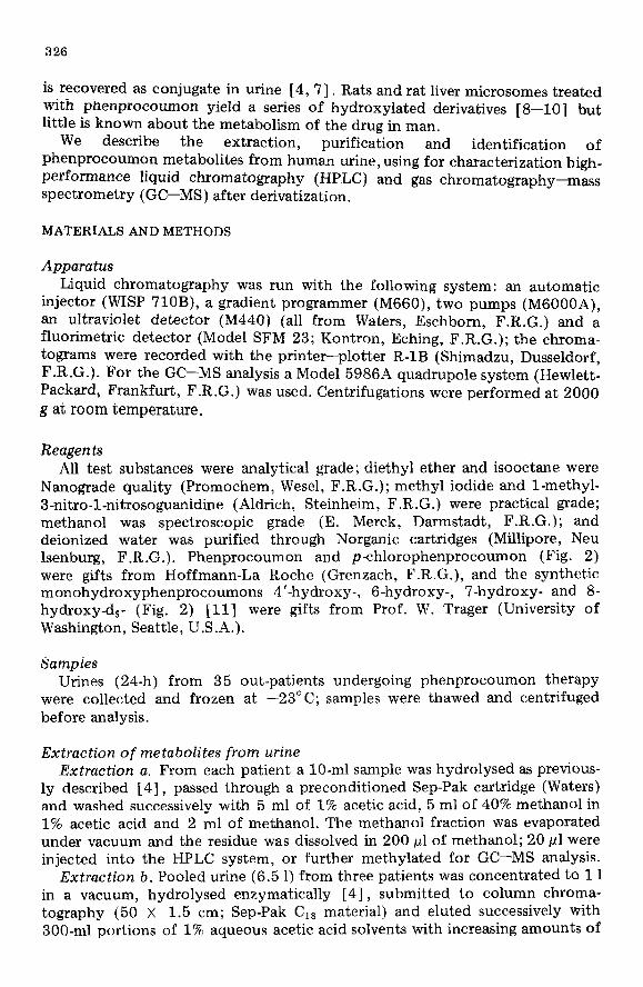
Nanograde quality (Promochem, Wesel, F.R.G.); methyl iodide and l-methyl- 3-nitro-l-nitrosoguanidine (Aldrich, Steinheim, F.R.G.) were practical grade; methanol was spectroscopic grade (E. Merck, Darmstadt, F.R.G.); and deionized water was purified through Norganic cartridges (Millipore, Neu Isenburg, F.R.G.). Phenprocoumon and p-chlorophenprocoumon (Fig. 2) were gifts from Hoffmann-La Roche (Grenzach, F.R.G.), and the synthetic monohydroxyphenprocoumons 4’-hydroxy-, 6-hydroxy-, 7-hydroxy- and 8- hydroxy-d5- (Fig. 2) [ll] were gifts from Prof. W. Trager (University of Washington, Seattle, U.S.A.).
Samples Urines (24-h) from 35 out-patients undergoing phenprocoumon therapy
were collected and frozen at -23°C; samples were thawed and centrifuged before analysis.
Extraction of metabolites from urine Extraction a. From each patient a lo-ml sample was hydrolysed as previous-
ly described [4], passed through a preconditioned Sep-Pak cartridge (Waters) and washed successively with 5 ml of 1% acetic acid, 5 ml of 40% methanol in 1% acetic acid and 2 ml of methanol. The methanol fraction was evaporated under vacuum and the residue was dissolved in 200 yl of methanol; 20 ~1 were injected into the HPLC system, or further methylated for GC-MS analysis.
Extraction b. Pooled urine (6.5 1) from three patients was concentrated to 1 1 in a vacuum, hydrolysed enzymatically [4], submitted to column chroma- tography (50 X 1.5 cm; Sep-Pak C is material) and eluted successively with 300-ml portions of 1% aqueous acetic acid solvents with increasing amounts of

327
1, Phenprocoumon H H H H
4. p- Chloro- phenprocoumon H H H h
5. 8- Hydroxy- I, OH H H H
6. 7- Hydroxy- 1 3 H OH H H
7 6- Hydroxy- 3, H H OH H
8 4’- Hydroxy- 5 1 H H H H
9, 8- Hydroxy - d5- (1 OH H H H
10. 4- Methyl - < , H H H H
11. 4- Methyl-7-methoxy- ?. H OMe H H
12. 4- Methyl -6-methoxy- (T H H OMe H
13. 4 - Methyl-4Lmethoxy- ., H H H H
Rl R2 R3 R4
14. 2 -Methyl- phenprocoumon H H H H
15. 2-Methyl-7-methoxy- 1% H OMe H H
16, 2 -Methyl -6-methoxy- 1. i-l H OMe H
17, 2- Methyl-4’-methoxy- *’ H H H H
R5
H
H
H
H
H
H
H
Me
Me
Me
Me
%
Me
Me
Me
Me
R6 R7
H H
Ii Cl
H H
H H
H H
l-l OH
D D
H H
H H
H H
H OMe
R6 R7
H H
H H
H H
H OMe
Fig. 2. Structures of phenprocoumon metabolites and derivatives. d, = pentadeutero (formula 9).
methanol (20, 40, 60, 80 and 100%). The 60% and 80% methanol fractions were evaporated, dissolved in methanol and centrifuged. The 60% methanol extract was used for the micropreparative isolation of 4’-hydroxyphen- procoumon (Fig. 2). 6-Hydroxyphenprocoumon, 7-hydroxyphenprocoumon and phenprocoumon (Fig. 2) were isolated after injecting the 80% methanol extract several times (5 X 20 ~1) into the HPLC system, peak collection and solvent evaporation.
Methylation
Synthetic reference compounds (Fig. 2; formulae 1, 4, 6, 7, 8, 9), urine extracts (extraction a or b), or fractions isolated by HPLC, were dissolved in methanol, dried under nitrogen in conical vials, dissolved in dry acetone (100 ~1) and alkylated with methyl iodide (50 ~1) in the presence of potassium carbonate (15 mg) and potassium iodide (2 mg) at 37°C overnight after capping with PTFE-lined discs. The solvent was evaporated, the residue was dissolved in

328
100 ~1 of water and 100 ~1 of 3 M hydrochloric acid, and extracted twice with 200 ~1 of chloroform. The chloroform extracts were evaporated; the residue
was dissolved in isooctane (lo-50 ~1) and injected into the GC-MS system. Diazomethane solutions obtained as described by Fales et al. [12] were also used for the methylations.
Chromatographic conditions High-performance liquid chromatography. Linear gradient ehtion (44-100%
solvent B in 50 min; flow-rate 2.0 ml/min) was used; solvent B was methanol and solvent A was a mixture of methanol-O.5% acetic acid (neutralized to pH 4.8 with ammonia) (100:900). Each run lasted 20 min (15 min analysis time and 5 min equilibration delay at initial conditions) and was performed at room temperature. A reversed-phase Cl8 column was employed for the separations (LiChrosorb RP-18 with lo-pm irregular particles; 250 X 4 mm I.D.; Hibar RT 250-4; Merck); a guard column (30 X 4 mm) was packed with Bondapak C,,-Corasil (35-50 pm spherical particles; Waters). After a series of analyses the column was washed with water and methanol. Peaks were detected fluorimetrically (excitation and emission wavelengths 310 and 390 nm, respectively; sensitivity setting low, 100%).
Gas chromatography-mass spectrometry. A gas chromatograph 5840A provided with a 183% capillary inlet system, splitless injector injector Iiner and a quartz capillary column (OV-1; 12.5 m X 0.2 mm) (all from Hewlett- Packard) were used for the separations. After the sample injection (1 pl), the oven temperature was maintained for 1 min at 80” C followed by a temperature gradient (30”C/min) to 250°C (injector temperature 250°C). The gas chromatograph was coupled to the mass spectrometer with an open split interface at 250°C. Purified helium was used as carrier gas, at a column pressure of 1.5 bars. The mass spectrometer was operated under electron-impact (EI) conditions both in the scanning mode for the mass spectra recording of the GC peaks and in the selected-ion monitoring (SIM) mode the specified ions. Typical parameters used for the EI settings were source temperature 2OO”C, emission 300 PA, electron energy 70 eV, multiplier 1400 V.
Thin-layer chromatography (TLC). HPTLC silica gel 60 FZs4 plates (E. Merck) were used with solvents 1 (dichloroethaneacetic acid, 96:4) and 2 (tert.-butanol-benzene-aqueous ammonia-water, 45:20:9:3) [Ill. The spots were visualized with UV light at 254 nm.
RESULTS AND DISCUSSION
The following parameters were determined for the patients, who received a weekly dose of 15.5 + 6.3 mg of phenprocoumon: plasma phenprocoumon concentration, 2.04 f 0.68 mg/l; phenprocoumon urinary excretion (as con- jugate), 0.44 * 0.22 mg per 24 h; thrombotest (9.6 ? 4.4%) [7]. The characterization of 7-hydroxyphenprocoumon as the main metabolite and of 4’-hydroxyphenprocoumon and 6-hydroxyphenprocoumon found in patients’ urine after enzymatic hydrolysis was accomplished mainly by HPLC and GC-MS.

329
High-performance liquid chromatography Fig. 3a shows the separation of a mixture of synthetic substances (4’-OH-,
6-OH-, &OH- and 7-OH-phenprocoumon, phenprocoumon and p-chloro- phenprocoumon) using radient elution HPLC and specific detection by fluorimetry; Fig. 3b shows the separation of an extract from blank urine spiked with 4’-OH-, 6-OH- and 7-OH-phenprocoumon and phenprocoumon. The fluorescence responses from the different metabolites at the same concentrations in relation to phenprocoumon were 0.40, 1.48 and 0.31, respectively. Fig. 3c shows the chromatogram from a patient’s urine after hydrolysis and work-up as described under Extraction a, in which 4’-OH-, 6-OI-I- and 7-OH-phen- procoumon and phenprocoumon could be detected. Each of the 35 patients’ urines was analysed in the same way with similar results. HPLC peaks from the urine extracts were qualitatively identified by their retention times compared to those of authentic substances, as well as by fractional collection of the peaks, methylation of each fraction as described above and analysis by GC-MS in the SIM mode as described below.
10
O-
:
Fig. 3. HPLC profiles of phenprocoumon and metabolites. (a) Mixture of pure compounds: 4’ = 4’-hydroxyphenprocoumon (retention time, tR = 4.24 min, capacity factor, k’ = 2.85),
6 = 6-hydroxyphenprocoumon ( tR = 5.07, k’ = 3.61), 8 = S-hydroxy-d,-phenprocoumon (TV = 6.05, k’ = 4.50), 7 = 7-hydroxyphenprocoumon (TV = 6.65, k’ = 5.05), P = phen- procoumon (TV = 7.45, 12’ = 5.77), C =pchloro-phenprocoumon (TV = 11.39, k’ = 9.33). (b) Extract from blank urine spiked with 1 pg/ml each of 4’-hydroxy-, 6-hydroxy- and ‘7- hydroxyphenprocoumon and phenprocoumon. (c) Extract from patient’s urine.
Although the analytical recoveries of phenprocoumon and metabolites in spiked urine using extraction a were high (90-95%) and reproducible (5 5%) and the fluorimetric detection was more specific than the ultraviolet previously used [4], accurate quantitative results could not be obtained with this method for patients’ urine due to interfering endogenous compounds and/or other drugs.
HPLC was also used for the micropreparative isolation of phenprocoumon and metabolites (extraction b) for further characterization by mass spectrometry (see below).
Gas chromatography-mass spectrome try Prior to the analysis of phenprocoumon and the hydroxylated metabolites

330
by GC-MS it was necessary to derivatize them by methylation; mixtures of the 4-O-methyl isomers (Fig. 2, formulae 10-13) with small amounts of the 2-0- methyl isomers (Fig. 2, formulae 14-17) were produced (due to the tautomerism in the 4-hydroxycoumarin series) [13,14] and could be separated with capillary GC columns. Methyl iodide alkylation [15] instead of the earlier diazomethane treatment [12] was used preferentially as higher yields of methylated products and less 2-O-methyl isomers were produced. The individual isomers could be characterized after methylating each synthetic substance (Fig. 2, formulae 1,6-g), submitting the reaction mixtures to HPLC and identifying each isolated peak by UV spectroscopy [13,14] and GC-MS. It was found that the 4-O-methyl isomers have longer GC retention times than the corresponding 2-O-methyl isomers.
By processing a large amount of patients’ urine using extraction b, purified samples were obtained which contained substances identified later by GC-MS as phenprocoumon, 7-OH-, 6-OH- and 4’-OH-phenprocoumon.
loo-
I a, (91
265
a)
C)
/, I / I, 1 I ,I 1
m/e 100 200 300 400
I 324 203
,\ Ai I I/ , I 1 I I I I I
200 3.x 400
Fig. 4. Mass spectra of phenprocoumon and metabolites isolated from patient’s urine (see under Extraction b) after methylation and CC-MS. (a) 4-0-methylphenprocoumon, (b) 4,7-dimethyl-7-hydroxyphenprocoumon, (c) 4,6-dimethyl-6-hydroxyphenprocoumon, (d) 4,4’-dimethyl-4’-hydroxyphenprocoumon.

331
Characteristic mass spectra were obtained for the 4-0-methylated
phenprocoumon (Fig. 4a) and the metabolites (Fig. 4b-d). The molecular ion of me thylated phenprocoumon (M + , m/e 294) was displaced to m/e 324 for
the methylated metabolites, indicating an additional oxygen in the latter. Fig. 4a-c shows an intense ion at m/e 91 characteristic for the benzylic side-chain, while Fig. 4d shows an m/e 121 peak indicating a methoxylated side-chain. This was corroborated by the appearance of the ion m+ - 91 for spectra a-c and M’ - 121 for spectrum d (m/e 203, 233, 233 and 203, respectively). More- over, all spectra show a characteristic and intense M+ - 29 peak (m/e 265 for phenprocoumon and 295 for the metabolites) from the loss of an ethyl group from the phenylpropyl side-chain, which was useful for GC-MS in the SIM mode. The retention times in GC-MS as well as their mass spectra (Fig. 4) were identical with those of synthetic phenprocoumon, and 7-OH-, 6-OH- and
4’-OH-phenprocoumon methylated by the same procedure. A detailed account of the mass spectrometric fragmentation of phenprocoumon and derivatives will be published elsewhere [16], The structures of the ions could be corroborated by the use of several substituted and deuterated compounds.
The main metabolite, 7-OH-phenprocoumon, as well as phenprocoumon, were also identified by the measurement of ultraviolet [ 111 and fluorescence spectra in acid and alkaline solution which are characteristic for the different isomers, and by TLC in several solvents, which were identical with those of authentic material [ 111.
Fig. 5. GC-MS analysis in the SIM mode of phenprocoumon and metabolites after methyla- tion. (a) Urine extract spiked with 1 @g/ml each of phenprocoumon (P), 6-hydroxyphen- procoumon (6), 4’-hydroxyphenprocoumon (4’) and 7-hydroxyphenprocoumon (7). (b) Extract from patient’s urine.

332
The same urine extracts from 35 patients previously analysed by HPLC were methylated and subjected to GC-MS analysis in the SIM mode. Fig. 5a shows a GC-MS chromatogram from a mixture of synthetic 6-OH-, 4’-OH- and 7-OH- phenprocoumon and phenprocoumon after methylation. The molecular ions at m/e 294 and 324 for the methylated phenprocoumon and metabolites, as well as the m/e 295 (M’ - 29) peak, characteristic of the methylated metabolites, were chosen for ion monitoring. Fig. 5b shows the chromatogram from a patient’s urine extract (the same shown in Fig. 3c) after methylation; the methylated 6-OH-, 4’-OH- and 7-OH-phenprocoumon and phenprocoumon were identified by their retention times and intensity ratios for the specified ions. All 35 samples showed the same qualitative pattern. &OH-phen- procoumon (whose 4-O-methyl derivative has a shorter GC retention time than the other metabolites) could not be detected. Detection limits were in the order of 10 ng/ml.
An evaluation of the relative amounts of metabolites was obtained by adding p-chlorophenprocoumon as internal standard to the extracts, methylating and analysing the products by GC-MS in the SIM mode. The m/e 299 trace (M+ - 29 for p-chlorophenprocoumon) was used for the normalization and the m/e 295 for monitoring the metabolites. This semi-quantitative method showed for the ratios metabolites/phenprocoumon median values [17] of 0.9, 0.2 and 0.1 (Table I) for the 7-OH, 4’-OH and 6-OH com- pounds, respectively, which has also been observed independently in volunteers [ 181. Considerable interindividual variability was seen for the patients, and median means were calculated to take into account the extreme values in the range [17] (Table I).
Analysis of urine before and after enzymatic hydrolysis showed that only trace amounts of unconjugated substances were eliminated. No methoxylated derivatives from phenprocoumon, 6-OH-, 4’-OH- or 7-OH-phenprocoumon could be detected as direct metabolites in patients’ urine by HPLC or GC-MS.
TABLE I
RATIO OF URINARY EXCRETED METABOLITES TO PHENPROCOUMON
Metabolite Median + S.E.M. Range
7-Hydroxyphenprocoumon 0.92 f 0.06 0.20-1.70 4’-Hydroxyphenprocoumon 0.23 f 0.03 0.02-1.90 6-Hydroxyphenprocoumon 0.10 + 0.03 0.02-2.00
The elimination of phenprocoumon, metabolites and warfarin In vivo metabolic studies with rats using radioactive phenprocoumon have
been published previously by Haddock et al. [8], who showed that the drug and hydroxylated metabolites were excreted in urine and faeces (20% and 59%, respectively, from the total radioactivity after twelve days). The elimination pattern was found to be 6-OH-phenprocoumon > 4’-OH-phenprocoumon > phenprocoumon > 7-OH-phenprocoumon > S-OH-phenprocoumon. Phen- procoumon is metabolized in the rat by liver microsomes and this drug has been used, as well as warfarin, as a probe to categorize the effect of inducing agents on microsomal hydroxylases [ 8--lo].
333
Earlier studies on the excretion of phenprocoumon in man [4, ‘71 have shown that only a fraction of the applied dose (20%) is eliminated in urine as conjugate; as we have shown here, 7-OH-, 6-OH- and 4’-OH-phenprocoumon are produced and also eliminated as conjugates. The total amount of phenprocoumon plus metabolites excreted in urine accounts for somewhat more than the half of the ingested drug; or unknown metabolites are produced which could not be detected, or a considerable amount is excreted in the faeces as has been previously described for rats [18]. Biliary excretion of phenprocoumon and metabolites has been reported for rats and is presumed in humans as studied in the interaction with cholestyramine [19] . Phenprocoumon and the metabolites are excreted in urine as conjugates, and enzymatic hydrolysis prior to extraction and analysis has to be done to measure the total amount eliminated.
Warfarin and metabolites have been characterized previously in human urine (warfarin, 7-hydroxy-warfarin, 6-hydroxy-warfarin, two diastereoisomeric warfarin alcohols and benzylic hydroxywarfarin) [20-241 and plasma (warfarin, warfarin alcohols, 7-hydroxywarfarin and 6-hydroxywarfarin [23, 25, 261. The acetonyl side-chain (instead of ethyl in phenprocoumon) is the main cause for the different physicochemical properties of these compounds [27-291, which is also reflected in differences in the pharmacokinetics and metabolism of both compounds.
Many drugs interact with warfarin, phenprocoumon and other coumarin anticoagulants [30-321. However, drug interactions with warfarin or phen- procoumon are sometimes quite different, as in the case of cimetidine and sulfinpyrazone [5, 6, 33-361 and are of clinical relevance.
Further work is being undertaken to develop a specific and sensitive assay for the quantitation of phenprocoumon metabolites for further clinicopharma- cological investigations.
ACKNOWLEDGEMENTS
We thank Prof. Dr. W. Trager (University of Washington, Seattle, WA, U.S.A.) for the synthetic phenprocoumon metabolites and for discussions, and Miss G. Fischer for technical assistance.
REFERENCES
1 R.A. O’Reilly, in A.G. Gilman, L.S. Goodman and A. Gilman (Editors), Goodman and Gilman’s The Pharmacological Basis of Therapeutics, Macmillan, New York, 6th ed., 1980, p. 1347.
2 A.S. Gallus, Drugs, 26 (1983) 543. 3 American Medical Association (Editors), AMA Drug Evaluations, W.B. Saunders,
Philadelphia, PA, 5th. ed., 1983, p. 811. 4 J.X. de Vries, J. Harenberg, E. Walter, R. Zimmermann and M. Simon, J. Chromatogr.,
231 (1982) 83. 5 J. Harenberg, R. Zimmermann, Ch. Staiger, J.X. de Vries, E. Walter and E. Weber, Eur.
J. Clin. Pharmacol., 23 (1982) 365. 6 J. Harenberg, Ch. Staiger, J.X. de Vries, E. Walter, E. Weber and R. Zimmermann, Brit.
J. Clin. Pharmacol., 14 (1982) 292. 7 J.X. de Vries, J. Harenberg, R. Zimmermann and M. Simon, J. Exp. Clin. Hematol.,
49 (1984) 138.

8 9
10 11 12 13 14 15 16 17
18 19
20 21 22 23
24
25 26
27
28 29 30 31 32 33
34 35
334
R.E. Haddock, W.F. Trager and L.R. Pohl, J. Med. Chem., 18 (1975) 519. W.R. Porter, C. Wheeler and W.F. Trager, Biochem. Pharmacol., 30 (1981) 3099. C. Wheeler, W.F. Trager and W.R. Porter, Biochem. Pharmacol., 30 (1981) 1785. L.R. Pohl, R. Haddock, W.A. Garland and W.F. Trager, J. Med. Chem., 18 (1975) 513. H.M. FaIes, T.M. Jaouni and J.F. Babashak, Anal. Chem., 43 (1973) 2302. F. Arndt, L. Loewe, R. Un and E. Ayca, Chem. Ber., 84 (1951) 319. J. Cieslak, S. Lewak and I. Chmielewska, Roczniki Chem., 33 (1959) 349. A, Hulshoff and A.D. FSrch, J. Chromatogr., 220 (1981) 275. J.X. de Vries and W.F. Trager, unpublished results. L. Sachs, Angewandte Statistik: Statistische Methoden und ihre Anwendungen, Springer, Berlin, 5th ed., 1978, p. 76. W.F. Trager, personal communication. T. Meinertz, H.J. Gilfrich, U. Groth, H.G. Jonen and E. Jfhnchen, Clin. Pharmacol. Ther., 21 (1977) 731. R.J. Lewis and W.F. Trager, J. Clin. Invest., 49 (1970) 907. W.F. Trager, J.R. Lewis and W.A. Garland, J. Med. Chem., 13 (1970) 1196. K.K. Chan, R.J. Lewis and W.F. Trager, J. Med. Chem., 15 (1972) 1265, R.J. Lewis, W.F. Trager, A. Breckenridge, M. Orme, M. Roland and W. Schary, J. Clin. Invest., 53 (1974) 1607. L.R. Pohl, S.D. Nelson, W.A. Garland and W.F. Trager, Biomed. Mass Spectrom., 2 (1975) 23. S.J. Lee, L.R. Field, W.N. Howaid and W.F. Trager, Anal. Chem., 53 (1981) 467. C. Banfield, R.O.‘Reiily, E. Chan and M. Rowland, Brit. J. Clin. Pharmacol., 16 (1983) 669. E.J. Valente, E.C. Lingafelter, W.R. Porter and W.F. Trager, J. Med. Chem., 20 (1977) 1489. E.J. Vaiente and W.F. Trager, J. Med. Chem., 21 (1978) 141. E.J. VaIente, W.R. Porter and W-F. Trager, J. Med. Chem., 21 (1978) 231. J. Koch Weser and E.M. Sellers, N. Engl. J. Med., 285 (1971) 487,547. SM. MacLeod and E.M. Sellers, Drugs, 11 (1976) 461. M.J. Serlin and A.M. Breckenridge, Drugs, 25 (1983) 610. M.J. Serlin, R.G. Sibeon, S. Mossman, A.M. Breckenridge, J.R.B. Williams, J.L. Atwood and J.M.T. Willoughby, Lancet, ii (1979) 317. R.A.O’Reilly, Circulation, 65 (1982) 202. J.O. Miners, T.Foenander, W. Wanwimolruk, AS. Gallus and D.J. Birkett, Eur. J. Ciin. Pharmacol., 22 (1982) 327.
36 R.A. O’Reilly, Arch, Int. Med., 143 (1982) 1634.