identification of developmental endothelial locus...
TRANSCRIPT

Biology of Human Tumors
Identification of Developmental EndothelialLocus-1 on Circulating Extracellular Vesicles as aNovel Biomarker for Early Breast CancerDetectionPyong-Gon Moon1, Jeong-Eun Lee1, Young-Eun Cho1, Soo Jung Lee2, Jin Hyang Jung3,Yee Soo Chae2, Han-Ik Bae4, Young-Bum Kim5, In-San Kim6, Ho Yong Park3, andMoon-Chang Baek1
Abstract
Purpose: Currently, there are no molecular biomarkers for theearly detection of breast cancer. This study focused on identifyingsurface proteins found on circulating extracellular vesicles (EVs)for detecting early-stage breast cancer.
Experimental Design: Circulating EVs, isolated from the plas-ma of 10 patients with breast cancer (stages I and II) and 5 healthycontrols, were analyzed using LC-MS/MS. Developmental endo-thelial locus-1 protein (Del-1) was selected as a candidate bio-marker. Twodifferent ELISAswereused tomeasureDel-1 inplasmasamples fromhealthy controls (n¼ 81), patientswithbreast cancer(n ¼ 269), breast cancer patients after surgical resection (n ¼ 50),patients with benign breast tumors (n ¼ 64), and patients withnoncancerous diseases (n ¼ 98) in two cohorts.
Results: Plasma Del-1 levels were significantly higher (P <0.0001) in patients with breast cancer than in all controls and
returned to almost normal after tumor removal. The diagnosticaccuracy of Del-1 was AUC, 0.961 [95% confidence interval (CI),0.924–0.983], sensitivity of 94.70%, and specificity of 86.36% intest cohort and 0.968 (0.933–0.988), 92.31%, and 86.62% invalidation cohort for early-stage breast cancer by one type ofELISA. Furthermore, Del-1 maintained diagnostic accuracy forpatients with early-stage breast cancer using the other type ofELISA [0.946 (0.905–0.972), 90.90%, and 77.14% in the testcohort; 0.943 (0.900–0.971), 89.23%, and 80.99% in the vali-dation cohort].
Conclusions:Del-1 on circulating EVs is a promising markerto improve identification of patients with early-stage breastcancer and distinguish breast cancer from benign breasttumors and noncancerous diseases. Clin Cancer Res; 22(7);1757–66. �2015 AACR.
IntroductionBreast cancer, the most common type of cancer in women,
causes up to 500,000 deaths yearly worldwide (1–3). Althoughcancer diagnosis and treatment have advanced in recent decades,only some molecular biomarkers, such as autoantibodies, withpoor values, for the early detection of breast cancer have been
reported (4–6). Identifying a biomarker to detect breast cancer atan early stage will enable the use of less-aggressive treatments andimprove clinical outcomes (7). CA 15-3 is the most widely usedserum marker in patients with metastatic breast cancer but has asensitivity of only 60% to 70% (5, 8, 9). Recently, variousmethods have been reported to detect circulating tumor cells,which are a promising biomarker (10, 11). The FDA has approvedtheCellSearch systemwith a sensitivity of about 65% for detectingcirculating tumor cells (�1 cell per 7.5 mL blood) in metastaticbreast cancer patients (10, 12). Furthermore, measuring circulat-ing tumor DNA showed superior sensitivity to measuring othercirculating biomarkers to monitor metastatic breast cancer andprovided the earliest index of treatment response (13, 14).
It is traditionally accepted that metastasis occurs at a late stageof cancer progression (15). However, metastatic dissemination ofcancer cells canoccur in preinvasive stages of tumor progression inmouse models of breast cancer and in human patients (16–18).This suggests that systemic spread is an early step in breast cancerand implies that unknown systemic factors secreted from theprimary tumor may prepare a premetastatic niche and promoteearly cancerous colonies.
Extracellular vesicles (EVs) are membranous vesicles secretedby various cells. EVs have been classified into several subcate-gories, including exosomes (50–100 nm in diameter) and micro-vesicles (100-1,000nm indiameter; refs. 19, 20). Cancer cells, andneighboring cells in the tumor microenvironment, secrete EVs
1DepartmentofMolecularMedicine,Cell andMatrixResearch Institute,School of Medicine, Kyungpook National University, Daegu, Republicof Korea. 2Department ofOncology/Hematology, KyungpookNation-al University Hospital, Daegu, Republic of Korea. 3Department ofBreast and Thyroid Surgery, Kyungpook National University Hospital,Daegu, Republic of Korea. 4Department of Pathology, KyungpookNational University Hospital, Daegu, Republic of Korea. 5Division ofEndocrinology,Diabetes andMetabolism, Beth Israel DeaconessMed-ical Center and Harvard Medical School, Boston, Massachusetts.6Department of Biochemistry and Cell Biology, School of Medicine,Kyungpook National University, Daegu, Republic of Korea.
Note: Supplementary data for this article are available at Clinical CancerResearch Online (http://clincancerres.aacrjournals.org/).
P.-G. Moon and J.-E. Lee contributed equally to this article.
Corresponding Author:Moon-Chang Baek, Kyungpook National University, 101Donging-Dong, Jung-Gu, Daegu 700-422, Republic of Korea. Phone: 82-53-420-4948; Fax: 82-53-426-4944; E-mail: [email protected]
doi: 10.1158/1078-0432.CCR-15-0654
�2015 American Association for Cancer Research.
ClinicalCancerResearch
www.aacrjournals.org 1757
Research. on September 28, 2018. © 2016 American Association for Cancerclincancerres.aacrjournals.org Downloaded from
Published OnlineFirst November 24, 2015; DOI: 10.1158/1078-0432.CCR-15-0654

that play important roles in pro-metastatic signaling, angiogen-esis, and immune suppression in an autocrine or paracrinemanner (21–24). Cancer cell–derived EVsmay have the potentialto be used as early biomarkers for various cancers because themembrane vesicles are secreted continuously into body fluids,including blood, from an early stage of the disease (25, 26).
Based on this background information, we hypothesized thatEV proteins can be used as early diagnostic biomarkers for breastcancer. We employed a proteomic approach to identify Del-1 asdisease-specific proteins on circulating EVs isolated from theplasma of patients with early-stage breast cancer. We then con-firmed that the plasma level of Del-1, measured by two differentELISAs, correlated with the presence of breast cancer.
Patients and MethodsStudy design and participants
The study design used to identify biomarkers of early-stagebreast cancer is illustrated in Fig. 1 and Supplementary Fig. S1.Plasma samples for test set were obtained from the KyungpookNational University Hospital, Daegu, Korea. An independentvalidation set was obtained from the Chonnam National Uni-versity Hwasun Hospital, Hwasun, Korea. The histologic cell typeand stage of breast cancer of the patients studied in test andvalidation sets are provided in Table 1. Plasmawas obtained fromfreshly drawn blood using plasma separate tube. Aliquots of eachplasma were deep-frozen at storage using liquid nitrogen. Allplasma samples were transported to laboratory on dry ice within24 hours. For analysis, plasma samples were thawed at 4�C.Thawed plasma samples were stored at 4�C for triplicate analysiswithin 2 weeks. Plasma samples were shaken at 1,500 rpm, 5minutes right before ELISA. These processes were same for casesand controls.
To identify early biomarker candidates for breast cancer, circu-lating EVs from 5 patients with stage I breast cancer, 5 with stage IIbreast cancer, and 5 healthy controls (discovery set, group 1) wereisolated, characterized (Supplementary Fig. S2), and analyzedusing LC-MS/MS. Del-1 was considered potential biomarker forbreast cancer. Plasmasamples from169patientswithbreast cancerand 35 healthy controls (test set, group 2) were used to testdifferential expression of diagnostic marker candidates using twodifferent ELISA methods. We mixed cases and age-matched con-trols in every plate, and each plate contained standard samples as
described previously to reduce the variation between plates.Group 2 contained 26 patients with benign breast tumors, 50breast cancer patients after surgical resection, and 40 patientswith noncancerous diseases (thyroiditis, gastritis, hepatitis B,and rheumatoid arthritis). The validation study was conductedwith a rigorous predefined statistical analysis plan with prespe-cified outcome. All validation cohorts were analyzed in a blindedfashion. Unblinding of clinical parameters and correspondingexperimental data was performed only after finishing all experi-ments. To validate biomarker candidates for breast cancer, Del-1levels were measured using ELISA in 100 patients with breastcancer, 46 healthy controls, 38 patients with benign breasttumors, and 58 patients with noncancerous diseases (validationset, group 3). The TNM Classification of Malignant Tumors(TNM) classification from the 7th edition of the American JointCommittee on Cancer (AJCC) was used to define early-stagebreast cancer (0, I, and II; refs. 27, 28). The Reporting Recom-mendations for Tumor Marker Prognostic Studies (REMARK)criteria, recommended by McShane and colleagues (29), were
Identification of biomarker candidates
Establishment of ELISA methods
ELISA Method 1
Diagnostic performances
Group 2 : Test set (n = 320)
Healthy volunteers (n = 35) Healthy volunteers (n = 46)
Breast cancer (n = 169) Breast cancer (n = 100)
Benign breast tumor (n = 26) Benign breast tumor (n = 38)
After surgery (n = 50)
Noncancerous diseases (n = 40)
Noncancerous diseases (n = 58)
Group 3 : Validation set (n = 242)
ELISA Method 2
Anti-Del-1Ab
Anti-CD63 Ab Anti-Del-1Ab
Anti-Del-1Ab
Group 1:Discovery set (n = 15)*HC (n = 5), stage I BC (n = 5), stage II TBC (n = 5)
Circulating EVs isolated from plasma of patients
LC/MS-MS
Del-1
Figure 1.Overview of the study design. Circulating EVs isolated fromplasma of healthycontrol patients (n¼ 5) and patients with early-stage breast cancer (BC; n¼10) were analyzed using LC-MS/MS to identify early biomarker candidates.Biomarker candidates amongproteins thatwere upregulated in breast cancerpatients compared with controls were selected based on three filteringcriteria (Fig. 2A). Del-1 was evaluated as a novel biomarker for the earlydetection of breast cancer using plasma (1 mL) from test cohort (n¼ 320) andvalidation cohort (n ¼ 242) by two types of ELISA with different primarycapture antibodies. � , Discovery set is included in the test set.
Translational Relevance
Breast cancer is the leading cause of death worldwide and isthe most common type of cancer in women. Molecular bio-markers for early detection of breast cancer have not beenreported, although survival rates for breast cancer increasewithearly diagnosis. Using two different ELISAs, the diagnosticperformance of Del-1 for breast cancer detection was 84% to99% for sensitivity and 74% to 86% for specificity. The level ofDel-1 on circulating extracellular vesicles (EVs) was signifi-cantly elevated at the earliest stage of breast cancer anddropped after surgical resection. Theminimally invasivemeth-od with high sensitivity and specificity based on themolecularbiomarker, Del-1–positive circulating EVs, is promising for theearly diagnosis of breast cancer using only one drop of blood.
Moon et al.
Clin Cancer Res; 22(7) April 1, 2016 Clinical Cancer Research1758
Research. on September 28, 2018. © 2016 American Association for Cancerclincancerres.aacrjournals.org Downloaded from
Published OnlineFirst November 24, 2015; DOI: 10.1158/1078-0432.CCR-15-0654

followed throughout this study. All individuals providedinformed consent for blood donation according to a protocolapproved by the Institutional Review Board of the KyungpookNational University Hospital.
Proteomic analysisEVs were purified by differential centrifugation as described
previously (30). EVs fromplasmaof 10patientswith breast cancer(stages I and II) and 5 control (healthy volunteer) patients wereanalyzed by nano-UPLC and tandem mass spectrometry (Q-TofPremier; Waters). Data processing, searching, and analyses wereperformed using Mascot server 2.2 (Matrix Science). Label-freeprotein quantification was performed using an IDEAL-Q software(31). The analysis was performed three times. EV proteins fromMDA-MB-231 cells were analyzed by the same methods. Theproteins were considered to be significantly upregulated with agreater than 2-fold difference versus healthy control and set withan adjusted P < 0.05.
ELISAFor quantification of Del-1 protein on EVs in plasma using
an ELISA method 1, 96-well plates were coated overnight withpolyclonal anti-CD63 (ab68418; Abcam)antibody at 100ng/wellin 0.2 mol/L sodium phosphate buffer (pH 6.5). The plates
were blocked for 1 hour at 37�C with 200 mL of PBS containing1% BSA and washed three times with PBS containing 0.05%Tween 20 (PBS-T). Plasma (10 mL) was diluted with blockingbuffer (90 mL). The diluted plasma (10 mL) was added to theplates in triplicate and incubated for 1 hour at 37�C. Followingthree washes with PBS-T, the plates were reacted with mono-clonal anti–Del-1 (ab88667; Abcam) antibody, preincubatedwith peroxidase-conjugated anti-mouse IgG antibody for 30min-utes, and developed with 3,30,5,50-tetramethylbenzidine contain-ing hydrogen peroxide. The reaction was stopped with 1 mol/Lphosphoric acid, and optical density values were measured at450 nm on an automated iMark plate reader (BioRad).
For quantification of Del-1 or CD63 proteins in plasma usingan ELISA Method 2, the same steps were performed as withMethod 1 except for coating the plates with polyclonal anti–Del-1 (ab151308; Abcam) or anti-CD63 (ab68418; Abcam)antibodies to capture each protein. Recombinant Del-1 (6046-ED-050; R&D Systems) or CD63 proteins (H00000967-G01;Abnova) were used to generate standard curves. The levels ofCD63 were measured using a monoclonal anti-CD63 (ab8219;Abcam) antibody. The CA 15-3 enzyme immunoassay was per-formed using an enzyme immunoassay kit (MYM Laboratory &Medical Supply) following the manufacturer's instructions. Eachdata point is the average of triplicate measurements.
Table 1. Characteristics of study population
Total Test ValidationCharacteristic N (%) Mean SD N (%) Mean SD N (%) Mean SD Pa
Number of study population 562 320 (56.9) 242 (43.1)Age, years 50 10.4 52.3 10.3 0.8090b
Breast cancer 269 169 (62.8) 100 (37.2)Histologic gradec 0.0148d
1 49 (18.2) 30 (17.9) 19 (19.0)2 136 (50.6) 96 (56.8) 40 (40.0)3 84 (31.2) 43 (25.4) 41 (41.0)
Stagee <0.0001d
0 42 (15.6) 37 (21.9) 5 (5.0)I 66 (24.5) 48 (28.4) 18 (18.0)II 89 (33.1) 47 (27.8) 42 (42.0)III 60 (22.3) 25 (14.8) 35 (35.0)IV 12 (4.5) 12 (7.1)
Estrogen receptor (ER)f 0.2045d
Negative 74 (27.5) 42 (24.9) 32 (32.0)Positive 195 (72.5) 127 (75.1) 68 (68.0)
Progesterone receptor (PgR)f 0.7049d
Negative 93 (34.6) 57 (33.7) 36 (36.0)Positive 176 (65.4) 112 (66.3) 64 (64.0)
HER2f 0.4544d
Positiveg 58 (21.6) 34 (20.1) 24 (24.0)Negative 211 (78.4) 135 (79.9) 76 (76.0)
Healthy control 81 35 (43.2) 46 (56.8)Noncancerous diseases 98 40 58 0.4635d
Thyroiditis 16 (16.3) 8 (20) 8 (13.8)Gastritis 14 (14.3) 7 (17.5) 7 (12.1)Hepatitis B 12 (12.2) 6 (15) 6 (10.3)Rheumatoid arthritis 56 (57.1) 19 (47.5) 37 (63.8)
After surgery 50 50Benign breast tumor 64 26 (40.6) 38 (59.4)aP value comparing test and validation groups.b t test.cmodified Scarff–Bloom–Richardson grading system.d x2 test.eAJCC staging system.faccording to immunohistochemical (IHC) staining of ER, PgR, and HER2.gIHC (3þ) or FISH (þ).
Biomarker for Early Breast Cancer Detection
www.aacrjournals.org Clin Cancer Res; 22(7) April 1, 2016 1759
Research. on September 28, 2018. © 2016 American Association for Cancerclincancerres.aacrjournals.org Downloaded from
Published OnlineFirst November 24, 2015; DOI: 10.1158/1078-0432.CCR-15-0654
Statistical analysesDescriptive statistics summarized clinical factors; x2 and t tests
were used to compare the test and validation groups. To estimatethe reproducibility and precision of the ELISA results, intra- andinterassay coefficients of variation (CV)were calculated, where CV(%) ¼ (SD/mean) � 100. For plasma Del-1, CD63, and CA 15-3levels, relationships were analyzed using the Kolmogorov–Smir-nov test to assess differences between individual variables fromtwo groups. Assessment of the correlation between tumor size andlevels of Del-1 or CD63 was performed using a nonparametricSpearman correlation. The diagnostic potential of Del-1 wasdetermined by calculating the receiver operating characteristic(ROC) curve plotted to evaluate the sensitivity and specificity ofthe biomarker measurements in predicting breast cancer. Dis-criminative efficacy of Del-1 and optimum diagnostic cutoff werecalculated by the area under the ROC curve (AUC) in test set.Using Del-1 threshold values in test set, we calculated diagnosticperformance of Del-1 for breast cancer detection in the validationcohort. All validation cohorts were analyzed in a blinded fashion.The Kruskal–Wallis one-way analysis was used to evaluate a
significant change of Del-1 in the presence of three types of breastcancer–related receptors. P values less than 0.05 indicated statis-tical significance. Analyses were conducted using MedCalc (Med-Calc Software) and Prism (GraphPad Software, Inc.). Data areexpressed as mean � SEs.
ResultsIdentification of Del-1 protein on circulating EVs in early-stagebreast cancer patients versus control
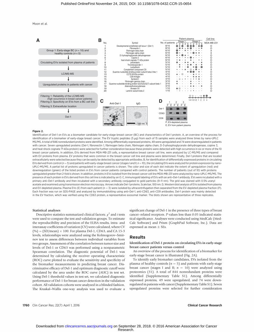
An overview of the process for identification of a biomarker forearly-stage breast cancer is illustrated (Fig. 2A).
To identify early biomarker candidates, EVs isolated from theplasma of healthy controls (n ¼ 5) and patients with early-stagebreast cancer (stages I and II; n ¼ 10) were analyzed usingproteomics (31). A total of 844 nonredundant proteins wereidentified (Supplementary Table S1). Among differentiallyexpressed proteins, 49 were upregulated, and 74 were down-regulated inpatientswith cancer (Supplementary Table S1). Sevenupregulated proteins were selected for further consideration
Group 1: Early-stage BC (n = 10) andhealthy controls (n = 5)
A B
C D
Circulating EVs isolated from plasma of patients
LC/MS-MS
No.123456789
1011121314151617181920
MDA-MB-231OOXXOOOOOOXOOOOXXOOO
Cell linePatient plasmaStage 1
1 2 3 4 5 1 2 3 4 5Stage 2
No. of patients8/108/106/106/106/106/106/105/105/105/105/104/104/104/104/103/103/103/101/101/10
SymbolDevelopmental endothelial cell locus-1 (Del-1)
Fibronectin 1Fibrinogen beta chainFibrinogen alpha chain
D-3-phosphoglycerate dehydrogenaseCopine 3
heat shock cognate 71 kDa proteinperoxidasin
Thrombospondin 1Lactadherin
Complement component C9Guanine nucleotide-binding protein subunit beta-2-like 1
CCT8 59 kDa proteinCD9 Antigen
Syntenin1Fibrinogen gamma chain
Guanine nucleotide-binding protein G(I)/G(S)/G(T) subunit beta 1Adenosylhomocysteinase
Annexin A1Laminin subunit beta 2
Upregulated proteins in patients with cancer
Filtering 1: Reliability of the LC/MS-MS
Filtering 2: Specificity on EVs from a BC cell line
Filtering 3: Extracellular location
Del-1
Anti-Del-1
100 nm 100 nm
Del-1
1P
P; EV-depleted plasmaE; EV from patient plasma
E P E P E2 3
1/4 1/3 1/2 1 2 3 4
CD63
CD9
IgG
(high occurrence in breast cancer patients)
Figure 2.Identification of Del-1 on EVs as a biomarker candidate for early-stage breast cancer (BC) and characteristics of Del-1 protein. A, an overview of the process foridentification of a biomarker of early-stage breast cancer. The EV tryptic peptides (5 mg) from each of 15 samples were analyzed three times by nano-UPLCMS/MS. A total of 846 nonredundant proteinswere identified. Among differentially expressed proteins, 49were upregulated and 74were downregulated in patientswith cancer. Seven upregulated proteins (Del-1, fibronectin 1, fibrinogen beta chain, fibrinogen alpha chain, D-3-phosphoglycerate dehydrogenase, copine 3,and heat shock cognate 71-kDa protein) were selected for further consideration because these proteins were detected with high occurrence in six or more of the 10breast cancer patients. In addition, EVs derived from MDA-MB-231 cells, a representative breast cancer cell line, were analyzed by LC-MS/MS and comparedwith EV proteins from plasma. EV proteins that were common in the breast cancer cell line and plasma were determined. Finally, Del-1 proteins that are locatedextracellularly were selected because they can be easily be detected by appropriate antibodies. B, for identification of differentially expressed proteins in circulatingEVsderived fromcontrol (n¼5) andpatientswith early-stage breast cancer (stages I and II;n¼ 10), the circulating EVswere analyzed for protein expression bynano-UPLC-MS/MS. A partial list of proteins upregulated in cancer patients is shown. The color and size of each dot indicate the extent of upregulation (red) anddownregulation (green) of the listed protein in EVs from cancer patients compared with control patients. The number of patients (out of 10) with proteinsupregulated greater than 2-fold is shown. In addition, proteins in EVs isolated from the breast cancer cell lineMDA-MB-231 were analyzed by nano-UPLC-MS/MS. Thepresence of each protein in EVs derived from this cell line is indicated by anO. C, Immunogold labeling of EVswith an anti–Del-1 antibody. EVswere incubated with aprimary anti–Del-1 antibody and then incubated with a secondary antibody conjugated to gold particles (9–11 nm). The grid was stained with 0.5% uranylacetate andexaminedusing transmission electronmicroscopy.Arrows indicateDel-1 proteins. Scalebar, 100nm.D,Western blot analysis of EVs isolated fromplasmaand EV-depleted plasma. Plasma EVs (E) from each patient (n¼ 3) were isolated by ultracentrifugation then separated from the EV-depleted plasma fraction (P).Each fraction was run on SDS-PAGE and analyzed by immunoblotting using anti–Del-1, anti-CD63, anti-CD9 antibodies. Del-1 protein was mainly detectedin the EV fraction, which was verified using the CD63 protein, a representative exosomal marker. The blots shown are representative of three replicates.
Moon et al.
Clin Cancer Res; 22(7) April 1, 2016 Clinical Cancer Research1760
Research. on September 28, 2018. © 2016 American Association for Cancerclincancerres.aacrjournals.org Downloaded from
Published OnlineFirst November 24, 2015; DOI: 10.1158/1078-0432.CCR-15-0654
because these proteinsweredetectedwithhighoccurrence of six ormore among 10 breast cancer patients (Fig. 2B). In addition,exosomes derived from MDA-MB-231 cells, a representativebreast cancer cell line, were analyzed by LC-MS/MS and comparedwith exosomal proteins from plasma (Fig. 2B; SupplementaryTable S2). Exosomal proteins that were common in the breastcancer cell line and plasma were determined. Finally, Del-1 thatare located extracellularly were selected because they can be easilydetected by appropriate antibodies (refs. 32, 33; Fig. 2C). Thelocation of Del-1 on the surface of exosomes was confirmed byimmunogold labeling analysis (Fig. 2C). The major location(exosome pellet or exosome-depleted plasma) was determinedfor Del-1. Del-1 was significantly enriched in the EV fraction (P <0.001 vs. EV-depleted plasma; Fig. 2D).
Levels of Del-1 determined by two different ELISAs in theplasma of breast cancer patients
Del-1 levels in 1mL of plasma from all patients were assessed bytwo different ELISAs using different primary capture antibodies(monoclonal anti-CD63 antibody for Method 1 and polyclonal
Del-1 antibody for Method 2; Fig. 1). The amount of protein inplasma was quantified based on standard curve generated fromrecombinant Del-1. Linearity and reproducibility for ELISAMeth-ods 1 and 2 are shown in Supplementary Fig. S3. Total CVs were7.16% using the ELISA Method 1 and 6.09% using the ELISAMethod 2.
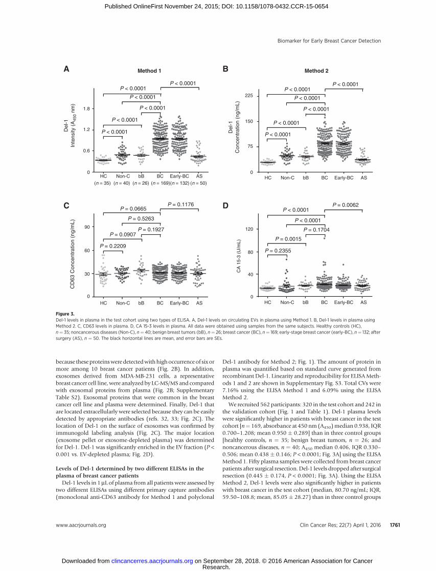
We recruited 562 participants: 320 in the test cohort and 242 inthe validation cohort (Fig. 1 and Table 1). Del-1 plasma levelswere significantly higher in patients with breast cancer in the testcohort [n¼ 169, absorbance at 450 nm (A450)median 0.938, IQR0.700–1.208; mean 0.950 � 0.289] than in three control groups[healthy controls, n ¼ 35; benign breast tumors, n ¼ 26; andnoncancerous diseases, n ¼ 40; A450 median 0.406, IQR 0.330–0.506; mean 0.438 � 0.146; P < 0.0001; Fig. 3A] using the ELISAMethod 1. Fifty plasma samples were collected from breast cancerpatients after surgical resection.Del-1 levels dropped after surgicalresection (0.445 � 0.174, P < 0.0001; Fig. 3A). Using the ELISAMethod 2, Del-1 levels were also significantly higher in patientswith breast cancer in the test cohort (median, 80.70 ng/mL; IQR,59.50–108.8; mean, 85.05� 28.27) than in three control groups
Method 1A B
C D
P < 0.0001
P < 0.0001
P < 0.0001
P < 0.0001
P < 0.0001
P = 0.0665
P = 0.5263
P = 0.1927P = 0.0907
P = 0.2209
P = 0.1176P < 0.0001
P < 0.0001
P = 0.0062
P = 0.1704
P = 0.0015
P = 0.2355
(n = 35) (n = 40) (n = 26) (n = 169)(n = 132) (n = 50)
HC0
0
30
CD
63 C
once
ntra
tion
(ng/
mL)
60
90
0
75
150
225
0.6
Inte
nsity
(A
450
nm)
Del
-1
Con
cent
ratio
n (n
g/m
L)D
el-1
1.2
1.8
Non-C bB BC Early-BC AS
HC Non-C bB BC Early-BC AS HC0
40
80
CA
15-
3 (U
/mL)
120
Non-C bB BC Early-BC AS
HC Non-C bB BC Early-BC AS
P < 0.0001P < 0.0001
P < 0.0001
P < 0.0001
P < 0.0001
P < 0.0001
P < 0.0001
Method 2
Figure 3.Del-1 levels in plasma in the test cohort using two types of ELISA. A, Del-1 levels on circulating EVs in plasma using Method 1. B, Del-1 levels in plasma usingMethod 2. C, CD63 levels in plasma. D, CA 15-3 levels in plasma. All data were obtained using samples from the same subjects. Healthy controls (HC),n¼ 35; noncancerous diseases (Non-C), n¼ 40; benign breast tumors (bB), n¼ 26; breast cancer (BC), n¼ 169; early-stage breast cancer (early-BC), n¼ 132; aftersurgery (AS), n ¼ 50. The black horizontal lines are mean, and error bars are SEs.
Biomarker for Early Breast Cancer Detection
www.aacrjournals.org Clin Cancer Res; 22(7) April 1, 2016 1761
Research. on September 28, 2018. © 2016 American Association for Cancerclincancerres.aacrjournals.org Downloaded from
Published OnlineFirst November 24, 2015; DOI: 10.1158/1078-0432.CCR-15-0654

(median, 39.22; IQR, 28.91–49.58; mean, 41.38 � 14.61; P <0.0001; Fig. 3B).
To demonstrate the utility of Del-1 as early diagnostic markerfor breast cancer relative to other biomarkers, the plasma levels ofCA 15-3, a molecular diagnostic biomarker currently used for thedetection of metastatic breast cancer (9), was measured for thesame subjects (Fig. 3D). In contrast with Del-1, but consistentwith previous work (34), CA 15-3 showed no significant changecompared with controls at breast cancer stages 0 to III (median,24.0 U/mL; IQR, 20.6–29.0; P ¼ 0.444; Supplementary Fig. S4)but was significantly increased at stage IV (median, 37.8 U/mL;IQR, 22.6–53.0; P ¼ 0.028; Supplementary Fig. S4). AlthoughDel-1 levels were higher in breast cancer patients than in controls,the levels of CD63, a representative exosomemarker protein, weresignificantly not changed (Fig. 3C). Similarly, the numbers ofcirculating EVs per same volume of plasma were significantly notchanged between healthy control and breast cancer patients (P ¼0.964; Supplementary Fig. S5A). These results suggested that thenumber of EVs is somewhat controlled in human plasma, asshown previously in melanoma patients (21).
The levels of Del-1 did not correlate with the size of the breasttumors (Spearman r¼0.079;P¼0.306; Supplementary Fig. S5B).Similarly, the levels of CD63 did not correlate with the size of thebreast tumors (Spearman r ¼ 0.055; P ¼ 0.447; SupplementaryFig. S5C). Moreover, Del-1 levels showed no significant correla-tion with the status of four types of receptors in breast cancers(ER/PRþHer2þ, ER/PRþHer2–, ER/PR–Her2þ, ER/PR–Her2–;P > 0.05; Supplementary Fig. S6).
Sensitivities and Specificities of Del-1 for breast cancerdiagnosis in test set
ROC curves showed predictive values, and likelihood ratios forDel-1 in the diagnosis of breast cancer are shown in Table 2. Theoptimum diagnostic cutoff for Del-1 was 0.512 (A450) [AUC,0.954; 95% confidence interval (CI), 0.923–0.975; sensitivity at95% specificity, 73.1%] using the ELISA Method 1 (Fig. 4Aand Table 2) and that was 53 ng/mL (AUC, 0.937; 95% CI,0.923–0.975; sensitivity at 95% specificity, 63.9%) using theELISA Method 2 (Fig. 4A and Table 2) in the test cohort.
A greater proportion of patients with breast cancer in the testcohort were positive for Del-1 using the ELISA Method 1 than theELISA Method 2 [164 (97.0%) vs. 156 (92.3%) of 169patients; Fig. 3A]. In the assessment of differential diagnosticaccuracy, plasma Del-1 using the ELISA Method 1 (AUC,0.936; 95% CI 0.899–0.963; sensitivity at 69.6% specificity) hadslightly higher AUC values than the ELISAMethod 2 (AUC, 0.912;95% CI 0.871–0.944; sensitivity at 95% specificity, 58.5%) inpatients with breast cancer compared with patients with benignbreast tumors and noncancerous diseases (Fig. 4B and Table 2).
In the test cohort, 132 (78%) of 169 patients with breast cancerhad early-stage disease (stages 0, I, and II). Del-1 plasma levelswere significantly higher in early-stage breast cancer than in threecontrol groups (P <0.0001; Fig. 3A andB). PlasmaDel-1 using theELISAMethod 1 (AUC, 0.961; 95%CI, 0.924–0.983; sensitivity at95% specificity, 73.5%) yielded a slightly higher differentialdiagnosis of early-stage breast cancer from three control groups,compared with the ELISAMethod 2 (AUC, 0.946; 95%CI, 0.905–0.972; sensitivity at 95% specificity, 65.0%; Fig. 4C and Table 2).Similarly, plasma Del-1 using the ELISA Method 1 (AUC, 0.939;95% CI, 0.890–0.971; sensitivity at 95% specificity, 68.8%)yielded a slightly higher differential diagnosis of early-stage breast Ta
ble
2.Results
formea
suremen
tofplasm
aDel-1usingELISAin
thediagno
sisofbreastcancer
Test
set
Validationset
AUC(95%
CI)
Sensitivity
(%)
Spec
ificity
(%)
PPV
(%)
NPV
(%)
Positive
LRNeg
ative
LRAUC(95%
CI)
Sensitivity
(%)
Spec
ificity
(%)
PPV
(%)
NPV
(%)
Positive
LRNeg
ative
LR
ELISA
Metho
d1
BCvs.b
B,n
on-C,a
ndHC
0.954
(0.923
–0.975
)95.27
81.19
98.98
51.87
4.67
0.05
0.961(0.931–0
.981)
99.00
86.62
99.34
77.30
6.15
0.01
BCvs.b
Ban
dNon-C
0.936
(0.899–0
.963)
88.75
76.24
98.42
33.67
4.27
0.23
0.948(0.911–0
.973
)99.00
81.2
598.85
81.2
34.69
0.01
Early-BCvs.b
B,N
on-C,a
ndHC
0.961(0.924
–0.983)
94.70
86.36
99.23
41.3
55.42
0.07
0.968(0.933
–0.988)
92.31
86.62
99.49
100.00
6.44
0.07
Early-BCvs.b
Ban
dNon-C
0.939
(0.890–0
.971)
89.39
77.27
99.46
33.26
12.00
0.21
0.957
(0.911–0
.983)
92.31
81.2
599.10
100.00
4.92
0.04
ELISA
Metho
d2
BCvs.b
B,N
on-C,a
ndHC
0.937
(0.923
–0.975
)93.49
76.24
98.96
45.3
4.71
0.08
0.920
(0.882–
0.950
)95.00
80.99
97.04
62.51
4.86
0.06
BCvs.b
Ban
dNon-C
0.912
(0.871–0
.944)
85.80
75.24
98.04
33.27
4.82
0.19
0.899(0.852
–0.935
)95.00
74.64
97.68
54.51
3.68
0.07
Early-BCvs.b
B,N
on-C,a
ndHC
0.946(0.905–
0.972
)90.90
77.14
99.34
25.17
8.54
0.09
0.943(0.900–0
.971)
89.23
80.99
98.83
100.00
5.12
0.05
Early-BCvs.b
Ban
dNon-C
0.924
(0.874
–0.959
)84.85
75.76
98.66
31.64
6.07
0.18
0.925
(0.871–0
.962)
89.23
74.64
97.95
100.00
3.88
0.06
Abbreviations:bB,b
enignbreasttumor;BC,b
reastcancer;HC,h
ealthy
controls;LR
,likelihoodratio;N
on-C,n
oncan
cerous
disea
ses;NPV,n
egativepredictive
value;
PPV,p
ositive
predictive
value.
Moon et al.
Clin Cancer Res; 22(7) April 1, 2016 Clinical Cancer Research1762
Research. on September 28, 2018. © 2016 American Association for Cancerclincancerres.aacrjournals.org Downloaded from
Published OnlineFirst November 24, 2015; DOI: 10.1158/1078-0432.CCR-15-0654
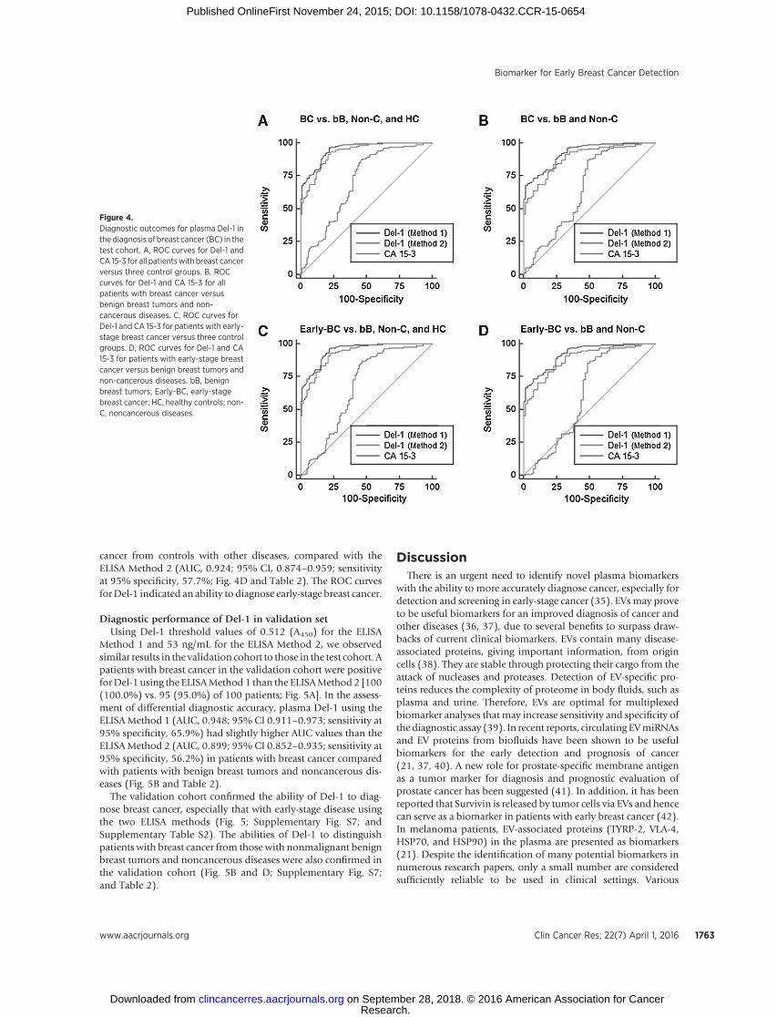
cancer from controls with other diseases, compared with theELISA Method 2 (AUC, 0.924; 95% CI, 0.874–0.959; sensitivityat 95% specificity, 57.7%; Fig. 4D and Table 2). The ROC curvesforDel-1 indicated an ability to diagnose early-stage breast cancer.
Diagnostic performance of Del-1 in validation setUsing Del-1 threshold values of 0.512 (A450) for the ELISA
Method 1 and 53 ng/mL for the ELISA Method 2, we observedsimilar results in the validation cohort to those in the test cohort. Apatients with breast cancer in the validation cohort were positiveforDel-1 using the ELISAMethod 1 than the ELISAMethod 2 [100(100.0%) vs. 95 (95.0%) of 100 patients; Fig. 5A]. In the assess-ment of differential diagnostic accuracy, plasma Del-1 using theELISAMethod 1 (AUC, 0.948; 95% CI 0.911–0.973; sensitivity at95% specificity, 65.9%) had slightly higher AUC values than theELISAMethod 2 (AUC, 0.899; 95% CI 0.852–0.935; sensitivity at95% specificity, 56.2%) in patients with breast cancer comparedwith patients with benign breast tumors and noncancerous dis-eases (Fig. 5B and Table 2).
The validation cohort confirmed the ability of Del-1 to diag-nose breast cancer, especially that with early-stage disease usingthe two ELISA methods (Fig. 5; Supplementary Fig. S7; andSupplementary Table S2). The abilities of Del-1 to distinguishpatients with breast cancer from those with nonmalignant benignbreast tumors and noncancerous diseases were also confirmed inthe validation cohort (Fig. 5B and D; Supplementary Fig. S7;and Table 2).
DiscussionThere is an urgent need to identify novel plasma biomarkers
with the ability to more accurately diagnose cancer, especially fordetection and screening in early-stage cancer (35). EVs may proveto be useful biomarkers for an improved diagnosis of cancer andother diseases (36, 37), due to several benefits to surpass draw-backs of current clinical biomarkers. EVs contain many disease-associated proteins, giving important information, from origincells (38). They are stable through protecting their cargo from theattack of nucleases and proteases. Detection of EV-specific pro-teins reduces the complexity of proteome in body fluids, such asplasma and urine. Therefore, EVs are optimal for multiplexedbiomarker analyses that may increase sensitivity and specificity ofthe diagnostic assay (39). In recent reports, circulating EVmiRNAsand EV proteins from biofluids have been shown to be usefulbiomarkers for the early detection and prognosis of cancer(21, 37, 40). A new role for prostate-specific membrane antigenas a tumor marker for diagnosis and prognostic evaluation ofprostate cancer has been suggested (41). In addition, it has beenreported that Survivin is released by tumor cells via EVs and hencecan serve as a biomarker in patients with early breast cancer (42).In melanoma patients, EV-associated proteins (TYRP-2, VLA-4,HSP70, and HSP90) in the plasma are presented as biomarkers(21). Despite the identification of many potential biomarkers innumerous research papers, only a small number are consideredsufficiently reliable to be used in clinical settings. Various
Figure 4.Diagnostic outcomes for plasma Del-1 inthe diagnosis of breast cancer (BC) in thetest cohort. A, ROC curves for Del-1 andCA 15-3 for all patientswith breast cancerversus three control groups. B, ROCcurves for Del-1 and CA 15-3 for allpatients with breast cancer versusbenign breast tumors and non-cancerous diseases. C, ROC curves forDel-1 and CA 15-3 for patients with early-stage breast cancer versus three controlgroups. D, ROC curves for Del-1 and CA15-3 for patients with early-stage breastcancer versus benign breast tumors andnon-cancerous diseases. bB, benignbreast tumors; Early-BC, early-stagebreast cancer; HC, healthy controls; non-C, noncancerous diseases.
Biomarker for Early Breast Cancer Detection
www.aacrjournals.org Clin Cancer Res; 22(7) April 1, 2016 1763
Research. on September 28, 2018. © 2016 American Association for Cancerclincancerres.aacrjournals.org Downloaded from
Published OnlineFirst November 24, 2015; DOI: 10.1158/1078-0432.CCR-15-0654

candidate exosomal proteins have been reported as biomarkersand confirmed by Western blot after EV purification from a smallnumber of clinical samples (21, 43, 44). In the current study, weshowed diagnostic accuracy of Del-1 with high sensitivity andspecificity in 562 participants from two cohorts using two differ-ent ELISAs. This assay is simple, reproducible, quantitative, andminimally invasive using a small amount of plasma (1 mL)without EV purification. The major location (exosome pellet orexosome-depleted plasma) was determined for Del-1, and thisprotein was significantly enriched in the exosome fraction (Fig.2D). Exosomal Del-1 using the ELISA Method 1 was a similardiagnostic performance, compared with plasma Del-1 using theELISA Method 2.
There are some studies for the function of the Del-1 protein incancer (43) and few relating to breast cancer. Del-1 was firstidentified as an extracellular matrix protein having 3 N-terminalepidermal growth factor–like domains and the discoidin I–like orfactor V C domains, C1 and C2 (45). Del-1 is expressed byendothelial cells during embryonic vascular development (46)and promotes the adhesion of endothelial cells through itsinteraction with integrin receptors (45). This protein is expressedin other cancers (43, 47, 48) as well as breast cancer (47),suggesting that this proteinmight have potential as cancer-specificbiomarker for various human cancers, including breast cancer.
We found that plasma Del-1 levels had no correlation with theAJCC stage (Fig. 3). This result raised the question as to why therewas no relationship between the circulating levels ofDel-1 and thetumor burden as one would expect larger tumors to secrete more
EVs than smaller tumors. The mechanisms that control the bal-ance between the secretion of EVs and their clearance are not wellknown in the circulating system. One potential mechanism toregulate EV secretion involves the ability of tumor cells to sensethe concentration of EVs in the microenvironment and then altertheir secretion (49). These researchers also found that labeled EVsfrom mammary epithelial cells are internalized into the tumorcells, suggesting a feedback regulatory mechanism for controllingEVs secretion. Another potential mechanism to regulate EV clear-ance involves special cells, such as endothelial cells, that eliminatecirculating EVs through uptake processes. Such scavenging cellsmight selectively take up EVs by recognizing specific epitopes,such as outer membrane proteins. Recently, a new pathway formicroparticle clearance involving Del-1–mediated integrin-dependent endothelial cell uptake from the circulation wasreported (32, 50). This strongly supports the possibility thatcirculating EVs with Del-1 from tumor cells could be selectivelyeliminated from the circulation by this mechanism. Based onthese mechanisms of EV secretion and clearance from the circu-lation, it is thought that steady-state EV levels in the circulationmight be maintained regardless of tumor size.
The patient groups differed to some degree in the diagnosticperformance results (Table 2). For example, the positive andnegative predictive values of plasma Del-1 for the differentialdiagnosis of early-stage breast cancer from controls were a bitdifferent because the validation cohort had only 65 patients withearly-stage breast cancer, compared with 132 in the test cohort.These findings can be explained by differences in the sample size
Figure 5.Diagnostic outcomes for plasma Del-1 inthe diagnosis of breast cancer in thevalidation cohort. A, ROC curves for Del-1 for all patients with breast cancerversus three control groups. B, ROCcurves for Del-1 for all patients withbreast cancer versus benign breasttumors and noncancerous diseases. C,ROC curves for Del-1 for patients withearly-stage breast cancer versus threecontrol groups. D, ROC curves for Del-1for patients with early-stage breastcancer versus benign breast tumors andnoncancerous diseases. bB, benignbreast tumors; Early-BC, early-stagebreast cancer; HC, healthy controls; non-C, noncancerous diseases.
Clin Cancer Res; 22(7) April 1, 2016 Clinical Cancer Research1764
Moon et al.
Research. on September 28, 2018. © 2016 American Association for Cancerclincancerres.aacrjournals.org Downloaded from
Published OnlineFirst November 24, 2015; DOI: 10.1158/1078-0432.CCR-15-0654

between the test and the validation cohorts (Table 1).Despite thesedifferences, the diagnostic capability of plasmaDel-1was generallysimilar in the two cohorts. The current study is retrospectiveanalysis of individuals with breast cancer. A prospective study willbe done in the future to assess whether Del-1 can be validated as invivomarker in patients with breast cancer. The striking decrease inDel-1 concentrations in plasma after surgery suggests that theseproteins could be useful surveillance biomarker to assess theresponse of breast cancer patients to cancer therapies. To furtherexplore this potential role, the long-term follow-upof breast cancerpatients who underwent surgery is planned.
To our knowledge, this is the first study to report the diagnosticrelevance of Del-1 on plasma EVs as a protein marker for breastcancer in a test cohort and an independent validation cohort.Combining measurement of Del-1 in plasma with imaging infor-mation, and other clinicopathologic characteristics, may improvethe identification of patients with early-stage breast cancer.
Disclosure of Potential Conflicts of InterestNo potential conflicts of interest were disclosed.
Authors' ContributionsConception and design: P.-G. Moon, J.-E. Lee, H. Park, M.-C. BaekDevelopment of methodology: P.-G. Moon, J.-E. Lee, M.-C. BaekAcquisition of data (provided animals, acquired and managed patients,provided facilities, etc.): P.-G. Moon, J.-E. Lee, Y.-E. Cho, S. J. Lee, Y. S. Chae,H.-I. Bae, M.-C. Baek
Analysis and interpretation of data (e.g., statistical analysis, biostatistics,computational analysis): P.-G. Moon, Y.-E. Cho, S. J. Lee, Y. S. Chae, I.-S. Kim,M.-C. BaekWriting, review, and/or revision of the manuscript: P.-G. Moon, J.-E. Lee,Y.-E. Cho, S. J. Lee, Y. S. Chae, Y.-B. Kim, I.-S. Kim, M.-C. BaekAdministrative, technical, or material support (i.e., reporting or organizingdata, constructing databases): P.-G. Moon, Y.-E. Cho, J. H. Jung, H.-I. Bae,M.-C. BaekStudy supervision: M.-C. Baek
AcknowledgmentsThe authors thank R. Santen and B. Smith for critiquing the article, M. Lee for
purification of extracellular vesicles, and S. Lee for helpful comments on thearticle. They also thank the National Biobank of Korea–Kyungpook NationalUniversity Hospital and the Chonnam National University Hwasun Hospitalfor providing plasma samples.
Grant SupportThis work was supported by aNational Research Foundation of Korea (NRF)
grant funded by the Korea Government (NRF-2012R1A2A2A01046512 and2014R1A5A2009242) and by a grant of the Korean Health Technology R&Dproject, Ministry of Health & Welfare, Republic of Korea (HI12C0534).
The costs of publication of this article were defrayed in part by thepayment of page charges. This article must therefore be hereby markedadvertisement in accordance with 18 U.S.C. Section 1734 solely to indicatethis fact.
Received March 17, 2015; revised October 20, 2015; accepted October 21,2015; published OnlineFirst November 24, 2015.
References1. Grayson M. Breast cancer. Nature 2012;485:S49.2. Maxmen A. The hard facts. Nature 2012;485:S50–1.3. Rice J. Metastasis: the rude awakening. Nature 2012;485:S55–7.4. Dalton WS, Friend SH. Cancer biomarkers–an invitation to the table.
Science 2006;312:1165–8.5. Henry NL, Hayes DF. Cancer biomarkers. Mol Oncol 2012;6:140–6.6. Cho-Chung YS. Autoantibody biomarkers in the detection of cancer.
Biochim Biophys Acta 2006;1762:587–91.7. Etzioni R, Urban N, Ramsey S, McIntosh M, Schwartz S, Reid B, et al. The
case for early detection. Nat Rev Cancer 2003;3:243–52.8. Duffy MJ, Evoy D, McDermott EW. CA 15-3: uses and limitation as a
biomarker for breast cancer. Clin Chim Acta 2010;411:1869–74.9. Harris L, FritscheH,Mennel R, Norton L, Ravdin P, Taube S, et al. American
Society of Clinical Oncology 2007 update of recommendations for the useof tumor markers in breast cancer. J Clin Oncol 2007;25:5287–312.
10. Cristofanilli M, Budd GT, Ellis MJ, Stopeck A, Matera J, Miller MC, et al.Circulating tumor cells, disease progression, and survival in metastaticbreast cancer. N Engl J Med 2004;351:781–91.
11. Parkinson DR, Dracopoli N, Petty BG, Compton C, Cristofanilli M,Deisseroth A, et al. Considerations in the development of circulatingtumor cell technology for clinical use. J Translat Med 2012;10:138.
12. MegoM,DeGiorgi U, Dawood S,Wang X, Valero V, Andreopoulou E, et al.Characterization of metastatic breast cancer patients with nondetectablecirculating tumor cells. Int J Cancer 2011;129:417–23.
13. Dawson SJ, Tsui DW, Murtaza M, Biggs H, Rueda OM, Chin SF, et al.Analysis of circulating tumor DNA to monitor metastatic breast cancer. NEngl J Med 2013;368:1199–209.
14. LippmanM,OsborneCK.Circulating tumorDNA–ready for prime time?NEngl J Med 2013;368:1249–50.
15. Valastyan S, Weinberg RA. Tumor metastasis: molecular insights andevolving paradigms. Cell 2011;147:275–92.
16. Hanahan D, Weinberg RA. Hallmarks of cancer: the next generation. Cell2011;144:646–74.
17. Husemann Y, Geigl JB, Schubert F, Musiani P, Meyer M, Burghart E, et al.Systemic spread is an early step in breast cancer. Cancer Cell 2008;13:58–68.
18. Weinberg RA. Leaving home early: reexamination of the canonical modelsof tumor progression. Cancer Cell 2008;14:283–4.
19. Muralidharan-Chari V, Clancy JW, Sedgwick A, D'Souza-Schorey C. Micro-vesicles: mediators of extracellular communication during cancer progres-sion. J Cell Sci 2010;123:1603–11.
20. Raposo G, Stoorvogel W. Extracellular vesicles: exosomes, microvesicles,and friends. J Cell Biol 2013;200:373–83.
21. Peinado H, Aleckovic M, Lavotshkin S, Matei I, Costa-Silva B, Moreno-Bueno G, et al. Melanoma exosomes educate bone marrow progenitorcells toward a pro-metastatic phenotype through MET. Nat Med 2012;18:883–91.
22. Luga V, Zhang L, Viloria-Petit AM,Ogunjimi AA, InanlouMR, Chiu E, et al.Exosomes mediate stromal mobilization of autocrine Wnt-PCP signalingin breast cancer cell migration. Cell 2012;151:1542–56.
23. Antonyak MA, Li B, Boroughs LK, Johnson JL, Druso JE, Bryant KL, et al.Cancer cell-derived microvesicles induce transformation by transferringtissue transglutaminase and fibronectin to recipient cells. Proc Natl AcadSci U S A 2011;108:4852–7.
24. Zhang HG, Grizzle WE. Exosomes and cancer: a newly described pathwayof immune suppression. Clin Cancer Res 2011;17:959–64.
25. D'Souza-Schorey C, Clancy JW. Tumor-derived microvesicles: sheddinglight on novel microenvironment modulators and prospective cancerbiomarkers. Genes Dev 2012;26:1287–99.
26. Moon PG, You S, Lee JE, Hwang D, Baek MC. Urinary exosomes andproteomics. Mass Spectrom Rev 2011;30:1185–202.
27. GnantM,Mlineritsch B, StoegerH, Luschin-EbengreuthG,HeckD,MenzelC, et al. Adjuvant endocrine therapy plus zoledronic acid in premeno-pausal women with early-stage breast cancer: 62-month follow-up fromthe ABCSG-12 randomised trial. Lancet Oncol 2011;12:631–41.
28. Houssami N, Hayes DF. Review of preoperative magnetic resonanceimaging (MRI) in breast cancer: should MRI be performed on all womenwith newly diagnosed, early stage breast cancer? CA Cancer J Clin 2009;59:290–302.
29. McShane LM, AltmanDG, Sauerbrei W, Taube SE, GionM, Clark GM, et al.Reporting recommendations for tumor marker prognostic studies(REMARK). J Natl Cancer Inst 2005;97:1180–4.
www.aacrjournals.org Clin Cancer Res; 22(7) April 1, 2016 1765
Biomarker for Early Breast Cancer Detection
Research. on September 28, 2018. © 2016 American Association for Cancerclincancerres.aacrjournals.org Downloaded from
Published OnlineFirst November 24, 2015; DOI: 10.1158/1078-0432.CCR-15-0654

30. Thery C, Amigorena S, RaposoG, ClaytonA. Isolation and characterizationof exosomes from cell culture supernatants and biological fluids. CurrProtoc Cell Biol 2006;Chapter 3:Unit 3 22.
31. Cho YE, Singh TS, Lee HC, Moon PG, Lee JE, Lee MH, et al. In-depthidentification of pathways related to cisplatin-induced hepatotoxicitythrough an integrative method based on an informatics-assisted label-freeprotein quantitation and microarray gene expression approach. Mol CellProteomics 2012;11:M111 010884.
32. Dasgupta SK, LeA,Chavakis T, Rumbaut RE, ThiagarajanP.Developmentalendothelial locus-1 (Del-1) mediates clearance of platelet microparticlesby the endothelium. Circulation 2012;125:1664–72.
33. Zhong J, Eliceiri B, Stupack D, Penta K, Sakamoto G, Quertermous T, et al.Neovascularization of ischemic tissues by gene delivery of the extracellularmatrix protein Del-1. J Clin Invest 2003;112:30–41.
34. Nowsheen S, Aziz K, Panayiotidis MI, Georgakilas AG. Molecular markersfor cancer prognosis and treatment: have we struck gold? Cancer Lett2012;327:142–52.
35. Wagner PD, Verma M, Srivastava S. Challenges for biomarkers in cancerdetection. Ann N Y Acad Sci 2004;1022:9–16.
36. Hanash SM, Baik CS, Kallioniemi O. Emerging molecular biomarkers–blood-based strategies to detect and monitor cancer. Nat Rev Clin Oncol2011;8:142–50.
37. Keller S, Sanderson MP, Stoeck A, Altevogt P. Exosomes: from biogenesisand secretion to biological function. Immunol Lett 2006;107:102–8.
38. Taylor DD, Gercel-Taylor C. MicroRNA signatures of tumor-derived exo-somes as diagnostic biomarkers of ovarian cancer. Gynecol Oncol2008;110:13–21.
39. Dijkstra S, Birker IL, Smit FP, Leyten GH, de Reijke TM, van Oort IM, et al.Prostate cancer biomarker profiles in urinary sediments and exosomes. JUrol 2014;191:1132–8.
40. Ohshima K, Inoue K, Fujiwara A, Hatakeyama K, Kanto K, Watanabe Y,et al. Let-7 microRNA family is selectively secreted into the extracellular
environment via exosomes in ametastatic gastric cancer cell line. PloS One2010;5:e13247.
41. Liu T, Mendes DE, Berkman CE. Functional prostate-specific membraneantigen is enriched in exosomes from prostate cancer cells. Int J Oncol2014;44:918–22.
42. Khan S, Bennit HF, Turay D, Perez M, Mirshahidi S, Yuan Y, et al. Earlydiagnostic value of survivin and its alternative splice variants in breastcancer. BMC Cancer 2014;14:176.
43. Beckham CJ, Olsen J, Yin PN, Wu CH, Ting HJ, Hagen FK, et al. BladderCancer Exosomes Contain EDIL-3/Del1 and Facilitate Cancer Progression.J Urol 2014;192:583–92.
44. Moon PG, Lee JE, You S, Kim TK, Cho JH, Kim IS, et al. Proteomic analysisof urinary exosomes from patients of early IgA nephropathy and thinbasement membrane nephropathy. Proteomics 2011;11:2459–75.
45. Schurpf T, Chen Q, Liu JH, Wang R, Springer TA, Wang JH. The RGD fingerof Del-1 is a unique structural feature critical for integrin binding. FASEB J2012;26:3412–20.
46. Hidai C, Zupancic T, Penta K,Mikhail A, KawanaM,Quertermous EE, et al.Cloning and characterization of developmental endothelial locus-1: anembryonic endothelial cell protein that binds the alphavbeta3 integrinreceptor. Genes Dev 1998;12:21–33.
47. Aoka Y, Johnson FL, Penta K, Hirata Ki K, Hidai C, Schatzman R, et al. Theembryonic angiogenic factor Del1 accelerates tumor growth by enhancingvascular formation. Microvasc Res 2002;64:148–61.
48. Sun JC, LiangXT, PanK,WangH,Zhao JJ, Li JJ, et al.High expression level ofEDIL3 in HCC predicts poor prognosis of HCC patients. World J Gastro-enterol 2010;16:4611–5.
49. Riches A, Campbell E, Borger E, Powis S. Regulation of exosome releasefrom mammary epithelial and breast cancer cells - a new regulatorypathway. Eur J Cancer 2014;50:1025–34.
50. Rautou PE, Mackman N. Del-etion of microvesicles from the circulation.Circulation 2012;125:1601–4.
Clin Cancer Res; 22(7) April 1, 2016 Clinical Cancer Research1766
Moon et al.
Research. on September 28, 2018. © 2016 American Association for Cancerclincancerres.aacrjournals.org Downloaded from
Published OnlineFirst November 24, 2015; DOI: 10.1158/1078-0432.CCR-15-0654

2016;22:1757-1766. Published OnlineFirst November 24, 2015.Clin Cancer Res Pyong-Gon Moon, Jeong-Eun Lee, Young-Eun Cho, et al. Detection
CancerExtracellular Vesicles as a Novel Biomarker for Early Breast Identification of Developmental Endothelial Locus-1 on Circulating
Updated version
10.1158/1078-0432.CCR-15-0654doi:
Access the most recent version of this article at:
Material
Supplementary
http://clincancerres.aacrjournals.org/content/suppl/2015/11/24/1078-0432.CCR-15-0654.DC1
Access the most recent supplemental material at:
Cited articles
http://clincancerres.aacrjournals.org/content/22/7/1757.full#ref-list-1
This article cites 49 articles, 10 of which you can access for free at:
E-mail alerts related to this article or journal.Sign up to receive free email-alerts
Subscriptions
Reprints and
To order reprints of this article or to subscribe to the journal, contact the AACR Publications Department at
Permissions
Rightslink site. Click on "Request Permissions" which will take you to the Copyright Clearance Center's (CCC)
.http://clincancerres.aacrjournals.org/content/22/7/1757To request permission to re-use all or part of this article, use this link
Research. on September 28, 2018. © 2016 American Association for Cancerclincancerres.aacrjournals.org Downloaded from
Published OnlineFirst November 24, 2015; DOI: 10.1158/1078-0432.CCR-15-0654