iconographia mycologicaverona — benedek
Post on 19-Aug-2016
223 views
TRANSCRIPT
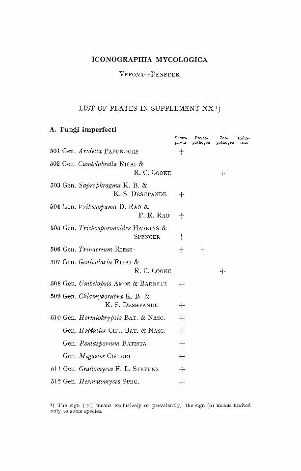
ICONOGRAPHIA MYCOLOGICA
V E R O N A - - B E N E D E K
LIST OF PLATES IN S U P P L E M E N T X X 1)
A. Fungi imperfecti Sapro- Phyto- Zoo- Indub, phyta pathogen pathogen trial
501 Gem Arxiella PAPENDORF @
502 Gem Candelabrella RIS-AI & R. C. COOKE
503 Gen. Sapr@hragma K. B. & K. S. DESHPANDE @-
504 Gen. Vriksh@ama D. RAo & P. R. RAo +
505 Gem Trichosporonoides HASKINS & SPENCER -t-
506 Gen. Trinacrium RIESS- +
507 Gen. Genicularia RIFAI & R. C. CooI,:E
508 Gen.
509 Gen.
Umbel@sis A~os & BARNETT @
Chlamydorubra K. B. & K. S. DESHPANDE @
Hormiokrypsis BAT. & NASC. @
Heptaster CIF., BAT. & NASC. +
Peuta@orium BATISTA +
Megaster CIFERRI @
Grallomyces F. L. STEVENS +
Hermatomyces SPEG. @
510 Gen.
Gen.
Gen.
G e n .
511 Gen.
512 Gen.
+
+
1) The sign ( + ) means exclusively or prevalently; the sign (o)means limited only to some species.
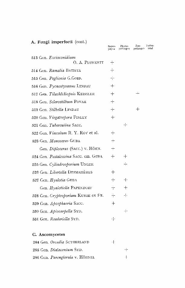
A. Fungi imperfect i (cont.)
513 Gen. Excioconidium O. A. 1)LUNKETT
514 Gen.
515 Gen.
516 Gen.
517 Gen.
518 Gen.
519 Gen.
520 Gen.
521 Gen.
522 Gen.
523 Gem
G e n .
524 Gen.
525 Gem
526 Gen.
527 Gen.
G e n .
528 Gem
529 Gem
530 Gen.
531 Gen.
Sapro- Phyto- Zoo- Indus- phyta pathogen pathogen trial
+
Ran,alia BATISTA ÷
Peglionia G.GolD. ÷
Pycnostysanus LINDAU +
TilachIidiopsis KEISSLER +
Sclerostilbum POVAH -{-
Stilbella LINDAU ÷
Virgatospora FINLEY +
Tuberculina SAte.
Vinculum R. Y. RoY et aI. +
Monoceras GUBA +
Diploceras (SAte.) v. HOHN. ÷
Pestalozzina SAcc. em. GuB~ +
Cylindrosporium UNGER
Libertella DESMAZI]~RES @
Hyalotia GuBA ÷
HyaIotiella PAPENDORF -~
Crypto@orium KUNZE ex FR. +
Aposphaeria SAte. +
A piocarpella SYD.
Readeriella SYD. ÷
+
+
+
+
+
+
÷
+
+
C. Ascomycetes
284 Gen. Orcadia SUTtlERLAND
285 Gen. Dialacenium Svn.
286 Gen. Parenglerula v. HOHNEL
+
+
+
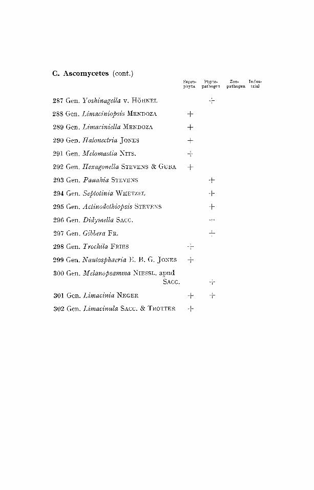
C. A s c o m y c e t e s (cont,)
287 Gen.
288 Gen.
289 Gen.
290 Gen.
291 Gen.
292 Gen.
293 Gen.
294 Gen.
295 Gem
296 Gen.
297 Gen.
Yoshinagella v. H6HNEL
Limaciniopsis MENDOZA
Limaciniella MENDOZA
Halonectria JONES
~Vf elomastia NITS.
Hexagonella STEVENS & GUBA
Pauahia STEVENS
Septotinia WHETZEL
Actinodoth@bsis STEVENS
Didymella SAcc.
Gibbera FR.
Sapro- Phyto- Zoo- Indus- phyta pathogen pathogeI1 trial
+
+
+
+
+
298 Gen. Trochila FRIES +
299 Gen. Nautosphaeria E. B. G. JONES +
300 Gen. Melanopsamma NIESSL. apud SAcc.
301 Gen. Limacinia NEGER +
302 Gen. Limacinula SACC. & TROTTEI~ +
+
+
+
+
+
+
+
+


F U N G I I M P E R F E C T I
M O N I L I A L E S
M O N I L I A C E A E
S A P R O P I - I Y T A
Gen. A r x i e l l a PAPENDORF
I C O N O G R A P H I A M Y C O L O G I C A
V E R O N A - - B E N E D E K
Pla te A 501
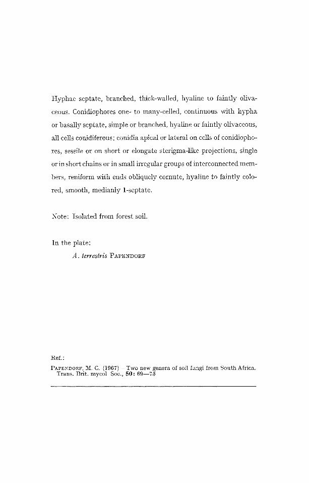
Hyphae septate, branched, thick-walled, hyaline to faintly oliva-
ceous. Conidiophores one- to many-celled, continuous with hypha
or basally septate, simple or branched, hyaline or faintly olivaceous,
all cells conidiferous; conidia apical or lateral on cells of conidiopho-
res, sessile or on short or elongate sterigma-like projections, single
or in short chains or in smMl irregular groups of interconnected mem-
bers, reniform with ends obliquely cornute, hyaline to faintly colo-
red, smooth, medianly 1-septate.
Note: Isolated from forest soil.
In the plate:
A. terrestris PAPENDOI~
Ref. :
t:~APENDORF, i~{[. C. (1967) - Two new genera of soil fungi f rom S o u t h Africa. Trans . Br i t . mycol Soc., 5 0 : 6 9 . - - 7 5
~J

F U N G I I M P E R F E C T I
M O N I L I A L E S
! ~ O N I L I A C E A E
Z O O P A T H O G E N
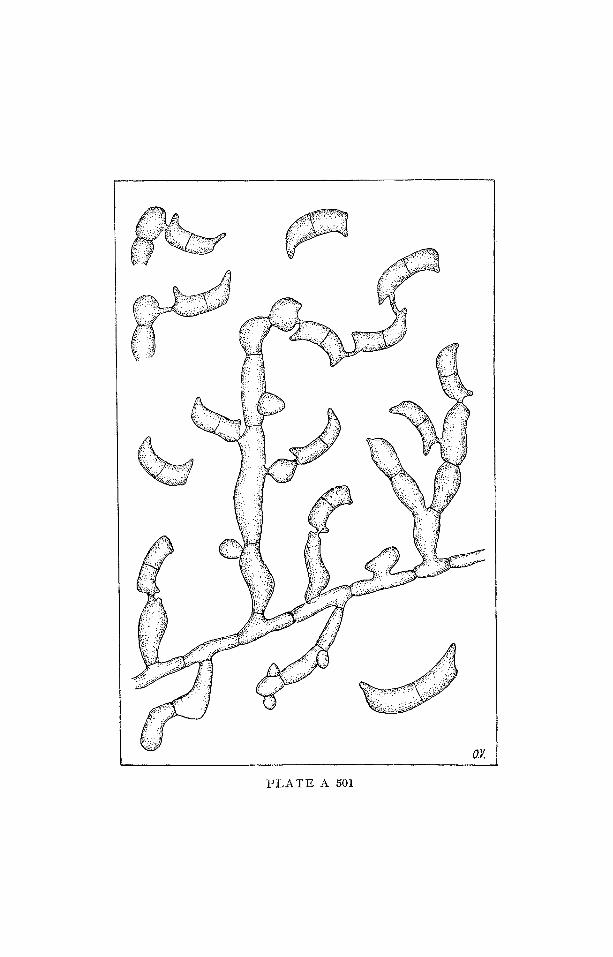
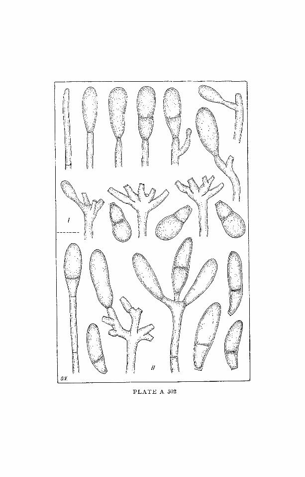
Gen. C a n d e l a b r e l l a RIVAl & R. C. COOKE
I C O N O G R A P H I A MYCOLOGICA
V E R O N A - - B E N E D E K
Plate A 502
Colonies effuse, pale-pink. Hyphae septate, hyaline, branched, smooth, sometimes aggregated to form creeping hyphal cords, some- times forming chlamydospores. Conidiophores erect, smooth, straight, septate and hyaline, termi- nated by a small candelabrum-like branching system arising by the subapical proliferations of the condiophores' apex. Conidia arise singly as blown out ends of the conidiophores' apices and at the ends of the successively produced new" growing points to form a lax head at the conidiophore apex, hyaline, obpyriform, ellipsoidal or curved, smooth-walled and 1-septate. Predacious of nematodes.
Note: The candelabrum-like branches and the long subcylindrical conidiat pegs, distinguish this genus from Arthrobotrys .
In the plate:
I - - C. favanica RIFAI & R. C. COOKE II - - C. m u s i / o r m i s (DREcHSLER) RIFAI & R. C. COOKE
Ref.:
RIFAI, M. A. & COOKE, R. C. (1966) - Studies on some didymosporous genera of nematode-trapping Hyphomycetes. Trans. Brit. mycol. Soc., 49: 147-- 168.
I !-

F U N G I I M P E R F E C T I
2 ¢ I O N I L I A L E S
M O N I L I A C E A E
S A P R O P H Y T A
Gen. S a p r o p h r a g m a K. B. & K. S. DESHPANDE
I C O N O G R A P H I A M Y C O L O G I C A
V E R O N A - - B E N E D E K
Plate A 503
Hyphomycete, saprogenous, producing abundant mycelium on
PDA, consisting of undifferentiated hyphae and phragmospores;
conidia borne directly on hyphae, solitary, hyaline, acerose, 2-8 sep-
tate; cells of spores formed as a result of septation during the deve-
lopment of conidium distinct from hyphae with close septation.
Note: isolated from soil in India.
In the plate:
S. acerosiae K. B. & K. S. DESHPANDE
1 hyphae bearing conidia
2-3 two celled fusoid conidia bearing similar conidia at their
tips
4 conidia
Ref. :
DESHPAND~, K.B. & DESHPANDE, K. S. (1966) -- Saprophragma, a new Hy- p h o m y c e t e f rom soils of M a r a t h w a d a . 1V~ycopat. & Mycol. appl, , 3 0 : 2 0 0 - - 202.
#
-p..

F U N G I I M P E R F E C T I
M O N I L I A L E S
M O N I L I A C E A E
S A P R O P H Y T A
Gen. V r i k s h o p a m a D. RAG & P. R. l~ao
I C O N O G R A P H I A M Y C O L O G I C A
VERONA -- BENEDEK
Plate A 504
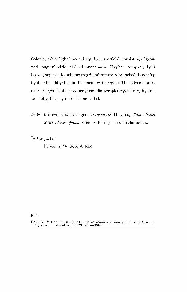
Colonies ash or light brown, irregular, superficial, consisting of grou-
ped long-cylindric, stalked synnemata. Hyphae compact, light
brown, septate, loosely arranged and ramosely branched, becoming
hyaline to subhyaline in the apical fertile region. The extreme bran-
ches are geniculate, producing conidia acropleurogenously, hyaline
to subhyaline, cylindrical one celled.
Note: the genus is near gen. Hans/ordia HUGHES, Tharo@ama
SUBR., Drum@area SUBR., differing for some characters.
In the plate:
V. swetasakha RAo & RA0
Ref.:
tZAO, D. & RAO, P. R. (1964) - gcdkshopama, a new genus of Stilbaceae. Mycopat. et Mycol. appl., 23: 286--290.

F U N G I I I ~ i P E R F E C T I
M O N I L I A L E S
M O N I L I A C E A E
S A P R O P H Y T A
Gen. Tr ichosporonoides HASKINS & SPENCER
I C O N O G R A P H I A M Y C O L O G I C A
V E R O N A - - B E N E D E K
Plate A 505
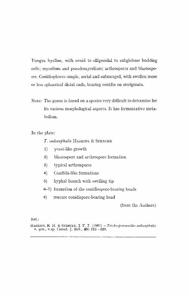
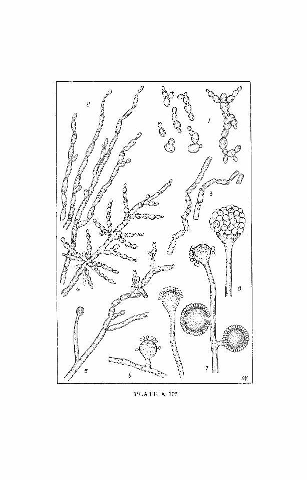
Fungus hyaline, with ovoid to ellipsoidal to subglobose budding
cells; mycelium and pseudomycelium; arthrospores and blastospo-
res. Conidiophores simple, aerial and submerged, with swollen more
or less sphaerical distal ends, bearing conidia on sterigmata.
Note: The genus is based on a species very difficult to determine for
its various morphological aspects. It has fermentative meta-
bolism.
In the plate:
T. oedocephalis HASKINS & SPENCER
1) yeast-like growth
2) blastospore and arthrospore formation
3) typical arthrospores
4) Candida-like formations
5) hyphal branch with swelling tip
6-7) formation of the conidiospore-bearing heads
8) mature conidispore-bearing head
(from the Authors)
Ref, :
HASKINS, R. H. & SPENCER, J. F. T. (1967) - Trichosporonoides oedocephalis n. gen., n.sp. Canad. J. Bot . , 4 5 : 515--520.
m~
~ ~ii ~

F U N G I I ~ I P E R F E C T I
M O N I L I A L E S
N I O N I L I A C E A E
S A P R O P H Y T A
or P H Y T O P A T H O G E N
(parasit ic on fungi)
Gen. T r i n a c r i u m RIEss
I C O N O G R A P H I A MYCOLOGICA
VERONA - - BENEDEK
Plate A 506
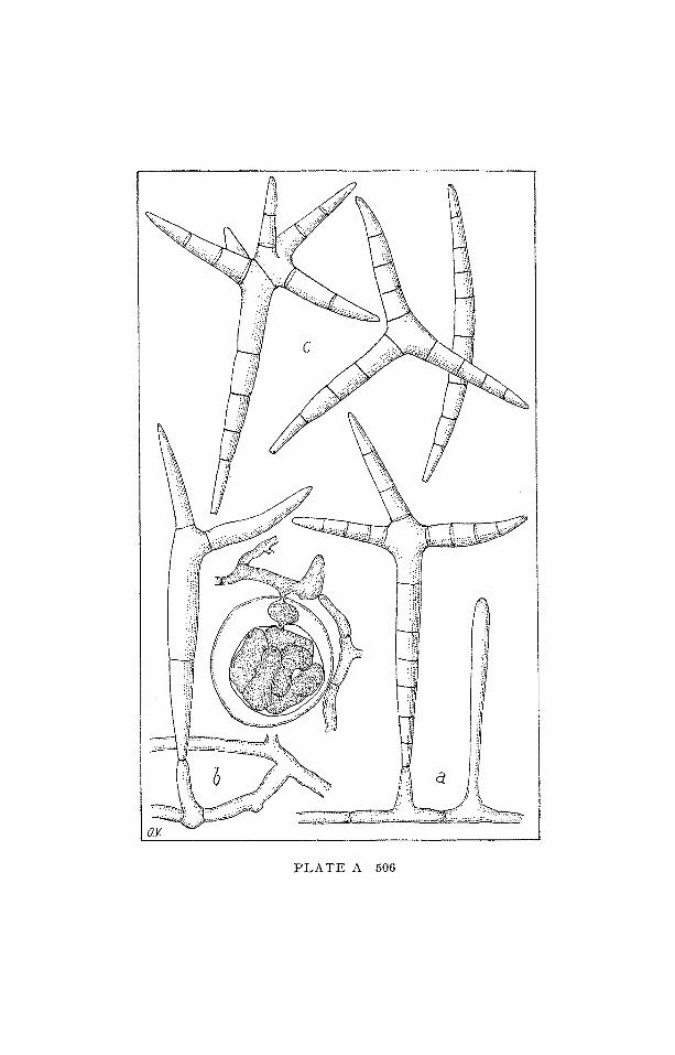
Conidiophores short, slender; conidia hyaline, multi-septate, un-
branched and long-spindle-shaped, or with one to four widely diver-
gent branches near the upper portion.
Note: On decaying vegetable material and parasitic on oospore of
Phythium.
In the plate:
T. subtile RIESS (from DRECHSLER)
a) A portion of superficial filament bearing a young coni-
diophorous process and a conidiophore supporting a ma-
ture cruciform conidium.
b) Portion of mycelium with a conidiophore bearing a some-
what immature conidium. Above, an oospore of Pythium
butleri occupied by the haustoriat system of T. subtile.
c) Some conidia in their various dimensions, shape and sep-
tation.
t{ef.:
I)R]~CI-ISLER, C. (1938) - Two Hyphomycetes parasitic on oospores of root- -rotting Oomycetes. Phytopathotogy, 28: 81--103.
C
P L A T E A 506

F U N G I I M P E R F E C T I
M O N I L I A L E S
M O N I L I A C E A E
ZOOPATHOGEN
Gen. G e n i c u l a r i a RIFAI & R. C. CooKE
I C O N O G R A P H I A M Y C O L O G I C A
V E R O N A - - B E N E D E K
Plate A 507
Colonies effuse, pale-pink. Mycelium with septate hyphae, hyaline,
branched, smooth. Conidiophores single, erect, straight at first, then
geniculate or flexuous, hyaline, smooth, successively growing by
subapical proliferation. Conidia single at apex of conidiophores, oh-
pyriform, smooth, hyaline or subhyaline, uniseptate, with the basal
cell smalJer. Predacious on nematodes.
Note: The predacious habit, the obpyriform, hyaline, uniseptate
conidia and the geniculate conidiophores which elongate by
subapical proliferation, serve to distinguish Genicularia from
other hyalodidymosporous genera (i.e. Trichothecium LINK).
The genus is based on Trichothecium cysto@orium DUDDING-
TON.
In the plate:
I - G. cystosporia (DUDDINGTON)
II - G. paucispora R. C. COOKE
n i - c. perpasta R. C. CooKE
RIFAI & R. C. COOKE
(from the Authors)
Ref.:
RIFAI, M. A. & COOKE, R. C. (1966) - Studies on some didymosporous genera of nematode-trapping Hyphomycetes. Trans. Brit. mycoh Soc., 49: I47 - - 168.

h~

FUNGI I1VfPERFECTI MONILIALES
MONILIACEAE
SAPROPHYTA
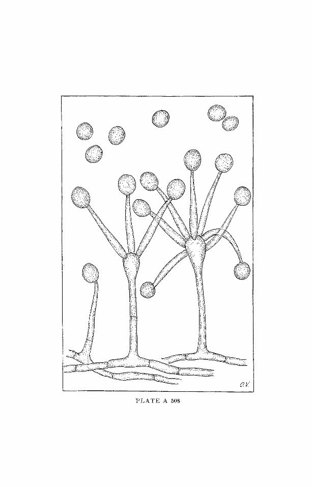
Gen. Umbelopsis AMOS & BARNETT
I C O N O G R A P H I A M Y C O L O G I C A
V E R O N A - - BENEDEK
Plate A 508
Conidiophores hyaline, often septate, bearing swollen head with
sporogenous branches at the apex; conidia one-celled, hyaline, glo-
bose.
Note: isolated from roots of quince, other plants, and soil.
In the plate:
U. versi/ormis AMos & BARNETT
(from the Authors)
Ref.:
AMos, R. E. & BARNETT, H.L. (1966) - Umbelopsis versi/ormis, a new genus and species of the Imperfects. Mycologia, 58: 805--808.
© © L ! f : :: z;~:. O0~: ::..!~;J
P L A T E A 508

F U N G I I M P E R F E C T I
~ O N I L I A L E S
DEMATIACEAE
S A P R O P H Y T A
Gen. C h l a m y d o r u b r a
K. B. DESHPANDE & K. S. DESHPANDE
ICONOGRAPHIA MYCOLOGICA
VERONA - - BENEDEK
Plate A 509
Colonies on PDA at first white, later green and finally red; young
hyphae hyaline with slight reddish tint; conidiophores absent;
chlamydospores thick-walled, dark-red, verrucose, arranged in
chains, single or intercalary.
Note: isolated from soil in India.
In the plate:
C. verrucosa K. B. ])ESH. & K. S. DEsm
(a and b from the authors)
Ref.:
DESHPANDE, K. B. ~¢ DESHPANDE, I~. S. (1966) -- CMamydorubra, a new ge- nus of I ) ema t i aceae in India . 1V[ycopat. & Mycol. appl. , 2 9 : 270- -272 .

F U N G I I M P E R F E C T I
M O N I L I A L E S
D E M A T I A C E A E
SAPROPHYTA
I - Gen. H o r m i o k r y p s i s BAT. & NASC.
II - Gen. H e p t a s t e r CIF., BAT. & 1NASC.
I I I - Gen. P e n t a s p o r i u m BATISTA
IV - G e n . M e g a s t e r Ct~ERRI
ICONOGRAPHIA MYCOLOGICA
VERONA -- BENEDEK
Plate A 510
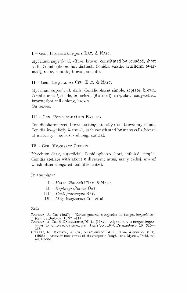
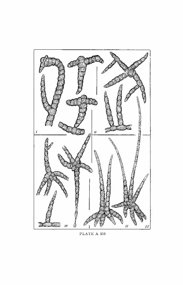
I - Gen. H o r m i o k r y p s i s BAT. & NASC.
Mycelium superficial, effuse, brown, cons t i tu ted b y rounded, short cells. Conidiophores not distinct. Conidia sessile, cruciform (4-ar- med), many-septa te , brown, smooth.
I I - Gen. H e p t a s t e r CIF., BAT. & ~NASC.
Mycelium superficial, dark. Conidiophores simple, septate, brown. Conidia apical, single, branched, (6-armed), irregular, many-cel led,
brown; foot cell oblong, brown. On leaves.
III - Gen. Pentasporium ]3ATISTA
Conidiophores erect, brown, arising laterally from brown mycelium. Conidia irregularly 5-armed, each constituted by many cells, brown at maturity. Foot cells oblong, conical.
IV - Gen. M e g a s t e r CIFERRI
Mycelium dark, superficial. Conidiophores short, inflated, simple. Conidia stellate with about 6 divergent arms, m a n y celled, one of which often elongated and a t tenua ted .
In the plate:
I - Horm. librocedri BAT. & NASC. I I - Hept.regmllianae BAT.
I I I -- Pent./oumroyae BAT. IV - Meg. iongicornis CIF. et al.
Ref. :
BATISTA, A. CH. (1957) - Xovos generos e especies de fungos imperfeitos. Rev. de Biologia, 1: 97--112.
B~TISTA, A. CH. & NASCIMENTO,'M. L. (1957) -Alguns novos fungos imper- feitos do comptexo de fumagina. Anais Soc. Biol. Pernambuco, 15: 345-- 353.
CIFERRI, R., BATISTA, A. CH., NASClMEN~O, ~1. L. & de AZEVEDO, P. C. (1956) - Another new genus of staurosporic fungi. Inst. Mycol., Publ. no. 48, Recife.
'F::

F U N G I I M P E R F E C T I
M O N I L I A L E S
D E M A T I A C E A E
S A P R O P H Y T A
Gen. Gra l lomyces F. L. STEVENS
I C O N O G R A P H I A M Y C O L O G I C A
V E R O N A - - B E N E D E K
Plate A 511
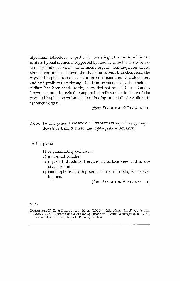
Mycelium foliicolous, superficial, consisting of a series of brown septate hyphal segments supported by, and attached to the substra- tum by stalked swollen attachment organs. Conidiophores short~ simple, continuous, brown, developed as lateral branches from the mycelial hyphae, each bearing a terminal conidium as a blown-out end and proliferating through the thin terminal scar after each co- nidium has been shed, leaving very distinct annellations. Conidia brown, septate, branched, composed of cells similar to those of the mycelial hyphae, each branch terminating in a stalked swollen at- tachment organ.
(from D~IGHTO~ & PIROZYNSKI)
Note: To this genus DEIGHTON & PIROZYNSKI report as synonym Phialetea BAT. & NASC. a n d Ophiopodium ARNAUD.
In the plate:
1) A germinating conidium; 2) abnormal conidia; 3) mycelial attachment organs, in surface view and in op-
tical section; 4) conidiophores bearing conidia in various stages of deve-
lopment. (from DEIGHTON • PIROZYNSKI)
IRef.:
I)EIGHTON, F. C. 6: PIROZYNSKI, K. A. (1966) - Microfungi II. Brooksia and Grallomyces; A crogenotheca ornata sp. nov.; the genus Xenosporium. Com- monw. Mycoh Inst., Mycol. Papers, no 105.
t~
t~
¢.7t
~
d


F U N G I I ~ i P E R F E C T I
l V I O N I L I A L E S
D E M A T I A C E A E
S A P R O P H Y T A
Gen. Hermatomyces SPEG.
I C O N O G R A P H I A 3 I Y C O L O G I C A
V E R O N A - - B E N E D E K
Plate A 512
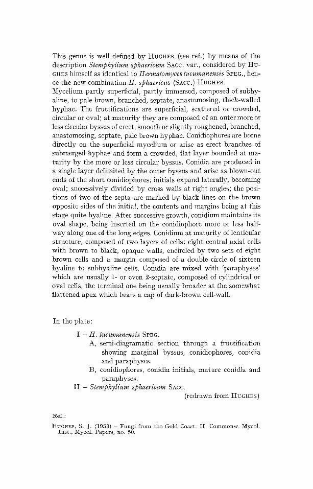
This genus is well defined by HUGHES (see ref.) by means of the description Sternphylium sphaericum SAcc. var., considered by HU- GHES himself as identical to Hermatomyces tucumanensis SPEG., hen- ce the new combination H. sflhaericus (SAcc.) HUGHES. Mycelium partly superficial, partly immersed, composed of subhy- aline, to pale brown, branched, septate, anastomosing, thick-walled hyphae. The fructifications are superficial, scattered or crowded, circular or oval; at matur i ty they are composed of an outer more or less circular byssus of erect, smooth or slightly roughened, branched, anastomosing, septate, pate brown hyphae. Conidiophores are borne directly on the superficial mycelium or arise as erect branches of submerged hyphae and form a crowded, flat layer bounded at ma- tur i ty by the more or less circular byssus. Conidia are produced in a single layer delimited by the outer byssus and arise as blown-out ends of the short conidiophores; initials expand laterally, becoming oval; successively divided by cross walls at right angles; the posi- tions of two of the septa are marked by black lines on the brown opposite sides of the initial, the contents and margins being at this stage quite hyaline. After successive growth, conidium maintains its oval shape, being inserted on the conidiophore more or less half- way along one of the long edges. Conidium at matur i ty of lenticular structure, composed of two layers of cells: eight central axial cells with brown to black, opaque walls, encircled by two sets of eight brown cells and a margin composed of a double circle of sixteen hyaline to subhyaline cells. Conidia are mixed with 'paraphyses' which are usually 1- or even 2-septate, composed of cylindrical or oval cells, the terminal one being usually broader at the somewhat flattened apex which bears a cap of dark-brown cell-wail.
In the plate:
I - H. tucumanensis SPEG. A, semi-diagramatic section through a fructification
showing marginal byssus, conidiophores, conidia and paraphyses.
]3, conidiophores, conidia initials, mature co11~dia and paraphyses.
II - Stemflhylium sphaericum Shcc. (redrawn from HUGHES)
Ref. :
HUGHES, S. J. (1953) - Fungi from the Gold Coast. II. Commonw. Mycol. Inst., Mycol. Papers, no. 50.
.g


F U N G I I M P E R F E C T I
M O N I L I A L E S
D F.iVIAT I A C E A E
S A P R O P H Y T A
Gen. E x c i o c o n i d i u m O. A. PLUNKETT
I C O N O G R A P H I A MYCOLOGICA
V E R O N A - - B E N E D E K
Plate A 513
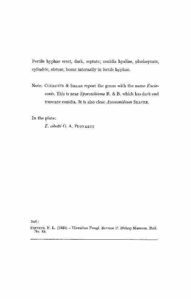
Fertile hyphae erect, dark, septate; conidia hyaline, pluriseptate,
cylindric, obtuse, borne internally in fertile hyphae.
Note: eLEMENTS & SHEAR report the genus with the name Excio-
conis. This is near S])oroschisma B. & B. which has dark and
truncate conidia. It is also close Ascoconidium SEAVER.
In the plate:
E. cibotti O. A. PLUNKETT
Ref.:
S~EVENS, F. L. (1925) - Hawaiian Fungi. Bernice P. Bishop Museum, Bull. No. 19.
t~


FUNGI I M P E R F E C T I MONILIALES
D EI~,IATIACEAE
SAPROPHYTA
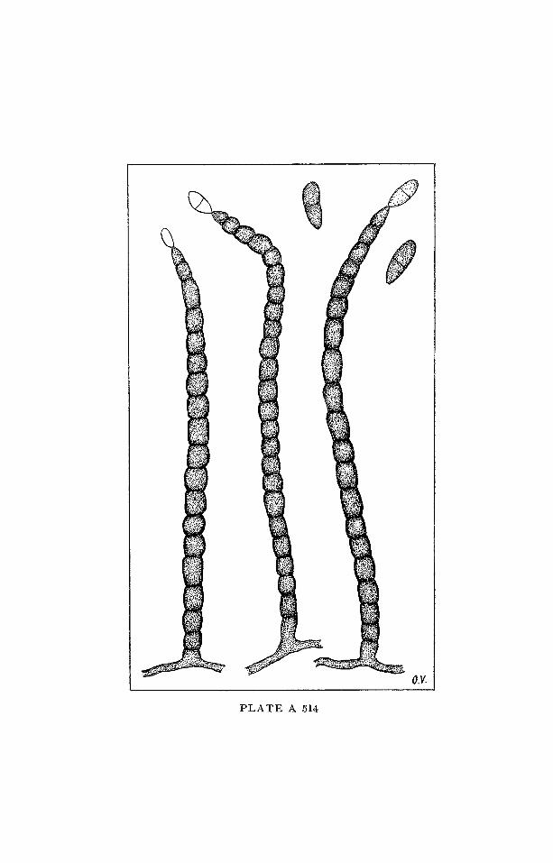
Gen. R a m a l i a BATISTA
I C O N O G R A P H I A M Y C O L O G I C A
VERONA - - BENEDEK
Plate A 514
Mycelium superficial, septate, brown. Conidiophores grouped, erect,
wavy or irregular, simple, brown. Conidia apical, single, at first hya-
line, later dark, 2-celled.
In the plate:
R. veronicae BAT. (from the Author)
Ref,:
BATISTA, A. CH. (1957) -Novos generos e especies de fungos imperfeitos. Rev. de ]3iologia, 1: 97--112.
t~
%

FUNGI IlVIPE RFECTI IVfONILIALES
DEMATIACEAE
SAPROPHYTA
Gen. P e g l i o n i a G. GOlD.
I C O N O G R A P H I A M Y C O L O G I C A
V E R O N A - - B E N E D E K
Plate A 515
Sterile hyphae erect, black, septate, rigid, verticillately ramose at
apex. Conidiophores short, flaskshaped, hyaline, inserted at base of
sterile hyphae. Conidia hyaline, bent, solitary, acrogenous.
Note: The genus differs from Circinotrichum NEES ex PERSO0~¢
(I.M. X-A-218) as the last one has circinate setae. I t is more
close to Gyrothrix (CDA.) CDA. which has setae 'repeatedly
branched, erect, straight, of flexuous, s e p t a t e . . . ' (PII~O-
ZYNSKI).
In the plate:
P. verticiclada G. Gon).
Ref. :
GOIDANICH, C*. (1934) - U n n u o v o genere di Demaz iaceae amerospore . Mal- pighia, 33.
PIROZYNSKI (1962) -- Circinotrichum and Gyrolhrix Commonw. Mycol. Ins t . , Mycol. Papers , no. 84.
~q

FUNGI I M P E R F E C T I MONILIALES
STILBACEAE
S A P R O P H Y T A
Gen. P y c n o s t y s a n u s LINDAU
I C O N O G R A P H I A M Y C O L O G I C A
VERONA ~ BENEDEK
Plate A 516
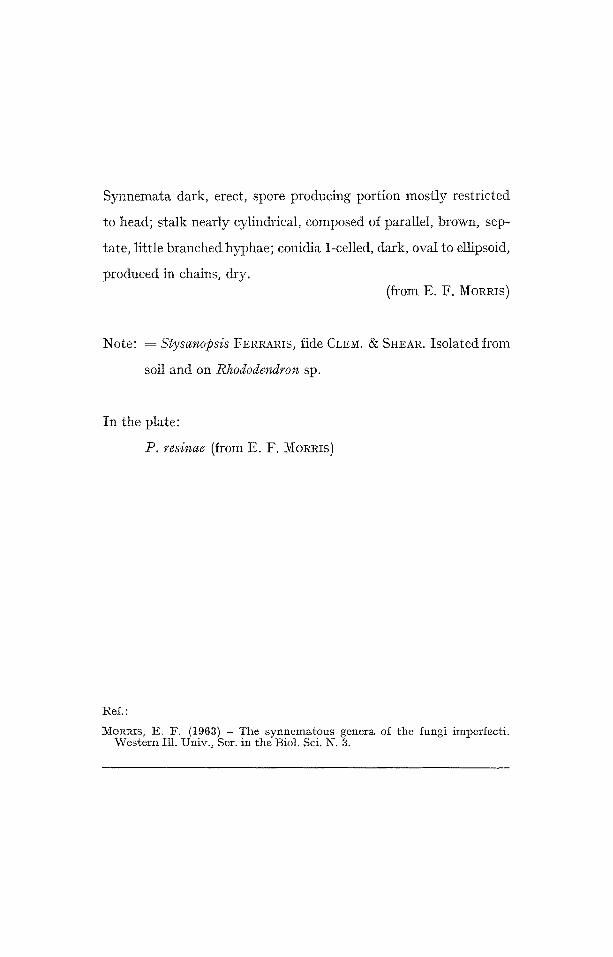
Synnemata dark, erect, spore producing portion mostly restricted
to head; stalk nearly cylindrical, composed of parallel, brown, sep-
tare, little branched hyphae; conidia 1-celled, dark, oval to ellipsoid,
produced in chains, dry. (from E. F. MORRIS)
Note: = Stysanopsis FERRARIS, fide CLEM. & SI~EAR. Isolated from
soil and on Rhododendron sp.
In the plate:
19. resinae (from E. F. MORRIS)
Ref.:
~V~ORRIS, E, F. ( 1 9 6 8 ) -- The synnematous genera of the fungi imperfecti. Western Ill. Univ., Ser, in the Biol. Sci. N. 3.

F U N G I I ~ P E R F E C T I M O N I L I A L E S
S T I L B A C E A E
ZOOPATHOGEN (Insecticole or S A P R O P H Y T A in soil)
Gen . T i l a c h l i d i o p s i s KEISSLER
I C O N O G R A P H I A M Y C O L O G I C A
V E R O N A - - B E N E D E K
Plate A 517

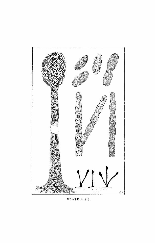
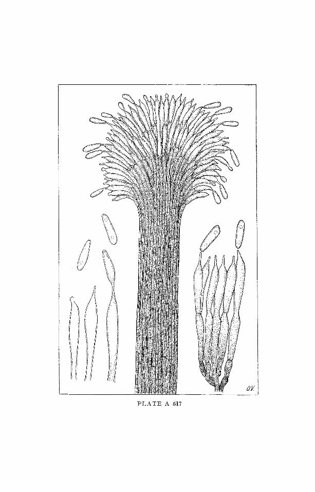
Synnemata brown below and lighter colored above, capitate, arising
from a brown rhizomorph-like, sterile, central axis; conidiophores
unbranched, diverging at apex, forming a palisade of phialides in
the head; conidia 1-celled, hyaline, ovate-oblong, gut tulate , pro-
duced singly.
Ref. :
MAINs, E. ]3. (1951) - Notes concerning entomogenous fungi. BuI1. Torrey Bot. Club, 78: 122--133.
MORRIS, E. F. (1963) -- The synnematous genera of the fungi Imperfecti. Western Ii1. Univ., Ser. in the Biol. Sci., n. 3.
~ ...
....
....
....
....
....
. f
t~
t~

F U N G I I M P E R F E C T I
3 / I O N I L I A L E S
S T I L B A C E A E
S A P R O P H Y T A
Gen. S c l e r o s t i l b u m PovAI~
I C O N O G R A P H I A MYCOLOGICA
V E R O N A - - B E N E D E K
Plate A 518
Synnemata arising from a sclerotium, with a sterile central axis,
lateral branches usually terminating in a depressed-globose head,
bearing conidia; conidiophores branched; conidia 1-celled, hyaline,
elliptical catenulate.
Note: POVAH advances that the genus might represent the imper-
fect state of a species of Xylaria. It is also given as imper-
fect state of Collybia (SINGER, 1951).
I
I
r

F U N G I I M P E R F E C T I
lVIOIgILIALES
S T I L B A C E A E
Z O O P A T H O G E N
(Insecticole and S A P R O P H Y T A )
Gen. S t i l b e l l a LINDAU
I C O N O G R A P H I A M Y C O L O G I C A
V E R O N A - - B E N E D E K
Pla te A 519
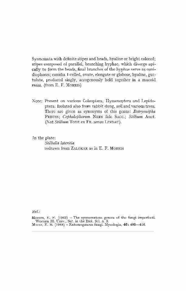
Synnemata with definite stipes and heads, hyaline or bright colored; stipes composed of parallel, branching hyphae, which diverge api- cally to form the heads, final branches of the hyphae serve as coni- diophores; conidia 1-celled, ovate, elongate or globose, hyaline, gut- tulate, produced singly, acrogenously held together in a mucoid mass. (from E. F. MORRIS)
Note: Present on various Coleoptera, Hymenoptera and Lepido- ptera. Isolated also from rabbit dung, soil and various trees. There are given as synonyms of this genus: Botryon@ha PREuss; Cephalophorum NEES fide SAcc.; Stilbum Auct. (Not Stilbum Tope ex FR. sensu LIXDAU).
In the plate: Stilbella laleri~ia redrawn from ZALOKAR as i n E . F . MORRIS
Ref. :
MORRIS, E. F. (1963) - The synnematous genera of the fungi imperfecti . Wes te rn Ill. Univ. , Set. in the Biol. Sci. n. 3.
~¢~AINS, E. t3. (1948) -- En tomogenous fungi. Mycologia, 40 : 402--416.
s
i
\,


F U N G I I 1 V f P E R F E C T I
M O N I L I A L E S
S T I L B A C E A E
S A P R O P H Y T A
Gen. V i r g a t o s p o r a FINLEY
ICONOGRAPHIA MYCOLOGICA
V E R O N A - - B E N E D E K
Plate A 520
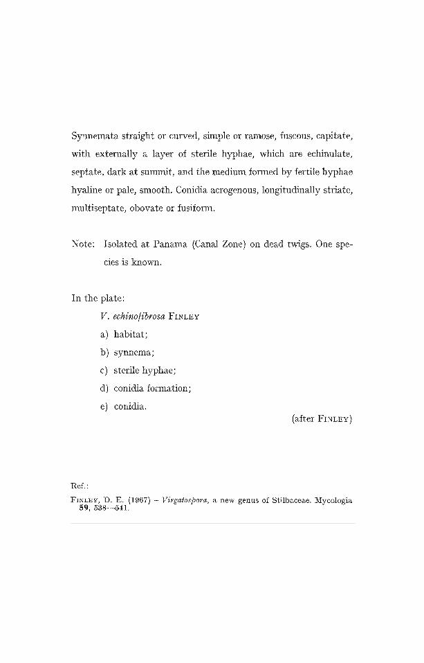
Synnemata straight or curved, simple or ramose, fuscous, capitate,
with externally a layer of sterile hyphae, which are echinutate,
septate, dark at summit, and the medium formed by fertile hyphae
hyaline or pale, smooth. Conidia acrogenous, longitudinally striate,
multiseptate, obovate or fusiform.
Note: Isolated at Panama (Canal Zone) on dead twigs. One spe-
cies is known.
In the plate:
V. echino/ibrosa FINLEY
a) habitat;
b) synnema;
e) sterile hyphae;
d) conidia formation;
e) conidia. (after FINLEY)
Ref."
FINLEY, I). E . (1967) -- Virgalospora, a n e w g e n u s o f S t i l b a c e a e . 1Kycologia 59 , 538-- -541.
t~
J~
I Iq
~ l
Jhm
~Jm
l
c~

F U N G I I M P E R F E C T I M O N I L I A L E S
T U B E R C U L A R I A C E A E
P H Y T O P A T H O G E N
(Hyperparasite)
Gen. T u b e r c u l i n a SAcc.
I C O N O G R A P H I A M Y C O L O G I C A
VERONA -- BENEDEK
Pla te A 521
Sporodochia small, arising within or immediately near the rust
fructifications. Conidiophores hyaline, simple, bearing a terminal
conidium. Conidia 1-celled, hyaline, globose or ovoid.
Note: Several species parasitic on rusts are known.
In the plate:
1 - T. m a x i m a ROSTR., parasitizing Cronar t ium ribicola
(LAscm) FISCHER de ~VALI)H.
g - T. pers ic ina (DIT.) SACC.
3 - T . vinosa SAcc. (2, 3, from SAVULESCU)
--t q zJ

F U N G I I M P E R F E C T I
M O N I L I A L E S
T U B E R C U L A R I A C E A E
S A P R O P t { Y T A
Gen. V i n c u t u m R. Y. RoY et al.
I C O N O G R A P H I A M Y C O L O G I C A
V E R O N A - - B E N E D E K
Plate A 522
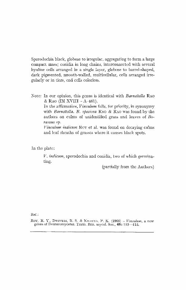
Sporodochia black, globose to irregular, aggregating to form a large compact mass; conidia in long chains, interconnected with several hyaline cells arranged in a single layer, globose to barrel-shaped, dark pigmented, smooth-walled, multicellular, cells arranged irre- gularly or in tiers, end cells colorless.
Note: In our opinion, this genus is identical with Barnettella RAO & RAO (IM x v i i i - A - 4 6 1 ) .
In the affirmative, Vinculum falls, for priority, in synonymy with Barnettella. B. @eciosa RAO & RAo was found by the authors on culms of unidentified grass and leaves of Bo- rassus sp. Vinculum indicum RoY et al. was found on decaying culms and leaf sheaths of grasses where it causes black spots.
In the plate:
V. indicum, sporodochia and conidia, two of which germina- ting.
(partially from the Authors)
Ref. :
ROY, R. Y., DWtV•DI, R. S. & •HANNA, P. K. (1965) -- Vinculum, a new genus of Deu te romyce tes . Trans . Br i t . mycol . Soc., 4 8 : 113--115.
L~

F U N G I I M P E R F E C T I
M E L A N C O N I A L E S
5 i E L A N C O N I A C E A E
S A P R O P t I Y T A
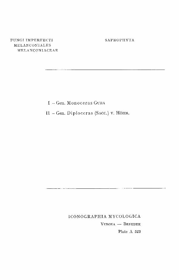
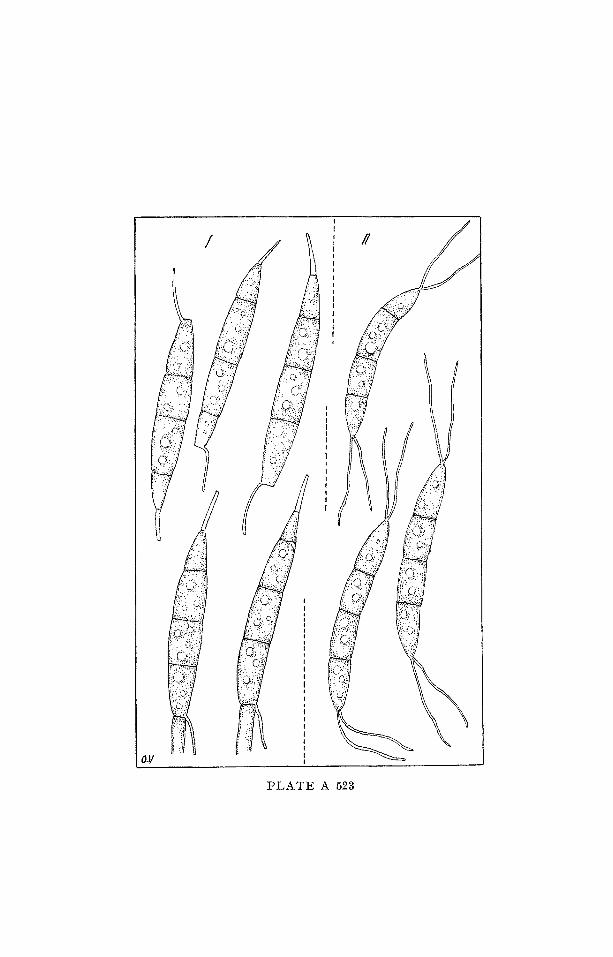
I - Gen. M o n o c e r a s GUBA
II - G e n . D i p l o c e r a s (SAcc.) v. HOHN.
I C O N O G R A P H I A MYCOLOGICA
V E R O N A - - B E N E D E K
Plate A 523
I - Gen. M e n o c e r a s GUBA
Acervuli simple, lenticular or globose-depressed, black. Conidia mul- tiseptate, usually 4-celled, narrow-fusiform to ob-lanceolate, broade- ned below, tapering toward the apices; apical ceils acute, basal cell cylindric, truncate, all cells with contents, at first subhyaline then pale yellow throughout; one filiform erect setula affixed at apices, distinct from acute apical cells, one filiform oblique persisting setula affixed at bases; pedicels thick, deciduous.
Note: The genus was created by GUBA on the species Pestalotia kriegeriana BRES., species early considered as Monochaetia kriegeriana (BREs.)ALLESCt{ER and Hyaloceras kriegerianum (BREs.) DIED. Such a species was described on leaves of Efli- lobium angusti/olium L. The genus is similar to D@loceras, except that the extremities of the conidia are 1-setulate.
II - G e n . D i p l o c e r a s (SAcc.) v. H6I-IN.
The genus is similar to Monoceras, differing by having bisetulate conidia at both extremities. Considered by SACCARDO as sub-genus (Syll. Fungorum, 10, 1484, (1892), was elevated to genus by yon H6HNEL (Syst. Fung. Imp., 342, (1923)) with the species Hyaloceras dilophosporum COOKE. To the genus was also referred Pestalotia hypericina CES. Actually these are indicated as Di~loceras dil@ho- @orum (COOKE) SAcc. and Diploceras hypericinum (CEs.) DIED.
In the plate:
I - Monoceras kriegerianum (BRES.) GUBA II - Diploceras hypericinum (CEs.) DIED.
Ref.:
CvUBA, E. M. (1981) - Monograph of Monochaetia and Pestalotia. Cambridge, Mass.
t~

F U N G I I 3 ~ P E R F E C T I
M E L A N C O N I A L E S M E L A N C O N I A C E A E
P H Y T O P A T t t O G E N
or S A P R O P H Y T A
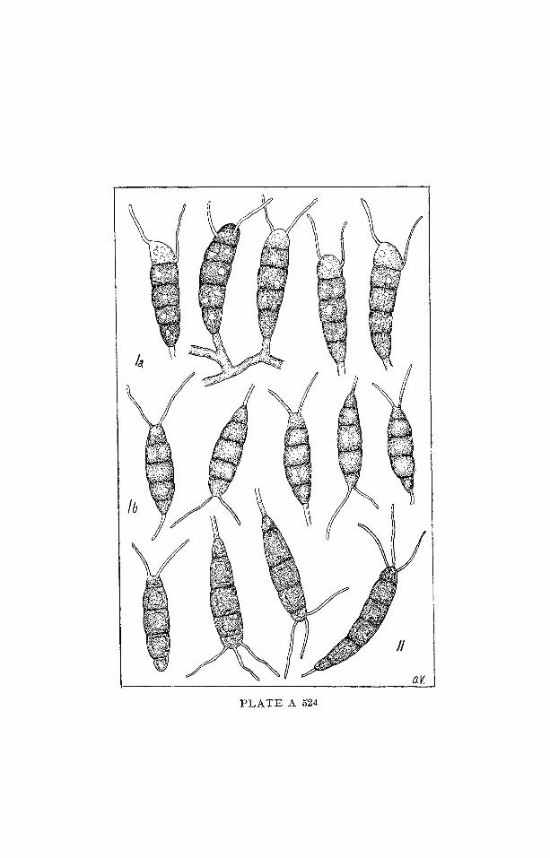
G e n . P e s t a l o z z i n a SACC. e m . GUBA
I C O N O G R A P H I A M Y C O L O G I C A
V E R O N A - - BENEDEK
Pla te A 524
Acervuli simple without stroma, dark-colored or black, oblong or globose-lenticular, depressed, at maturity breaking and freeing the contents. Conidia 5-celled, cuneiform, clavate or somewhat fusiform, somewhat unequilateral, yellow or pale yellow-brown, color dimi- nishing toward the extremities, all cells with contents; slightly con- stricted at septa, in form comparable to slugs or horned insect lar- vae; apical cells hemisphaericaI; setulae flexuous, acropleurogenous (projecting from the side, base and apex of the apical cells); basal cells conoid, rounded at base, resting in a short pedicel.
Note: Pestalozzina was at first considered by SACCARDO (1884) as a sub-genus of Pestalotia. It encluded species with hyaline con- idia. Later he elevated it (1892) to genus. GUBA maintains Pestalozzina in the range of genus, emending the diagnosis, and encludes species with hyaline conidia within the parallel genus Hyalotia (see: IM XX-A-527).
In the plate:
I a - P. thuemenii (SPEG.) GUBA (=Pestalotia thuemenii SPEG.)
I b - P. thuemenii (SPEG.) GUBA (=Pestalotia monochroa TAssi)
II - P . unicoIor (BERK. • CURT.) SACC.
Ref.:
GUBA, E. IVL (1961) - ~onograph of Monochaelia and ~°estalotia. Cambridge, Mass.
~j

FUNGI IMPER]?ECTI
MELANCONIALES
5IELAXCONIACEAE
PKYTOPATHOGEN
Gen. C y l i n d r o s p o r i u m UN6ER
I C O N O G R A P H I A M Y C O L O G I C A
V E R O N A - - B E N E D E K
Plate A 525
Acervuli subepidermal white or pale; conidiophores short, simple;
conidia hyaline, filiform, straight or curved, 1-celled or becoming
septate.
Note: It is parasitic on leaves. Many species are metagenetically
related with Mycosphaerella Join. or even Gnomonia CEs. &
Dz NOT., i.e.: C. castaneicolum (DESM.) BEI~L. (Mycosphae-
rella maculi/ormis (PERS.) SCHR.);
C. pruni-cerasi MASSAL (Gnomonia erythrostoma (PERs.)
AtJEI~SW.) Other species belong to the life cycle of Entyloma
DE gARY and Doassansia CORNU.

~J
m~
~j


F U N G I I M P E R F E C T I
M E L A N C O N I A L E S
l V I E L A N C O N I A C E A E
S A P R O P H Y T A
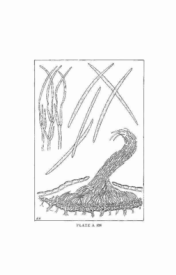
Gen. L i b e r t e l l a DESMAZIi~RES
I C O N O G R A P H I A M Y C O L O G I C A
VERONA - - BENEDEK
P l a t e A 526
Stroma always subepidermal. At the moment of spore formation,
the epidermis slightly heaves up and rends, liberating the spore
mass which often forms curls. Conidiophores branched, hyaline,
bearing filiform, hyaline, i-celled conidia.
Note: Caulicole species, some of which are considered as conidial
forms of Diatrype FR. and Q¢~aternaria TL~L.
b~

F U N G I I M P E R F E C T I
M E L A N C O N I A L E S
M E L A N C O N I A C E A E
P H Y T O P A T H O G E N
or S A P R O P H Y T A
I - Gen. H y a t o t i a GUBA
II - Gen. H y a l o t i e l l a PAPENDORF
I C O N O G R A P H I A M Y C O L O G I C A
VERONA - - BENEDEK
Plate A 527
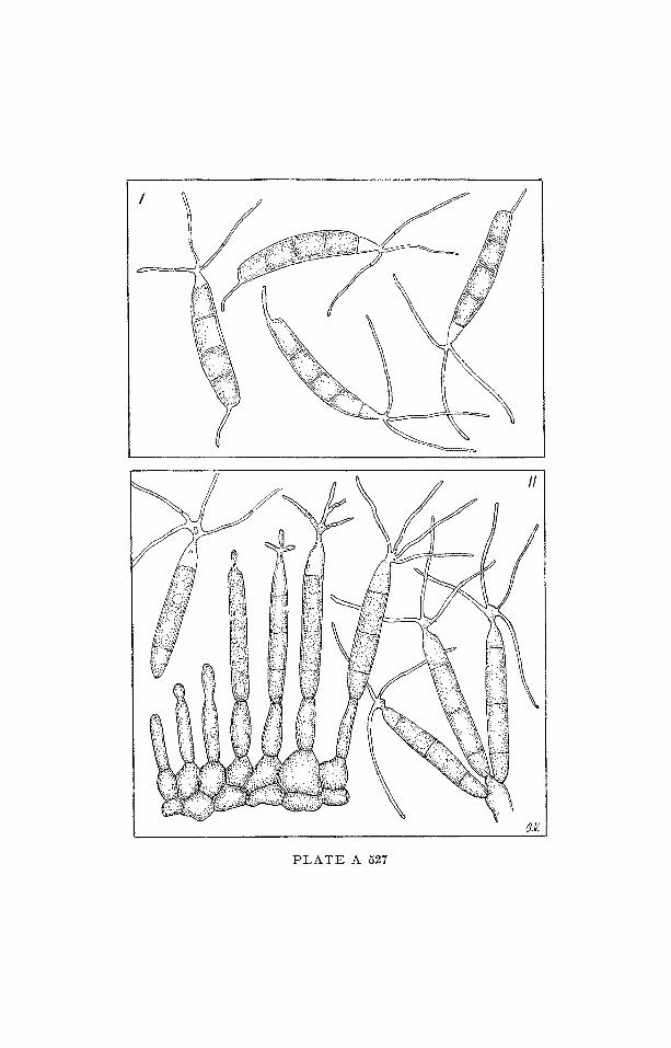
I - Gen. H y a l o t i a GUBA
Acervuli simple, hemisphaerical, globose-depressed, monoloculate, black or dark colored, innate-erumpent, exposing the contents. Conidia narrow-fusiform, at first continuous, then usually 4-septate, rarely 5-septate, all cells hyaline with contents, pointed apices cres- ted with 2 or more, usually 3 setulae; basal cells t runcate at base, resting obliquely on hyaline, short, filiform pedicels.
Note: This genus has been created on a species (h r. laurina (MONT.) GUBA) earlier denominated P estalozzina lauri na. P estalozzina was earlier considered by SACCARDO a sub-genus of Pesta- lotia then elevated to genus, with differential character against Pestalotia the hyaline color of spores. GUBA emends Gen. Pestalozzina, referring it to the phaeophragmous Melan- coniaceae, and includes, on the contrary, species with hyaline conidia, into gen. Hyalotia.
In the plate: H. laurina (MONT.) GUBA = Conidia.
I I - Gen. H y a l o t i e l l a PAPENDORF
Fruit ing pustule pycnidium-like, dark-brown to black, irregular, globose or lobate, finally discoid; conidiophores elongate, cylindrical, simple or branched; conidia (ateuriospores) cylindrical at first, con- tinuous, then 3-septate, rarely 4-septate, subhyaline, apical cell hyaline, clear, narrow-cylindrical, pointed and crested with two to four simple or rarely branched setulae, basal cell rounded or t run- cate at the base, without supporting pedicel.
Note: The genus is similar to Hyalotia, differing by the absence of the basal pedicel. Isolated from soil. Hyalotia and Hyalo- tiella both differ from Pestalotia by the hyaline color of spores (cfr. Pestalozzina, I.M. XX-A-524)
In the plate: [t. transvalensis PAPENDORF
Rcf, :
GUBA, E. F. (1961) - Monograph of Monochaetia and Pestalotia. Cambridge, Mass.
PAPENDORF, M. C. (1967) - Two new genera of soil fungi from South Africa. Trans. Brit. myeol. Soc., 50: 69--75.
t~
~q
b,.9

F U N G I I M P E R F E C T I
M E L A N C O N I A L E S
M E L A N C O N I A C E A E
S A P R O P I - t Y T A
or P H Y T O P A T H O G E N
Gen. C r y p t o s p o r i u m KUNZE ex FR.
I C O N O G R A P H I A M Y C O L O G I C A
V E R O N A ~ B E N E D E K
Pla t e A 528
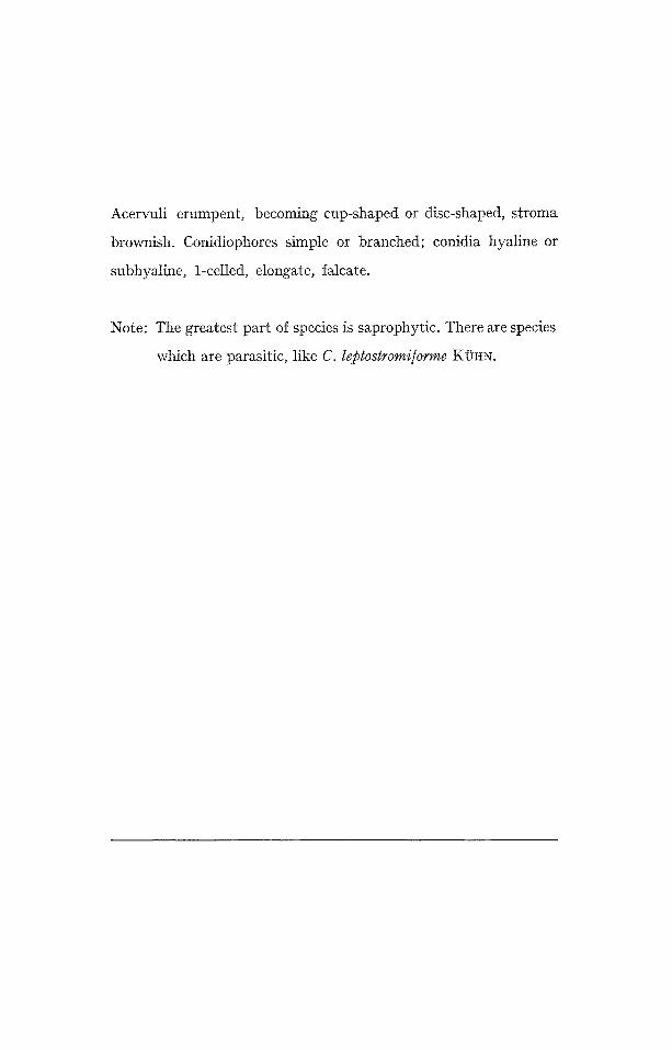
Acervuli erumpent, becoming cup-shaped or disc-shaped, stroma
brownish. Conidiophores simple or branched; conidia hyaline or
subhyaline, 1-celled, elongate, falcate.
Note: The greatest part of species is saprophytic. There are species
which are parasitic, like C. leptostromiforme KOHN.
1
07 ,
P L A T E A 528

F U N G I I M P E R F E C T I
SPHAEROPSIDALFS SAPROPHYTA
Gen. Aposphaeria SAcc.
I C O N O G R A P H I A M Y C O L O G I C A
V E R O N A - - B E N E D E K
Plate A 529
Pycnidia erumpent to superficial, single or clustered, dark, globose
with a short papillate ostiole; conidiophores short, 1-celled; conidia
hyaline, 1-celled, globose, ovoid or elongate.
Note: Saprophytic in wood.
~.~
t,~
o 0
o 0 0
0

F U N G I I M P E R F E C T I
S P H A E R O P S I D A L E S
P H Y T O P A T H O G E N
Gen. A p i o c a r p e l l a SYDOW
ICONOGRAPHIA MYCOLOGICA
V E R O N A - - B E N E D E K
Plate A 530
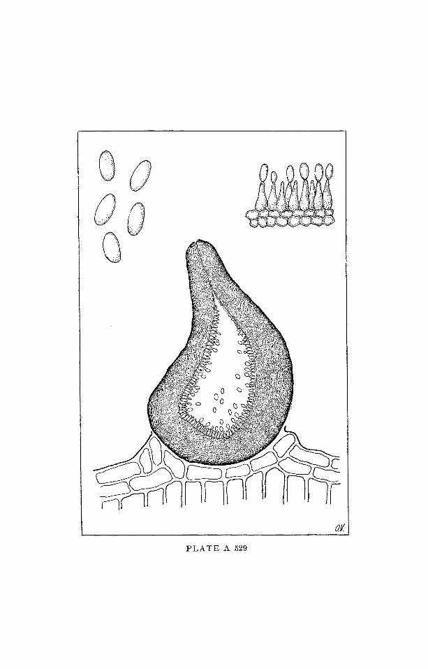
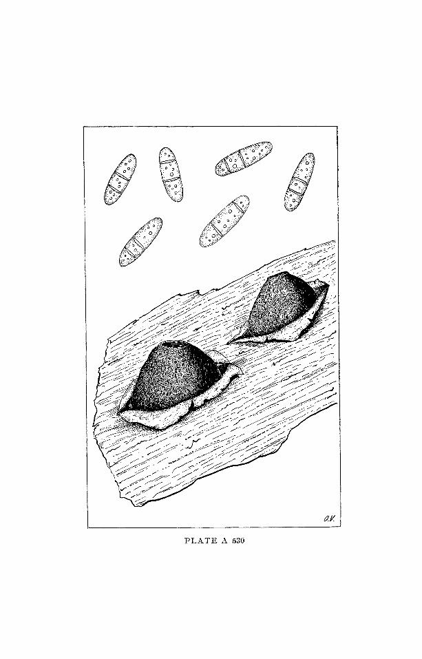
Pycnidia globose, erumpent, brown, ostiolate. Conidia hyaline,
unequally 2-celled, ellipsoid or cylindrical.
Note: Causing leaf spots on grasses.

F U N G I I M P E R F E C T I
S P H A E R O P S I D A L E S
P H O M A C E A E
S A P R O P H Y T A
Gen. R e a d e r i e l l a SYDow
I C O N O G R A P H I A M Y C O L O G I C A
V E R O N A - - B E N E D E K
Plate A 531
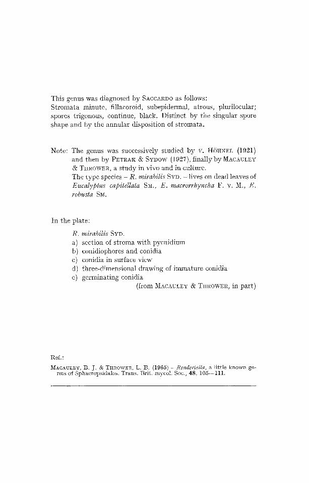
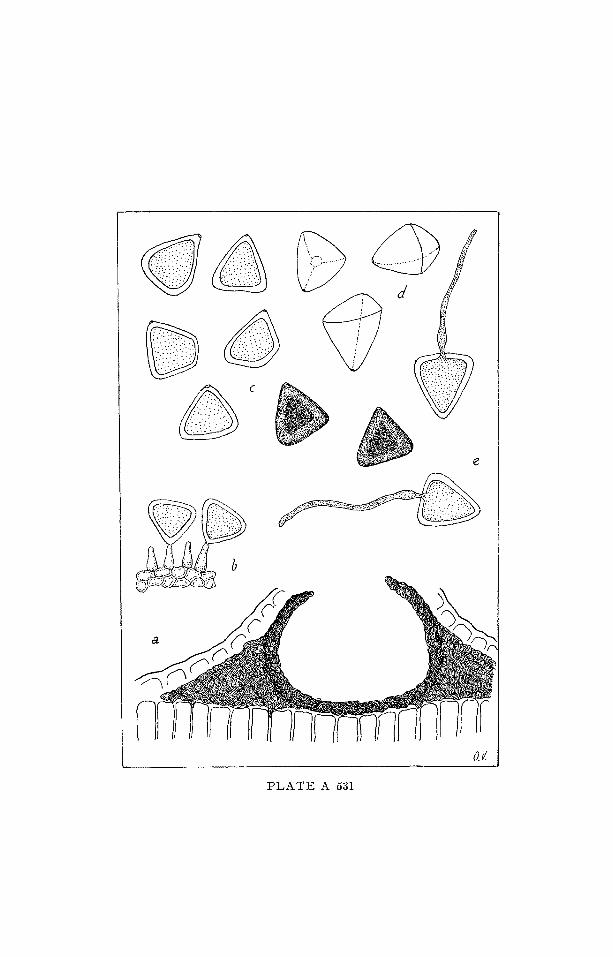
This genus was diagnosed by SACCARI)O as follows: Stromata minute, fillacoroid, subepiderma~, atrous, plurilocular; spores trigonous, continue, black. Distinct by the singular spore shape and by the annular disposition of stromata.
Note: The genus was successively studied by v. H{SttNEL (1921) and then by PETRAK & SYI)OW (1927), finally by MACAIJLE¥ & T~tROWER, a s tudy in vivo and in culture. The type species - R. mirabilis SYD. - lives on dead leaves of Eucalyptus capitdlata S~a., E. rnacrorrhyncha F. v. M., E. robusta SM.
In the plate:
R. mirabilis SYD. a) section of stroma with pycnidium b) conidiophores and conidia c) conidia in surface view d) three-dimensional drawing of immature conidia e) germinating conidia
(from MACA~LEY & THROWER, in part)
Ref.:
MACAULEY, B. J. • TItROW~ER, L. 13. (1965) - Reade~'ietla, a little known ge- nus of Sphaeropsidales. Trans. Brit. mycoI. Soc., 48, 105--111.
P L A T E A 531

ASCO3/[YCETES
H Y P O C R E A L E S
N E C T R I A C E A E
S A P R O P I - t Y T A
Gen. O r c a d i a SUTHERLAND
I C O N O G R A P H I A M Y C O L O G I C A
V E R O N A - - B E N E D E K
Plate C 284
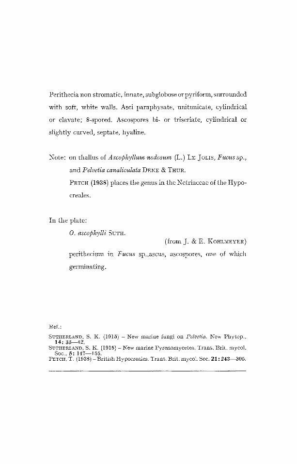
Perithecia non stromatic, innate, subglobose or pyriform, surrounded
with soft, white walls. Asci paraphysate, unitunicate, cylindricaI
or clavate; 8-spored. Ascospores bi- or triseriate, cylindrical or
slightly curved, septate, hyaline.
Note: on thallus of Ascophyllum nodosum (L.) LE JoLIS, Fucus sp.,
and Pelvetia canaliculata D~,Z~F~ & THUR.
PETC~ (1938) places the genus in the Netriaceae of the Hypo-
creales.
In the plate:
O. ascophylli SUTH. (from J. & E. KOHLMEYER)
perithecium in Fucus sp.,ascus, ascospores, one of which
germinating.
~ef."
SUTHERLAND, S. K. (1915) - New marine fungi on Petvetia. New Phytop., 14: 33--42.
SUTHERLAND, S. K. (1915) - New marine Pyrenomycetes. Trans. Brit. mycol. Soc., 5: 147--155.
Px~cH, T. (1938) -Br i t i sh Hypocreales. Trans. Brit. mycol. Soc. 21 : 243--305.
"-t "3
~0

ASCOMYCETES DOTHIORALES or MELIOLALES
E N G L E R U L A C E A E (MELIOLACEAE) a f t e r HANSFORD
P H Y T O P A T H O G E N
(Ectophyta)
Gen. D i a l a c e n i u m SYD.
I C O N O G R A P H I A M Y C O L O G I C A
V E R O N A - - - B E N E D E t ~
Plate C 285
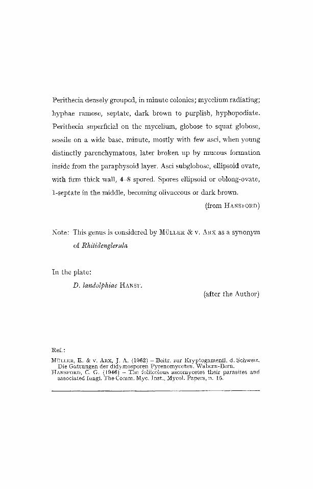
Perithecia densely grouped, in minute colonies; mycelium radiating;
hyphae ramose, septate, dark brown to purplish, hyphopodiate.
Perithecia superficial on the mycelium, globose to squat globose,
sessile on a wide base, minute, mostly with few asci, when young
distinctly parenchymatous, later broken up by mucous formation
inside from the paraphysoid layer. Asci subglobose, ellipsoid ovate,
with firm thick wall, 4-8 spored. Spores ellipsoid or oblong-ovate,
1-septate in the middle, becoming olivaceous or dark brown.
(from HANSrORD)
Note: This genus is considered by MOLLER & V. ARX as a synonym
of Rhitidenglerula
In the plate:
D. landolphiae HANS,. (after the Author)
Ref. :
M~'LLER, g . • V. ARX, J. A, (1962) - Beitr . zur Kryptogamenf l . d. Schweiz. Die Ga t tungen der d idymosporen Pyrenomyceten . Wabern-Bern .
HA•SrORD, C. G. (1946) - The foliieolous ascomycetes their parasi tes and associated fungi. The Comm, Myc. Inst. , Mycol. Papers, n. 15.

t~
C~
L~
C~o
!, J
P

A S C O M Y C E T E S D O T H I O R A L E S or 3 I E L I O L A L E S
E N G L E R U L A C E A E ( M E L I O L A C E A E , after I-IANSrORD)
P H Y T O P A T H O G E N (Ectophyta)
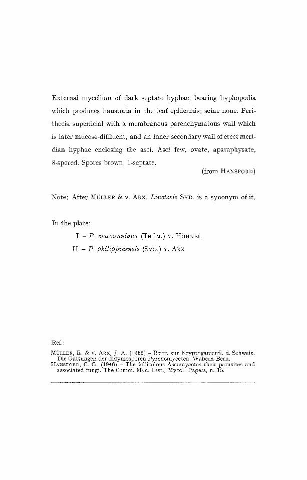
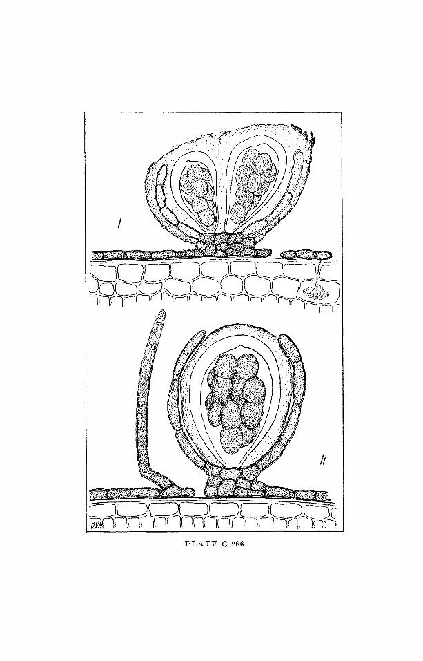
Gen. P a r e n g l e r u l a v. H6HNEL
I C O N O G R A P H I A M Y C O L O G I C A
V E R O N A - - BE~EDEK
Pla te C 286
External mycelium of dark septate hyphae, bearing hyphopodia
which produces haustoria in the leaf epidermis; setae none. Peri-
thecia superficial with a membranous parenchymatous wall which
is later mucose-diffluent, and an inner secondary wall of erect meri-
dian hyphae enclosing the asci. Asci few, ovate, aparaphysate,
8-spored. Spores brown, 1-septate. (from HANSFORD)
Note: After MI3LLER & V. AleX, L i n o t e x i s SYD. is a synonym of it.
In the plate:
I - P . macowaniana (THuM.) V. H 6 H N E L
II - P . p h i l i p p i n e n s i s (SYD.) V. ARX
1Ref. :
MgLL1ZR, E. 8c V. ARX, J. A. (1962) - Beitr , zur Kryptogamenf t . d. Schweiz. Die Ga t tungen der d idymosporen Pyrenomyceten , Wabern-t3ern.
I-IANsFORD, C. G, (1946) - The foliicolous Ascomycetes their parasi tes and associated fungi. The Comm. iViyc. Inst . , A~ycoI. Papers , n. 15.
P L A T E C 286


A S C O M Y C E T E S
D O T I ~ I D E A L E S
D O T H I D E A C E A E
P H Y T O P A T H O G E N
Gen. Y o s h i n a g e l l a v. HOHN.
I C O N O G R A P H I A M Y C O L O G I C A
VERONA -- BENEDEK
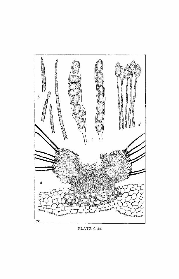
Plate C 287
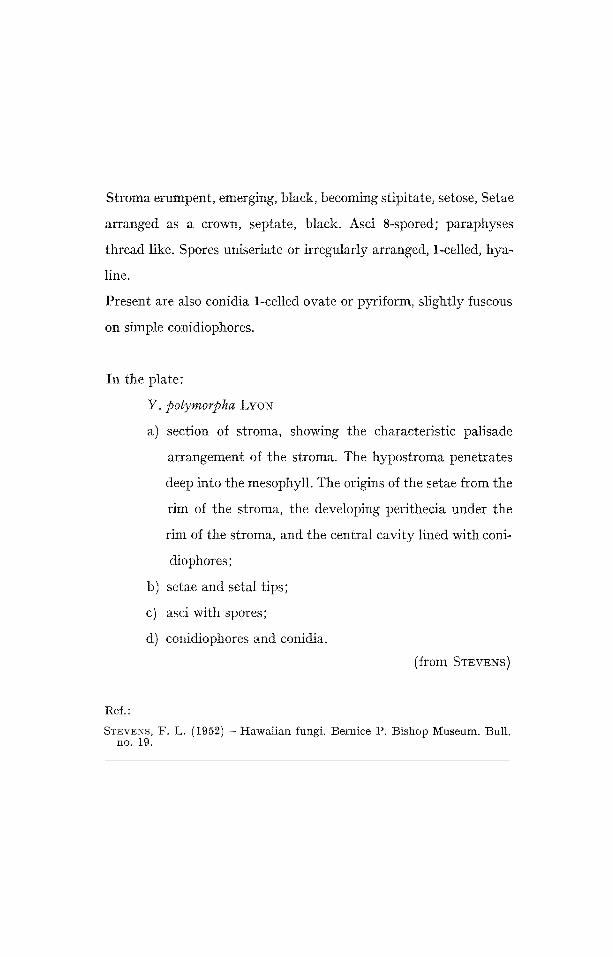
Stroma erumpent, emerging, black, becoming stipitate, setose, Setae
arranged as a crown, septate, black. Asci 8-spored; paraphyses
thread like. Spores uniseriate or irregularly arranged, 1-celled, hya-
line.
Present are also conidia 1-celled ovate or pyriform, slightly fuscous
on simple conidiophores.
In the plate:
Y. polymorpha LYON
a) section of stroma, showing the characteristic palisade
arrangement of the stroma. The hypostroma penetrates
deep into the mesophyll. The origins of the setae from the
rim of the stroma, the developing perithecia under the
rim of the stroma, and the central cavity lined with coni-
diophores;
b) setae and setal tips;
c) asci with spores;
d) conidiophores and conidia.
(from STEVENS)
Ref. :
STEVENS, F. L. (1952) -Hawaiian fungi. Bernice P. Bishop Museum. Bull. no. 19.
c~o

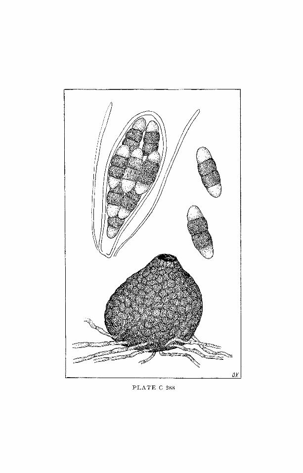
A S C O ~ { Y C E T E S
D O T H I D E A L E S
C A P N O D I A C E A E
SAPROPHYTA
Gen. L i m a c i n i o p s i s MENDOZA
ICONOGRAPHIA MYCOLOGICA
V E R O N A - - B E N E D E K
Plate C 288
Mycelium perisporoid; perithecia globular, ostiolate, without se-
tae; asci 8-spored, aparaphysate; spores 4-celled, brown with 2 end
cells hyaline.
Note: In AINSWORTH'S Dictionary of Fungi the genus is reported
as dubitatively equal to Phragmocapnias TI~ElSS. & SYD.
Found associated to filamentous blue-green algae.
STEVENS notices that the genus, ill its species L. rollandiae,
is closely related to Limacinia except for the presence of
paraphyses and the color of the end cells of the spores. I t is
strange that in the diagnosis of the genus asci are recalled as
aparaphysate, while L. rollandiae is described and figured
with paraphysate asci.
In the plate:
L. rollandiae MENDOZIk
Ref.:
ST~ZVENS, F. L. (1925) - Hawaiian fungi. Bernice P. Bishop Museum, Bulh N. 19.
.q

ASCOMYCETES D O T H I D E A L E S
CAPNODIACEAE
SAPROPHYTA (Epiphyta)
Gen. L i m a c i n i e l l a MENDOZA
I C O N O G R A P H I A M Y C O L O G I C A
V E R O N A - - B E N E D E K
Plate C 289
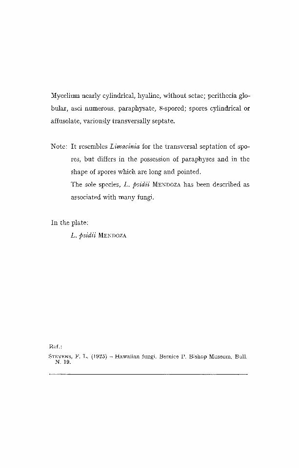
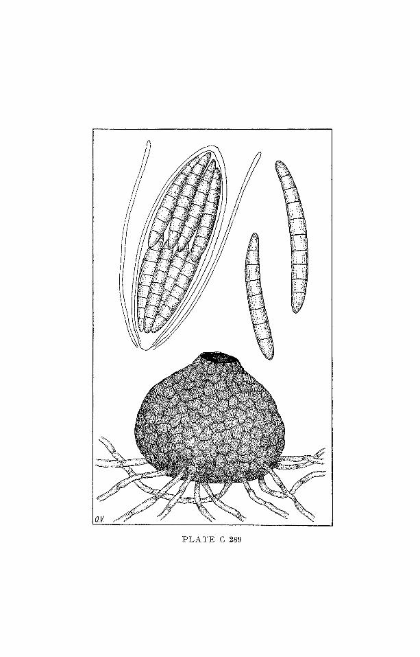
Mycelium nearly cylindrical, hyaline, without setae; perithecia glo-
bular, asci numerous, paraphysate, 8-spored; spores cylindrical or
affusolate, variously transversally septate.
Note: It resembles Limacinia for the transversal septation of spo-
res, but differs in the possession of paraphyses and in the
shape of spores which are long and pointed.
The sole species, L. psidii MENOOZA has been described as
associated with many fungi.
In the plate:
L. psidii M~NDOZA
Ref. :
STEVENS, F. L. (1925) - Hawai ian fungi. Bernice P. Bishop Museum, Bull. N. 19.
-d
7~

A S C O M Y C E T E S
S P H A E R I A L E S H A L O S P H A E R I A C E A E
S A P R O P H Y T A
Gen. Halonectria JoNEs
I C O N O G R A P H I A M Y C O L O G I C A
V E R O N A - - B E N E D E K
P l a t e C 290
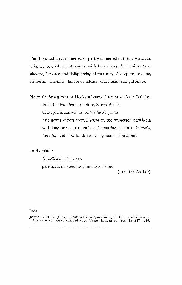
Perithecia solitary, immersed or partly immersed in the substratum,
brightly colored, membranous, with long necks. Asci unitunicate,
clavate, 8-spored and deliquescing at maturity. Ascospores hyaline,
fusiform, sometimes lunate or falcate, unicellular and guttulate.
Note: On Scotspine test blocks submerged for 24 weeks in Dalefort
Field Centre, Pembrokeshire, South Wales.
One species known: H. mil/ordensis Jo~Es
The genus differs from Nectria in the immersed perithecia
with long necks. It resembles the marine genera Lulworthia,
Orcadia and Trailia,differing by some characters.
In the plate:
H. mil/ordensis JONES
perithecia in wood, asci and ascospores. (from the Author)
Ref. :
JoNEs, E. B. G. ( t965) - Halo~ectric~ mil[orde~sis gen. & sp. nov. a m a r i n e P y r e n o m y c e t e on s u b m e r g e d wood. Trans . Br i t . mycol . Soc., 48, 287- -290 .
O.k.
P L A T E C 290

A S C O M Y C E T E S
P L E O S P O R A L E S
P L E O S P O I R A C E A E
S A P R O P H Y T A
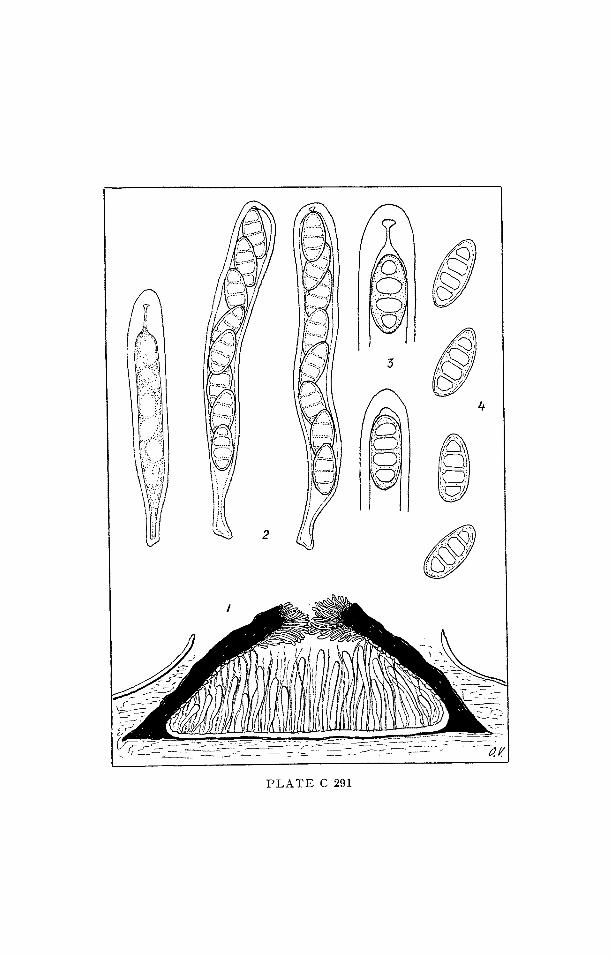
Gen. M e l o m a s t i a NITS.
I C O N O G R A P H I A M Y C O L O G I C A
V E R O N A - - BENEDEK
Plate C 291
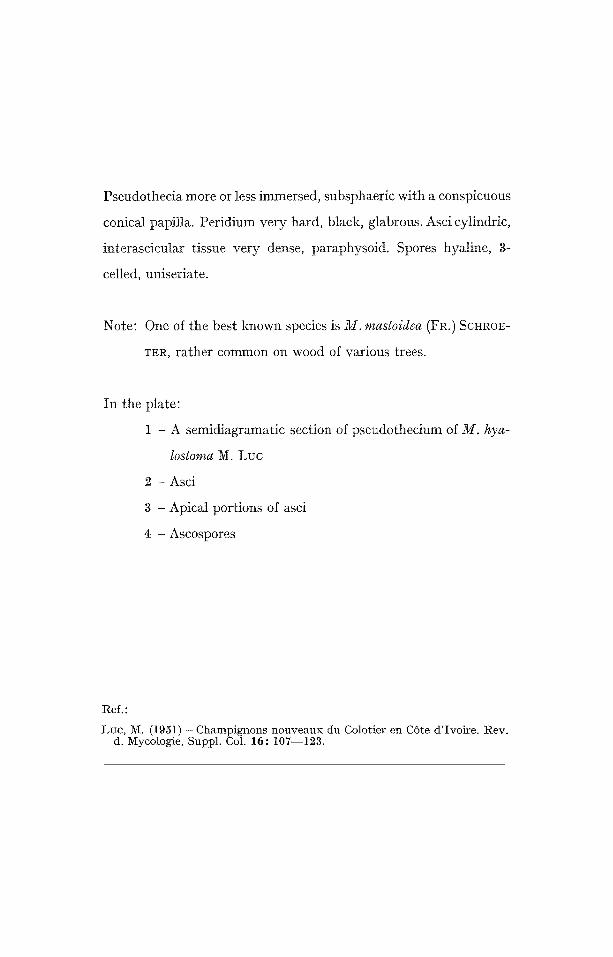
Pseudothecia more or less immersed, subsphaeric with a conspicuous
conical papilla. Peridium very hard, black, glabrous. Asci cylindric,
interascicular tissue very dense, paraphysoid. Spores hyaline, 3-
celled, uniseriate.
Note: One of the best known species is M. mastoidea (FI<) SCHROE-
TER, rather common on wood of various trees.
In the plate:
1 - A semidiagramatic section of pseudothecium of M. hya-
lostoma M. Luc
2 - Asci
3 - Apical portions of asci
4 - Ascospores
Ref. :
Luc, ~. (1951) - Champignons nouveaux du Colofier en C6te d'Ivoire. Rev. d. l~ycologie, Suppl. Col. 16: 107--123.
"d
L'~
'}' I'll /',ii!
,I 'Jl .%1.~

ASCO1VIYCETES E U R O T I A L E S
(HE~ilSPHAE1RIA LES)
GYMNOASCACEAE ( t tEMISPHAERIACEAE)
SAPROPHYTA
Gen. H e x a g o n e l l a STEVENS • GUBA
t C O N O G R A P H I A MYCOLOGICA
VERONA - - BENEDE1K
Plate C 292
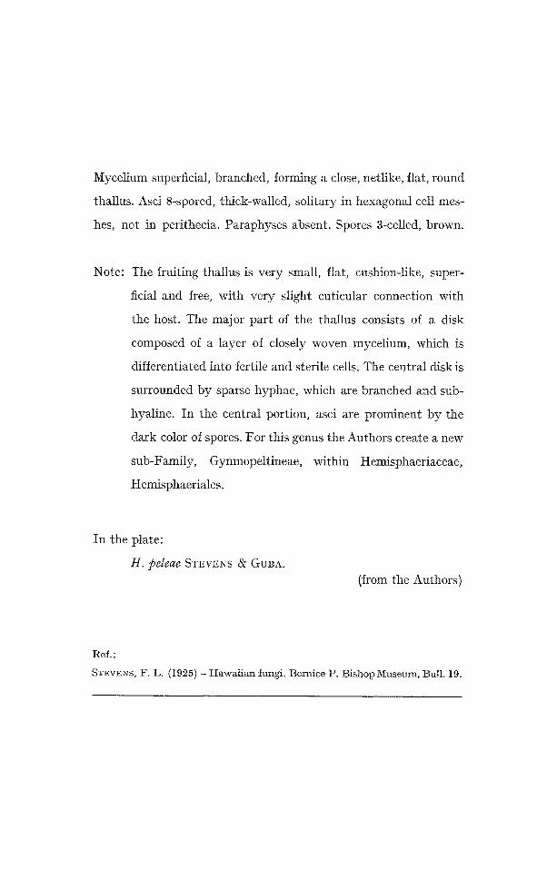
Mycelium superficial, branched, forming a close, netlike, flat, round
thallus. Asci 8-spored, thick-walled, solitary in hexagonal cell mes-
hes, not in perithecia. Paraphyses absent. Spores 3-celled, brown.
Note: The fruiting thallus is very small, flat, cushion-like, super-
ficial and free, with very slight cuticular connection with
the host. The major part of the thallus consists of a disk
composed of a layer of closely woven mycelium, which is
differentiated into fertile and sterile cells. The central disk is
surrounded by sparse hyphae, which are branched and sub-
hyaline. In the central portion, asci are prominent by the
dark color of spores. For this genus the Authors create a new
sub-Family, Gymnopeltineae, within Hemisphaeriaceae,
Hemisphaeriales.
In the plate:
H. peleae STEVENS ~Y GUBA. (from the Authors)
Ref. :
STEVENS, F. L. (1925) -IIawaiian fungi. Bernice P. Bishop Museum, But1.19.
J~

ASCOMYCETES P L E C T A S C A L E S
M E L I O L A C E A E
P H Y T O P A T H O G E N (Epiphyta)
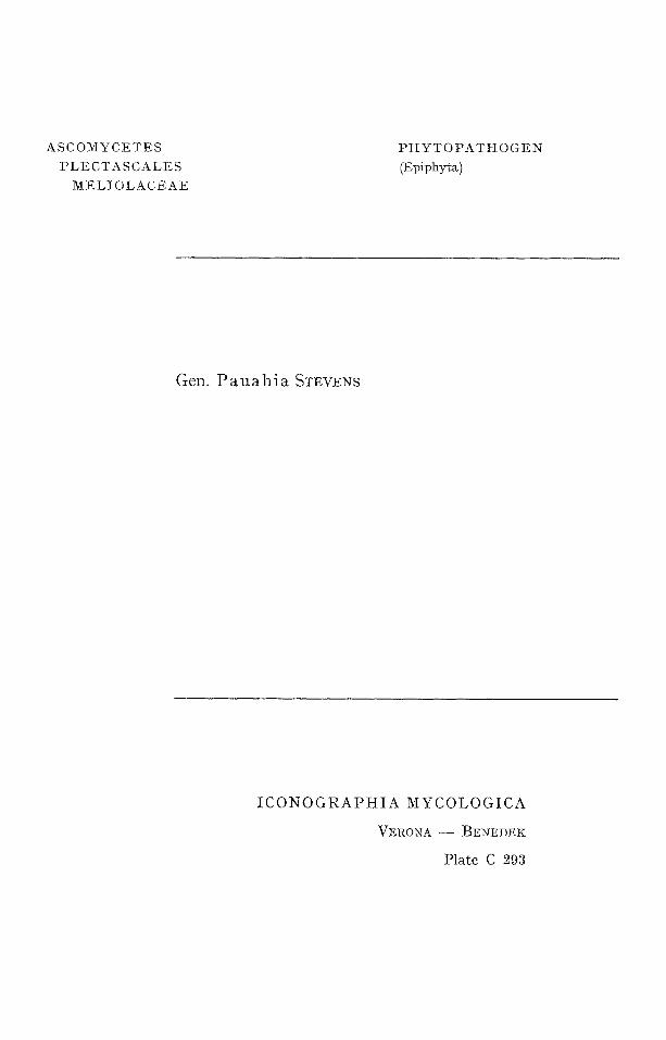
Gen. Pauahia STEVENS
I C O N O G R A P H I A M Y C O L O G I C A
V E R O N A - - BENEDEK
Pla te C 293

Stroma superficial, wi th a s t ructure at palisade, with several locules.
Asci evanescent, spores brown, 3-septate.
In the plate:
P. sideroxyli STEVENS (from the Author)
Ref. :
STEVENS, F. L, (1925) - Hawaiian fungi. Bernice P. Bishop Museum. Bull. 19.
i d
J

A SCOI~[YCETES
H E L O T I A L E S
H E L O T I A C E A E
P H Y T O P A T H O G E N
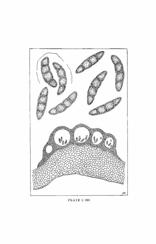
Gen. Septot in ia WHETZEL
I C O N O G R A P H I A M Y C O L O G I C A
VERONA - - BENEDEK
Pla te C 294
Apothecia shallow, cup-shaped, stipitate, arising in the spring from sclerotia in the soil or leafmold. Asci slender, cylindrical. Ascospores hyaline, ovoid. Paraphyses simple or branched, tips swollen. Conidia hyaline, elongate, septate, borne on branches, hyaline co- nidiophores massed to form a typical sporodochium. Sclerotia angular, elongate or circular, thin, black, formed in the invaded tissues of the affected plant parts, usually after they have fallen to the ground. Spermatia (microconidia) ovate, very minute, produced on short In- dian-club-shaped spermatophores, clustered to form minute sper- modochia on the decaying tissue; accompanying formation of scle- rotia.
Note: The genus was based on the species Sept. podophyllina (ELL. & Ev.), ascigerons state of Gloeosporium podophyllinum (ELL. & Ev.). This last species was retained by SACCARDO as be- longing to Gen. Septogloeum, until BUCHWALD (1949) crea- ted for the conidial forms of Septotinia the new genus Sep- totis.
In the plate: Septotinia podophyllina WHETZEL 1 -- apothecia on sclerotium; 2 - asci and paraphyses; 3 - fructification of the conidial state of Septotis; 4 - formation of microconidia.
(from WtlETZEL, in part)
Ref. :
WHETZ~L, H. H. (1937) - Septotinia, a new genus of tile Ciborioideae. Myco- logia, 29: t78--146.
ARX, J. A. v. (1957) - Revision der zu Gtoeosporium gestellten Pilze. N.V. Noord-Hollandsche Uitg. Maatsch., Amsterdam.
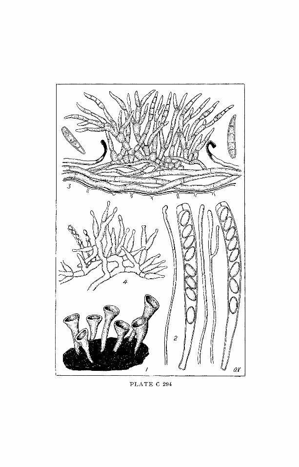
PLATE C 294

A S C O M Y C E T E S
P S E U D O S P H A E R I A L E S
V E N T U R I A C E A E
P H Y T O P A T H O G E N
Gen. Act inodoth iops is STEVENS
I C O N O G R A P t I I A M Y C O L O G I C A
VERONA - - BENEDEK
P l a t e C 295
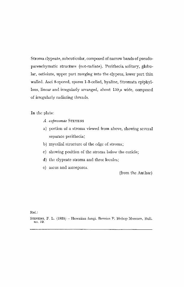
Stroma clypeate, subcuticular, composed of narrow bands of pseudo-
parenchymatic structure (not-radiate). Perithecia solitary, globu-
lar, ostiolate, upper part merging into the clypeus, lower part thin
walled. Asci 8-spored, spores 1-3-celled, hyaline, Stromata epiphyl-
tous, linear and irregularly arranged, about 150/, wide, composed
of irregularly radiating threads.
In the plate:
A. coprosmae STEVENS
a) portion of a stroma viewed from above, showing several
separate perithecia;
b) mycelial structure of the edge of stroma;
c) showing position of the stroma below the cuticle;
d) the clypeate stroma and three locules;
e) ascus and ascospores. (from the Author)
Ref.:
STEVENS, F. L. (1925) - Hawaiian fungi. Bernice P. Bishop Museum, Bulh no. 19.
t

ASCOMYCETES
PSEUDOSPHAERIALES
M¥COSPHAERELLACEAE
P H Y T O P A T H O G E N
Gen. Didymel la SAcc.
I C O N O G R A P H I A M Y C O L O G I C A
VERONA -- BENEDEK
Plate C 296
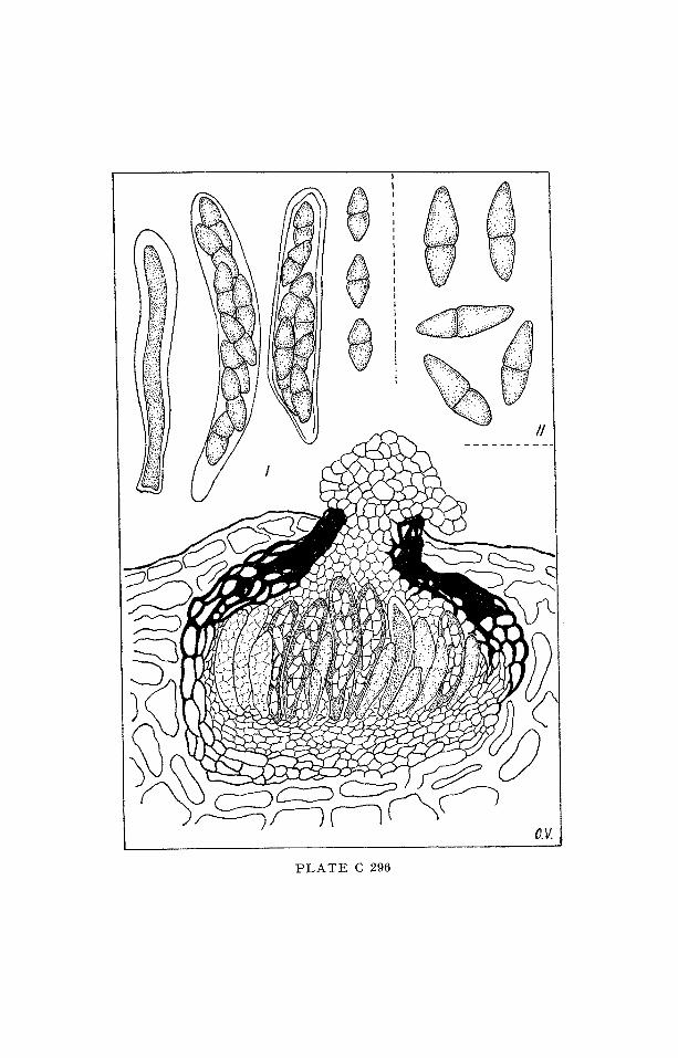
Pseudothecia small, scattered, immersed, mainly in dead herbace- ous stems. Ascospores 2-celled, hyaline.
Note: Many authors divide the genus into two sections: I section: (Eu-Didymella), pseudothecia of an ordinary, simple structure; peridium uniform. Spores generally equal- ly 2-celled. II section: (Mycosphaerellopsis v. H6HN:EL). Pseudothecia somewhat sclerotioid, with a lateral thickening of the peri- dium. Spores more or less unequally 2-celled. For this section MUNK, in 1953, created the genus Didymo- lepta, on the basis of Didymella winteriana (SAcc.) PETRAK, V. H6ttNEL had previously established for this type-species, the genus Mycosphaerellopsis. To Didymella are added, as imperfect forms, species of Ascochyta, (IM V-A-113), Diplodina (IM VII-A-155), Phoma (IM IV-A-97) Some species (like D. ligulicola, and others) are of certain economical interest.
In the plate:
I - D. ligulicola (BAKER et aI.) V. ARX II - Spores of D. winteriana (SAcc.) PETRAK
Ref. :
SAUTHOFF, W. (1969~) - Didymella ligulicola (BAKER & al.) v. ARX als Krank- heitserreger an Chrysan themen in Deutschland. Phy t . Z. 48 : 240--250.
MOLLER, E. & VON ARX. J. A. (1962) - Die Oa t tungen der d idymosporen Pyrenomyceten , in Beitr . z. Kryptogamenf l . der Schweiz, Band II , H e f t 2, Wabern-]3ern.
MUNK, A. (1957) - Danish Pyrenomycetes . Dansk ]3ot. Arkiv, 17: no. 1.
"d
t~
r~
00
~

A S C O M Y C E T E S
P S E U D O S P H A E R I A L E S
V E N T U R I A C E A E
P H Y T O P A T H O G E N
Gen. G i b b e r a FR.
I C O N O G R A P H I A MYCOLOGICA
V E R O N A - - B E N E D E K
Plate C 297
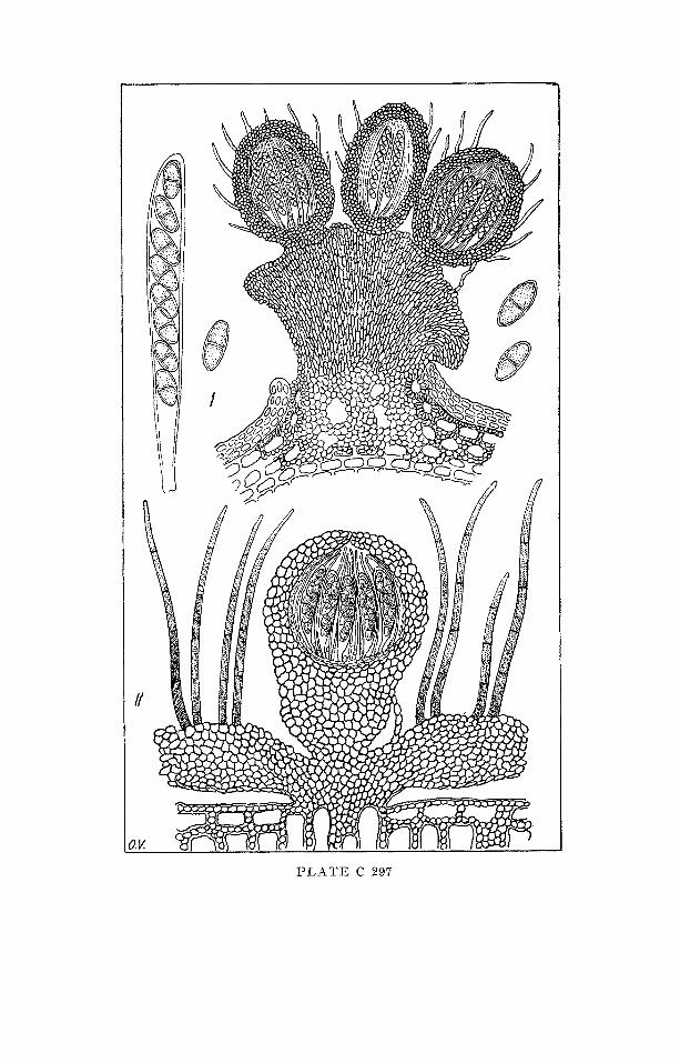
Basal stroma formed by prismatic, coarse, vertically seriate cells.
Peridium bristly, with thick-walled cells at the outside. Asci cylin-
dric; interascicular tissue well developed, paraphysoid. Spores 1-
seriate, 2-celled, light-brown.
In the plate:
I - G. vaccinii (Sow.) FR. (from Mt~LLEI~ & V. AleX)
II - G. pr insep iae (CttoNA & al.) M/JLLER (from MULLER)
Ref.:
M/~ILLER, [E. 6; VON ARK, J. A. (1962) - Die Ga t tungen der d idymosporen Pyrenomyce ten . In: Bei t r~ge zur Kryptogamenf l . der Schweiz. W a b e m - -Bern.
MOLLER, E. (1958) - Pilze aus dem H i m a l a y a II . Sydowia, Ann. Mycol. ser. I I , 12: 160--184.
C3
.,~
,~\
--
~ .
f~
%%

ASCOMYCETES DISCON[YCETES
(HELOTIALES)
SAPROPHYTA
Gem T r o c h i l a FRIES
I C O N O G R A P H I A M Y C O L O G I C A
VERONA- BENEDEK
Plate C 298
Apothecia sunken at first, then exposed by rupture of the epidermis;
inserted on a basal layer of dark cells. Asci cylindric, 8-spored,
with an apical pore. Ascospores continue, elliptical, hyaline, or even
more or less brown and 2-celled.
Note: present in the tissues of dead leaves.
In the plate:
I - T . craterium FR.
I I - T. i l ic ina (NEES ex
JONES
FR.) GREENHALGI-I & MORGAN-
Ref. :
GREENI-tALOI-I, G. N. & MORGAN-JoNEs, O. (1964) -- Some species of Trochila and an undescribed Discomycete on leaves of P r u n u s laurocerasus. Trans, Brit. mycol. Soc., 47: 311--320.
$6~ D ~ZV'~d
.
f.-(
7/~ j
h '-C ),
....... ll.l.~] j
//


A S C O M Y C E T E S
S P F I A E R I A L E S
I ~ A L O S P H A E R I A C E A E
S A P R O P H Y T A
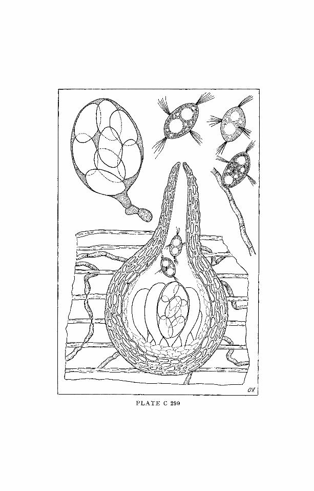
Gen. Nau tosphae r i a E. B. G. JoNEs
I C O N O G R A P H I A M Y C O L O G I C A
V E R O N A - - B E N E D E K
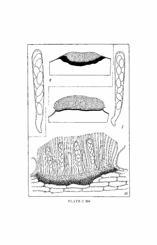
Plate C 299
Perithecia solitary, immersed in the substratum, hyaline or cream,
membranaceous, neck long. Asci unitunicate, 8-spored, clavate,
soon deliquescing. Ascospores grey or fuscous, unicellular, ellipsoid,
apically and laterally appendiculate.
Note: The genus is compared by the author with other marine ge-
nera, like Remispora, Halosibhaeria, Corollospora, Ceriospo-
ropsis (respectively, IM VI-C-88; VII-C-95; VI-C-85).
The type species - N . crista minuta JONES -- was found on a
beech test block submerged for 66 weeks in the Irish Sea.
In the Plate:
N. crista minuta JONES
section of a perithecium, ascus and ascospores, one of which
germinating.
R e f . :
JONES, E. ]~. G. (1964) -Nautosphaeria cristaminuta gen. & sp. nov., a marine l~renomycete on submerged wood. Trans. Brit. mycol, soc. 47: 97--101.
.%
7~
7~
r_y

A S C O M Y C E T E S S P F I A E R I A L E S
S P H A E R I A C E A E
P H Y T O P A T H O G E N
Gen. Me lanopsamma NIESSL. apud SACCARDO
I C O N O G R A P H I A M Y C O L O G I C A
V E R O N A - - B E N E D E K
Pla te C 300
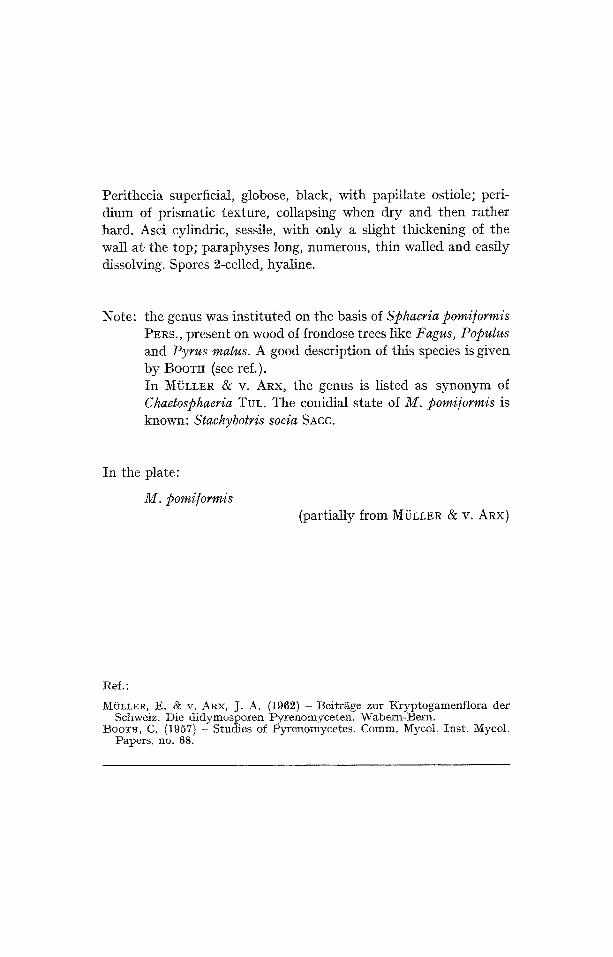
Perithecia superficial, globose, Mack, with papillate ostiole; peri- dium of prismatic texture, collapsing when dry and then rather hard. Asci cylindric, sessile, with only a slight thickening of the wall at~ the top; paraphyses long, numerous, thin walled and easily dissolving. Spores 2-celled, hyaline.
Note: the genus was instituted on the basis of Sphaeria pomi/ormis PEns., present on wood of frondose trees like Fagus, Populus and Pyrus malus. A good description of this species is given by BOOTtt (see ref.). In MI~LLER & V. ARX, the genus is listed as synonym of Chaetosphaeria TuL. The conidial state of M. pomi/ormis is known: Stachybotris socia SAcc.
In the plate:
M. pomi/ormis (partially from M/2LLER & V. Anx)
Ref. :
MOLLER, E. & v. ARX, J. A. (1962) - Beitr~ge zur Kryptogamenflora der Schweiz. Die didymosporen Pyrenomyceten. Wa.bern-Bern.
BOOTH, C. (1957) - Studies of Pyrenomycetes. Comm. Mycol. Inst. Mycoh Papers, no. 68.
c~
.<
%

ASC OMYCETES
C A P N O D I A L E S
C A P N O D I A C E A E
S A P R O P H Y T A
or P H Y T O P A T H O G E N
(Sooty-Molds)
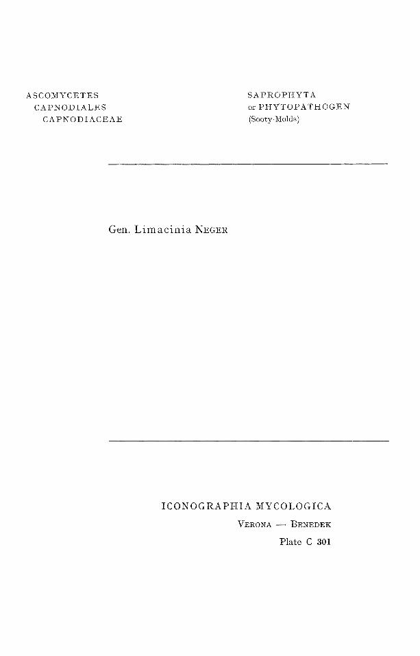
Gen. L i m a c i n i a NECER
I C O N O G R A P H I A M Y C O L O G I C A
V E R O N A - - B E N E D E K
Plate C 301
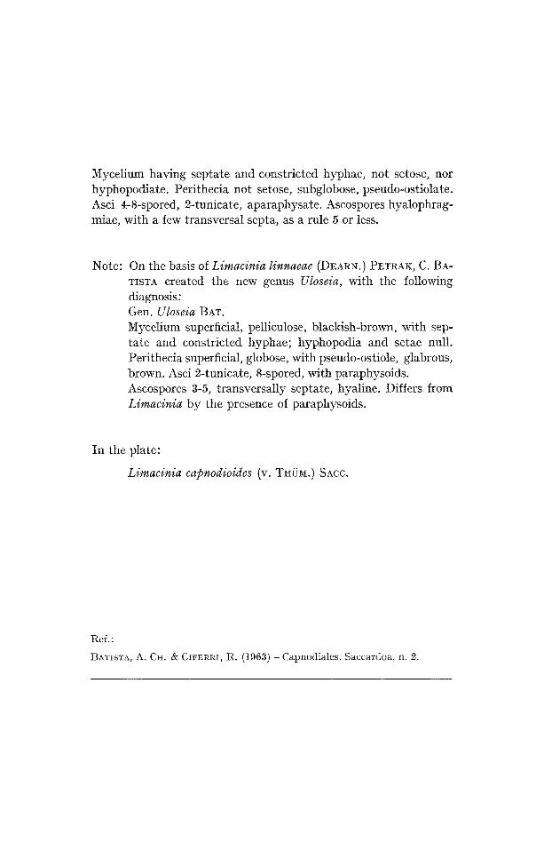
Mycelium having septate and constricted hyphae, not setose, nor hyphopodiate. Perithecia not setose, subglobose, pseudo-ostiolate. Asci 4-8-spored, 2-tunicate, aparaphysate. Ascospores hyalophrag- miae, with a few transversal septa, as a rule 5 or less.
Note: On the basis of Limacinia linnaeae (DEARN.) PETRAK, C. BA- TISTA created the new genus Uloseia, with the following diagnosis: Gen. Uloseia BAT. Mycetium superficiM, pelliculose, blackish-brown, with sep- tate and constricted hyphae; hyphopodia and setae null. Perithecia superficial, globose, with pseudo-ostiole, glabrous, brown. Asci 2-tunicate, 8-spored, with paraphysoids. Ascospores 3-5, transversally septate, hyaline. Differs from Limacinia by the presence of paraphysoids.
In the plate:
Limacinia capnodioides (v. THOM.) SACC.
Ref.:
BATISTA, A. CH. & CIFERRI, It. (1963) -- Capnodiales. Saccardoa, n. 2.
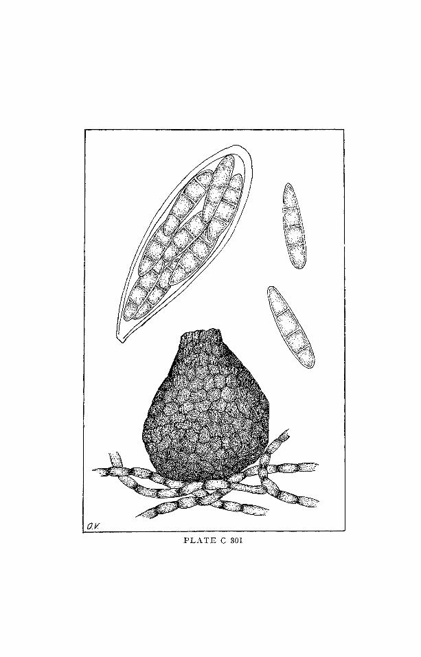
a V
P L A T E C 301

A S C O M Y C E T E S
C A P N O D I A L E S
C A P N O D I A C E A E
S A P R O P H Y T A
(Sooty-Molds)
Gen. L i m a c i n u l a (SAcc. ut sub genus) SACC. & TROTTER
I C O N O G R A P H I A M Y C O L O G I C A
V E R O N A - - B E N E D E K
Plate C a02
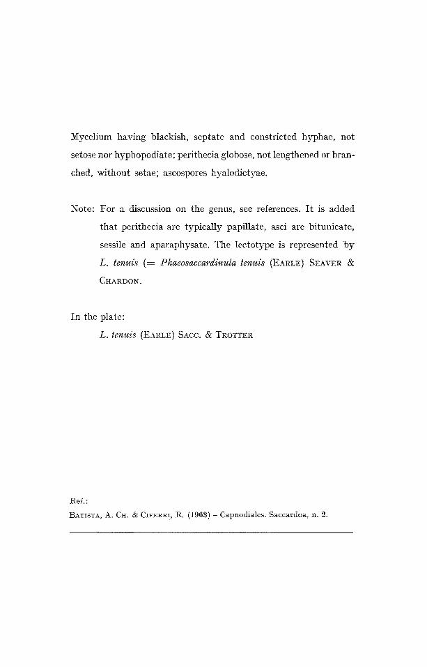
Mycelium having blackish, septate and constricted hyphae, not
setose nor hyphopodiate; perithecia globose, not lengthened or bran-
ched, without setae; ascospores hyalodictyae.
Note: For a discussion on the genus, see references. It is added
that perithecia are typically papillate, asci are bitunicate,
sessile and aparaphysate. The lectotype is represented by
L. tenuis (= Phaeosaccardinula tenuis (EARLE) SEAVER &
CHARDON.
In the plate:
L. tenuis (EARLE SACC. & TROTTER
Ref.:
BATI'
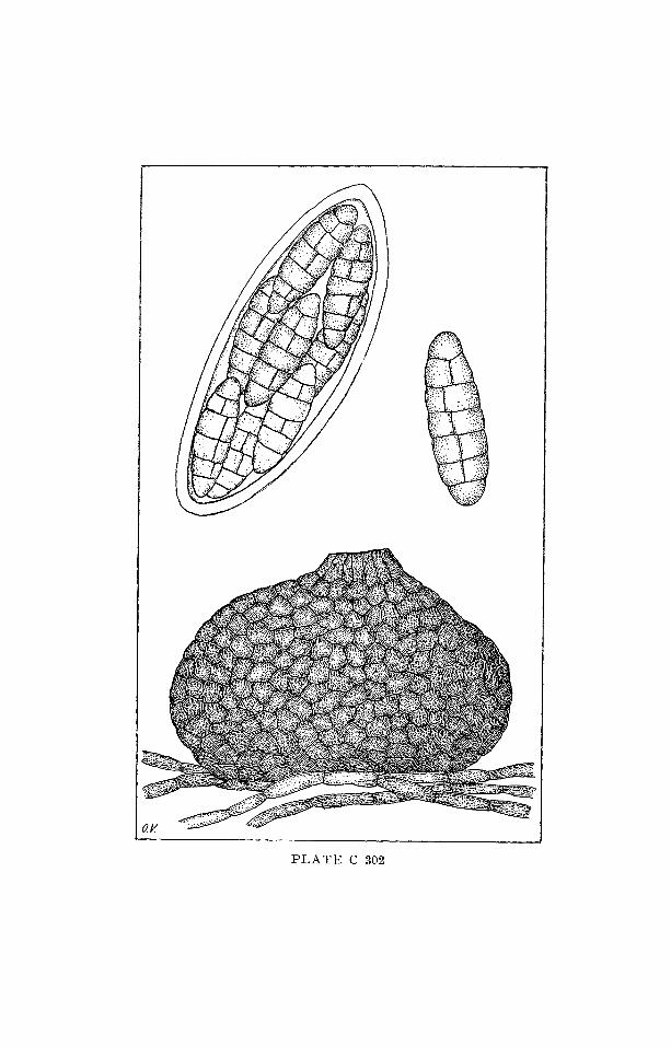
PLATE C 302