iarc monographs – 108 such as piper wichmanni, piper aduncum and piper auritum have also been...
TRANSCRIPT
117
1. Exposure Data
The kava plant is indigenous to Oceania (Lebot et al., 1997; Ramzan & Tran, 2004) and has been used both ceremonially and recreation-ally in certain cultures of the South Pacific for at least 1500 years. Europeans documented its use when they travelled to Polynesia in the 18th century (WHO, 2007). The cultural history of the use of kava has been reviewed by Singh (1992).
In the past, traditional use of kava was widespread, but, in certain cultures, custom determined who could use kava and for what purposes. In recent years, as part of the processes of modernization, major changes have occurred with regard to who uses kava, and where and how it is consumed. In some places, kava is now being consumed much like alcohol in western countries, as a beverage that is drunk socially (McDonald & Jowitt, 2000).
1.1 Identification of the agent
1.1.1 Botanical data
(a) Nomenclature
Chem. Abstr. Serv. Reg. No.: 9000-38-8Chem. Abstr. Name: Kava-kava resin (8Cl)Botanical name: Piper methysticum G. ForstFamily: PiperaceaeGenus: PiperPlant part: Rhizome
(WHO, 2004; O’Neil et al., 2006; NTP, 2012; and SciFinder, 2013)
According to WHO (2007), some other species such as Piper wichmanni, Piper aduncum and Piper auritum have also been marketed as “kava.”
Common names: Kava; Kava-kava; Ava-ava; Antares; Ava; Ava pepper; Ava pepper shrub; Ava root; Awa; Fijian kava; Gea; Gi; Grog; Intoxicating long pepper; Intoxicating pepper; Kao; Kava kava rhizome; Kava root; Kavapiper; Kavapyrones; Kavarod; Kavasporal forte; Kave-kave; Kawa; Kawa kawa; Kawa pepper; Kawa Pfeffer; Kew; Long pepper; Macropiper latifo-lium; Malohu; Maluk; Maori kava; Meruk; Milik; Pepe kava; Piperis methystici rhizome; Rhizoma piperis methystici; Sakaua; Sakau; Tonga; Yagona; Yangona; Yaqona; Yongona
(b) Description
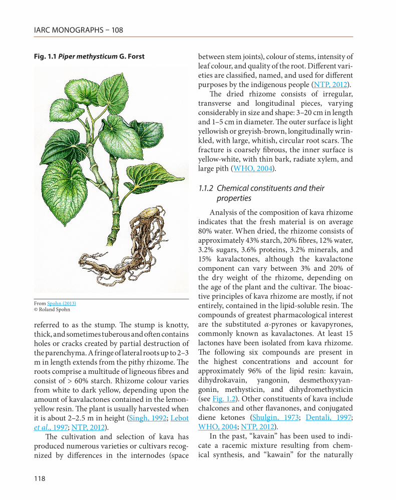
See Fig. 1.1The tropical shrub Piper methysticum is a
hardy, fairly succulent, slow-growing perennial that is widely cultivated in Oceania. The species is sterile and reproduces asexually. Due to its traditional use as a ritual beverage known for promoting relaxation and a sense of well-being, the kava plant spread widely throughout Oceania, in Polynesia, Melanesia, and the Federated States of Micronesia (Norton & Ruze, 1994; NTP, 2012).
The leaves are heart-shaped, pointed, 8–25 cm in length, and smooth and green on both sides. Kava is cultivated for its rootstock (rhizome), also
KAVA
IARC MONOGRAPHS – 108
118
referred to as the stump. The stump is knotty, thick, and sometimes tuberous and often contains holes or cracks created by partial destruction of the parenchyma. A fringe of lateral roots up to 2–3 m in length extends from the pithy rhizome. The roots comprise a multitude of ligneous fibres and consist of > 60% starch. Rhizome colour varies from white to dark yellow, depending upon the amount of kavalactones contained in the lemon-yellow resin. The plant is usually harvested when it is about 2–2.5 m in height (Singh, 1992; Lebot et al., 1997; NTP, 2012).
The cultivation and selection of kava has produced numerous varieties or cultivars recog-nized by differences in the internodes (space
between stem joints), colour of stems, intensity of leaf colour, and quality of the root. Different vari-eties are classified, named, and used for different purposes by the indigenous people (NTP, 2012).
The dried rhizome consists of irregular, transverse and longitudinal pieces, varying considerably in size and shape: 3–20 cm in length and 1–5 cm in diameter. The outer surface is light yellowish or greyish-brown, longitudinally wrin-kled, with large, whitish, circular root scars. The fracture is coarsely fibrous, the inner surface is yellow-white, with thin bark, radiate xylem, and large pith (WHO, 2004).
1.1.2 Chemical constituents and their properties
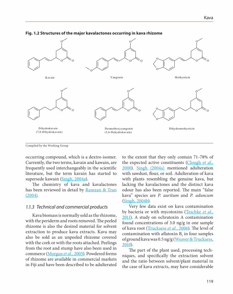
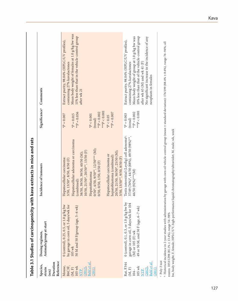
Analysis of the composition of kava rhizome indicates that the fresh material is on average 80% water. When dried, the rhizome consists of approximately 43% starch, 20% fibres, 12% water, 3.2% sugars, 3.6% proteins, 3.2% minerals, and 15% kavalactones, although the kavalactone component can vary between 3% and 20% of the dry weight of the rhizome, depending on the age of the plant and the cultivar. The bioac-tive principles of kava rhizome are mostly, if not entirely, contained in the lipid-soluble resin. The compounds of greatest pharmacological interest are the substituted α-pyrones or kavapyrones, commonly known as kavalactones. At least 15 lactones have been isolated from kava rhizome. The following six compounds are present in the highest concentrations and account for approximately 96% of the lipid resin: kavain, dihydrokavain, yangonin, desmethoxyyan-gonin, methysticin, and dihydromethysticin (see Fig. 1.2). Other constituents of kava include chalcones and other flavanones, and conjugated diene ketones (Shulgin, 1973; Dentali, 1997; WHO, 2004; NTP, 2012).
In the past, “kavain” has been used to indi-cate a racemic mixture resulting from chem-ical synthesis, and “kawain” for the naturally
Fig. 1.1 Piper methysticum G. Forst
From Spohn (2013)© Roland Spohn
Kava
119
occurring compound, which is a dextro-isomer. Currently, the two terms, kavain and kawain, are frequently used interchangeably in the scientific literature, but the term kavain has started to supersede kawain (Singh, 2004a).
The chemistry of kava and kavalactones has been reviewed in detail by Ramzan & Tran (2004).
1.1.3 Technical and commercial products
Kava biomass is normally sold as the rhizome, with the periderm and roots removed. The peeled rhizome is also the desired material for solvent extraction to produce kava extracts. Kava may also be sold as an unpeeled rhizome covered with the cork or with the roots attached. Peelings from the root and stump have also been used in commerce (Morgan et al., 2005). Powdered forms of rhizome are available in commercial markets in Fiji and have been described to be adulterated
to the extent that they only contain 71–78% of the expected active constituents (Clough et al., 2000). Singh (2004a) mentioned adulteration with sawdust, flour, or soil. Adulteration of kava with plants resembling the genuine kava, but lacking the kavalactones and the distinct kava odour has also been reported. The main “false kava” species are P. auritum and P. aduncum (Singh, 2004b).
Very few data exist on kava contamination by bacteria or with mycotoxins (Teschke et al., 2011). A study on ochratoxin A contamination found concentrations of 3.0 ng/g in one sample of kava root (Trucksess et al., 2006). The level of contamination with aflatoxin B1 in four samples of ground kava was 0.5 ng/g (Weaver & Trucksess, 2010).
The part of the plant used, processing tech-niques, and specifically the extraction solvent and the ratio between solvent/plant material in the case of kava extracts, may have considerable
Fig. 1.2 Structures of the major kavalactones occurring in kava rhizome
O O
O
O O
O
O
O O
O
O
O
Kavain Yangonin Methysticin
O O
O
O O
O
O O
O
O
O
D ihydrokavain(7,8-Dihydrokavain)
Desmethoxyyangonin(5,6-Dehydrokavain)
Dihydromethysticin
Compiled by the Working Group
IARC MONOGRAPHS – 108
120
influence on the chemical composition of the end product. For example, the alkaloid pipermethys-tine was not detectable in some commercial kava extracts (Teschke et al., 2011).
[The Working Group noted that the influences on composition mentioned above may hinder the comparison of studies, especially if the applied kava material was not specified exactly.]
1.2 Analysis
The chemical analysis and quality control of both kava and its extracts obtained by aqueous acetone or aqueous methanol, and supercritical fluid extraction – typically with carbon dioxide modified with methanol as solvent – were reviewed by Bilia et al. (2004). Both gas chro-matography (GC) and high-performance liquid chromatography (HPLC) can be used for the analysis of kavalactones with some advantages and disadvantages for each method. Using GC analysis, methysticin and yangonin, which are two of the major components, are generally not separated. In addition, the high temperature of the injection port causes the decomposition of methysticin. Concerning HPLC analyses, reversed-phase separation is generally better because it is highly reproducible with a very low detection limit for all compounds even if the quantitative analysis of the kavalactones by HPLC needs to be carried out in the absence of light to prevent the cis/trans isomerization of yangonin (Bilia et al., 2004). Besides various chromatographic approaches reviewed by Bilia et al. (2004), near infrared spectroscopy and nuclear magnetic resonance spectroscopy have been suggested to directly determine kavalac-tones without the need for separation (Table 1.1).
1.3 Use
1.3.1 Indications
(a) Medicinal use
According to WHO (2004), the medicinal uses supported by clinical data are short-term symptomatic treatment of mild states of anxiety or insomnia due to nervousness, stress or tension; the medicinal uses described in pharma-copoeias and in traditional systems of medicine are to induce relaxation, reduce weight, and treat fungal infections. Uses described in traditional medicine, but not supported by experimental or clinical data, are treatment of asthma, common cold, cystis, gonorrhoea, headaches, menstrual irregularities, urinary infections, and warts.
The German Commission E has approved kava for use in conditions of nervous anxiety, stress, and restlessness (Anonymous, 2000).
(b) Traditional food and recreational use
A local traditional drink, also known under the name kava, is obtained from the root or rhizome of the kava plant. The kava drink is made from water extracts, with water-insoluble substances made available to the drinker by emulsification, which may be accomplished by pounding or chewing of the rhizome (WHO, 2007).
On some islands in the South Pacific, fresh kava root or rhizome is used to prepare the traditional drink, while on others it is the dried and ground roots or rhizomes that are used. For fresh preparations, the root is chewed by young women, who spit the juice into the kava bowl without swallowing it themselves. The juice is then mixed with water or coconut milk and further processed. Most people drink only the water extracts of kava. This is obtained by adding water to kava roots which are finely ground and then filtered using cheese-cloth (WHO, 2007).
The kava drink has been described to have a psychoactive activity, and potency can vary
Kava
121
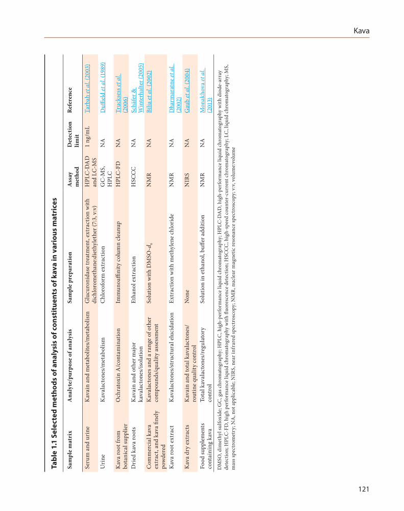
Tabl
e 1.
1 Se
lect
ed m
etho
ds o
f ana
lysi
s of
con
stit
uent
s of
kav
a in
var
ious
mat
rice
s
Sam
ple
mat
rix
Ana
lyte
/pur
pose
of a
naly
sis
Sam
ple
prep
arat
ion
Ass
ay
met
hod
Det
ecti
on
limit
Ref
eren
ce
Seru
m a
nd u
rine
Kav
ain
and
met
abol
ites/
met
abol
ism
Glu
curo
nida
se tr
eatm
ent,
extr
actio
n w
ith
dich
loro
met
hane
:die
thyl
ethe
r (7:
3, v
:v)
HPL
C-D
AD
an
d LC
-MS
1 ng
/mL
Tarb
ah et
al.
(200
3)
Uri
neK
aval
acto
nes/
met
abol
ism
Chl
orof
orm
ext
ract
ion
GC
-MS,
H
PLC
NA
Duffi
eld
et a
l. (1
989)
Kav
a ro
ot fr
om
bota
nica
l sup
plie
rO
chra
toxi
n A
/con
tam
inat
ion
Imm
unoa
ffini
ty c
olum
n cl
eanu
pH
PLC
-FD
NA
Truc
kses
s et a
l. (2
006)
Dri
ed k
ava
root
sK
avai
n an
d ot
her m
ajor
ka
vala
cton
es/is
olat
ion
Etha
nol e
xtra
ctio
nH
SCC
CN
ASc
häfe
r &
Win
terh
alte
r (20
05)
Com
mer
cial
kav
a ex
trac
t, an
d ka
va fi
nely
po
wde
red
Kav
alac
tone
s and
a ra
nge
of o
ther
co
mpo
unds
/qua
lity
asse
ssm
ent
Solu
tion
with
DM
SO-d
6N
MR
NA
Bilia
et a
l. (2
002)
Kav
a ro
ot e
xtra
ctK
aval
acto
nes/
stru
ctur
al e
luci
datio
nEx
trac
tion
with
met
hyle
ne c
hlor
ide
NM
RN
AD
harm
arat
ne et
al.
(200
2)K
ava
dry
extr
acts
Kav
ain
and
tota
l kav
alac
tone
s/ro
utin
e qu
ality
con
trol
Non
eN
IRS
NA
Gau
b et
al.
(200
4)
Food
supp
lem
ents
co
ntai
ning
kav
aTo
tal k
aval
acto
nes/
regu
lato
ry
cont
rol
Solu
tion
in e
than
ol, b
uffer
add
ition
NM
RN
AM
onak
hova
et a
l. (2
013)
DM
SO, d
imet
hyl s
ulfo
xide
; GC
, gas
chr
omat
ogra
phy;
HPL
C, h
igh-
perf
orm
ance
liqu
id c
hrom
atog
raph
y; H
PLC
-DA
D, h
igh-
perf
orm
ance
liqu
id c
hrom
atog
raph
y w
ith d
iode
-arr
ay
dete
ctio
n; H
PLC
-FD
, hig
h pe
rfor
man
ce li
quid
chr
omat
ogra
phy
with
fluo
resc
ence
det
ectio
n; H
SCC
C, h
igh-
spee
d co
unte
r-cu
rren
t chr
omat
ogra
phy;
LC
, liq
uid
chro
mat
ogra
phy;
MS,
m
ass s
pect
rom
etry
; NA
, not
app
licab
le; N
IRS,
nea
r inf
rare
d sp
ectr
osco
py; N
MR
, nuc
lear
mag
netic
reso
nanc
e sp
ectr
osco
py; v
:v, v
olum
e:vo
lum
e
IARC MONOGRAPHS – 108
122
considerably. Kava drinking initially produces a slight numbing of the tongue. Delayed effects have been described as relief of fatigue, reduc-tion of anxiety, and production of a pleasant, cheerful, and sociable attitude in the drinker (WHO, 2007).
The consumption of kava is part of everyday life on islands such as Fiji, Tonga, and Vanuatu, and occurs during important events or social gatherings (Singh, 1992).
It is difficult to compare the psychopharmaco-logical effects of kava between published studies as methods of preparation, means of ingestion, and the potency and quantity of dosages actu-ally consumed vary considerably (Cairney et al., 2002).
Kava bars, at which prepared kava can be purchased to drink on the spot or to take away, are an increasingly common feature throughout Oceania (McDonald & Jowitt, 2000).
(c) Non-traditional food use
Non-traditional kava products are marketed in Europe and North America typically as food or dietary supplements in tablet form (Morris & Avorn, 2003; Teschke & Lebot, 2011). Interestingly, these food supplements are often marketed over the internet (Morris & Avorn, 2003). In some countries this may be due to difficulties with regulatory acceptance. For example, in Europe this practice is illegal, but kava products are nevertheless available (Monakhova et al., 2013).
(d) Cosmetic use
Kava extracts from various parts of the plant may be used as skin-conditioning agents in cosmetics. However, the USA Cosmetic Ingredient Review expert panel concluded that the available data were insufficient to support the safety of kava extracts for cosmetic use (Robinson et al., 2009).
1.3.2 Dosage
(a) Medicinal use
The comminuted crude drug and extracts are used for oral use. Daily dosage for crude drug and extracts is equivalent to 60–210 mg of kava-lactones (WHO, 2004). The recommended oral dose for use of commercial kava extracts as an anxiolytic is 50–70 mg of kavalactones, two to four times per day and, as a hypnotic, 150–210 mg in a single oral dose before bedtime (Bilia et al., 2002b).
The pharmaceutical industry was primarily interested in the organic solvent (such as 95% ethanol or acetone) extracts of kava containing the organic compounds of commercial interest. Some marketed products, referred to as “synthetic,” consist of a single kavalactone, l-ka-vain (WHO, 2007).
A review of standardized kava brands in the USA found an approximate equivalence of actual [measured] and labelled amounts of kavalactones in 13 products that listed amounts of constitu-ents. Kavalactones per tablet or capsule ranged from 50 to 110 mg. Two brands that did not label amounts of constituents contained 10–15 mg per tablet or capsule (Ulbricht et al., 2005).
Typical usage has ranged from 70 to 280 mg of kavalactones per day as a single bedtime dose or divided doses (60–120 mg of kavalactones per day). Many practitioners allegedly start at a lower dose and titrate up as needed (Ulbricht et al., 2005).
(b) Traditional food and recreational use
Only rough estimations exist on the dosage of traditional food and in recreational use of kava. Heavy consumers may drink the equiva-lent of at least 610 g/week of kava powder, which, with an estimated kavalactone content of 12.5%, may equate to approximately 76 g of lactones per week or more than 50 times the recommended therapeutic dose (Cairney et al., 2002).
Kava
123
In Arnhem Land, Australia, weekly per capita consumption was estimated as 145 g of powder for 1989–90 and 368 g of powder for 1990–91. When seven cups of 100 mL are consumed in 1 hour, about 3.8 g of lactones may be consumed. In a detailed review of the literature on weekly consumption levels and possible lactone contents, the estimations encompassed a wide variation from 39 to 1840 g of kava powder consumed, and from 4.1 g to 188.6 g of lactones consumed per week (Clough et al., 2000).
Typical dosage of dried root or by decoction was reported to be 6–12 g per day (Morgan et al., 2005).
(c) Non-traditional food use
The Dietary Supplements Label Database lists 11 products that contain kava as active ingredient in amounts of 60–1000 mg. Of the 11 products, 4 are listed as discontinued (NLM, 2012).
Kava food supplements, illegally sold over the internet in Germany, contained 8–10 mg of kava-lactones per capsule (Monakhova et al., 2013).
(d) Cosmetic use
The Cosmetic, Toiletry, and Fragrance Association (CTFA) provided a use concentra-tion of 0.0001–0.01% for leaf/root/stem extract, and of 0.1% for root extract (Robinson et al., 2009).
1.4 Production, sales, and consumption
1.4.1 Production
(a) Production process
Kava production including cultivation, diseases and pests, harvesting and processing has been reviewed by Singh (2004b).
(b) Production volume
Kava was one of the most extensively used herbal products in the USA in the 1990s (NTP, 2012). According to Morris & Avorn (2003), sales of kava were US$ 69 million in 2000. In 2003, 62 retail sites were identified that sold kava over the internet (Morris & Avorn, 2003).
In Australia, trade in kava rhizome in Arnhem Land was approximately 28 tonnes in 1992, and between 27 and 36 tonnes in 1997. At the end of 1999, by which time trade in kava was illegal, trade was estimated to be 20 tonnes, while in 2000 the trade was approximately 15 tonnes (Clough, 2003).
1.4.2 Sales
By the mid-1990s, North Americans, Europeans, and Australians had begun using kava products as an alternative medicine and herbal relaxant. Commercial kava bars promoted recreational kava drinking, which can often occur for extended periods. Drug stores and supermarkets offered a variety of kava products in pill, capsule, tea, and liquid form. In addition, powdered kava root was available by mail order from several internet sites. Most of this exported kava derived from Fiji and Vanuatu, and to a lesser extent, Samoa and Tonga (Lindstrom, 2004). Kava abuse has been reported, especially in Pacific Island nations, leading to significant health and social problems (McDonald & Jowitt, 2000; Rychetnik & Madronio, 2011).
Current use in North America, Europe, and Australia may have been influenced by regula-tory measures (see Section 1.6) and reports of adverse events in the popular press.
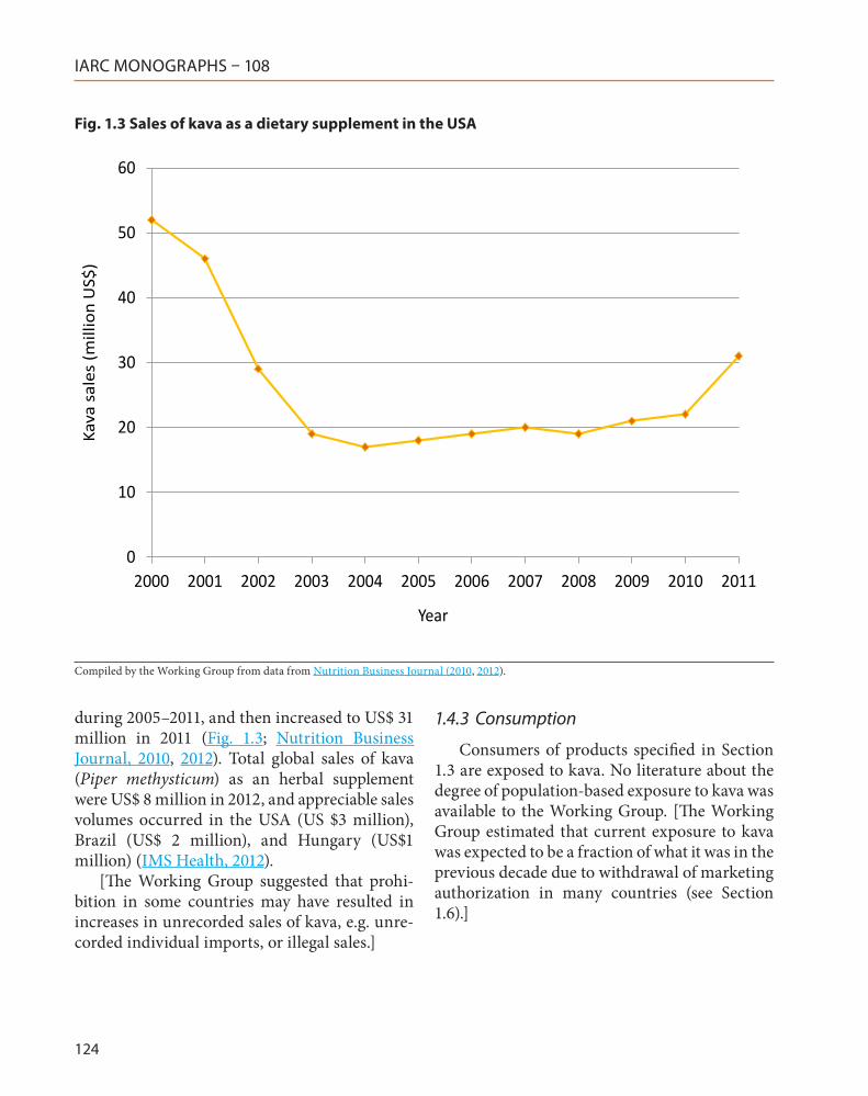
According to the 2012 Nutrition Business Journal Annual Report, kava was the 36th best-selling dietary supplement in the USA in 2011. There has been a considerable decline in kava sales in the USA from US$ 52 million in 2000 to US$ 17 million in 2004. Sales remained at a similar level between US$ 18 and 22 million
IARC MONOGRAPHS – 108
124
during 2005–2011, and then increased to US$ 31 million in 2011 (Fig. 1.3; Nutrition Business Journal, 2010, 2012). Total global sales of kava (Piper methysticum) as an herbal supplement were US$ 8 million in 2012, and appreciable sales volumes occurred in the USA (US $3 million), Brazil (US$ 2 million), and Hungary (US$1 million) (IMS Health, 2012).
[The Working Group suggested that prohi-bition in some countries may have resulted in increases in unrecorded sales of kava, e.g. unre-corded individual imports, or illegal sales.]
1.4.3 Consumption
Consumers of products specified in Section 1.3 are exposed to kava. No literature about the degree of population-based exposure to kava was available to the Working Group. [The Working Group estimated that current exposure to kava was expected to be a fraction of what it was in the previous decade due to withdrawal of marketing authorization in many countries (see Section 1.6).]
Fig. 1.3 Sales of kava as a dietary supplement in the USA
0
10
20
30
40
50
60
2000 2001 2002 2003 2004 2005 2006 2007 2008 2009 2010 2011
Kava
sal
es (m
illio
n U
S$)
Year
Compiled by the Working Group from data from Nutrition Business Journal (2010, 2012).
Kava
125
1.5 Occupational exposure
No specific studies on occupational exposure were available to the Working Group. It can be assumed that workers in kava production for food, cosmetic, or medicinal use may be exposed.
1.6 Regulations and guidelines
Several cases of liver damage have been asso-ciated with exposure to kava in Europe, and have led to withdrawal of the product license (NTP, 2012). Reviews on the cases of adverse effects potentially caused by exposure to kava have been compiled by Schmidt et al. (2005) (detailed anal-ysis of 83 cases), as by WHO (2007) (analysis of 93 cases). Speculations about the causes of the adverse effects included the use of less expensive stem peelings in commercial materials instead of the usual peeled rhizomes (Teschke et al., 2011).
Sales of kava have been suspended or withdrawn in several countries, including Australia, Canada, France, Germany, Spain, and Switzerland, and due to reported association with hepatotoxicity in humans (Russmann et al., 2001, 2003; Campo et al., 2002; De Smet, 2002; Parkman, 2002; Clough et al., 2003; Humberston
et al., 2003; Teschke et al., 2003; Ulbricht et al., 2005; NTP, 2012).
Although sales of kava were not regulated or controlled in the USA in 2012 (NTP, 2012), the Food and Drug Administration (FDA) had issued a public warning in 2001 that kava might be be associated with serious liver damage, including hepatitis, cirrhosis, and liver failure (FDA, 2002). The regulatory action taken by various countries around the world from the year 2000 after concerns about hepatotoxicity is summarized in WHO (2007). Current regula-tory status was summarized by Teschke & Lebot (2011), and included suggested chemical stand-ards and agricultural standardizations. WHO (2004) provided some guidelines for the quality control of kava (see Table 1.2).
2. Cancer in Humans
Steiner (2000) investigated the association between cancer incidence and consumption of kava in an ecological study of six countries in the South Pacific. Exposure was estimated by a surrogate measure of consumption based on the number of kava plants under cultivation in
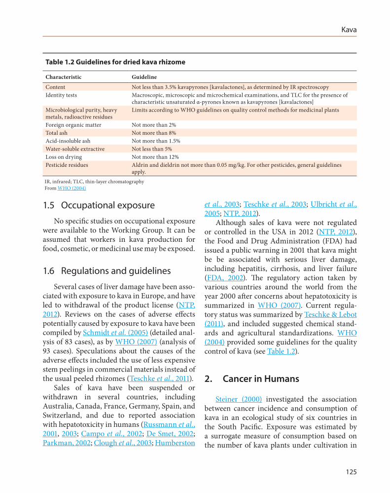
Table 1.2 Guidelines for dried kava rhizome
Characteristic Guideline
Content Not less than 3.5% kavapyrones [kavalactones], as determined by IR spectroscopyIdentity tests Macroscopic, microscopic and microchemical examinations, and TLC for the presence of
characteristic unsaturated α-pyrones known as kavapyrones [kavalactones]Microbiological purity, heavy metals, radioactive residues
Limits according to WHO guidelines on quality control methods for medicinal plants
Foreign organic matter Not more than 2%Total ash Not more than 8%Acid-insoluble ash Not more than 1.5%Water-soluble extractive Not less than 5%Loss on drying Not more than 12%Pesticide residues Aldrin and dieldrin not more than 0.05 mg/kg. For other pesticides, general guidelines
apply.IR, infrared; TLC, thin-layer chromatographyFrom WHO (2004)
IARC MONOGRAPHS – 108
126
each country. Exposure estimates and data on cancer incidence for men in the 1980s were used in the analysis on the assumption that all kava produced before 1990 was consumed locally, and primarily by men. An inverse correlation was observed between the incidence of all cancers in men and estimated exposure, but no test of statistical significance or confidence intervals, was reported. [The Working Group considered this study as uninformative because of its ecolog-ical design, the use of crude measures of expo-sure and outcome, and inadequate assessment of the role of chance.]
3. Cancer in Experimental Animals
3.1 Mouse
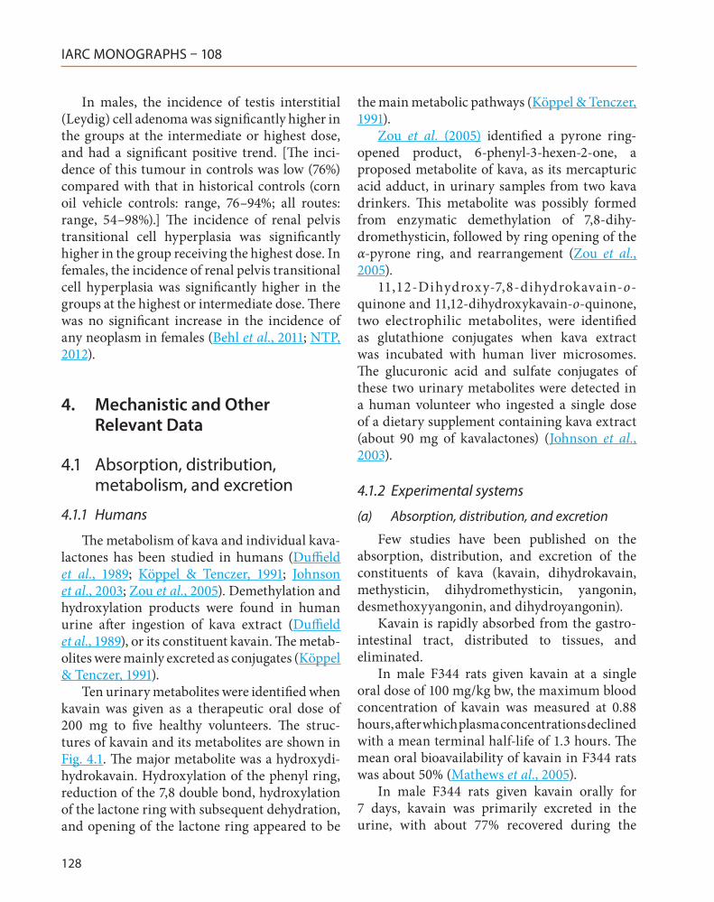
See Table 3.1In one study of oral administration, groups
of 50 male and 50 female B6C3F1 mice (age, 5–6 weeks) were given kava extract at a dose of 0 (corn oil vehicle, 10 mL/kg body weight, bw), 0.25, 0.5, or 1.0 g/kg bw per day by gavage, 5 days per week, for 105 weeks. The purity of the kava extract was 98.04% by high-performance liquid chromatography/ultraviolet (HPLC/UV) profiles. The extract contained 27% kavalactones identified as kavain, dihydrokavain, methysticin, dihydromethysticin, yangonin, and desmethoxy-yangonin. In males, the mean body weight of the dosed groups was similar to that in the control group. In females, the mean body weight of the group at 1.0 g/kg bw was 11% less than that in the control group after week 21. The mean survival time of male and female mice in the dosed groups was similar to that of the controls.
In males, the incidence of hepatoblastoma was significantly higher in the groups receiving the intermediate and highest dose, and had a significant positive trend. The incidence of hepatocellular carcinoma and hepatoblastoma (combined) was significantly higher in the group
receiving the intermediate dose. The incidence of eosinophilic hepatocyte foci, a preneoplastic hepatocyte lesion, was significantly higher in the groups receiving the intermediate or highest dose.
In females, the incidence of hepatocellular carcinoma was significantly higher in the group receiving the lowest dose. The incidence of hepato-cellular adenoma or carcinoma (combined) was significantly higher in the groups receiving the lowest and intermediate doses. The incidence of hepatocellular carcinoma and hepatoblas-toma (combined) was significantly higher in the group receiving the lowest dose. [The Working Group noted that reduced body weight may have reduced one tumour response in females at the highest dose.] The incidence of eosinophilic hepatocyte foci was significantly higher in the group receiving the highest dose. The incidence of squamous cell hyperplasia of the forestomach was significantly higher in the groups receiving the lowest or intermediate doses (Behl et al., 2011; NTP, 2012).
3.2 Rat
See Table 3.1In one study of oral administration, groups
of 49 or 50 male and 50 female F344/N rats (age, 6–7 weeks) were given kava extract at 0 (corn-oil vehicle, 5 mL/kg bw), 0.1, 0.3, or 1.0 g/kg bw per day by gavage, 5 days per week, for 104 (male rats) or 105 (female rats) weeks. The purity of the kava extract was 98.04% by HPLC/UV profiles. The extract contained 27% kavalactones identified as kavain, dihydrokavain, methysticin, dihy-dromethysticin, yangonin, and desmethoxyyan-gonin. The mean body weight of the groups at 1.0 g/kg bw was 10% less than that of the control group after week 65 in males and after week 41 in females. The mean survival time for rats in the dosed groups was similar to that of controls for both sexes.
Kava
127
Tabl
e 3.
1 St
udie
s of
car
cino
geni
city
wit
h ka
va e
xtra
cts
in m
ice
and
rats
Spec
ies,
st
rain
(s
ex)
Dur
atio
n R
efer
ence
Dos
ing
regi
men
, A
nim
als/
grou
p at
star
tIn
cide
nce
of tu
mou
rsSi
gnifi
canc
eaC
omm
ents
Mou
se,
B6C
3F1
(M, F
) 10
5 w
k N
TP
(201
2),
Behl
et a
l. (2
011)
0 (c
ontr
ol),
0.25
, 0.5
, or 1
.0 g
/kg
bw
by g
avag
e in
cor
n oi
l, 5
days
/wk
for
105
wk
50 M
and
50
F/gr
oup
(age
, 5–6
wk)
Hep
atoc
ellu
lar c
arci
nom
a:
3/50
, 13/
50*,
8/50
, 8/5
0 (F
)*P
= 0
.007
Extr
act p
urity
, 98.
04%
(HPL
C/U
V p
rofil
es),
cont
aini
ng 2
7% k
aval
acto
nes
Mea
n bo
dy w
eigh
t of f
emal
es a
t 1.0
g/k
g bw
was
11
% le
ss th
an th
at in
the
vehi
cle-
cont
rol g
roup
aft
er w
k 21
Hep
atoc
ellu
lar a
deno
ma
or c
arci
nom
a (c
ombi
ned)
: 38
/50,
39/
50, 3
9/50
, 40/
50 (M
);
10/5
0, 2
1/50
*, 20
/50*
*, 13
/50
(F)
*P =
0.0
15
**P
= 0.
036
Hep
atob
last
oma:
0/
50*,
4/50
, 9/5
0**,
12/5
0***
(M);
0/
50, 0
/50,
1/5
0, 0
/50
(F)
*P <
0.0
01
(tren
d)
**P
= 0.
002
**
*P <
0.0
01H
epat
ocel
lula
r car
cino
ma
or
hepa
tobl
asto
ma
(com
bine
d):
20/5
0, 2
1/50
, 30/
50*,
25/5
0 (M
);
3/50
, 13/
50**
, 9/5
0, 8
/50
(F)
*P <
0.0
5 **
P =
0.00
7
Rat,
F344
(M
, F)
104–
105
wk
NTP
(2
012)
, Be
hl et
al.
(201
1)
0 (c
ontr
ol),
0.1,
0.3
, or 1
.0 g
/kg
bw b
y ga
vage
in c
orn
oil,
5 da
ys/w
k fo
r 104
(M
) or 1
05 (F
) wk
49 o
r 50
M, a
nd 5
0 F
(age
, 6–7
wk)
Test
is in
ters
titia
l (Le
ydig
) cel
l ade
nom
ab : 37
/49
(76%
)*, 4
4/50
(88%
), 49
/50
(98%
)**,
46/5
0 (9
2%)*
**(M
)
*P =
0.0
03
(tren
d)
**P
= 0.
002
**
*P <
0.0
01
Extr
act p
urity
, 98.
04%
(HPL
C/U
V p
rofil
es),
cont
aini
ng 2
7% k
aval
acto
nes
Mea
n bo
dy w
eigh
t of g
roup
at 1
.0 g
/kg
bw w
as
10%
less
than
that
of t
he v
ehic
le-c
ontr
ol g
roup
aft
er w
k 65
(M) a
nd w
k 41
(F)
No
signi
fican
t inc
reas
e in
the
inci
denc
e of
any
ne
opla
sm in
fem
ales
a Po
ly-3
test
b H
isto
rica
l inc
iden
ce in
2-y
ear s
tudi
es w
ith a
dmin
istr
atio
n by
gav
age
with
cor
n oi
l veh
icle
-con
trol
gro
up (m
ean
± st
anda
rd d
evia
tion)
: 176
/199
(88.
4% ±
8.6
%),
rang
e 76
–94%
; all
rout
es: 1
053/
1298
(81.
1% ±
13.
4%),
rang
e 54
–98%
bw, b
ody
wei
ght;
F, fe
mal
e; H
PLC
/UV,
hig
h-pe
rfor
man
ce li
quid
chr
omat
ogra
phy/
ultr
avio
let;
M, m
ale;
wk,
wee
k
IARC MONOGRAPHS – 108
128
In males, the incidence of testis interstitial (Leydig) cell adenoma was significantly higher in the groups at the intermediate or highest dose, and had a significant positive trend. [The inci-dence of this tumour in controls was low (76%) compared with that in historical controls (corn oil vehicle controls: range, 76–94%; all routes: range, 54–98%).] The incidence of renal pelvis transitional cell hyperplasia was significantly higher in the group receiving the highest dose. In females, the incidence of renal pelvis transitional cell hyperplasia was significantly higher in the groups at the highest or intermediate dose. There was no significant increase in the incidence of any neoplasm in females (Behl et al., 2011; NTP, 2012).
4. Mechanistic and Other Relevant Data
4.1 Absorption, distribution, metabolism, and excretion
4.1.1 Humans
The metabolism of kava and individual kava-lactones has been studied in humans (Duffield et al., 1989; Köppel & Tenczer, 1991; Johnson et al., 2003; Zou et al., 2005). Demethylation and hydroxylation products were found in human urine after ingestion of kava extract (Duffield et al., 1989), or its constituent kavain. The metab-olites were mainly excreted as conjugates (Köppel & Tenczer, 1991).
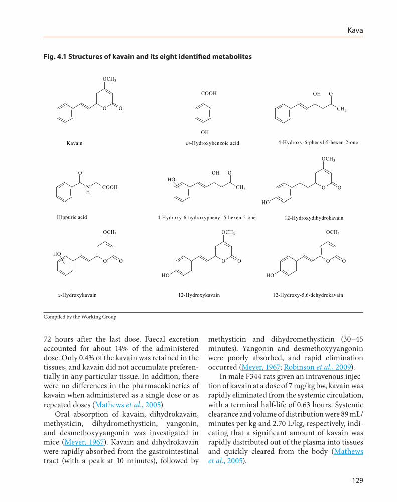
Ten urinary metabolites were identified when kavain was given as a therapeutic oral dose of 200 mg to five healthy volunteers. The struc-tures of kavain and its metabolites are shown in Fig. 4.1. The major metabolite was a hydroxydi-hydrokavain. Hydroxylation of the phenyl ring, reduction of the 7,8 double bond, hydroxylation of the lactone ring with subsequent dehydration, and opening of the lactone ring appeared to be
the main metabolic pathways (Köppel & Tenczer, 1991).
Zou et al. (2005) identified a pyrone ring-opened product, 6-phenyl-3-hexen-2-one, a proposed metabolite of kava, as its mercapturic acid adduct, in urinary samples from two kava drinkers. This metabolite was possibly formed from enzymatic demethylation of 7,8-dihy-dromethysticin, followed by ring opening of the α-pyrone ring, and rearrangement (Zou et al., 2005).
11,12- Dihydroxy -7,8-dihydrokavain-o- quinone and 11,12-dihydroxykavain-o-quinone, two electrophilic metabolites, were identified as glutathione conjugates when kava extract was incubated with human liver microsomes. The glucuronic acid and sulfate conjugates of these two urinary metabolites were detected in a human volunteer who ingested a single dose of a dietary supplement containing kava extract (about 90 mg of kavalactones) (Johnson et al., 2003).
4.1.2 Experimental systems
(a) Absorption, distribution, and excretion
Few studies have been published on the absorption, distribution, and excretion of the constituents of kava (kavain, dihydrokavain, methysticin, dihydromethysticin, yangonin, desmethoxyyangonin, and dihydroyangonin).
Kavain is rapidly absorbed from the gastro-intestinal tract, distributed to tissues, and eliminated.
In male F344 rats given kavain at a single oral dose of 100 mg/kg bw, the maximum blood concentration of kavain was measured at 0.88 hours, after which plasma concentrations declined with a mean terminal half-life of 1.3 hours. The mean oral bioavailability of kavain in F344 rats was about 50% (Mathews et al., 2005).
In male F344 rats given kavain orally for 7 days, kavain was primarily excreted in the urine, with about 77% recovered during the
Kava
129
72 hours after the last dose. Faecal excretion accounted for about 14% of the administered dose. Only 0.4% of the kavain was retained in the tissues, and kavain did not accumulate preferen-tially in any particular tissue. In addition, there were no differences in the pharmacokinetics of kavain when administered as a single dose or as repeated doses (Mathews et al., 2005).
Oral absorption of kavain, dihydrokavain, methysticin, dihydromethysticin, yangonin, and desmethoxyyangonin was investigated in mice (Meyer, 1967). Kavain and dihydrokavain were rapidly absorbed from the gastrointestinal tract (with a peak at 10 minutes), followed by
methysticin and dihydromethysticin (30–45 minutes). Yangonin and desmethoxyyangonin were poorly absorbed, and rapid elimination occurred (Meyer, 1967; Robinson et al., 2009).
In male F344 rats given an intravenous injec-tion of kavain at a dose of 7 mg/kg bw, kavain was rapidly eliminated from the systemic circulation, with a terminal half-life of 0.63 hours. Systemic clearance and volume of distribution were 89 mL/minutes per kg and 2.70 L/kg, respectively, indi-cating that a significant amount of kavain was rapidly distributed out of the plasma into tissues and quickly cleared from the body (Mathews et al., 2005).
Fig. 4.1 Structures of kavain and its eight identified metabolites
O
OCH3
O
COOH
OH
CH3
OOH
O
NH
COOH CH3
OOHHO
O O
HO
OCH3
O O
OCH3
HOO O
HO
OCH3
O O
HO
OCH3
Kavain m-Hydroxybenzoic acid 4-Hydroxy-6-phenyl-5-hexen-2-one
Hippuric acid 4-Hydroxy-6-hydroxyphenyl-5-hexen-2-one 12-Hydroxydihydrokavain
x-Hydroxykavain 12-Hydroxykavain 12-Hydroxy-5,6-dehydrokavain
Compiled by the Working Group
IARC MONOGRAPHS – 108
130
Keledjian et al. (1988) observed a peak concentration at 5 minutes in brain for kavain and 7,8-dihydrokavain; the compounds were rapidly eliminated after intraperitoneal admin-istration (100 mg/kg bw) of individual kava constituents in male Balb/c mice. The maximum concentrations of kavain and 7,8-dihydroka-vain were 64.7 and 29.3 ng/mg wet brain tissue, respectively. The maximum concentrations of desmethoxyyangonin and yangonin were 10.4 and 1.2 ng/mg wet brain tissue, lower than those of kavain or 7,8-dihydrokavain. When kava extract was given intraperitoneally to male Balb/c mice, the maximum concentrations of kavain and yangonin increased in the brain, while the concentrations of 7,8-dihydrokavain and desmethoxyyangonin were similar to those measured after they were injected separately (Keledjian et al., 1988).
(b) Metabolism
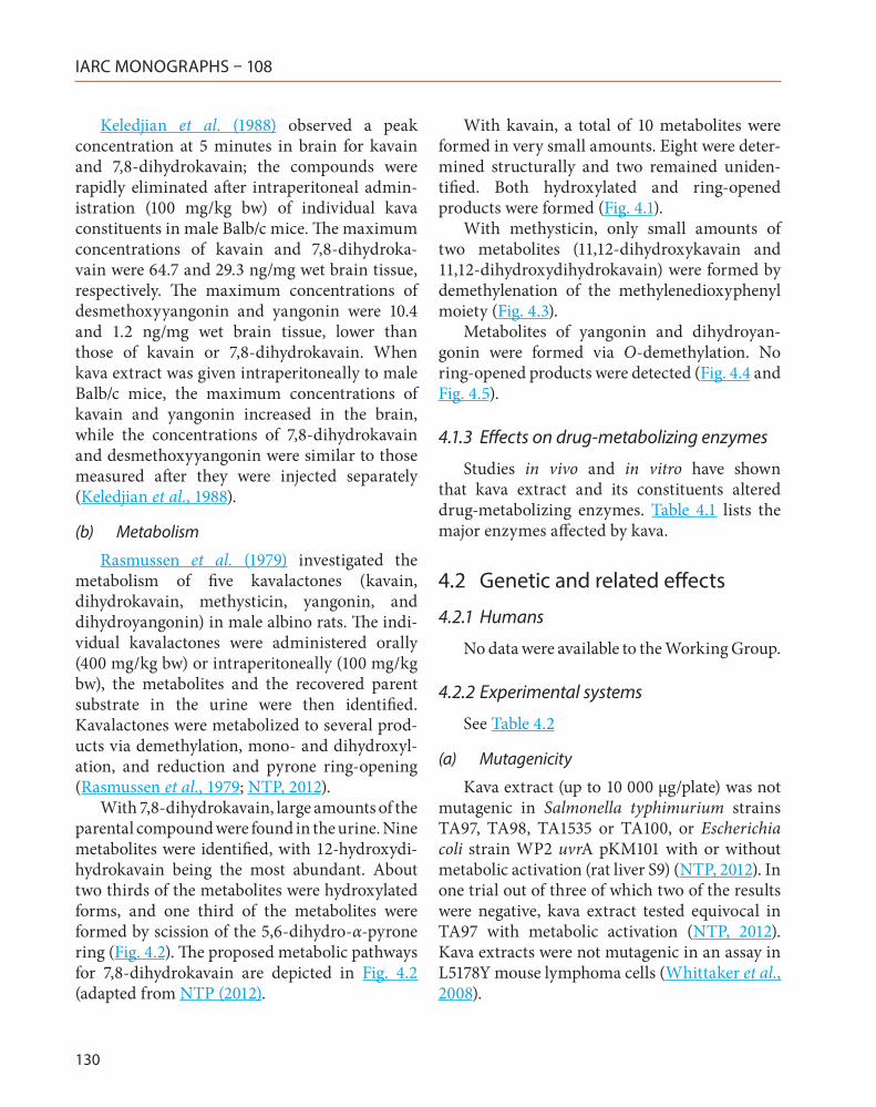
Rasmussen et al. (1979) investigated the metabolism of five kavalactones (kavain, dihydrokavain, methysticin, yangonin, and dihydroyangonin) in male albino rats. The indi-vidual kavalactones were administered orally (400 mg/kg bw) or intraperitoneally (100 mg/kg bw), the metabolites and the recovered parent substrate in the urine were then identified. Kavalactones were metabolized to several prod-ucts via demethylation, mono- and dihydroxyl-ation, and reduction and pyrone ring-opening (Rasmussen et al., 1979; NTP, 2012).
With 7,8-dihydrokavain, large amounts of the parental compound were found in the urine. Nine metabolites were identified, with 12-hydroxydi-hydrokavain being the most abundant. About two thirds of the metabolites were hydroxylated forms, and one third of the metabolites were formed by scission of the 5,6-dihydro-α-pyrone ring (Fig. 4.2). The proposed metabolic pathways for 7,8-dihydrokavain are depicted in Fig. 4.2 (adapted from NTP (2012).
With kavain, a total of 10 metabolites were formed in very small amounts. Eight were deter-mined structurally and two remained uniden-tified. Both hydroxylated and ring-opened products were formed (Fig. 4.1).
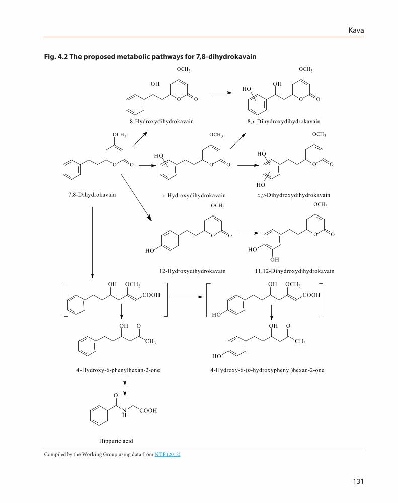
With methysticin, only small amounts of two metabolites (11,12-dihydroxykavain and 11,12-dihydroxydihydrokavain) were formed by demethylenation of the methylenedioxyphenyl moiety (Fig. 4.3).
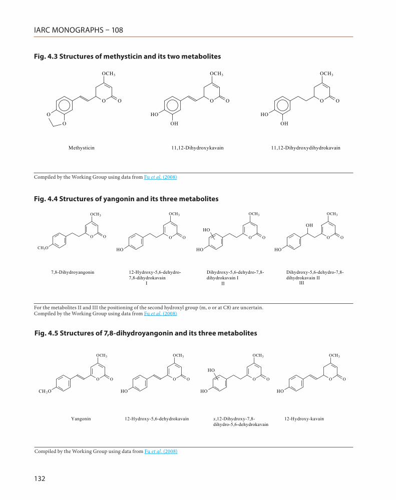
Metabolites of yangonin and dihydroyan-gonin were formed via O-demethylation. No ring-opened products were detected (Fig. 4.4 and Fig. 4.5).
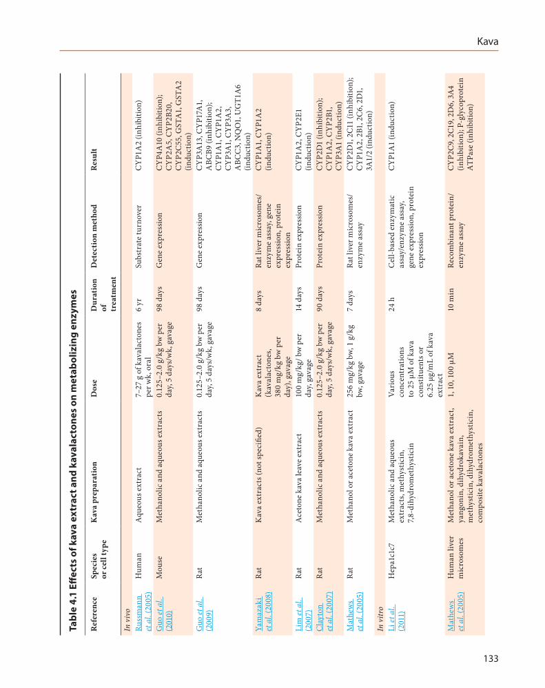
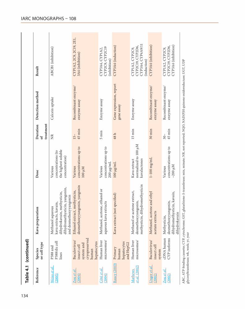
4.1.3 Effects on drug-metabolizing enzymes
Studies in vivo and in vitro have shown that kava extract and its constituents altered drug-metabolizing enzymes. Table 4.1 lists the major enzymes affected by kava.
4.2 Genetic and related effects
4.2.1 Humans
No data were available to the Working Group.
4.2.2 Experimental systems
See Table 4.2
(a) Mutagenicity
Kava extract (up to 10 000 µg/plate) was not mutagenic in Salmonella typhimurium strains TA97, TA98, TA1535 or TA100, or Escherichia coli strain WP2 uvrA pKM101 with or without metabolic activation (rat liver S9) (NTP, 2012). In one trial out of three of which two of the results were negative, kava extract tested equivocal in TA97 with metabolic activation (NTP, 2012). Kava extracts were not mutagenic in an assay in L5178Y mouse lymphoma cells (Whittaker et al., 2008).
Kava
131
Fig. 4.2 The proposed metabolic pathways for 7,8-dihydrokavain
HO
O O
OCH3
O O
OCH3
O O
OCH3
O O
OCH3
HO
O O
OCH3
HO
OH
HO HO
HO
COOH
OH OCH3
HO
COOH
OH OCH3
CH3
OH O
NH
COOH
O
8-Hydroxydihydrokavain 8,x-Dihydroxydihydrokavain
7,8-Dihydrokavain x-Hydroxydihydrokavain x,y-Dihydroxydihydrokavain
12-Hydroxydihydrokavain 11,12-Dihydroxydihydrokavain
4-Hydroxy-6-phenylhexan-2-one 4-Hydroxy-6-(p-hydroxyphenyl)hexan-2-one
Hippuric acid
CH3
OH O
HO
O O
OCH3
OH
O O
OCH3
OH
Compiled by the Working Group using data from NTP (2012).
IARC MONOGRAPHS – 108
132
Fig. 4.3 Structures of methysticin and its two metabolites
O
O
O O
OCH3
OH
HO
O O
OCH3
OH
HO
O O
OCH3
Methysticin 11,12-Dihydroxykavain 11,12-Dihydroxydihydrokavain
Compiled by the Working Group using data from Fu et al. (2008)
Fig. 4.4 Structures of yangonin and its three metabolites
CH3O
O O
OCH3
O O
OCH3
HO
O O
OCH3
HO
O O
OCH3
HO
OHHO
7,8-Dihydroyangonin 12-Hydroxy-5,6-dehydro-7,8-dihydrokavain
Dihydroxy-5,6-dehydro-7,8-dihydrokavain I
Dihydroxy-5,6-dehydro-7,8-dihydrokavain II
I II III
For the metabolites II and III the positioning of the second hydroxyl group (m, o or at C8) are uncertain. Compiled by the Working Group using data from Fu et al. (2008)
Fig. 4.5 Structures of 7,8-dihydroyangonin and its three metabolites
CH 3O
O O
OCH3
HO
O O
OCH3
HO
O O
OCH3
HO
O O
OCH3
HO
Yangonin 12-Hydroxy-5,6-dehydrokavain x,12-Dihydroxy-7,8-dihydro-5,6-dehydrokavain
12-Hydroxy-kavain
Compiled by the Working Group using data from Fu et al. (2008)
Kava
133
Tabl
e 4.
1 Eff
ects
of k
ava
extr
act a
nd k
aval
acto
nes
on m
etab
oliz
ing
enzy
mes
Ref
eren
ceSp
ecie
s or
cel
l typ
eK
ava
prep
arat
ion
Dos
eD
urat
ion
of
trea
tmen
t
Det
ecti
on m
etho
dR
esul
t
In v
ivo
Russ
man
n et
al.
(200
5)H
uman
Aqu
eous
ext
ract
7–27
g o
f kav
alac
tone
s pe
r wk,
ora
l6
yrSu
bstr
ate
turn
over
CY
P1A
2 (in
hibi
tion)
Guo
et a
l. (2
010)
Mou
seM
etha
nolic
and
aqu
eous
ext
ract
s0.
125–
2.0
g/kg
bw
per
da
y, 5
days
/wk,
gav
age
98 d
ays
Gen
e ex
pres
sion
CY
P4A
10 (i
nhib
ition
); C
YP2
A5,
CY
P2B2
0,
CY
P2C
55, G
STA
1, G
STA
2 (in
duct
ion)
Guo
et a
l. (2
009)
Rat
Met
hano
lic a
nd a
queo
us e
xtra
cts
0.12
5–2.
0 g/
kg b
w p
er
day,
5 da
ys/w
k, g
avag
e98
day
sG
ene
expr
essio
nC
YP3
A13
, CY
P17A
1,
ABC
B9 (i
nhib
ition
); C
YP1
A1,
CY
P1A
2,
CY
P3A
1, C
YP3
A3,
A
BCC
3, N
QO
1, U
GT1
A6
(indu
ctio
n)Ya
maz
aki
et a
l. (2
008)
Rat
Kav
a ex
trac
ts (n
ot sp
ecifi
ed)
Kav
a ex
trac
t (k
aval
acto
nes,
380
mg/
kg b
w p
er
day)
, gav
age
8 da
ysRa
t liv
er m
icro
som
es/
enzy
me
assa
y, ge
ne
expr
essio
n, p
rote
in
expr
essio
n
CY
P1A
1, C
YP1
A2
(indu
ctio
n)
Lim
et a
l. (2
007)
Rat
Ace
tone
kav
a le
ave
extr
act
100
mg/
kg/ b
w p
er
day,
gava
ge14
day
sPr
otei
n ex
pres
sion
CY
P1A
2, C
YP2
E1
(indu
ctio
n)C
layt
on
et a
l. (2
007)
Rat
Met
hano
lic a
nd a
queo
us e
xtra
cts
0.12
5–2.
0 g/
kg b
w p
er
day,
5 da
ys/w
k, g
avag
e90
day
sPr
otei
n ex
pres
sion
CY
P2D
1 (in
hibi
tion)
; C
YP1
A2,
CY
P2B1
, C
YP3
A1
(indu
ctio
n)M
athe
ws
et a
l. (2
005)
Rat
Met
hano
l or a
ceto
ne k
ava
extr
act
256
mg/
kg b
w, 1
g/k
g bw
, gav
age
7 da
ysRa
t liv
er m
icro
som
es/
enzy
me
assa
yC
YP2
D1,
2C1
1 (in
hibi
tion)
; C
YP1
A2,
2B1
, 2C
6, 2
D1,
3A
1/2
(indu
ctio
n)In
vitr
oLi
et a
l. (2
011)
Hep
a1c1
c7M
etha
nolic
and
aqu
eous
ex
trac
ts, m
ethy
stic
in,
7,8-
dihy
drom
ethy
stic
in
Vari
ous
conc
entr
atio
ns
to 2
5 µM
of k
ava
cons
titue
nts o
r 6.
25 µ
g/m
L of
kav
a ex
trac
t
24 h
Cel
l-bas
ed e
nzym
atic
as
say/
enzy
me
assa
y, ge
ne e
xpre
ssio
n, p
rote
in
expr
essio
n
CY
P1A
1 (in
duct
ion)
Mat
hew
s et
al.
(200
5)H
uman
live
r m
icro
som
esM
etha
nol o
r ace
tone
kav
a ex
trac
t, ya
ngon
in, d
ihyd
roka
vain
, m
ethy
stic
in, d
ihyd
rom
ethy
stic
in,
com
posit
e ka
vala
cton
es
1, 1
0, 1
00 µ
M10
min
Reco
mbi
nant
pro
tein
/en
zym
e as
say
CY
P2C
9, 2
C19,
2D
6, 3
A4
(inhi
bitio
n); P
-gly
copr
otei
n A
TPas
e (in
hibi
tion)
IARC MONOGRAPHS – 108
134
Ref
eren
ceSp
ecie
s or
cel
l typ
eK
ava
prep
arat
ion
Dos
eD
urat
ion
of
trea
tmen
t
Det
ecti
on m
etho
dR
esul
t
Wei
ss et
al.
(200
5)P3
88 a
nd
P388
/dx
cell
lines
Met
hano
l aqu
eous
ka
va e
xtra
cts,
kava
in,
dihy
drok
avai
n, m
ethy
stic
in,
dihy
drom
ethy
stic
in, y
ango
nin,
an
d de
smet
hoxy
yang
onin
Vari
ous
conc
entr
atio
ns (t
o th
e hi
ghes
t sol
uble
co
ncen
trat
ion)
NR
Cal
cein
upt
ake
ABC
B1 (i
nhib
ition
)
Zou
et a
l. (2
004)
Bacu
lovi
rus/
inse
ct c
ell
syst
em a
nd
cryo
pres
erve
d hu
man
he
pato
cyte
s
Etha
nol e
xtra
ct, m
ethy
stic
in,
desm
etho
xyya
ngon
in, y
ango
nin
Vari
ous
conc
entr
atio
ns u
p to
10
0 µM
15–
45 m
inRe
com
bina
nt e
nzym
e/en
zym
e as
say
CY
P1A
2, 2
C9,
2C1
9, 2
E1,
3A4
(inhi
bitio
n)
Côt
é et
al.
(200
4)H
uman
live
r m
icro
som
es/
Met
hano
l, ac
eton
e, e
than
ol o
r aq
ueou
s kav
a ex
trac
tsVa
riou
s co
ncen
trat
ions
up
to
200
μg/m
L
5 m
inEn
zym
e as
say
CY
P3A
4, C
YP1
A2,
C
YP2
C9,
CY
P2C1
9 (in
hibi
tion)
Rauc
y (2
003)
Prim
ary
hum
an
hepa
tocy
tes
and
Hep
G2
Kav
a ex
trac
t (no
t spe
cifie
d)10
0 μg
/mL
48 h
Gen
e ex
pres
sion,
repo
rt
gene
ass
ayC
YP3
A4
(indu
ctio
n)
Mat
hew
s et
al.
(200
2)H
uman
live
r m
icro
som
es/
Met
hano
l or a
ceto
ne e
xtra
ct,
desm
etho
xyya
ngon
in,
met
hyst
icin
, dih
ydro
met
hyst
icin
Kav
a ex
trac
t no
rmal
ized
to 1
00 μ
M
kava
lact
ones
15 m
inEn
zym
e as
say
CY
P1A
2, C
YP2
C9,
C
YP2
C19,
CY
P2D
6,
CY
P3A
4, C
YP4
A9/
11
(indu
ctio
n)U
nger
et a
l. (2
002)
Bacu
lovi
rus/
inse
ct c
ell
syst
em
Met
hano
l, ac
eton
e an
d et
hyl
acet
ate
extr
acts
1–10
0 m
g/m
L30
min
Reco
mbi
nant
enz
yme/
enzy
me
assa
yC
YP3
A4
(inhi
bitio
n)
Zou
et a
l. (2
002)
cDN
A h
uman
C
YP
isof
orm
sM
ethy
stic
in,
desm
etho
xyya
ngon
in,
dihy
drom
ethy
stic
in, k
avai
n,
dihy
drok
avai
n
Vari
ous
conc
entr
atio
ns u
p to
~2
00 µ
M
30–
45 m
inRe
com
bina
nt e
nzym
e/en
zym
e as
say
CY
P1A
2, C
YP2
C9,
C
YP2
C19,
CY
P2D
6,
CY
P3A
4 (in
hibi
tion)
ABC
, ATP
-bin
ding
cas
sett
e; C
YP,
cyt
ochr
ome;
GST
, glu
tath
ione
-S-t
rans
fera
se; m
in, m
inut
e; N
R, n
ot re
port
ed; N
QO
, NA
D(P
)H q
uino
ne o
xido
redu
ctas
e; U
GT,
UD
P gl
ycos
yltr
ansf
eras
e; w
k, w
eek;
yr,
year
Tabl
e 4.
1 (
cont
inue
d)
Kava
135
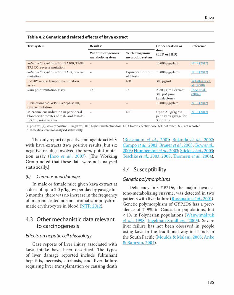
The only report of positive mutagenic activity with kava extracts (two positive results, but six negative results) involved the umu point muta-tion assay (Jhoo et al., 2007). [The Working Group noted that these data were not analysed statistically.]
(b) Chromosomal damage
In male or female mice given kava extract at a dose of up to 2.0 g/kg bw per day by gavage for 3 months, there was no increase in the frequency of micronucleated normochromatic or polychro-matic erythrocytes in blood (NTP, 2012).
4.3 Other mechanistic data relevant to carcinogenesis
Effects on hepatic cell physiology
Case reports of liver injury associated with kava intake have been described. The types of liver damage reported include fulminant hepatitis, necrosis, cirrhosis, and liver failure requiring liver transplantation or causing death
(Russmann et al., 2001; Bujanda et al., 2002; Campo et al., 2002; Brauer et al., 2003; Gow et al., 2003; Humberston et al., 2003; Stickel et al., 2003; Teschke et al., 2003, 2008; Thomsen et al., 2004).
4.4 Susceptibility
Genetic polymorphisms
Deficiency in CYP2D6, the major kavalac-tone-metabolizing enzyme, was detected in two patients with liver failure (Russmann et al., 2001). Genetic polymorphism of CYP2D6 has a prev-alence of 7–9% in Caucasian populations, but < 1% in Polynesian populations (Wanwimolruk et al., 1998; Ingelman-Sundberg, 2005). Severe liver failure has not been observed in people using kava in the traditional way in islands in the South Pacific (Moulds & Malani, 2003; Anke & Ramzan, 2004).
Table 4.2 Genetic and related effects of kava extract
Test system Resultsa Concentration or dose (LED or HID)
Reference
Without exogenous metabolic system
With exogenous metabolic system
Salmonella typhimurium TA100, TA98, TA1535, reverse mutation
– – 10 000 μg/plate NTP (2012)
Salmonella typhimurium TA97, reverse mutation
– Equivocal in 1 out of 3 tests
10 000 μg/plate NTP (2012)
L5178Y mouse lymphoma mutation assay
– NR 300 μg/mL Whittaker et al. (2008)
umu point mutation assay +a +a 2330 μg/mL extract 300 μM pure kavalactones
Jhoo et al. (2007)
Escherichia coli WP2 uvrA/pKM101, reverse mutation
– – 10 000 μg/plate NTP (2012)
Micronucleus induction in peripheral blood erythrocytes of male and female B6C3F1 mice in vivo
– NT Up to 2.0 g/kg bw per day by gavage for 3 months
NTP (2012)
+, positive; (+), weakly positive; –, negative; HID, highest ineffective dose; LED, lowest effective dose; NT, not tested; NR, not reported a These data were not analysed statistically.
IARC MONOGRAPHS – 108
136
4.5 Mechanistic considerations
Kava extract is not mutagenic based on the results of numerous studies of genotoxicity, including tests for mutagenicity in bacteria, induction of micronuclei in vivo (NTP, 2012), and the mouse lymphoma assay (Whittaker et al., 2008). The reported carcinogenicity in mice is most probably mediated through nongenotoxic mechanisms.
5. Summary of Data Reported
5.1 Exposure data
The kava (or kava kava) plant Piper methysticum is a perennial tropical shrub that is widely cultivated in Oceania. The rhizome of the plant was originally used as an ingre-dient in local traditional drinks with psychop-harmacological properties, and as traditional folk medicine. More recently, rhizome extracts have been used in medicinal products, food or dietary supplements, and cosmetics. Important chemical constituents of the resin contained in the kava rhizome are kavalactones, kavain being the major compound. The medicinal uses of kava supported by clinical data are short-term symp-tomatic treatment of mild states of anxiety or insomnia due to nervousness, stress, or tension. Use of kava was popular worldwide, but several case reports of liver damage associated with exposure to kava reduced sales, and caused kava to be banned in several countries.
5.2 Human carcinogenicity data
The Working Group was able to identify only one epidemiological study of cancer and kava consumption. This ecological study found an inverse correlation between all cancers in men and a proxy measure of kava consumption, but no confidence intervals or test of statistical
significance were reported. The Working Group regarded the study as uninformative because the ecological design provided only weak support for causal inference at the individual level, the meas-ures of exposure and outcome were crude, and the role of chance was not evaluated.
5.3 Animal carcinogenicity data
A kava extract was tested for carcinogenicity in one study in mice and one study in rats treated by gavage. In mice, the extract caused a signifi-cant increase in the incidence of hepatoblastoma in males, and of hepatocellular adenoma or carcinoma (combined), and hepatocellular carci-noma, in females. In male rats, the same extract caused a significant increase in the incidence of testis interstitial (Leydig) cell adenoma; however, the incidence in controls was low compared with that in historical controls. There was no signifi-cant increase in the incidence of any neoplasm in female rats.
5.4 Mechanistic and other relevant data
The major components of kava extract, kava-lactones, are extensively metabolized in humans and experimental animals. Among the numerous metabolites are products from demethylation, hydroxylation, and ring-opening.
Kava extract gave negative results in several standard bacterial assays for mutation in the absence or presence of exogenous metabolic activation. Kavalactones gave negative results in most of these assays.
The reported carcinogenicity of kava in mice is most likely to be mediated through a nongen-otoxic mechanism.
Kava
137
6. Evaluation
6.1 Cancer in humans
There is inadequate evidence in humans for the carcinogenicity of kava extract.
6.2 Cancer in experimental animals
There is sufficient evidence in experimental animals for the carcinogenicity of kava extract.
6.3 Overall evaluation
Kava extract is possibly carcinogenic to humans (Group 2B).
References
Anke J & Ramzan I (2004). Kava hepatotoxicity: Are we any closer to the truth? Planta Med, 70(3):193–6. doi:10.1055/s-2004-815533 PMID:15114493
Anonymous (2000). Kava kava rhizome (root). In: Blumenthal M, Goldberg A, Brinckmann J, editors. Expanded Commission E Monographs. Herb Monographs, based on those created by a special expert committee of the German Federal Institute for Drugs and Medicinal Devices. Newton (MA), USA: Integrative Medicine Communications
Behl M, Nyska A, Chhabra RS, Travlos GS, Fomby LM, Sparrow BR et al. (2011). Liver toxicity and carcino-genicity in F344/N rats and B6C3F1 mice exposed to Kava Kava. Food Chem Toxicol, 49(11):2820–9. doi:10.1016/j.fct.2011.07.067 PMID:21871523
Bilia AR, Bergonzi MC, Lazari D, Vincieri FF (2002). Characterization of commercial kava-kava herbal drug and herbal drug preparations by means of nuclear magnetic resonance spectroscopy. J Agric Food Chem, 50(18):5016–25. doi:10.1021/jf020049j PMID:12188601
Bilia AR, Gallon S, Vincieri FF (2002b). Kava-kava and anxiety: growing knowledge about the efficacy and safety. Life Sci, 70(22):2581–97. doi:10.1016/S0024-3205(02)01555-2 PMID:12269386
Bilia AR, Scalise L, Bergonzi MC, Vincieri FF (2004). Analysis of kavalactones from Piper methysticum (kava-kava). J Chromatogr B Analyt Technol Biomed Life Sci, 812(1-2):203–14. doi:10.1016/j.jchromb.2004.07.038 PMID:15556499
Brauer RB, Stangl M, Stewart JR, Pfab R, Becker K (2003). Acute liver failure after administration of herbal tranquilizer kava-kava (Piper methysticum). J Clin Psychiatry, 64(2):216–8. doi:10.4088/JCP.v64n0215c PMID:12633134
Bujanda L, Palacios A, Silvariño R, Sánchez A, Muñoz C (2002). [Kava-induced acute icteric hepatitis] Gastroenterol Hepatol, 25(6):434–5. doi:10.1016/S0210-5705(02)70281-1 PMID:12069710
Cairney S, Maruff P, Clough AR (2002). The neurobehav-ioural effects of kava. Aust N Z J Psychiatry, 36(5):657–62. doi:10.1046/j.1440-1614.2002.01027.x PMID:12225450
Campo JV, McNabb J, Perel JM, Mazariegos GV, Hasegawa SL, Reyes J (2002). Kava-induced fulminant hepatic failure. J Am Acad Child Adolesc Psychiatry, 41(6):631–2. doi:10.1097/00004583-200206000-00001 PMID:12049436
Clayton NP, Yoshizawa K, Kissling GE, Burka LT, Chan PC, Nyska A (2007). Immunohistochemical analysis of expressions of hepatic cytochrome P450 in F344 rats following oral treatment with kava extract. Exp Toxicol Pathol, 58(4):223–36. doi:10.1016/j.etp.2006.08.002 PMID:17059882
Clough A (2003). Enough! or too much. What is ‘excessive’ kava use in Arnhem Land? Drug Alcohol Rev, 22(1):43–51. doi:10.1080/0959523021000059820 PMID:12745358
Clough AR, Bailie RS, Currie B (2003). Liver function test abnormalities in users of aqueous kava extracts. J Toxicol Clin Toxicol, 41(6):821–9. doi:10.1081/CLT-120025347 PMID:14677792
Clough AR, Burns CB, Mununggurr N (2000). Kava in Arnhem Land: a review of consumption and its social correlates. Drug Alcohol Rev, 19(3):319–28. doi:10.1080/713659370
Côté CS, Kor C, Cohen J, Auclair K (2004). Composition and biological activity of traditional and commercial kava extracts. Biochem Biophys Res Commun, 322(1):147–52. doi:10.1016/j.bbrc.2004.07.093 PMID:15313185
De Smet PA (2002). Safety concerns about kava not unique. Lancet, 360(9342):1336 doi:10.1016/S0140-6736(02)11347-X PMID:12414243
Dentali SJ (1997). Herb safety review: Kava: Piper methysticum Forster F. (Piperaceae). Boulder (CO), USA: Herb Research Foundation.
Dharmaratne HR, Nanayakkara NP, Khan IA (2002). Kavalactones from Piper methysticum, and their 13C NMR spectroscopic analyses. Phytochemistry, 59(4):429–33. doi:10.1016/S0031-9422(01)00443-5 PMID:11830162
Duffield AM, Jamieson DD, Lidgard RO, Duffield PH, Bourne DJ (1989). Identification of some human urinary metabolites of the intoxicating beverage kava. J Chromatogr A, 475(2):273–81. doi:10.1016/S0021-9673(01)89682-5 PMID:2777959
FDA (2002). Kava-containing dietary supplements may be associated with severe liver injury. US Food and Drug

IARC MONOGRAPHS – 108
138
Administration. Available at: http://www.fda.gov/food/resourcesforyou/consumers/ucm085482.htm, accessed 18/04/2012.
Fu PP, Xia Q, Guo L, Yu H, Chan PC (2008). Toxicity of kava kava. J Environ Sci Health C Environ Carcinog Ecotoxicol Rev, 26(1):89–112. doi:10.1080/10590500801907407 PMID:18322868
Gaub M, Roeseler Ch, Roos G, Kovar KA (2004). Analysis of plant extracts by NIRS: simultaneous determina-tion of kavapyrones and water in dry extracts of Piper methysticum Forst. J Pharm Biomed Anal, 36(4):859–64. doi:10.1016/j.jpba.2004.06.030 PMID:15533680
Gow PJ, Connelly NJ, Hill RL, Crowley P, Angus PW (2003). Fatal fulminant hepatic failure induced by a natural therapy containing kava. Med J Aust, 178(9):442–3. PMID:12720510
Guo L, Li Q, Xia Q, Dial S, Chan PC, Fu P (2009). Analysis of gene expression changes of drug metabolizing enzymes in the livers of F344 rats following oral treat-ment with kava extract. Food Chem Toxicol, 47(2):433–42. doi:10.1016/j.fct.2008.11.037 PMID:19100306
Guo L, Shi Q, Dial S, Xia Q, Mei N, Li QZ et al. (2010). Gene expression profiling in male B6C3F1 mouse livers exposed to kava identifies–changes in drug metabolizing genes and potential mechanisms linked to kava toxicity. Food Chem Toxicol, 48(2):686–96. doi:10.1016/j.fct.2009.11.050 PMID:19948201
IMS Health (2012) Multinational Integrated Data Analysis (MIDAS). Plymouth Meeting, 2012, Pennsylvania: IMS Health.
Humberston CL, Akhtar J, Krenzelok EP (2003). Acute hepatitis induced by kava kava. J Toxicol Clin Toxicol, 41(2):109–13. doi:10.1081/CLT-120019123 PMID:12733846
Ingelman-Sundberg M (2005). Genetic polymorphisms of cytochrome P450 2D6 (CYP2D6): clinical conse-quences, evolutionary aspects and functional diver-sity. Pharmacogenomics J, 5(1):6–13. doi:10.1038/sj.tpj.6500285 PMID:15492763
Jhoo JW, Ang CY, Heinze TM, Deck J, Schnackenberg LK, Beger RD et al. (2007). Identification of C-glycoside flavonoids as potential mutagenic compounds in kava. J Food Sci, 72(2):C120–5. doi:10.1111/j.1750-3841.2007.00278.x PMID:17995826
Johnson BM, Qiu SX, Zhang S, Zhang F, Burdette JE, Yu L et al. (2003). Identification of novel electrophilic metabolites of Piper methysticum Forst (kava). Chem Res Toxicol, 16(6):733–40. doi:10.1021/tx020113r PMID:12807356
Keledjian J, Duffield PH, Jamieson DD, Lidgard RO, Duffield AM (1988). Uptake into mouse brain of four compounds present in the psychoactive beverage kava. J Pharm Sci, 77(12):1003–6. doi:10.1002/jps.2600771203 PMID:3244102
Köppel C & Tenczer J (1991). Mass spectral characteriza-tion of urinary metabolites of D,L-kawain. J Chromatogr
A, 562(1-2):207–11. doi:10.1016/0378-4347(91)80578-Z PMID:2026693
Lebot V, Merlin M, Lindstrom L (1997). Kava-the Pacific elixir: the definitive guide to its ethnobotany, history, and chemistry. Rochester (VT), USA: Healing Arts Press.
Li Y, Mei H, Wu Q, Zhang S, Fang JL, Shi L et al. (2011). Methysticin and 7,8-dihydromethysticin are two major kavalactones in kava extract to induce CYP1A1. Toxicol Sci, 124(2):388–99. doi:10.1093/toxsci/kfr235 PMID:21908763
Lim ST, Dragull K, Tang CS, Bittenbender HC, Efird JT, Nerurkar PV (2007). Effects of kava alkaloid, piper-methystine, and kavalactones on oxidative stress and cytochrome P450 in F-344 rats. Toxicol Sci, 97(1):214–21. doi:10.1093/toxsci/kfm035 PMID:17329236
Lindstrom L (2004). History, folklore, traditional and current uses of kava. In: Singh YN, editor. Kava: from ethnology to pharmacology. Boca Raton (FL), USA: CRC Press; pp. 10–28.
Mathews JM, Etheridge AS, Black SR (2002). Inhibition of human cytochrome P450 activities by kava extract and kavalactones. Drug Metab Dispos, 30(11):1153–7. doi:10.1124/dmd.30.11.1153 PMID:12386118
Mathews JM, Etheridge AS, Valentine JL, Black SR, Coleman DP, Patel P et al. (2005). Pharmacokinetics and disposition of the kavalactone kawain: interac-tion with kava extract and kavalactones in vivo and in vitro. Drug Metab Dispos, 33(10):1555–63. doi:10.1124/dmd.105.004317 PMID:16033948
McDonald D & Jowitt A (2000). Kava in the Pacific Islands: a contemporary drug of abuse? Drug Alcohol Rev, 19(2):217–27. doi:10.1080/713659319
Meyer HJ (1967). Pharmacology of kava. 1. Psychopharmacol Bull, 4(3):10–1. PMID:5616309
Monakhova YB, Kuballa T, Löbell-Behrends S, Maixner S, Kohl-Himmelseher M, Ruge W et al. (2013). Standardless 1H NMR determination of pharmaco-logically active substances in dietary supplements and medicines that have been illegally traded over the internet. Drug Test Anal, 5(6):400–11. doi:10.1002/dta.1367 PMID:22550015
Morgan M, Bone K, Mills S et al. (2005). Kava. Safety monograph. In: Mills S, Bone K, editors. The essential guide to herbal safety. St. Louis (MO), USA: Elsevier Churchill Livingstone; pp. 484–492.
Morris CA & Avorn J (2003). Internet marketing of herbal products. JAMA, 290(11):1505–9. doi:10.1001/jama.290.11.1505 PMID:13129992
Moulds RF & Malani J (2003). Kava: herbal panacea or liver poison? Med J Aust, 178(9):451–3. PMID:12720513
NTP (2012). Toxicology and carcinogenesis studies of kava kava extract (CAS No. 9000–38–8) in F344/N rats and B6C3F1 mice (gavage studies). Natl Toxicol Program Tech Rep Ser, 571(571):1–186. PMID:22441424

Kava
139
NLM (2012). Products that contain active ingredient - Kava Kava. Dietary supplements labels database. United States National Library of Medicine. Available from: http://www.dsld.nlm.nih.gov/dsld/rptQSearch.jsp?item=Kava+Kava&db=adsld, accessed 7 July 2014.
Norton SA & Ruze P (1994). Kava dermopathy. J Am Acad Dermatol, 31(1):89–97. doi:10.1016/S0190-9622(94)70142-3 PMID:8021378
Nutrition Business Journal (2010). NBJ’s Supplement Business Report. An analysis of markets, trends, competition and strategy in the U.S. dietary supple-ment industry. New York (NY), USA: Penton Media, Inc.
Nutrition Business Journal (2012). NBJ’s Supplement Business Report. An analysis of markets, trends, competition and strategy in the U.S. dietary supple-ment industry. . New York (NY), USA: Penton Media, Inc. Available from: http://newhope360.com/2012-supplement-business-report; accessed 4 September 2014.
O’Neil MJ, Heckelman PE, Koch CB et al. (2006). The Merck Index - An Encyclopedia of Chemicals, Drugs, and Biologicals. 14th ed. Version 14.6. Whitehouse Station (NJ), USA: Merck & Co., Inc.
Parkman CA (2002). Another FDA warning: Kava supplements. Case Manager, 13(4):26–8. doi:10.1067/mcm.2002.126437 PMID:12131903
Ramzan I, Tran VH (2004). Chemistry of kava and kava-lactones. In: Singh YN, editor. Kava: from ethnology to pharmacology. Boca Raton (FL), USA: CRC Press; pp. 76–103.
Rasmussen AK, Scheline RR, Solheim E, Hänsel R (1979). Metabolism of some kava pyrones in the rat. Xenobiotica, 9(1):1–16. doi:10.3109/00498257909034699 PMID:760318
Raucy JL (2003). Regulation of CYP3A4 expression in human hepatocytes by pharmaceuticals and natural products. Drug Metab Dispos, 31(5):533–9. doi:10.1124/dmd.31.5.533 PMID:12695340
Robinson V, Bergfeld WF, Belsito DV, Klaassen CD, Marks JG Jr, Shank RC et al.; Cosmetic Ingredient Review Expert Panel (2009). Final report on the safety assessment of Piper methysticum leaf/root/stem extract and Piper methysticum root extract. Int J Toxicol, 28(6):Suppl: 175S–88S. doi:10.1177/1091581809350934 PMID:19966149
Russmann S, Barguil Y, Cabalion P, Kritsanida M, Duhet D, Lauterburg BH (2003). Hepatic injury due to traditional aqueous extracts of kava root in New Caledonia. Eur J Gastroenterol Hepatol, 15(9):1033–6. doi:10.1097/00042737-200309000-00015 PMID:12923378
Russmann S, Lauterburg BH, Barguil Y, Choblet E, Cabalion P, Rentsch K et al. (2005). Traditional aqueous kava extracts inhibit cytochrome P450 1A2 in humans: Protective effect against environmental carcinogens?
Clin Pharmacol Ther, 77(5):453–4. doi:10.1016/j.clpt.2005.01.021 PMID:15900292
Russmann S, Lauterburg BH, Helbling A (2001). Kava hepatotoxicity. Ann Intern Med, 135(1):68–9. doi:10.7326/0003-4819-135-1-200107030-00036 PMID:11434754
Rychetnik L & Madronio CM (2011). The health and social effects of drinking water-based infusions of kava: a review of the evidence. Drug Alcohol Rev, 30(1):74–83. doi:10.1111/j.1465-3362.2010.00184.x PMID:21219501
Schäfer K & Winterhalter P (2005). Application of high speed countercurrent chromatography (HSCCC) to the isolation of kavalactones. J Liquid Chromatogr Relat Technol, 28(11):1703–16. doi:10.1081/JLC-200060451
Schmidt M, Morgan M, Bone K et al. (2005). Kava: a risk-benefit assessment. In: Mills S, Bone K, editors. The essential guide to herbal safety. St. Louis (MO), USA: Elsevier Churchill Livingstone; pp. 155–221.
SciFinder (2013). CAS Registry Number 9000-38-8 (accessed: 15/01/2013). Columbus, Ohio, USA: Chemical Abstracts Service, American Chemical Society.
Shulgin AT (1973). The narcotic pepper - the chemistry and pharmacology of Piper methysticum and related species. Bull Narc, 25:59–74.
Singh YN (1992). Kava: an overview. J Ethnopharmacol, 37(1):13–45. doi:10.1016/0378-8741(92)90003-A PMID:1453702
Singh YN (2004a). An introduction to Kava Piper methysticum. In: Singh YN, editor. Kava: from ethnology to pharmacology. Boca Raton (FL), USA: CRC Press; pp. 1–9.
Singh YN (2004b). Kava: production, marketing and quality assurance. In: Singh YN, editor. Kava: from ethnology to pharmacology. Boca Raton (FL), USA: CRC Press; pp. 29–49.
Spohn R (2013). Water colour of Piper methysticum. Available from: http://www.spohns.de/heilpflanzenillus/pipermethysticum.html, accessed 13 March 2013.
Steiner GG (2000). The correlation between cancer incidence and kava consumption. Hawaii Med J, 59(11):420–2. PMID:11149250
Stickel F, Baumüller HM, Seitz K, Vasilakis D, Seitz G, Seitz HK et al. (2003). Hepatitis induced by Kava (Piper methysticum rhizoma). J Hepatol, 39(1):62–7. doi:10.1016/S0168-8278(03)00175-2 PMID:12821045
Tarbah F, Mahler H, Kardel B, Weinmann W, Hafner D, Daldrup T (2003). Kinetics of kavain and its metab-olites after oral application. J Chromatogr B Analyt Technol Biomed Life Sci, 789(1):115–30. doi:10.1016/S1570-0232(03)00046-1 PMID:12726850
Teschke R, Gaus W, Loew D (2003). Kava extracts: safety and risks including rare hepatotoxicity. Phytomedicine, 10(5):440–6. doi:10.1078/0944-7113-00314 PMID:12834011
IARC MONOGRAPHS – 108
140
Teschke R & Lebot V (2011). Proposal for a kava quality standardization code. Food Chem Toxicol, 49(10):2503–16. doi:10.1016/j.fct.2011.06.075 PMID:21756963
Teschke R, Qiu SX, Lebot V (2011). Herbal hepatotoxicity by kava: update on pipermethystine, flavokavain B, and mould hepatotoxins as primarily assumed culprits. Dig Liver Dis, 43(9):676–81. doi:10.1016/j.dld.2011.01.018 PMID:21377431
Teschke R, Schwarzenboeck A, Hennermann KH (2008). Kava hepatotoxicity: a clinical survey and critical anal-ysis of 26 suspected cases. Eur J Gastroenterol Hepatol, 20(12):1182–93. doi:10.1097/MEG.0b013e3283036768 PMID:18989142
Thomsen M, Vitetta L, Schmidt M, Sali A (2004). Fatal fulminant hepatic failure induced by a natural therapy containing kava. Med J Aust, 180(4):198–9, author reply 199. PMID:14960147
Trucksess M, Weaver C, Oles C, D’Ovidio K, Rader J (2006). Determination of aflatoxins and ochratoxin A in ginseng and other botanical roots by immuno-affinity column cleanup and liquid chromatography with fluorescence detection. J AOAC Int, 89(3):624–30. PMID:16792061
Ulbricht C, Basch E, Boon H, Ernst E, Hammerness P, Sollars D et al. (2005). Safety review of kava (Piper methysticum) by the Natural Standard Research Collaboration. Expert Opin Drug Saf, 4(4):779–94. doi:10.1517/14740338.4.4.779 PMID:16011454
Unger M, Holzgrabe U, Jacobsen W, Cummins C, Benet LZ (2002). Inhibition of cytochrome P450 3A4 by extracts and kavalactones of Piper methysticum (Kava-Kava). Planta Med, 68(12):1055–8. doi:10.1055/s-2002-36360 PMID:12494328
Wanwimolruk S, Bhawan S, Coville PF, Chalcroft SC (1998). Genetic polymorphism of debrisoquine (CYP2D6) and proguanil (CYP2C19) in South Pacific Polynesian populations. Eur J Clin Pharmacol, 54(5):431–5. doi:10.1007/s002280050488 PMID:9754989
Weaver CM & Trucksess MW (2010). Determination of aflatoxins in botanical roots by a modification of AOAC Official Method 991.31: single-laboratory validation. J AOAC Int, 93(1):184–9. PMID:20334179
Weiss J, Sauer A, Frank A, Unger M (2005). Extracts and kavalactones of Piper methysticum G. Forst (kava-kava) inhibit P-glycoprotein in vitro. Drug Metab Dispos, 33(11):1580–3. doi:10.1124/dmd.105.005892 PMID:16051732
Whittaker P, Clarke JJ, San RH, Betz JM, Seifried HE, de Jager LS et al. (2008). Evaluation of commercial kava extracts and kavalactone standards for mutagenicity and toxicity using the mammalian cell gene mutation assay in L5178Y mouse lymphoma cells. Food Chem Toxicol, 46(1):168–74. doi:10.1016/j.fct.2007.07.013 PMID:17822821
WHO (2004). Rhizoma Piperis Methystici. In: WHO monographs on selected medicinal plants. Vol. 2,
Geneva, Switzerland: World Health Organization. Available at http://apps.who.int/medicinedocs/en/d/Js4927e/23.html.
WHO (2007). Assessment of the risk of hepatotoxicity with kava products. Geneva, Switzerland: World Health Organization.
Yamazaki Y, Hashida H, Arita A, Hamaguchi K, Shimura F (2008). High dose of commercial products of kava (Piper methysticum) markedly enhanced hepatic cytochrome P450 1A1 mRNA expression with liver enlargement in rats. Food Chem Toxicol, 46(12):3732–8. doi:10.1016/j.fct.2008.09.052 PMID:18930106
Zou L, Harkey MR, Henderson GL (2002). Effects of herbal components on cDNA-expressed cytochrome P450 enzyme catalytic activity. Life Sci, 71(13):1579–89. doi:10.1016/S0024-3205(02)01913-6 PMID:12127912
Zou L, Harkey MR, Henderson GL (2005). Synthesis, in vitro, reactivity, and identification of 6-phenyl-3-hexen-2-one in human urine after kava-kava (Piper methysticum) ingestion. Planta Med, 71(2):142–6. doi:10.1055/s-2005-837781 PMID:15729622
Zou L, Henderson GL, Harkey MR, Sakai Y, Li A (2004). Effects of kava (Kava-kava, ’Awa, Yaqona, Piper methysticum) on c-DNA-expressed cytochrome P450 enzymes and human cryopre-served hepatocytes. Phytomedicine, 11(4):285–94. doi:10.1078/0944711041495263 PMID:15185840