hypothalamus: functions posterior pituitarywahoo.nsm.umass.edu/histology/sites/wahoo.nsm... ·...
TRANSCRIPT

1
Hormone production & release: Feedback Mechanisms
-Response to a stimulus has an effect on the original stimulus-
Negative feedback: produces decrease in original stimulus
Positive feedback: produces increase in original stimulus
Hypothalamus and Pituitary
Hypothalamus: Functions
Hypothalamus
Sensory signals
Hormone release from The Pituitary Gland
Activity of Autonomic Nervous System
Behavioral Responses
Posterior Pituitary NEURAL NOT ENDOCRINE
Oxytocin targets: uterus- contraction mammary glands- milk ejection (let-down)
Vasopressin (ADH) targets: kidneys- water retention increase blood volume decrease [Na+]

2
Posterior Pituitary
Infundibulum
Pars nervosa
Magnocellular
neurosecretory
neurons
Posterior Pituitary: Pars nervosa
Herring bodies: dilations of the axon filled with neurosecretory granules (blue arrows) Fenestrated capillaries Pituicytes: astrocyte-like cells , associated with capillaries (red arrow)
http://som.umdnj.edu/histology/lab22/lab22pituitary.html http://medpics.ucsd.edu
Hypothalamic Control of the Anterior Pituitary
Anterior Pituitary is an endocrine gland.
Its hormone release controlled by hypothalamic hormones.
Parvocellular neurosecretory
cells
Hypothalamus
Release control hormones
into 1st capillary bed of infundibulum
Target cells
Anterior Pituitary
Stimulate or inhibit hormone release into 2nd capillary bed- portal system
Target cells
Throughout Body
Anterior Pituitary Hormones
Follicle-stimulating hormone* Luteinizing hormone * Thyroid-stimulating hormone * Adrenocorticotropin * Growth Hormone Prolactin
*tropic hormones: regulate hormone release of target cells

3
Anterior Pituitary
Pars tuberalis Pars intermedia Pars distalis
Parvocellular
Neurosecretory
Cells
Anterior Pituitary: Pars Distalis
Cell Classification based on staining characteristics:
1)! Chromophobes- light staining; Yellow arrows
2)! Acidophils- bright red staining; Blue arrows
3)! Basophils- intermediately-stained; Red arrows
Cell Classification based on functional characteristics:
1)! Somatotropes: 50%, round nuclei, acidophilic, release growth hormone (somatotropin); +release GHRH and ghrelin, - release somatostatin
2)! Lactotropes: 15-20%, large polygonal,oval nuclei, acidophilic vesicles -when released-chromophobe; prolactin; +THR and VIP, -dopamine
3)! Corticotropes: 15-20%, medium polygonal, round eccentric nuclei, basophils, PAS+, produce Adrenocorticotropin (ACTH), MSH, enkephalin from precursor molecule POMC (proopiomelanocortin); +CRH
4) Gonadotropes: 10%, small oval w/round eccentric nuclei, basophil and PAS+, release follicle-stimulating hormone (FSH) and luteinizing hormone (LH); +GnRH
5) Tyrotropes: 5%, large, polygonal, basophilia, PAS+, thyroid-stimulating hormone (TSH), +TRH
Altered Hypothalamic/Pituitary Function
Excessive Growth hormone
Giantism/Acromegaly
news.bbc.co.uk/.../south_east/6092086.stm?ls
www.pbs.org/.../special_dwarfism_ety.html
Insufficient Growth hormone
Growth Hormone Deficiency Dwarfism
Tom Thumb (2 feet 1 inch) & P. T. Barnum
Hussain Bisad
7feet 9 inches

4
Thyroid Thyroid Follicles Epithelium: simple cuboidal or
low columnar
Follicular Cells - produce thyroid hormone T3 and T4 - spherical nuclei - basophilic - microvilli
Parafollicular ( C ) Cells - produce calcitonin - follicule periphery - within basal lamina - pale staining
Fenestrated capillaries
Follicular Cells
Arranged around lumen filled with acidophilic colloid (PAS positive)
Parafollicular (C) Cells
Calcitonin:
-! Calcitonin secreted in response to increase in blood calcium
-! Suppresses osteoclast function, increase osteiod calcification
-! Decreases blood calcium levels
-! Physiological antagonist to parathyroid hormone
THYROID: Follicular and Parafollicular cells

5
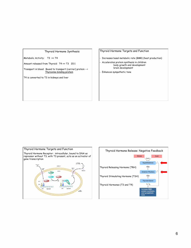
Thyroid Hormone Synthesis
1) Follicular cells produce thyroglobulin and secrete it into lumen
2)! Follicular cells actively take up iodide and it is oxidized to iodine and released into the colloid.
3)! Tyrosine residues on thyroglobulin are iodinated to form MIT and DIT. Mediated by thyroid peroxidase on apical surface.
MIT= monoiodotyrosine
DIT= diiodotyrosine
thyroid peroxidase
Thyroid Hormone Synthesis
4)! MIT + DIT form T3 DIT + DIT form T4
Still part of thyroglobulin in colloid
Thyroid Hormone Synthesis
5) Upon stimulation with TSH (Thyroid Stimulating Hormone), thyroglobulin is reabsorbed by receptor mediated endocytosis and combine with lysosomes, releasing T3 and T4.
6
Metabolic Activity: T3 >>> T4
Amount released from Thyroid: T4 >>> T3 20:1
Transport in blood: Bound to transport (carrier) protein --> Thyroxine-binding protein
T4 is converted to T3 in kidneys and liver
Thyroid Hormone Synthesis Thyroid Hormone Targets and Function
- Increases basal metabolic rate (BMR) (heat production)
- Accelerates protein synthesis in children body growth and development brain development
- Enhances sympathetic tone
Thyroid Hormone Targets and Function
Thyroid Hormone Receptor: intracellular, bound to DNA as repressor without T3; with T3 present, acts as an activator of gene transcription
Thyroid Hormone Release: Negative Feedback
Thyroid Releasing Hormone (TRH)
Thyroid Stimulating Hormone (TSH)
Thyroid Hormones (T3 and T4)

7
Diseases of the Thyroid
Iodine Deficiency: low T3 + T4, elevated TSH, goiter Hypothyroidism
Diseases of the Thyroid
Hypothyroidism
Diseases of the Thyroid
Hyperthyroidism
Diseases of the Thyroid
Graves Disease

8
Graves Disease:
T3 and T4
TSH
Digestive System
Alimentary Canal and Associated Organs
Mouth Tongue Esophagus Teeth Stomach Salivary Glands Small Intestine Pancreas Large Intestine Liver Gall Bladder
Alimentary Canal
Barrier: between internal and external environments
Motility: movement of food
Secretion: enzymes, mucous, acid, antibodies
Absorption: products of digestion
Immunological Defense: site of lymphatic tissue
Alimentary Canal
General Structure from Esophagus ---> Anus
Mucosa: Epithelium Lamina Propria Muscularis Mucosa (smooth muscle)
Submucosa: Dense irregular connective tissue
Muscularis externa: Two layers of smooth muscle
Serosa:simple squamous epithelium, connective tissue

9
Barrier- Epithelium
Oral Cavity: parakeratinized epithelium- most superficial cells do not lose nuclei
tongue, gums, hard palate
Connective tissue papilla
Barrier- Epithelium
Esophagus: stratified squamous epithelium
Small and Large Intestine- tight junctions between columnar cells of simple epithelium
Immunological Defense
Tonsils: ring of lymphatic tissue (lymphatic nodules or follicles) at entrance to respiratory and digestive tracts
micro.magnet.fsu.edu/optics/intelplay/gallery...
Adenoids: lymphatic tissue located high on the posterior wall of the pharynx.
- similar to tonsils
-! clear antigens from air
- reduced in adults
- can be enlarged / inflamed
SYMPTOMS: -!mouth breathing -!snoring -!bad breath -!chronic runny nose -!sleep apnea -!pulmonary hypertension -!right-sided heart failure

10
Immunological Defense
Gut-associated lymphatic tissue (GALT): diffuse lymphatic tissue and lymphatic nodules in lamina propria of small and large intestine Striking in Appendix and Ileum=> Peyer’s Patches
MALT=Mucous associated lymphatic Tissue
Immunological Defense
Plasma Cells secrete a special form of antibody, ==> secreted IgA
-Dimeric
-Linked via J chain and secretory component
-More stable
-More resistant to enzymatic digestion
-in saliva, milk, and mucous membranes of respiratory and digestive tracts
Possible modes of defense mediated by IgA binding to its receptor, pIgR, (the secretory component , SC).
(a)! pIgR-driven export of dimeric IgA with J chain (IgA+J)
(b)! Neutralization of infecting virus and transport of viral products from the lumen.
(c)! Intracellular neutralization of endotoxin (LPS) from Gram-negative bacteria.
(d)! Clearance of antigen (Ag) that has breached the mucosal barrier.
From Trends Immunol. 2004, 25:150-57.
Immunological Defense
Peyer’s Patches
Lymph nodules capped by specialized epithelial cells, =>M Cells
www.bu.edu/histology/p/12001oba.htm

11
M Cells - Follicle-Associated Epithelium (FAE): epithelial cells associated with lymph nodules of MALT
-! look for absence of goblet cells over Peyer’s Patch
-! apical surface microfolds rather than microvilli
- connected to neighbors with tight junctions
M Cells -! have extensive inpocketings of basal membrane containing
T and B lymphocytes
www.rcai.riken.go.jp/eng/group/epi/
M Cells: specialized for transepithelial transport: deliver intact foreign antigens and microorganisms from lumen to immune cells
Motility
Muscularis Mucosa: 2 layers of smooth muscle inner-circular, outer-longitudinal responsible for moving the mucosa

12
Motility
Muscularis Externa: mixes, propels contents of lumen
2 thick layers of smooth muscle
inner layer=> circularly-oriented layer -tight spiral
outer layer=>longitudinally-oriented layer -loose spiral
Between muscle layers- Nervous innervation
Myenteric plexis (Auerbach’s plexis)
Motility: Muscularis Externa
Motility: Muscularis Externa Motility
MUSCULARIS EXTERNA EXCEPTIONS:
Striated muscle in proximal esophagus (upper 1/3) and anus

13
MUSCULARIS EXTERNA EXCEPTIONS:
Teniae Coli: 3 thickened bands of longitudinal layer large intestine-
Secretion
-! carried out by epithelial cells and associated glands
-! secretions include:
Antibodies: IgA
Lubrication substances
Aid for digestion: hydrochloric acid & enzymes
Hormones
Water
-!secretions from salivary glands, stomach, small and large intestine
Anatomy of the Stomach
3 regions:
Cardiac
Pyloric
Fundic
Rugae: longitudinal folds or ridges on inner surface
Anatomy of the Stomach
3 regions:
Cardiac
Pyloric
Fundic
Rugae: longitudinal folds or ridges on inner surface
Simple columnar epithelium

14
Each stomach region
has distinctive glands.
•!Cardiac glands
•!Pyloric glands
•!Fundic glands
-gastric pits
-isthmus cell replication
-neck
-base or fundus
Anatomy of the Small Intestine
3 components: Duodenum, Jeunum, Ileum
- Plicae circularis - Villi - Microvilli
- Simple columnar epithelium
Anatomy of the Small Intestine