hypothalamic metallic deposition and the production of experimental obesity
TRANSCRIPT
Physiology and Behavior, Vol. 10, pp. 677-681. Brain Research Publications Inc., 1973. Printed in the U.S.A.
Hypothalamic Metallic Deposition and the Production of Experimental Obesity
PAUL U. DUBUC
Department o f Metabolism and Endocrinology, Oty of Hope National Medical Center Duarte, California 91010
AND
ROBERT W. REYNOLDS
Department o f Psychology, University o f California, Santa Barbara Santa Barbara, California 93106
(Received 8 August 1972)
DUBUC, P. U. AND R. W. REYNOLDS. Hypothalamic metallic deposition and the production of experimental obesity. PHYSIOL. BEHAV. 10(4) 677-681, 1973.-The effectiveness of the deposition of metallic ions in the medial hypothalamus in producing experimental obesity in rats was investigated. Both the direct deposition to the hypothalamus of Fe ~-~ (as FeC! 3. 6H=O) or Cu ~ (as CuCI 2. 2H20) via bilateral stainless steel cannulae or via anodal electrolysis with stainless steel or copper electrodes resulted in similar significant increases in daily weight gain and the development of obesity. NaCI deposition via cannulae, sham lesions, and empty cannulae insertion were all ineffective in altering body weight. These results suggest that: (a) the electrical events associated with anodal direct current lesions using stainless steel or copper electrodes are secondary to the deposition of metallic ions in the production of experimental obesity; and, (b) the tissue destruction in the ventromedial hypothalamic region coupled with the presence of heavy metal ions apparently 'add' to produce a very high probability of the development of experimental obesity.
Experimental ohesity Ventromedial hypothalamus Satiety center Body weight
Electrolytic lesions Irritative focus Hyperphagia
THE TRADITIONAL technique for the production of discrete lesions in the central nervous system includes the passing of low, constant, direct current through a stainless steel anode located in the proposed lesion site. Invariably this technique results in the deposition of a sphere of ferric ions in the tissue proximal to the uninsulated anode tip.
Studies by Everett and his colleagues [2, 3, 4] have suggested that lesions depositing ferric ions (as demon- strated by precipitation with acidified potassium ferrocy- anide [5]) result in physiological responses qualitatively different from nonferric depositing techniques. Specifically these authors have shown that ovulation can be induced in anovulatory pentobarbital treated rats by anodal d.c. lesions or by the direct application of 5% FeCI3. However, these authors reported that ovulation did not follow anodal platinum lesions of comparable size, radio frequency lesions, nor acidified NaC1 application. Furthermore, the direct application of CuCI 2 also resulted in ovulatory responses in a high proportion of animals. Thus, Everett
suggested that some characteristics of the presence of Fe ÷~* or other heavy metal ions in the tissue resulted in the generation of prolonged neural signals, that ultimately initiate the pituitary release sufficient for ovulation. This phenomenon has been labeled electrochemical stimulation by Everett, and apparently is related to the presence of an irritative focus of heavy metal ions, which acts to stimulate tissue in the immediate vicinity of the lesion site.
Similar results are reported by a number of investigators who have studied the effects of hypothalamic lesions on food intake and body weight regulation in laboratory rats. Anodal (stainless steel) direct current lesions of the ventromedial hypothalamic area (VMH) invariably produce a profound increase in food intake and body weight in experimental animals [7]. However, much like the data reported by Everett and his associates above, noniron- depositing lesions placed in the VMH apparently result in a significantly reduced probability that obesity will occur. Radio frequency lesions [6, 10, 12. 13], platinum anodal
Supported in part by Grant No. NB05583 from the National Institutes of Health.
677
678 DUBUC AND REYNOLI)S
lesions, stainless steel cathodal lesions [1 2], and suction lesions [10] though producing substantial destruction of the so-called satiety center have generally been reported to result in a significantly reduced proportion of animals developing hyperphagia when compared to anodal lesions using stainless steel electrodes. Furthermore, even lesions produced by anodal electrolysis do not reliably produce hyperphagia unless demonstrable Fe **+ is present in associ- ation with the lesions [ 1 ].
These physiological and behavioral data, coupled with the finding by MacIntyre, et al. [9] that a used electrode in contact with CNS tissue, without current flow, will result in significant tissue destruction (i.e., passive transfer effect) suggest that the deposition of heavy metals alone in CNS tissue will produce histological and behavioral effects similar to those reported to follow anodal, d.c. lesions. The following study compares the effectiveness of electrolytic lesions and the direct application of heavy metal ions in producing (a) tissue destruction in the ventromedial hypo- thalamus and (b) experimental obesity in laboratory rats.
METHOD
Animals
Thirty-three female Sprague-Dawley rats ranging in age from 120-220 days participated in the experiment. All animals were allowed free access to Purina Laboratory Chow and tap water throughout the course of the experi- ment. All animals were housed individually in the colony room with temperature maintained at 76°±2°F. and room lighting on a twelve hour cycle (7:00 a.m. to 7:00 p.m.).
Procedure
Animals were divided into the following experimental groups: (1) N=4 bilateral copper electrode lesions of the VMH; (2) N=7 bilateral stainless steel electrode lesions of the VMH; (3) N=8 sham bilateral lesions of the VMH; (4) N=4 bilateral VMH application of FeC13-6H 2 O (reagent grade); (5) N=2 bilateral VMH application of CuCI~ • 2H~ O (reagent grade); (6) N=2 bilateral VMH application of NaCI (reagent grade); (7) N=2 bilateral VMH cannulation, no drug; and (8) N=4 unoperated control animals.
Lesions
All surgery was performed under anesthesia [20] with the aid of a Kopf stereotaxic instrument. With skull surface leveled between lambda and bregma, the bared tip of an insulated stainless steel 00 insect pin or insulated copper wire (reagent grade) was positioned 2.5 mm posterior to bregma, 0.8 mm lateral to the midline of the sagittal sinus, and 9.0 mm below the dural surface. Two mA, for 15 sec (30 mC), was passed through the anode to the cathode (ear bars) from a constant current lesion maker. Following the lesions the animal was injected with procaine penicillin (50,000 U, IM) and mikedimide (5 mg, IP) and returned to its home cage. Sham lesions were performed by lowering the appropriate electrode with no passage of current.
Cannulae
Bilateral cannulae were fashioned of 21 gauge stainless guide shafts and 26 gauge inserts. The tops of the guide shafts were cemented together (dental acrylic) 1.6 mm apart for bilateral VMH placement. The inserts were
fashioned to fit snugly into the guide shafts and to extend beyond the guide shafts approximately 3 ram. During surgery the bilateral cannulae were centered directly over the sagittal sinus, and lowered into the brain so that the insert tips were approximately 0.2 mm above VMH. The guide shafts were then fixed to the skull with stainless steel screws and dental acrylic. Postsurgical procedures were identical to those for lesioned animals. Twelve days following surgery the cannula inserts were removed and cleaned. Approximately 0.5 mm (2.5 x 10 -~ ram31 of fe r r ic c h l o r i d e (FeCI3 . 6 H : O ) , cupric chloride (CuCI:. 2H~ O), or NaC1 was tamped into the lumen of the 26 gauge insert. The excess was wiped from the cannula and it was replaced in the animal.
Body weights for all animals to the nearest gram were recorded daily in the afternoon.
Drug Dosages
Theoretical maximum values of the deposition of each cation were calculated. These values served only as rough indicators of the relative effectiveness of the drug for producing tissue destruction or for inducing the behavioral responses.
.4. Lesions. If it is assumed that the anodal reaction is: Fe-,Fe *÷÷ + 3e- and Cu~Cu *÷ + 2e- [12,13] then approxi- mately 5.5 ug of Fe **÷ and 13.0 ug of Cu ÷* were deposited from 30 mC lesions.
However, implicit in the description by Madntyre, et al. [9], of the passive transfer effect is the removal of some unknown portion of the cation with the removal of the lesioning electrode. Thus, only rough relative comparisons between the Cu ÷÷ and the Fe *÷÷ lesions can be offered.
B. Cannulae. The calculation of the amount of cation loaded into the cannulae can serve as only an approximate estimation of the amount of drug in contact with the tissue. However, if it is assumed that all drug in the cannula insert is released at the implantation site, then approximately 7 ug Fe ~*÷ and 17 ug Cu ÷÷ were deposited.
Histology
Approximately 35 days following lesion or chemical implantation, the animals were sacrificed with a lethal dose of sodium pentobarbital. One percent Formalin followed by 10% Formalin was perfused through the brain via the common carotid artery. The cannulae were removed with the brain in situ. The brains were removed and fixed in 10% Formalin for a minimum of a week. Brain sections were cut at 40 u through the region of the lesion or cannula tip, Sections were mounted on slides and stained with cresyl violet, cresyl violet/Luxol fast blue, or after the method of Well. Counter staining with potassium ferrocyanide or rubeanic acid [5,19] was performed to determine the presence of heavy metal ions.
RESULTS
Histology
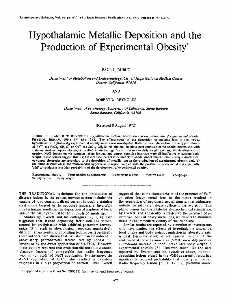
Stainless steel electrode electrolysis resulted in bilateral ablation of the VMH, periventricular iron deposition, and ventricular distortion typical of lesions of this area (Fig. 1, top). Ferric chloride cannulated animals showed massive deposition of iron and tissue ablation extending laterally to. and in most cases including, the fornix (Fig. 1, bottom). Comparison of tissue destruction for these two techniques
HYPOTHALAMIC METALLIC DEPOSITION AND OBESITY 679
/ '
' ) - ' L ' " , "
FIG. 1. Typical hypothalamic destruction produced by the deposi- tion of Fe ~'+. Top: Anodal electrolysis via stainless steel electrode (30 mC). Bottom: Cannulation of approximately 7 ~g crystalline
FeC13 - 6H 20.
demonstrated that the cannulated animals typical ly had approximate ly three times as much tissue destruct ion in the coronal plane as the animals with stainless steel electrode lesions.
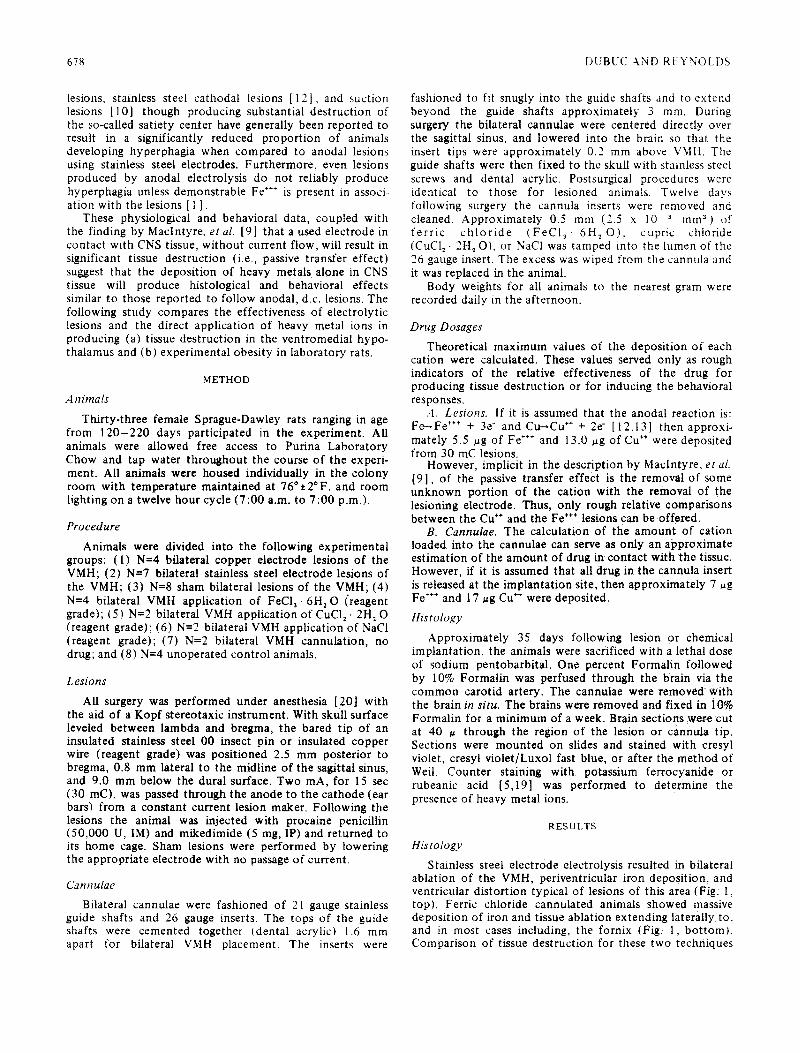
Copper electrode electrolysis produced lesions larger in all dimensions in comparison to Fe lesions (Fig. 2, top). The anticipated presence of cupriferrocyanide or copper rubeanate at the cannula tips or lesion site was not demonstrated. However, finely grained copper salts were apparently present in the area of gliosis immediately surrounding the lesion or cannula tips. Cupric chloride application, via cannula, resulted in massive destruction of the basal hypothalamic region (Fig. 2, bot tom). Close examination of the mounted sections suggested that some percentage of the tissue destruction resulted from the passage of the microtome knife. Apparent ly the large deposits of copper salts associated with the cannula were not sliced, but tended to be forced through the tissue. Consequently, much addit ional damage seemingly occurred with the histological procedures. However, rough indica- tions of the deposit ion induced damage were provided by assessing the extent of the finely grained copper salt particles invading the glial cells surrounding the lesion site. These deposits suggest that copper dzposi t ion via cannula led to the greatest lesion extent both in the coronal and sagittal planes. Copper electrode lesions were comparable in destruction to the smaller Pie cannula lesions, and in
FIG. 2. Hypothalamic destruction as a result of the presence of Cu ~ ions in tissue. Top: Anodal electrolysis with copper electrode (30 mC). Bottom: Deposition of approximately 17 ~g Cu * as
CuC12 .2H: O.
general, were substantially larger than Fe electrolysis destruction. Ranking coronal tissue destruction by tech- nique, from greatest to least, found Cu cannulae greater than Fe cannulae greater than Cu lesions greater than Fe lesions greater than NaCI cannulae. However, the extent of tissue destruction, especially of Cu treated animals was somewhat arbitrary.
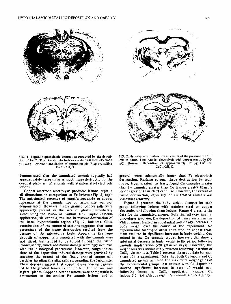
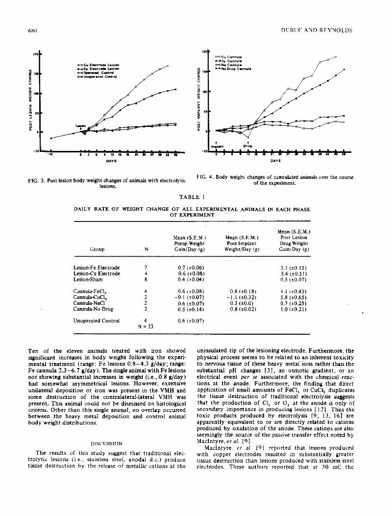
Figure 3 presents the body weight changes for each group following lesions with stainless steel or copper electrodes or following sham lesions. Figure 4 presents the data for the cannulated groups. Note that all experimental procedures involving the deposit ion of heavy metals in the VMH region resulted in substantial and steady increases in body weight over the course of the experiment. No experimental technique other than iron or copper treat- merit resulted in significant increases in body weight. One animal in the Cu cannula group, however, did show a substantial decrease in body weight in the period following cannula implantat ion (-20 g/twelve days). However, this weight loss was immediately reversed following insertion of CuC12 via cannula. Table 1 presents the group data for each phase of the experiment. Note that both Cu lesions and Cu cannulated groups achieved the maximum weight gains of the experimental groups. All animals with Cu deposition showed significant increases in rate of weight gain following lesion or CuCI~ application (range: Cu lesions 3 .2 -8 .6 g/day; range: Cu cannula 4 .5 -7 .1 g/day).
680 DUBUC AND REYNOLDS
200
lSg Z
t0e
Z 2
so
o
- So
20¢
"|2 0 ] 6 0 |2 lS IS ~1 34 ~7 ~ U N
DAY S
O-O Cu Electrode Lesion o-.F, s J ~ t r ~ Lesio. ~ o-~O~rated Coetrm ~ z ~s(
-$0
o"o Cu Cannula ~ F e Canntlta ~ I ' : ~ ' I ~ O-ONa Cannula / H N O Drug Cannuie / ~
i ~ s Drug
DAYS
FIG. 3. Post lesion body weight changes of animals with electrolytic lesions.
FIG. 4. Body weight changes of cannulated animals over the course of the experiment.
TABLE 1
DALLY RATE OF WEIGHT CHANGE OF ALL EXPERIMENTAL ANIMALS IN EACH PHASE OF EXPERIMENT
Group N
Mean (S.E.M.) Mean (S.E.M.) Mean (S.E.M.) Post Lesion Preop Weight Post Implant Drug Weight Gain/Day (g) Weight/Day (g) Gain/Day (g)
Lesion-Fe Electrode 7 Lesion-Cu Electrode 4 Lesion-Sham 8
Cannula-FeCl 3 4 Cannula-CuCl~ 2 Cannula-NaCi 2 Cannula-No Drug 2
Unoperated Control 4 N=33
0.7 (±0.06) 3.l (±0.15) 0.6 (±0.08) 5.4 (±0.51) 0.4 (±0.04) 0.5 (±0.07)
0.6 (-*0.08) 0.8 (±0.18) 4.1 (±0.45) -0.1 (±0.07) - t .1 (±0.32) 5.8 (±0.65)
0.6 (±0.07) 0.3 (±0.0) 0.7 (±0.25) 0.5 (±0.14) 0.8 (±0.02) 1.0 (±0.21)
0.6 (±0.07)
Ten of the eleven animals treated with iron showed significant increases in body weight following the experi- mental treatment (range: Fe lesions 0 .8 -4 .3 g/day; range: Fe cannula 2 .3-6 .7 g/day). The single animal with Fe lemons not showing substantial increases in weight (i.e., 0.8 g/day) had somewhat asymmetrical lesions. However, extensive unilateral deposition or iron was present in the VMH and some destruction of the contralateral-lateral VMH was present. This animal could not be dismissed on histological criteria. Other than this single animal, no overlap occurred between the heavy metal deposition and control animal body weight distributions.
DISCUSSION
The results of this study suggest that traditional elec- trolytic lesions (i.e., stainless steel, anodal d.c.) produce tissue destruction by the release of metallic cations at the
uninsulated tip of the lesioning electrode. Furthermore, the physical process seems to be related to an inherent toxicity to nervous tissue of these heavy metal ions rather than the substantial pH changes [3], an osmotic gradient, or an electrical event p e r se associated with the chemical.reac- tions at the anode. Furthermore, the finding that direct application of small amounts of FeCI 3 or CuCtl duplicates the tissue destruction of traditional electrolysis suggests that the production of Cl: or O 2 at the anode is only of secondary importance in producing lesions [ 12]. Thus the toxic products produced by electrolysis [9, 13, 16] are apparently equivalent to or are directly related to cations produced by oxidation of the anode. These cations are also seemingly the source of the passive transfer effect noted by MacIntyre, e t al. [9].
MacIntyre, e t al. [9] reported that lesions produced with copper electrodes resulted in substantially greater tissue destruction than lesions produced with stainless steel electrodes. These authors reported that at 30 mC the
HYPOTHALAMIC METALLIC DEPOSITION AND OBESITY 681
volume indices (cross-sectional area x length) of lesion extent were 0.95 for stainless steel vs. 3.42 for copper. The data from the present study suggest that iron and copper lesions are comparable in size when the different equivalent weights and approximate iron content in the stainless steel electrode are considered. Equal 30 mC lesions deposit approximately 5.5 ug Fe ÷÷÷ from stainless steel electrodes vs. 13 ug of Cu ÷÷ from reagent grade copper electrodes. These values approximate the volume index differences reported by Maclntyre, et al., and also are roughly correlated to the tissue destruction differences reported in the present study. Thus, it is concluded that the extent of tissue destruction resulting from heavy metal electrolytic deposition is directly related to the equivalent weight of the deposited ion.
The similarities of the lesion conditions which produce ovulation in anovulatory pentobarbital treated rats and the lesion conditions which result in high incidences of obesity in experimental animals suggest that analogous neural mechanisms may underlie both behavioral phenomena. Everett has contended that a necessary requirement for the induction of ovulation is an irritative or electrochemical stimulus sufficiently prolonged to augment gonadotropin release and thus to induce ovulation [3] . In contrast, the relationship between heavy metal deposition and the
production of hyperphagia and/or experimental obesity is much less conclusive.
Dahl and Ursin [1], Rabin and Smith [12], and Reynolds [ 14] have suggested that an important factor in the development of hypothalamic hyperphagia is the presence of an irritative focus that is generated by electrolytic lesioning. However, the use of theoretically nonmetallic-depositing techniques (platinum electrode, d. c. lesions [18]; radio frequency current [6, 8, 10, 12, 13]; aspiration lesions [10] to lesion VMH do result in experimental obesity, albeit with a reduced probability of its occurrence. Thus, it is apparent from these studies and the present data that the combination of metallic deposi- tion from electrolysis, or by direct chemical application, in combination with VMH destruction, are additive in produ- cing a very high incidence of experimental obesity (> 90%). The increased effectiveness of lesions that deposit metallic ions suggests that metallic ions might serve as a sustained irritative or electrochemical stimulus to CNS tissue [ 11, 15, 17] and potentiate the effects of VMH ablation.
AC KNOWL EDGEM ENT
The authors are indebted to Miss Marion Dalin for her technical assistance.
REFERENCES
1. Dahl, E. and H. Ursin. Obesity produced by iron and tissue destruction in the ventromedial hypothalamus. Physiol. Behav. 4: 315-317, 1969.
2. Everett, J. W. Preoptic stimulative lesions and ovulation in the rat: 'Thresholds' and LH-release time in late diestrus and proestrus. In: Major Problems in Neuroendocrinology, edited by E. Bajusz and G. Jasmin. Bas~! and New York: S. Karger, 1964, pp. 346-366.
3. Everett, J. W. and H. M. Radford. Irritative deposits from stainless steel electrodes in the preoptic rat brain causing release of pituitary gonadotropin. Proc. Soc. exp. Biol. Med. 108: 604-609, 1961.
4. Everett, J. W., H. M. Radford and J. Holsinger. Electrolytic irritative lesions in the hypothalamus and other forebrain areas. In: Proceedings o f the First International Congress on Hor- monal Steroids, Volume 1, edited by L. Martini and A. Pecile. New York: Academic Press, 1964, pp. 235-249.
5. Gomori, G. Microtechnical demonstration of iron. Am. J. Pathot. 12: 655-663, 1936.
6. Herrero, S. Radio-frequency-current and direct-current lesions in the ventromedial hypothalamus. Am. J. Physiol. 217: 403-410, 1969.
7. Hetherington, A. W. and S. W. Ranson. Hypothalamic lesions and adiposity in the rat. Anat. Rec. 78: 149-172, 1942.
8. Hoebel, B. G. Hypothalamic lesions by electrocauterization: Disinhibition of feeding and self-stimulation. Science 149: 452-453, 1965.
9. Maclntyre, W. J., T. G. Bidder and V. Rowland. The production of brain lesions with electric currents. In: Pro- ceedings o f the First National Biophysical Conference, edited by H. Quastler and H. J. Morowitz. New Haven: Yale University Press, 1959, pp. 723-732.
10. Pool, R. H. An investigation of hypothalamic hyperphagia. Ph.D. Dissertation, University of Washington, Seattle, 1963.
11. Rabin, B. M. Investigation of the electrical activity of the ventrolateral hypothalamus following irritative and non- irritative lesions of the ventromedial hypothalamus. Ph.D. Dissertation, State University of New York, Buffalo, 1968.
12. Rabin, B. M. and C. J. Smith. Behavioral comparison of the effectiveness of irritative and non-irritative lesions in producing hypothalamic hyperphagia. Physiol. Behav. 3: 417 -420, 1968.
13. Reynolds, R. W. Ventromedial hypothalamic lesions without hyperphagia. Am. J. Physiol. 204: 60-62, 1963.
14. Reynolds, R. W. An irritative hypothesis concerning the hypothalamic regulation of food intake. Psychol. Rev. 72: 105-116, 1965.
15. Rolls, B. J. Drinking by rats after irritative lesions in the hypothalamus. Physiol. Behav. 5: 1385 - 1393, 1970.
16. Rowland, V., W. J. Maclntyre and T. G. Bidder. The production of brain lesions with electric currents II. Bidirec- tional Currents. J. Neurosurg. 17:55 -69, 1960.
17. Simons, B. J. Cause of excessive drinking in diabetes insipidus. Nature 219:1061-1062, 1968.
18. Teitelbaum, P. Sensory control of hypothalamic hyperphagia. J. eomp. physiol. Psychol. 48: 156-163, 1955.
19. Uzmann, L. L. Histochemical localization of copper with rubeanic acid. Lab. Invest. 5: 299-305, 1956.
20. Valenstein, E. S. A note on anaesthetizing rats and guinea pigs. J. exp. Analysis Behav. 4: 6, 1961.