hypothalamic demyelination induced by gold thioglucose
TRANSCRIPT
Micron, 1980, Vol.: 11, pp. 7-8. 0047-7206/80/0201-0007 $02.00/0 © Pergamon Press Ltd. Printed in Great Britain.
HYPOTHALAMIC DEMYELINATION INDUCED BY GOLD THIOGLUCOSE
James S. Hatfield and Ben Pansky
Dept. of Anatomy, Medical College of Ohio, C.S. 10008, Toledo, Ohio 43699
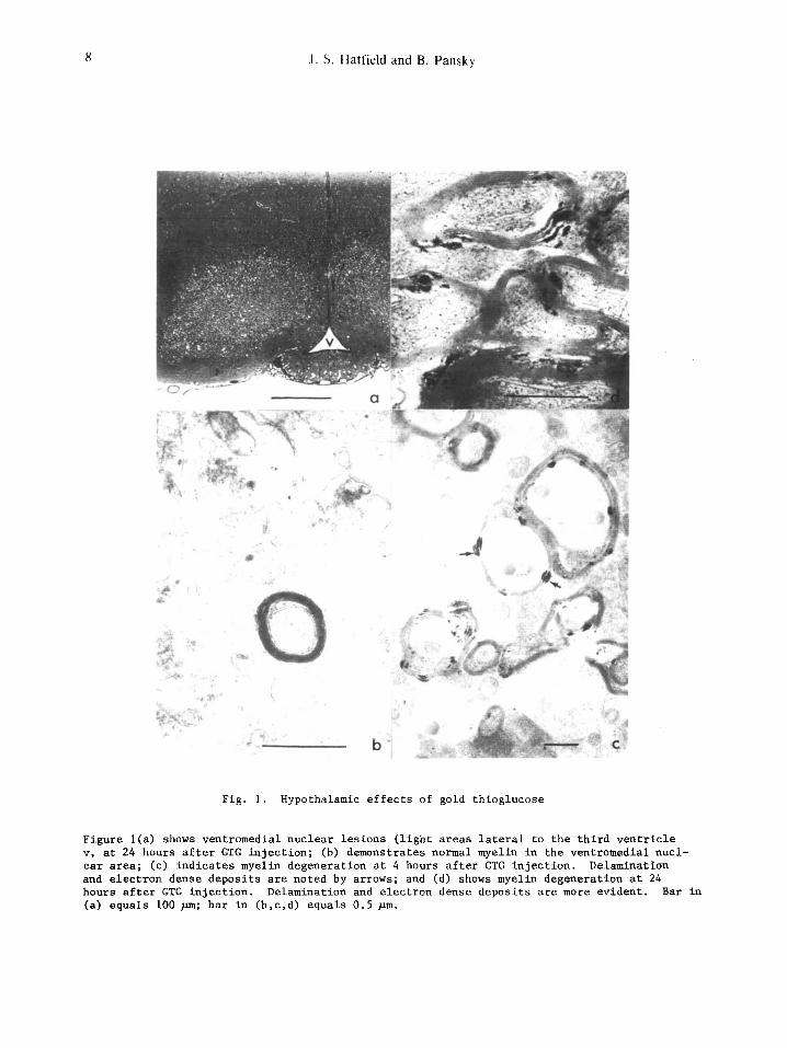
The compound gold thioglucose (GTG) has been frequently used to produce specific lesions in the glucoreceptive ventromedial hypothalamus (VMH) of mice in order to study neural metabolic and food intake control mechanisms (1,2). The administration of GTG to adult mice by intra- peritoneal injection, at a dosage of 0.4 mg/gram body weight results, within 48 hours, in the progressive induction of glial swelling, neuronal shrinkage, cellular necrosis, and gliosis (glial scar tissue) confined primarily to the VMH (3)(Fig. la). In addition to the above phenomena, we have found, in hypothalamic tissue prepared routinely for transmission electron microscopy, marked uptake of GTG in cell processes, nerve terminals, and myelin sheaths in the vicinity of the VMH, arcuate, and periventricular nuclei, as early as four hours and in- creasing in concentration through 24 hours post-injection. These findings verify earlier studies of GTG-induced hypothalamic degeneration (4). The presence of GTG was denoted by the accumulation of highly electron-dense floccular and particulate inclusions. Delamination of myelin and membrane dissolution accompanied the deposition of such inclusions (Fig. ic). Following GTG injection, these alterations in normal myelin configuration appeared to incre- ase in severity with time (Fig. id). In contrast, myelinated axons in cortical and thalamic areas demonstrated no GTG accumulation (Fig. ib). The intralaminar deposition of GTG (or its metabolites) is presumed to have occurred via intracytoplasmic translocation along the Schmidt Lanterman clefts, i.e., cytoplasmic extensions of the myelin-forming glial cells. GTG, which otherwise is highly diffusible, is apparently held in situ within the myelin laminae until exposed by sectioning. The presence of GTG was also noted in vacuoles within phagocytic Kupffer cells in hepatic tissue. Uninjected control tissues exhibited no similar inclusions. These observations point to differential sensitivity to GTG by the myelin-forming glial cells in various brain regions. Additionally, the influence of the disruption of autonomic tracts in the vicinity of the VMH must now be included in any explanation of the metabolic phenomena associated with GTG lesions.
(I) B.
(2) J.
(3) R.
(4) L.
Brecher and S. H. Waxier, Obesity in albino mice due to single injections of goldthio- glucose, Proc. Soc. Exp. Biol. Med. 70, 498 (1949).
Mayer and N. B. Marshall, Specificity of gold thioglucose for ventromedlal hypothala- mic lesions and hyperphagla, Nature 178, 1399 (1956).
A. Liebelt and J. H. Perry: Hypothalamic lesions associated with goldthloglucose- induced obesity, Proc. Soc. Exp. Biol. Med. 95, 774 (1957).
Zaborsky, C. Leranth, and M. Palkovits, Gold thioglucose terminal degeneration in mouse hypothalamus, Experientia 30, 811 (1974).
Supported in part by Biomedical Research Support Grant No. SO 7 RR 05700-08.
8 J .S. Hatfield and B, Pansky
~o
Fig. i. Hypothalamic effects of gold thioglucose
Figure l(a) shows ventromedial nuclear lesions (light areas lateral to the third ventricle v, at 24 hours after GTG injection; (b) demonstrates normal myelin in the ventromedial nucl- ear area; (c) indicates myelin degeneration at 4 hours after GTG injection. Delamination and electron dense deposits are noted by arrows; and (d) shows myelin degeneration at 24 hours after GTG injection. Delamination and electron dense deposits are more evident. Bar in (a) equals I00 ~m; bar in (b,c,d) equals 0.5 ~m.