hypomagnesemia with secondary hypocalcemia due to a missense
TRANSCRIPT
Hypomagnesemia with Secondary Hypocalcemia due to aMissense Mutation in the Putative Pore-formingRegion of TRPM6*□S
Received for publication, December 4, 2006 Published, JBC Papers in Press, December 29, 2006, DOI 10.1074/jbc.M611117200
Vladimir Chubanov‡, Karl P. Schlingmann§, Janine Waring‡, Jolanta Heinzinger§, Silke Kaske‡,Siegfried Waldegger§, Michael Mederos y Schnitzler‡, and Thomas Gudermann‡1
From the ‡Institute for Pharmacology and Toxicology and the §University Children’s Hospital, Philipps-University Marburg,Karl-von-Frisch-Strasse 1, 35033 Marburg, Germany
Hypomagnesemia with secondary hypocalcemia is an auto-somal recessive disorder caused by mutations in the TRPM6gene. Current experimental evidence suggests that TRPM6may function in a specific association with TRPM7 by meansof heterooligomeric channel complex formation. Here, wereport the identification and functional characterization of anew hypomagnesemia with secondary hypocalcemia mis-sense mutation in TRPM6. The affected subject presentedwith profound hypomagnesemia and hypocalcemia caused bycompound heterozygous mutation in the TRPM6 gene:1208(�1)G > A affecting the acceptor splice site precedingexon 11, and 3050C > G resulting in the amino acid change(P1017R) in the putative pore-forming region of TRPM6. Toassess the functional consequences of the P1017R mutation,TRPM6P1017R and wild-type TRPM6 were co-expressed withTRPM7 in Xenopus oocytes and HEK 293 cells, and currentswere assessed by two-electrode voltage clamp and whole cellpatch clamp measurements, respectively. Co-expression ofwild-type TRPM6 and TRPM7 resulted in a significant increasein the amplitude of TRPM7-like currents. In contrast,TRPM6P1017R suppressed TRPM7 channel activity. In line withthese observations, TRPM7, containing the correspondingmutation P1040R, displayed a dominant-negative effect uponco-expressionwithwild-typeTRPM7.Confocalmicroscopy andfluorescence resonance energy transfer recordings demon-strated that the P1017R mutation neither affects assembly ofTRPM6 with TRPM7, nor co-trafficking of heteromultimericchannel complexes to the cell surface.We conclude that a func-tional defect in the putative pore of TRPM6/7 channel com-plexes is sufficient to impair body magnesium homeostasis.
Mg2� plays a vital role in virtually all cellular pathways as acofactor of enzymes, an essential structural element of proteinsand nucleic acids, and a modulator of receptors and ion chan-nels (1–4). At present, the molecular mechanisms controlling
Mg2� homeostasis are poorly understood. Thus, clinical andmolecular characterizations of hereditary disorders associatedwith Mg2� handling provide a promising point of entry toinvestigate the molecular components pertinent to cellularMg2� handling.Recently, it was discovered that loss-of-function mutations
in the gene of melastatin-related transient receptor potentialcation channel 6 (TRPM6)2 result in hypomagnesemia withsecondary hypocalcemia (HSH) (5–8). HSH is an autosomalrecessive disorder characterized by low serumMg2� levels dueto defective intestinal absorption and increased renal Mg2�
wasting (3, 9, 10). Hypocalcemia results from a secondary insuf-ficiency of the parathyroid glands in the presence of profoundhypomagnesemia. Supplementation with high Mg2� dosescompensates for theMg2� deficiency ofHSHpatients (3, 9, 10).
TRPM6 belongs to the melastatin-related group of the TRPion channel family (11, 12). Like other TRP channels, TRPMproteins contain six transmembrane helices (S1–S6) flanked bycytoplasmic N and C termini; TRPMs most likely function astetrameric channel complexes (12). Hydrophobic segmentslocated between the S5 and S6 helices of four channel subunitsare thought to contribute to a channel pore. In contrast to otherknown ion channels, TRPM6 and its closest family member,TRPM7, display the unique structural feature of being cationchannels fused to Ser/Thr kinase domains at their C termini(13).TRPM7 is a ubiquitously expressed protein, which is essen-
tial for Mg2� homeostasis. Disruption of the TRPM7 gene inDT40 chicken lymphocytes and the zebrafish Danio rerio,resulted in Mg2� deficiency (14, 15). Functional characteriza-tion of heterologously expressed TRPM7 revealed a constitu-tively active cation channel permeable to a broad range of diva-lent cations, including Mg2� and Ca2� (16–18). TRPM7channel activity is tightly regulated by the intracellular free con-
* This work was supported by the Deutsche Forschungsgemeinschaft andthe German Kidney Foundation. The costs of publication of this articlewere defrayed in part by the payment of page charges. This article musttherefore be hereby marked “advertisement” in accordance with 18 U.S.C.Section 1734 solely to indicate this fact.
□S The on-line version of this article (available at http://www.jbc.org) containssupplemental Figs. S1–S3.
1 To whom correspondence should be addressed: Tel.: 49-6421-2865-000;Fax: 49-6421-2865-600; E-mail: [email protected].
2 The abbreviations used are: TRPM6, melastatin-related transient receptorpotential cation channel 6; TRPM7, melastatin-related transient receptorpotential cation channel; TRPC6, canonical transient receptor potentialcation channel 6; HSH, hypomagnesemia with secondary hypocalcemia;KvAP, a voltage-dependent K� channel from the thermophilic archae-bac-terium Aeropyrum pernix; GFP, enhanced green fluorescent protein; CFP,enhanced cyan fluorescent protein; YFP, enhanced yellow fluorescent pro-tein; FRET, fluorescence resonance energy transfer; DIC, differential inter-ference-contrast; HEK 293 cells, human embryonic kidney 293 cells; MIC,Mg2�-inhibited cation; RT, reverse transcription; PBS, phosphate-bufferedsaline.
THE JOURNAL OF BIOLOGICAL CHEMISTRY VOL. 282, NO. 10, pp. 7656 –7667, March 9, 2007© 2007 by The American Society for Biochemistry and Molecular Biology, Inc. Printed in the U.S.A.
7656 JOURNAL OF BIOLOGICAL CHEMISTRY VOLUME 282 • NUMBER 10 • MARCH 9, 2007
by guest on February 6, 2018http://w
ww
.jbc.org/D
ownloaded from
centrations of Mg2� ([Mg2�]i), MgATP ([MgATP]i) and agrowing number of other stimuli (19–25). Annexin A1 andmyosin II have been identified as physiological substrates of theTRPM7 kinase domain (23, 26). However, the functional inter-play between kinase and channel domains inTRPM7 is far frombeing understood (15, 20, 27, 28).Conflicting findings have been reported concerning func-
tional characteristics of TRPM6 expressed in heterologous cellsystems. According to Voets et al. (29), TRPM6 forms homo-oligomeric channel complexes with biophysical characteristicssimilar to those of TRPM7. More recently, Li et al. (30)described a number of TRPM6 properties different fromTRPM7, including single channel conductance, divalent cationpermeability, and pH sensitivity. In contrast, two other studiessuggest that TRPM6 functions only in association with TRPM7by means of heterooligomeric channel complex formation (8,28). Consistently, in DT40 cells, which are Mg2�-deficient dueto a genetically disrupted TRPM7 gene, recombinant humanTRPM6 (unlike TRPM7) is unable to compensate for TRPM7(28). Within the formed channel complex, TRPM6 potentiatesTRPM7 channel activity (8) and is able to cross-phosphorylateTRPM7, but not vice versa (28). Moreover, in terms of biophys-ical characteristics, heterooligomeric TRPM6/7 channels werefound to be different from TRPM7 homooligomers (30). Atpresent, the stoichiometry of TRPM6/7 in native channel com-plexes remains unknown.Most HSH mutations in TRPM6 introduce stop codons,
deletions of exons, frame shifts, or affect splice sites, thus result-ing in complete loss of function (7). Thus, it remains elusivewhether a lack of channel and/or kinase activities of TRPM6 isresponsible for the HSH phenotype. Therefore, the molecularanalysis of HSH mutations affecting distinct domains ofTRPM6/7 (for instance, the kinase domain and pore-formingsegments) should be instrumental in dissecting the specific roleof different TRPM6 domains for Mg2� handling. For example,we recently discovered the disease-causing molecular mecha-nism of a TRPM6 missense mutation, which disrupts the het-eromeric assembly of TRPM6 and TRPM7 thereby elicitingHSH (8).Here, we report the identification and functional character-
ization of a novel point mutation in the TRPM6 gene. We dis-close a novel molecular mechanism underlying HSH: TheP1017R mutation neither affects the expression of the TRPM6protein, nor its co-assembly with TRPM7, but impairs channelactivity ofTRPM6/7 complexes by dominant-negative suppres-sion. The effect exerted by TRPM6P1017R strongly supports thenotion that both channel subunits contribute to functionalTRPM6/7 complexes. Thus, a suppression of cation fluxes viabi-functional TRPM6/7 complexes is sufficient for the develop-ment of HSH.
EXPERIMENTAL PROCEDURES
Subjects, Genotyping, and Clinical Studies—Identificationand clinical characterization of an HSH subject (Family F14)was reported previously (7). Subsequent genotyping, reportedhere, was performed using direct sequencing of the entire cod-ing region of the TRPM6 gene from genomic DNA extractedfrom blood leukocytes as described (5, 7). Assessment of renal
magnesiumhandling, aswell as determination of serum [Mg2�]and [Ca2�] levels were performed as reported (5, 7).Molecular Biology and Generation of TRPM6 and TRPM7
Antibodies—Human TRPM6 (TRPM6, variant a, AY333285)andmouse TRPM7 (NM_021450) cDNAs were cloned into thepcDNA3.1/V5-His TA-TOPO eukaryotic expression vector(Invitrogen) as described previously (8). TRPM6P1017R andTRPM7P1040R mutations were introduced by site-directedmutagenesis using the QuikChange system (Stratagene). Togenerate TRPM6 and TRPM7 C-terminally fused to enhancedgreen fluorescent protein (GFP), enhanced yellow fluorescentprotein (YFP) and enhanced cyan fluorescent protein (CFP),STOP codons were replaced by XhoI restriction sites throughsite-directed mutagenesis followed by in-frame subcloning ofYFP cDNAs. Previously, we demonstrated that YFP tags do notinfluence trafficking and channel properties of TRPM6 andTRPM7 (8). Expression of TRPM6 and TRPM7 inXenopus lae-vis oocytes was performed using cDNAs subcloned into thepOGII vector (a pBluescript derivative with the 5�- and 3�-un-translated regions of Xenopus �-globin). All cDNA constructsused in the present work were confirmed by sequencing. Apolyclonal TRPM6 antiserum was obtained as recentlyreported (8). To generate a polyclonal TRPM7-specific anti-body, rabbits were immunized with the following peptidecoupled to keyhole limpet hemacyanin: H2N-DSPEVDS-KAALLP-COOH (Eurogentec, Brussels). Subsequently, theTRPM7-specific antibody was purified by peptide affinitychromatography.Structural Analysis of the TRPM6 Pore-forming Region—
Three-dimensional coordinates of the S1–S6 segment of thehuman TRPM6 protein (Q9BX84) were acquired from MOD-BASE (salilab.org/modbase), a data base of annotated compar-ative protein structuremodels (31). The pore-forming segmentof TRPM6 (amino acids 872–1074) was matched to the struc-ture of KvAP, a voltage-dependent K� channel from the ther-mophilic archae-bacterium Aeropyrum pernix (32), PDB code1orq (chain C, amino acids 28–240). Accordingly, the 1orqtemplate was applied for comparative modeling, and annotatedcoordinateswereused togeneratemolecular graphics imageswiththe University of California at San Francisco Chimera package(33). For simplicity, only �-carbon traces of the S5–S6 regionsfrom TRPM6 and KvAP are shown (Fig. 1B). For sequence align-ment ClustalW (www.ebi.ac.uk/clustalw/) was used.Cell Culture, Transient Expression, and Generation of Stable
Cell Lines—Human embryonic kidney (HEK) 293 cells weremaintained at 37 °C under 5% CO2 in Earle’s minimal essentialmedium supplemented with 10% fetal calf serum, 100 �g/mlstreptomycin, and 100 units/ml penicillin (PAA Laboratories,Pasching, Austria). Cells were transiently transfected using theFuGENE6 transfection reagent (Roche Molecular Biochemi-cals) according to the manufacturer’s instructions.Stable cell lines expressing TRPM6 and TRPM7 were gener-
ated using an ecdysone-inducible expression system (Invitro-gen) according to the supplier’s instructions. Briefly, TRPM6andTRPM7cDNAswere subcloned into the pIND(SP1)/Hygroexpression vector (Invitrogen) and transfected into the EcR293cell line (Invitrogen). Stable transfectants were selected by cul-turing in 400 �g/ml hygromycin B (Invitrogen), propagated,
Novel Missense Mutation in TRPM6
MARCH 9, 2007 • VOLUME 282 • NUMBER 10 JOURNAL OF BIOLOGICAL CHEMISTRY 7657
by guest on February 6, 2018http://w
ww
.jbc.org/D
ownloaded from
and analyzed for ponasterone A-inducible expression by RT-PCR and Western blotting. Two clones were selected for fur-ther analysis: M6pIND (clone 2-1, expressing human TRPM6)and M7pIND (clone 2-19, expressing mouse TRPM7). TheM6pIND and M7pIND cell lines were maintained at 37 °C under5% CO2 in Dulbecco’s modified Earle’s medium (PAA Labora-tories) supplemented with 10% fetal calf serum, 100 �g/mlstreptomycin, 100 units/ml penicillin, 400 �g/ml hygromycinB, and 400 �g/ml Zeocin (Invitrogen).Western Blotting—M6pIND and M7pIND cells grown in
35-mm dishes to �80% confluence were induced by differentconcentrations of ponasterone A (Invitrogen) as indicated inthe figure legends. 24 h after induction, cells were washed twicewith phosphate-buffered saline (PBS: 137 mM NaCl, 2.7 mMKCl, 4.3 mM Na2HPO4, 1.4 mM KH2PO4, pH 7.3). Next, 1 ml of2� Laemmli buffer was added and samples were heated for 5min at 65 °C. Cell lysates (20 �l) were subjected to SDS-PAGE(8%) and blotted on nitrocellulose membranes (Protran,What-man).After incubation in blocking buffer (5%nonfat drymilk inPBS) for 1 h at room temperature, blots were probed eitherwithanti-TRPM6 antiserum (1:1000) or anti-TRPM7 antibody (2�g/ml) in blocking buffer at 4 °C overnight. We used peroxi-dase-conjugated anti-rabbit IgG (1:1000) (Sigma) as secondaryantibody. For a loading control, blots were re-probed using arabbit polyclonal anti-�-actin antibody (1:1000, Abcam). Per-oxidase activity was detected with a chemiluminescence sub-strate (SuperSignal West Pico Substrate, Pierce) and X-OmatUV Plus Films (Kodak).RT-PCR—M6pIND and M7pIND cells grown in 35-mm dishes
to �80% confluence were induced by different concentrationsof ponasterone A. RT-PCR was performed as reported previ-ously (8). To specifically monitor ponasterone A-inducibleexpression of TRPM6 and TRPM7 transcripts, a low number ofPCR cycles (20) was performed with the isoform-specific(human TRPM6a) or the species-specific (mouse TRPM7)primer pairs: M6for, 5�-AGGGCCTGCAAATCAAAG-3�;M6rev, 5�-GAGGGCATAGTAAAGTTCTGGA-3�; M7for, 5�-AAGATTTGCCCGTGATACCCCAGAG-3�; and M7rev,5�-TTCACTATATCCAGCAGCACCCACAT-3�.Immunofluorescence Staining andConfocalMicroscopy—For
subcellular localization of YFP-tagged TRPM6 and TRPM7,HEK 293 cells grown on 25-mm glass coverslips in 35-mmdishes were transiently transfected with plasmid DNAs (2�g/dish) as indicated in the figure legends. In co-expressionexperiments, a 1:3 cDNA mixture (YFP-tagged/untaggedcDNAs) was used to increase the probability of the incorpora-tion of YFP-labeled channel subunits into heterotetramers. Liv-ing cells were directly examined 16–24 h after transfection.Confocal and differential interference-contrast (DIC) images ofliving cells were obtained with a LSM510META confocal laserscanningmicroscope (Carl Zeiss).Weused a Plan-Apochromat63�/1.4 oil objective, the 488 nm excitation wavelength of anargon laser, and a 505 nm long-pass filter. The pinhole diameterwas set to yield optical sections of 0.6 �m.
For immunofluorescent staining, M6pIND or M7pIND cellsgrown on 25-mm glass coverslips were induced with 5 �M pon-asterone A. 24 h after induction, cells were washed twice withPBS, fixed with ice-cold methanol for 20 min at �20 °C, and
blocked for 1 h with 10% (v/v) normal goat serum in PBS atroom temperature. The TRPM7-specific antibody (0.3 �g/ml)or the TRPM6-specific antiserum (1:1000) were applied. Thesecondary antibody (0.5 �g/ml) was goat anti-rabbit IgG con-jugated to Alexa488 (Molecular Probes). Each incubation wasperformed in PBS containing 5% (v/v) normal goat serum for1 h at room temperature followed by a triple washing step withPBS. After the final washing step, coverslips were placed onglass slides using mounting medium (DakoCytomation), andexamined using a LSM510 META confocal laser scanningmicroscope as indicated above.Fluorescence Resonance Energy Transfer (FRET) Recordings—
Our static FRET protocol was based on donor (CFP) fluores-cence recovery after acceptor (YFP) bleach as reported previ-ously (8, 34). Briefly, HEK 293 cells grown on 25-mm glass cov-erslips in 35-mm dishes were transiently transfected with 4�g/dish plasmid DNAs (1 �g of plasmid cDNA encoding theCFP-taggedTRPMchannel subunit and 3�g of plasmid encod-ing the respective YFP-tagged subunit). Cells were examined40–48 h after transfection at room temperature in HEPES-buffered saline (140 mM NaCl, 6 mM KCl, 1 mM MgCl2, 2 mMCaCl2, 10 mM HEPES, 5 mM glucose, 0.1% bovine serum albu-min, pH 7.4) with an Olympus IX70 inverted microscopeequipped with a polychrome IV monochromator (TILL Pho-tonics), anOlympus 63�/1.4 UApo objective, a Sensicam char-ge-coupled device camera, and a Lambda 10-2 emission filterwheel (Sutter Instruments). FRET efficiencies were determinedby monitoring the increase in the CFP (FRET donor) fluores-cence emission during selective YFP (FRET acceptor) photo-bleaching (8, 34). In each experiment, data from four to sixsingle cells were averaged. Means � S.E. were calculated fromthree to four independent transfections for each combinationof CFP- and YFP-fused TRPM6/7 subunits. Statistical analysiswas performed using Student’s t test.Electrophysiological Techniques—M6pIND or M7pIND cells
grown in 35-mmdishes to�70% confluencewere inducedwith5 �M ponasterone A for 18–24 h before performing electro-physiological experiments. For transient expression ofTRPM6wt-GFP, TRPM6P1017R-YFP, TRPM7wt-GFP, andTRPM7P1040R-YFP in the pcDNA3.1/V5-His vector (Invitro-gen) or TRPM7P1040R in pIRES2-EGFP vector (Clontech) inM7pIND cells, corresponding plasmid DNAs (2 �g/dish) weretransfected with the FuGENE6 transfection reagent. �24 hafter transfection, culture media were replaced by fresh mediacontaining 5 �M ponasterone A. Only GFP- or YFP-positivecells were measured 18–24 h after induction.Whole-cell patchclamp recordings were carried out at room temperature(22 °C). Cellswere superfusedwith bath solution: 140mMNaCl,5 mM CsCl, 2 mM CaCl2, 1 mM MgCl2, 10 mM glucose, 10 mMHEPES, titrated to pH 7.4 with NaOH. Data from whole-cellrecordingswere collectedwith an EPC10 patch clamp amplifier(HEKA) using the Pulse software (HEKA). Patch pipettes weremade of borosilicate glass (Science Products) and had resis-tances between 1.8 and 3.1 M� when filled with intracellularsolution: 120 mM CsCl, 10 mM NaCl, 0.635 mM CaCl2 (5.5 nMfree Ca2�, calculated with the CaBuf program (ftp.cc.kuleuven.ac.be/pub/droogmans/cabuf.zip)), 10 mM 1,2-bis(2-amino-phenoxy)ethane-N,N,N�,N�-tetraacetic acid tetrakis, 1 mM
Novel Missense Mutation in TRPM6
7658 JOURNAL OF BIOLOGICAL CHEMISTRY VOLUME 282 • NUMBER 10 • MARCH 9, 2007
by guest on February 6, 2018http://w
ww
.jbc.org/D
ownloaded from
HEDTA, 10 mM HEPES, titrated to pH 7.2 with CsOH. Theliquid junction potential was�5.1mV andwas corrected for bythe Pulse v8.7 software. Current-voltage relationships wererecorded during voltage ramps from �100 to �100 mV with aslope of 0.5 V s�1 applied at a frequency of 2 Hz. Using the Pulsesoftware, series resistance and capacity were estimated and cor-rected automatically before each ramp. Series resistance compen-sation of 80%was used to reduce voltage errors in all experiments.Data were acquired at a frequency of 5 kHz after filtering at 1.67kHz. Statistical analysis was performed using Student’s t test.Expression of TRPM Proteins in X. laevis Oocytes—TRPM6
and TRPM7 cDNAs inserted into the pOGII expression vec-
tor were transcribed in vitro withthe mMessage mMachine system(Ambion). 10 ng of cRNA for eachTRPM construct was injected intodefolliculated Xenopus oocytes. Forco-expression experiments, a 1:1ratio of different cRNAs (10 ng foreach TRPM)was used. In a subset ofexperiments, 10 ng of TRPM7cRNA were co-injected with 5 ng ofwild-type TRPM6 plus 5 ng ofTRPM6P1017R cRNAs. Oocyteswere kept in ND96 solution con-taining 96 mM NaCl, 2 mM KCl, 1.8mM CaCl2, 1 mM MgCl2, 5 mMHEPES (pH 7.4), 2.5 mM sodiumpyruvate, 0.5 mM theophylline, and20 �g/ml gentamicin at 16 °C. 2–5days after injection, two-electrodevoltage-clamp measurements wereperformed with a GeneClamp 500amplifier (Axon Instruments) atroom temperature. Currents wererecorded in ND96 solution withoutsodium pyruvate, theophylline, andgentamicin. Data were reproducedin at least four different batches ofoocytes derived from differentfrogs. Statistical analysis was per-formed on current recordingsderived from one batch of oocyteswith at least seven oocytes per datapoint using Student’s t test.
RESULTS
Identification and Clinical Char-acterization of a Novel HSH Mis-sense Mutation—Recently, we de-scribed the genotyping and clinicalcharacterization of an HSH patient(F14.1) with a heterozygous muta-tion in the TRPM6 gene (7). In brief,the patientwas born to non-consan-guineous parents and presented atthe age of 7 months with general-ized convulsions. HSH was diag-
nosed based on profound hypomagnesemia (0.29 mM serum[Mg2�]) and hypocalcemia (1.6mM serum [Ca2�]) as comparedwith normal serum levels of these cations (0.7–1.1 mM [Mg2�]and 2.2–2.9mM [Ca2�]). Sequence analysis revealed a splice sitemutation at the acceptor splice site preceding exon 11[1208(�1)G � A)] of the TRPM6 gene (7).Here, we extended the genotyping analysis of the F14 family
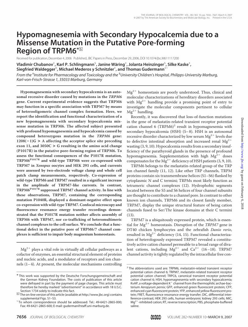
(Fig. 1A) and detected a second affected allele: a single nucleo-tide exchange (3050C � G) leading to a non-conservativeamino acid exchange from proline to arginine at position 1017(P1017R). The patient F14.1 presented with cerebral seizuresdue to severe hypomagnesemia (0.29 mM) and currently
FIGURE 1. Isolation of a novel missense HSH mutation located in the putative pore helix of TRPM6. A, mutationanalysis of TRPM6 by direct sequencing. Genomic sequences of exon 22 in the mother (top), the father (middle),and the affected individual (bottom) are shown. B, molecular model of the TRPM6 S5–S6 segment comparedwith the structure of the corresponding region in the KvAP channel. The �-carbon traces of TRPM6 (white) andKvAP (green) are shown. Outer (S5), inner (S6), and pore (P) helices are indicated. The P1017 residue in TRPM6 islabeled in red. C, sequence alignment of the S5–S6 segments from KvAP and TRPM6. Outer (S5), inner (S6), andpore (P) helices are defined on the basis of B. Identical (*), conserved (:), and semi-conserved (.) amino acids areindicated. P1017 in TRPM6 is labeled by a red-dotted box. KvAP residues in the pore loop known to be importantfor K� selectivity are in a blue-dotted box.
Novel Missense Mutation in TRPM6
MARCH 9, 2007 • VOLUME 282 • NUMBER 10 JOURNAL OF BIOLOGICAL CHEMISTRY 7659
by guest on February 6, 2018http://w
ww
.jbc.org/D
ownloaded from
receives continuous oral magnesium supplementation at a typ-ical dosage for HSH patients (0.6 mmol/kg/day). As expected,each of the mutations was observed in a heterozygous state inthe parents (Fig. 1A). Notably, the father carrying the alleleP1017R presented with a normal serum [Mg2�] level (0.93mM)and fractional magnesium excretion (2.4%). Thus, we classifiedthe patient as compound heterozygous, with a phenotype indis-tinguishable from previously described HSH patients (5, 7).The identified HSH mutation affects a residue located
between the predicted S5 and S6 domains of TRPM6 (supple-mental Fig. S1). This region displays only weak sequencehomology to other TRP channels (35). Therefore, we tookadvantage of the MODBASE resource to map P1017 in theannotated three dimensional coordinates of the TRPM6 S5–S6segment (see “Experimental Procedures”). As shown in Fig. 1 (Band C), TRPM6 has amino acid and predicted structural simi-larity to a voltage-gated K� channel from the archae-bacteriumAeropyrumpernix (KvAP) (32). The comparison ofTRPM6andKvAP suggested that P1017 may be located in the center of theputative pore helix (Fig. 1B). This domain is thought to be anessential structural element of the channel pore in a large num-ber of ion channels (35). Consequently, we hypothesized thatthe substitution of P1017 by a positively charged Arg couldchange the conformation of the pore helix and thus interferewith ion permeation properties of TRPM6.TRPM6P1017R Suppresses TRPM7-mediated Currents in
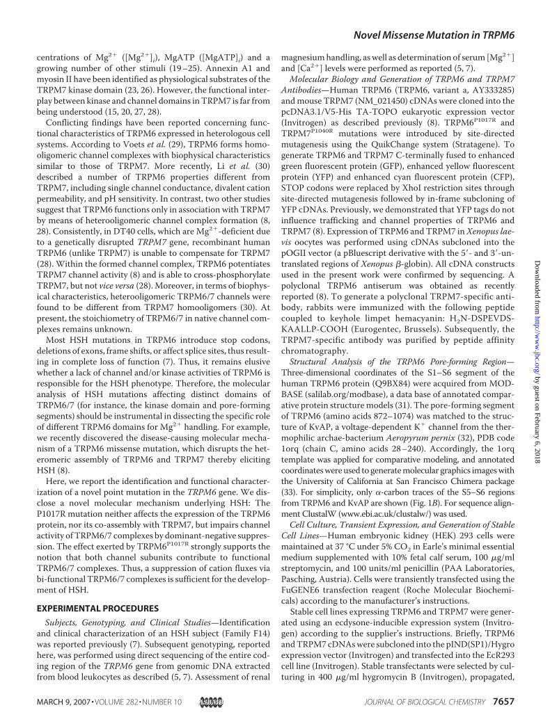
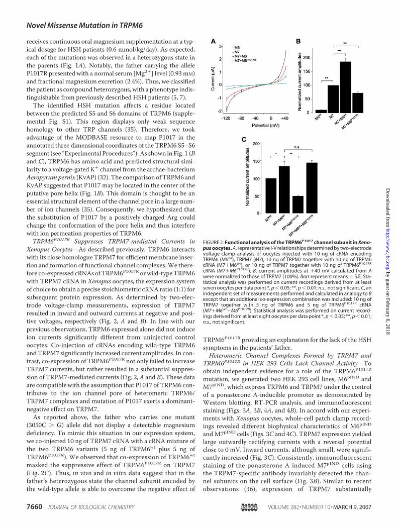
Xenopus Oocytes—As described previously, TRPM6 interactswith its close homologue TRPM7 for efficientmembrane inser-tion and formation of functional channel complexes.We there-fore co-expressed cRNAs ofTRPM6P1017R orwild-typeTRPM6with TRPM7 cRNA in Xenopus oocytes, the expression systemof choice to obtain a precise stoichiometric cRNA ratio (1:1) forsubsequent protein expression. As determined by two-elec-trode voltage-clamp measurements, expression of TRPM7resulted in inward and outward currents at negative and posi-tive voltages, respectively (Fig. 2, A and B). In line with ourprevious observations, TRPM6 expressed alone did not induceion currents significantly different from uninjected controloocytes. Co-injection of cRNAs encoding wild-type TRPM6andTRPM7 significantly increased current amplitudes. In con-trast, co-expression of TRPM6P1017R not only failed to increaseTRPM7 currents, but rather resulted in a substantial suppres-sion of TRPM7-mediated currents (Fig. 2,A and B). These dataare compatible with the assumption that P1017 of TRPM6 con-tributes to the ion channel pore of heteromeric TRPM6/TRPM7 complexes and mutation of P1017 exerts a dominant-negative effect on TRPM7.As reported above, the father who carries one mutant
(3050C � G) allele did not display a detectable magnesiumdeficiency. To mimic this situation in our expression system,we co-injected 10 ng of TRPM7 cRNA with a cRNAmixture ofthe two TRPM6 variants (5 ng of TRPM6wt plus 5 ng ofTRPM6P1017R). We observed that co-expression of TRPM6wtmasked the suppressive effect of TRPM6P1017R on TRPM7(Fig. 2C). Thus, in vivo and in vitro data suggest that in thefather’s heterozygous state the channel subunit encoded bythe wild-type allele is able to overcome the negative effect of
TRPM6P1017R providing an explanation for the lack of the HSHsymptoms in the patients’ father.Heteromeric Channel Complexes Formed by TRPM7 and
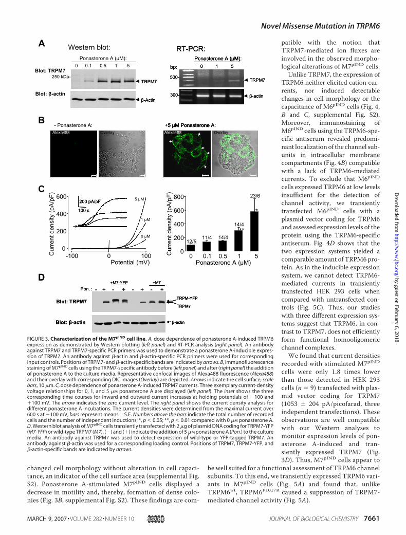
TRPM6P1017R in HEK 293 Cells Lack Channel Activity—Toobtain independent evidence for a role of the TRPM6P1017Rmutation, we generated two HEK 293 cell lines, M6pIND andM7pIND, which express TRPM6 and TRPM7 under the controlof a ponasterone A-inducible promoter as demonstrated byWestern blotting, RT-PCR analysis, and immunofluorescentstaining (Figs. 3A, 3B, 4A, and 4B). In accord with our experi-ments with Xenopus oocytes, whole-cell patch clamp record-ings revealed different biophysical characteristics of M6pIND
andM7pIND cells (Figs. 3C and 4C). TRPM7 expression yieldedlarge outwardly rectifying currents with a reversal potentialclose to 0 mV. Inward currents, although small, were signifi-cantly increased (Fig. 3C). Consistently, immunofluorescentstaining of the ponasterone A-induced M7pIND cells usingthe TRPM7-specific antibody invariably detected the chan-nel subunits on the cell surface (Fig. 3B). Similar to recentobservations (36), expression of TRPM7 substantially
FIGURE 2. Functional analysis of the TRPM6P1017 channel subunit in Xeno-pus oocytes. A, representative I-V relationships determined by two-electrodevoltage-clamp analysis of oocytes injected with 10 ng of cRNA encodingTRPM6 (M6wt), TRPM7 (M7), 10 ng of TRPM7 together with 10 ng of TRPM6cRNA (M7�M6wt), or 10 ng of TRPM7 together with 10 ng of TRPM6P1017R
cRNA (M7�M6P1017R). B, current amplitudes at �40 mV calculated from Awere normalized to those of TRPM7 (100%). Bars represent means � S.E. Sta-tistical analysis was performed on current recordings derived from at leastseven oocytes per data point *, p 0.05; **, p 0.01; n.s., not significant. C, anindependent set of measurements performed and calculated in analogy to Bexcept that an additional co-expression combination was included: 10 ng ofTRPM7 together with 5 ng of TRPM6 and 5 ng of TRPM6P1017R cRNA(M7�M6wt�M6P1017R). Statistical analysis was performed on current record-ings derived from at least eight oocytes per data point *, p 0.05; **, p 0.01;n.s., not significant.
Novel Missense Mutation in TRPM6
7660 JOURNAL OF BIOLOGICAL CHEMISTRY VOLUME 282 • NUMBER 10 • MARCH 9, 2007
by guest on February 6, 2018http://w
ww
.jbc.org/D
ownloaded from
changed cell morphology without alteration in cell capaci-tance, an indicator of the cell surface area (supplemental Fig.S2). Ponasterone A-stimulated M7pIND cells displayed adecrease in motility and, thereby, formation of dense colo-nies (Fig. 3B, supplemental Fig. S2). These findings are com-
patible with the notion thatTRPM7-mediated ion fluxes areinvolved in the observed morpho-logical alterations of M7pIND cells.
Unlike TRPM7, the expression ofTRPM6 neither elicited cation cur-rents, nor induced detectablechanges in cell morphology or thecapacitance of M6pIND cells (Fig. 4,B and C, supplemental Fig. S2).Moreover, immunostaining ofM6pIND cells using the TRPM6-spe-cific antiserum revealed predomi-nant localization of the channel sub-units in intracellular membranecompartments (Fig. 4B) compatiblewith a lack of TRPM6-mediatedcurrents. To exclude that M6pIND
cells expressed TRPM6 at low levelsinsufficient for the detection ofchannel activity, we transientlytransfected M6pIND cells with aplasmid vector coding for TRPM6and assessed expression levels of theprotein using the TRPM6-specificantiserum. Fig. 4D shows that thetwo expression systems yielded acomparable amount of TRPM6 pro-tein. As in the inducible expressionsystem, we cannot detect TRPM6-mediated currents in transientlytransfected HEK 293 cells whencompared with untransfected con-trols (Fig. 5C). Thus, our studieswith three different expression sys-tems suggest that TRPM6, in con-trast to TRPM7, does not efficientlyform functional homooligomericchannel complexes.We found that current densities
recorded with stimulated M7pIND
cells were only 1.8 times lowerthan those detected in HEK 293cells (n 9) transfected with plas-mid vector coding for TRPM7(1053 � 204 pA/picofarad, threeindependent transfections). Theseobservations are well compatiblewith our Western analyses tomonitor expression levels of pon-asterone A-induced and tran-siently expressed TRPM7 (Fig.3D). Thus, M7pIND cells appear to
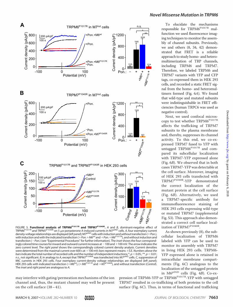
be well suited for a functional assessment of TRPM6 channelsubunits. To this end, we transiently expressed TRPM6 vari-ants in M7pIND cells (Fig. 5A) and found that, unlikeTRPM6wt, TRPM6P1017R caused a suppression of TRPM7-mediated channel activity (Fig. 5A).
FIGURE 3. Characterization of the M7pIND cell line. A, dose dependence of ponasterone A-induced TRPM6expression as demonstrated by Western blotting (left panel) and RT-PCR analysis (right panel). An antibodyagainst TRPM7 and TRPM7-specific PCR primers was used to demonstrate a ponasterone A-inducible expres-sion of TRPM7. An antibody against �-actin and �-actin-specific PCR primers were used for correspondinginput controls. Positions of TRPM7- and �-actin-specific bands are indicated by arrows. B, immunofluorescencestaining of M7pIND cells using the TRPM7-specific antibody before (left panel) and after (right panel) the additionof ponasterone A to the culture media. Representative confocal images of Alexa488 fluorescence (Alexa488)and their overlay with corresponding DIC images (Overlay) are depicted. Arrows indicate the cell surface; scalebars, 10 �m. C, dose dependence of ponasterone A-induced TRPM7 currents. Three exemplary current-densityvoltage relationships for 0, 1, and 5 �M ponasterone A are displayed (left panel). The inset shows the threecorresponding time courses for inward and outward current increases at holding potentials of �100 and�100 mV. The arrow indicates the zero current level. The right panel shows the current density analysis fordifferent ponasterone A incubations. The current densities were determined from the maximal current over600 s at �100 mV; bars represent means �S.E. Numbers above the bars indicate the total number of recordedcells and the number of independent inductions; *, p 0.05; **, p 0.01 compared with 0 �M ponasterone A.D, Western blot analysis of M7pIND cells transiently transfected with 2 �g of plasmid DNA coding for TRPM7-YFP(M7-YFP) or wild-type TRPM7 (M7). (�) and (�) indicate the addition of 5 �M ponasterone A (Pon.) to the culturemedia. An antibody against TRPM7 was used to detect expression of wild-type or YFP-tagged TRPM7. Anantibody against �-actin was used for a corresponding loading control. Positions of TRPM7, TRPM7-YFP, and�-actin-specific bands are indicated by arrows.
Novel Missense Mutation in TRPM6
MARCH 9, 2007 • VOLUME 282 • NUMBER 10 JOURNAL OF BIOLOGICAL CHEMISTRY 7661
by guest on February 6, 2018http://w
ww
.jbc.org/D
ownloaded from

P1017 in TRPM6 is a conserved residue in the TRPM genefamily (supplemental Fig. S1). Three-dimensional modeling ofthe pore-forming region of TRPM7mapped the correspond-ing P1040 in TRPM7 to an equivalent position to that of
P1017 in the TRPM6 model, i.e.the putative pore helix (data notshown). Thus, we reasoned that ananalogous mutation in TRPM7(TRPM7P1040R) would entail func-tional consequences similar to thoseobserved in TRPM6P1017R. To testthis hypothesis, we transientlyexpressed TRPM7P1040R in M7pIND
cells and found that, similar toTRPM6P1017R, TRPM7P1040R com-pletely suppressed channel activityof wild-type TRPM7 in the ponas-terone A-stimulated M7pIND cells(Fig. 5B).TRPM7 is considered to be the
molecular substrate of Mg2�-inhib-ited cation (MIC) currents readilydetectable in various cells whencytoplasmic [Mg2�] is loweredduring whole-cell recordings, thecommonly used maneuver todetect TRPM7-mediated currentsin heterologous expression sys-tems (16, 37). We asked whetherTRPM6P1017R and TRPM7P1040Rwould be able to suppress nativeMIC currents. Similar to otherreports (16, 36), our recordings onuntransfected HEK 293 cellsrevealed TRPM7-like inward andoutward currents (Fig. 5C). In fact,unlike TRPM6wt, transient expres-sion of both mutants caused a sub-stantial reduction of TRPM7-likecurrents. Interestingly, the suppres-sive effect of TRPM7P1040R was sig-nificantly more pronounced thanthat ofTRPM6P1017R (Fig. 5C). Alto-gether, our biophysical experimentsprovide an independent line of evi-dence that TRPM6P1017R andTRPM7P1040R function as domi-nant-negative channel subunits.TRPM6P1017R and TRPM7P1040R
Are Assembled in HeteromericChannel Complexes on the CellSurface—Missense mutations asso-ciated with dominant-negativeeffects on multimeric ion channelscan be classified into two types.The first class of mutations, typi-cally located in cytoplasmic seg-ments of a subunit, may impair its
trafficking. Consequently, channel complexes, includingwild-type subunits, are retained in intracellular membranecompartments (38–41). Alternatively, mutations located ina transmembrane segment or in the pore-forming region
FIGURE 4. Characterization of the M6pIND cell line. A, dose dependence of ponasterone A-induced TRPM6 expres-sion as demonstrated by Western blotting (left panel) and RT-PCR analysis (right panel). An antiserum against TRPM6and TRPM6-specific PCR primers were used to demonstrate a ponasterone A-inducible expression of TRPM6. Theantibody against �-actin and the �-actin-specific PCR primers were used for corresponding input controls. Positionsof TRPM6- and �-actin-specific bands are indicated by arrows. B, immunofluorescence staining of M6pIND cells usingthe TRPM6-specific antiserum before (left panel) and after (right panel) the addition of ponasterone A to the culturemedia. Representative confocal images of Alexa488 fluorescence (Alexa488) and their overlay with correspondingDIC images (Overlay) are depicted. Scale bars are 10 �m. C, dose dependence of ponasterone A-induced TRPM6currents. Three exemplary current-density voltage relationships for 0, 1, and 5 �M ponasterone A are displayed(left panel). The inset shows the three corresponding time courses for inward and outward current increases at�100 and �100 mV. The arrow indicates the zero current level. The right panel shows the current densityanalysis for different ponasterone A incubations. The current densities were determined from the maximalcurrent over 600 s at �100 mV; bars represent means �S.E. Numbers above the bars indicate the total numberof recorded cells and the number of independent inductions. D, Western blot analysis of TRPM6 expression inM6pIND cells transiently transfected with 2 �g of plasmid DNA coding for wild-type TRPM6. (�) and (�) indicatetransient transfection of TRPM6 (M6) or the addition of 5 �M ponasterone A (Pon.) to the culture media. Anantiserum against TRPM6 was used to detect expression of TRPM6. An antibody against �-actin was used for acorresponding loading control. Positions of TRPM6 and �-actin-specific bands are indicated by arrows.
Novel Missense Mutation in TRPM6
7662 JOURNAL OF BIOLOGICAL CHEMISTRY VOLUME 282 • NUMBER 10 • MARCH 9, 2007
by guest on February 6, 2018http://w
ww
.jbc.org/D
ownloaded from
may interfere with gating/permeationmechanisms of the ionchannel and, thus, the mutant channel may well be presenton the cell surface (38–41).
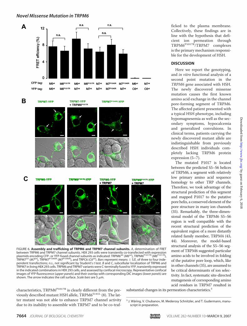
To elucidate the mechanismsresponsible for TRPM6P1017R mal-function we used fluorescence imag-ing techniques tomonitor the assem-bly of channel subunits. Previously,we and others (8, 34, 42) demon-strated that FRET is a reliableapproach to study homo- and hetero-multimerization of TRP channels,including TRPM6 and TRPM7.Therefore, we labeled TRPM6 andTRPM7 variants with YFP and CFPtags, co-expressed them in HEK 293cells, and recorded a static FRET sig-nal from the homo- and heteromul-timers formed (Fig. 6A). We foundthat wild-type and mutated subunitswere indistinguishable in FRET effi-ciencies (human TRPC6 was used asnegative control).Next, we used confocal micros-
copy to test whether TRPM6P1017Raffects the trafficking of TRPM7subunits to the plasma membraneand, thereby, suppresses its channelactivity. To this end, we co-ex-pressed TRPM7 fused to YFP withuntagged TRPM6P1017R and com-pared its subcellular localizationwith TRPM7-YFP expressed alone(Fig. 6B). We observed that in bothcasesTRPM7-YFPwas detectable atthe cell surface. Moreover, imagingof HEK 293 cells transfected withTRPM7P1040R-YFP demonstratedthe correct localization of themutant protein at the cell surface(Fig. 6B). Alternatively, we useda TRPM7-specific antibody forimmunofluorescence staining ofHEK 293 cells expressing wild-typeor mutated TRPM7 (supplementalFig. S3). This approach also demon-strated a correct cell surface local-ization of TRPM7P1040R.
As shown previously (8), the sub-cellular localization of TRPM6labeled with YFP can be used tomonitor its assembly with TRPM7in living HEK 293 cells. TRPM6-YFP expressed alone is retained inintracellular membrane compart-ments (Fig. 6C) analogous to thelocalization of the untagged proteinin M6pIND cells (Fig. 4B). Co-ex-
pression of TRPM6-YFP or TRPM6P1017R-YFP with untaggedTRPM7 resulted in co-trafficking of both proteins to the cellsurface (Fig. 6C). Thus, in terms of functional and trafficking
FIGURE 5. Functional analysis of TRPM6P1017R and TRPM7P1040R. A and B, dominant-negative effect ofTRPM6P1017R and TRPM7P1040R on 5 �M ponasterone A-induced currents in M7pIND cells. A, four exemplary currentdensity-voltage relationships are displayed (left panel); M7pIND cells with induction and without transfection (�Pon.),with induction and with the indicated transfection (�Pon.�M6wt and �Pon.�M6P1017R), and without induction andtransfection (�Pon.) (see “Experimental Procedures” for further information). The inset shows the four correspond-ingly colored time courses for inward and outward current increases at �100 and �100 mV. The arrow indicates thezero current level. The right panel shows the correspondingly colored current density analysis. Current densitieswere determined from the maximal current over 600 s at �100 mV; bars represent means �S.E. Numbers above thebars indicate the total number of recorded cells and the number of independent inductions; *, p 0.05.; **, p 0.01;n.s., not significant. B, in analogy to A, except that TRPM7P1040R was transfected into M7pIND cells. C, suppression ofMIC currents in HEK 293 cells. Four exemplary current-density voltage relationships are displayed (left panel);HEK 293 cells with indicated transfection (�M6wt), (�M6P1017R and �M7P1040R), and without transfection (Control).The inset and right panel are analogous to A.
Novel Missense Mutation in TRPM6
MARCH 9, 2007 • VOLUME 282 • NUMBER 10 JOURNAL OF BIOLOGICAL CHEMISTRY 7663
by guest on February 6, 2018http://w
ww
.jbc.org/D
ownloaded from
characteristics, TRPM6P1017R is clearly different from the pre-viously described mutant HSH allele, TRPM6S141L (8). The lat-ter mutant was not able to enhance TRPM7 channel activitydue to its inability to assemble with TRPM7 and to be co-traf-
ficked to the plasma membrane.Collectively, these findings are inline with the hypothesis that defi-cient ion permeation throughTRPM6P1017R/TRPM7 complexesis the primary mechanism responsi-ble for the development of HSH.
DISCUSSION
Here we report the genotyping,and in vitro functional analysis of asecond point mutation in theTRPM6 gene associated with HSH.The newly discovered missensemutation causes the first knownamino acid exchange in the channelpore-forming segment of TRPM6.The affected patient presented witha typical HSH phenotype, includinghypomagnesemia as well as the sec-ondary symptoms, hypocalcemiaand generalized convulsions. Inclinical terms, patients carrying thenewly discovered mutant allele areindistinguishable from previouslydescribed HSH individuals com-pletely lacking TRPM6 proteinexpression (5–7).The mutated P1017 is located
between the predicted S5–S6 helicesof TRPM6, a segment with relativelylow primary amino acid sequencehomology to other TRP channels.Therefore, we took advantage of thestructural prediction of this segmentand mapped P1017 to the putativepore helix, a conserved element of thepore structure in many ion channels(35). Remarkably, the three-dimen-sional model of the TRPM6 S5–S6region is well compatible with therecent structural prediction of theequivalent region of a more distantlyrelated family member, TRPM4 (43,44). Moreover, the model-basedstructural analysis of the S5–S6 seg-ment of TRPM6 suggested particularamino acids to be involved in foldingof the putative pore loop, which, likein other channels (35), are assumed tobe critical determinants of ion selec-tivity. In fact, systematic site-directedmutagenesis of corresponding aminoacid residues in TRPM7 resulted in
substantial changes in its permeation characteristics.3
3 J. Waring, V. Chubanov, M. Mederosy Schnitzler, and T. Gudermann, manu-script in preparation.
FIGURE 6. Assembly and trafficking of TRPM6 and TRPM7 channel subunits. A, determination of FRETbetween TRPM6 and TRPM7 channel subunits. HEK 293 cells were transiently co-transfected with expressionplasmids encoding CFP- or YFP-fused channel subunits as indicated: TRPM6wt (M6wt), TRPM6P1017R (M6P1017R),TRPM7wt (M7wt), TRPM7P1040R (M7P1040R), and TRPC6 (C6wt). Bars represent means � S.E. of three to four inde-pendent transfections; n.s., not significant by Student’s t test. B and C, subcellular localization of TRPM6 andTRPM7 in living HEK 293 cells. TRPM6 and TRPM7 variants were C-terminally fused to YFP, transiently expressedin the indicated combinations in HEK 293 cells, and assessed by confocal microscopy. Representative confocalimages of YFP fluorescence (upper panels) and their overlay with corresponding DIC images (lower panels) areshown. The arrow indicates the cell surface. Scale bars are 5 �m.
Novel Missense Mutation in TRPM6
7664 JOURNAL OF BIOLOGICAL CHEMISTRY VOLUME 282 • NUMBER 10 • MARCH 9, 2007
by guest on February 6, 2018http://w
ww
.jbc.org/D
ownloaded from
Based on the structural prediction, we hypothesized that theP1017Rmutation in TRPM6may alter the overall folding of thepore helix, thereby affecting its ion permeation characteristics.To evaluate the missense mutation functionally, we used twoexperimental systems: Xenopus oocytes and HEK 293 cells. Inboth expressionmodels TRPM6,when expressed alone, did notshow a significant level of channel activity, whereas co-expres-sion of wild-type TRPM6 and TRPM7 resulted in amplificationof TRPM7-mediated currents. However, unlike wild-typeTRPM6, TRPM6P1017R caused a suppression of TRPM7-medi-ated ion currents. Confocal microscopy of living HEK 293 cellsshowed that TRPM6P1017R/TRPM7 complexes were normallyco-targeted to the cell surface. Thus, biophysical and traffickingcharacteristics of TRPM6P1017R are noticeably different fromthe TRPM6S141L allele described previously: the S141L muta-tion severely impaired trafficking of TRPM6without detectableimpact on TRPM7 channel activity (8). Together, we concludethat TRPM6P1017R was efficiently assembled with TRPM7 butexerted a dominant-negative effect on channel activity primar-ily due to functional defects in the channel pore of theheterotetramer.The affected proline in TRPM6 is a highly conserved residue
in TRPM proteins. Accordingly, the incorporation of a HSH-like mutation in the closely related TRPM7 channel had func-tional consequences similar to those observed inTRPM6P1017R.TRPM7P1040R was properly targeted to the cell surface where itelicited a dominant-negative effect on ion currentsmediated bywild-type TRPM7. Thus, these experiments provide additionalevidence in favor of our molecular explanation for the func-tional defect in TRPM6P1017R.
None of the HSH families with loss-of-functionmutations inthe TRPM6 gene examined so far did display a haploinsuffi-ciency in heterozygous subjects, i.e. only a complete lack ofTRPM6 causes clinical symptoms of HSH (7). However, it islikely that mutations affecting exon splicing sites or truncatingthe coding sequence due to a frameshift (7) result in expressionof dominant-negative channel subunits. If so, such a situationwould be similar to the P1017R allele. Of note, our genetic andclinical data indicate clearly that the patient’s father carrying asingle mutation in the gene (P1017R) has normal renal magne-sium handling. A plausible explanation for the lack of symp-toms in the patient’s father is that TRPM6P1017R is not able tocompletely suppress the function of proteins produced by thewild-type allele. Apparently, a functional role of TRPM6may bemaintained by a relatively low amount of the protein, a phe-nomenon frequently observed with genes associated with auto-somal recessive disorders (38–41, 45). In fact, our experimentswith Xenopus oocytes showed that TRPM6wt co-injected withTRPM6P1017R at a 1:1 ratio reversed the dominant negativeeffect of TRPM6P1017R on TRPM7. The following scenario canbe proposed to explain this observation. Wild-type TRPM6may heteromultimerize more efficiently than the mutant pro-tein. Thus, the actual number of functional channel complexeson the cell surface may be different from that calculated from astoichiometric expression ratio of TRPM6wt/TRPM6P1017R inthe heterozygote. Also, we cannot exclude that our heterolo-gous expression system does not comprehensively reproducethe pathophysiological situation in native epithelial cells. Yet,
different isoforms of TRPM6 (8) and TRPM7 (17) may shapethe net effect of P1017R in vivo. It is also possible that, in theheterozygous state, the expression of both alleles may be up-regulated raising the number of heterooligomers containingonly wild-type subunits to a sufficient level.Apart from the clinical importance, analysis of the TRPM6
and TRPM7 mutants highlighted a number of issues concern-ing the function of these proteins. First, we found that tran-siently expressed TRPM6P1017R and TRPM7P1040R are able tosuppress native TRPM7-like, Mg2�-inhibited (MIC) cationcurrents inHEK293 cells. In linewith recent data obtainedwitha short hairpin RNA approach (36), these results provide inde-pendent evidence that TRPM7 channel complexes are molecu-lar substrates of endogenous MIC channels. Second, the dom-inant-negative effect exerted by TRPM6P1017R on endogenousMIC channels supports our previous notion (8) that inTRPM6/7 heterooligomers both channel subunits contributeto the channel pore. Third, analyses of HSH missense muta-tions affecting different functional modules of TRPM6 mayprovide valuable information permitting to dissect the biologi-cal role played by its channel and kinase domains. At present,the functional interplay between channel and kinase activitiesof TRPM6/7 complexes remains elusive. Our functional analy-sis of TRPM6P1017R indicates that suppression of the channelfunction in TRPM6/7 complexes is sufficient to elicit the char-acteristic HSH phenotype. This finding is well compatible withthe biophysical analysis of heterologously expressed TRPM7lacking the kinase domain or the TRPM7 mutants deficient inthe kinase activity (15, 27, 46). These studies showed that prin-cipally TRPM7 kinase activity is not required for channel activ-ity. Moreover, wild-type and “kinase-dead” TRPM7 variantswere found to be equivalent in their ability to rescue the viabil-ity andMg2� deficiency of DT40 lymphocytes carrying a genet-ically disrupted TRPM7 gene (15). More recently, Clark et al.(23) reported that TRPM7-mediatedCa2� influx is required forkinase domain-dependent actomyosin contractility. In sum-mary, our analysis of the new missense HSH mutation inTRPM6, together with in vitro experiments onTRPM7, suggestthat in TRPM6/7 the kinase domains may function down-stream of their channel activity. Accordingly, TRPM6P1017Rand TRPM7P1040R, characterized here, represent valuablemolecular tools to further test this hypothesis.Recent reports on the functional characterization of TRPM7
and TRPM6 yielded controversial results. Interestingly, smallvariations in the composition of pipette solutions used forwhole-cell recordings of TRPM7-mediated currents are suffi-cient to produce contradictory observations (16, 17, 46, 47). It isstill a moot issue whether TRPM6 homomultimers form func-tional ion channels in native cells. Here, we used two alternativeheterologous expression systems, Xenopus oocytes and HEK293 cells, and could not detect significant TRPM6 channelactivity, fully consistent with the channel’s predominant intra-cellular localization. These findings are in line with our previ-ous observations (7, 8, 12) andwith a recent report of Schmitz etal. (28) who demonstrated that TRPM6 requires TRPM7 forefficient cell surface localization. However, studies of two othergroups (29, 30) reported that transfection of a plasmid vectorcoding for human TRPM6 resulted in the formation of func-
Novel Missense Mutation in TRPM6
MARCH 9, 2007 • VOLUME 282 • NUMBER 10 JOURNAL OF BIOLOGICAL CHEMISTRY 7665
by guest on February 6, 2018http://w
ww
.jbc.org/D
ownloaded from
tional ion channels. Notably, attempts to record native currentsmediated solely by TRPM6 homooligomers have not been suc-cessful (30). Therefore, we used an independent approach tofunctionally evaluate TRPM6 and TRPM7 proteins. There isevidence to show that TRPM7-mediated cation entry regulatesadhesion and spreading of cells (23, 36). In accord with thesefindings (36), we observed that ponasterone A-induced expres-sion of TRPM7, in contrast to TRPM6, resulted in pronouncedalterations of themorphology and spreading of cells. Hence, thedifferent morphological characteristics of TRPM6- andTRPM7-expressing cells corroborate our biophysical and traf-ficking data demonstrating that TRPM6 subunits are very inef-ficient in the formation of functional homomultimers.We cannot exclude that a specific overexpression maneuver
or application of “chemical chaperone” strategies may over-come the intracellular retention mechanism of TRPM6. How-ever, a growing number of examined tissues and cell linesinvariably show co-expression of TRPM6 with TRPM7 (4, 8,48–50) raising the question whether TRPM6 can functionalone. The specific and efficient assembly of TRPM6 withTRPM7, reproduced in various alternative expression models,most probably reflects the intrinsicmode ofTRPM6 function asa subunit of TRPM6/7 complexes. Such a scenario would agreewell with an indispensable role ofTRPM7 inDT40 lymphocytes(15).In conclusion, we identified a second point mutation in
TRPM6, P1017R, resulting in HSH. The dominant-negativeeffect exerted by TRPM6P1017R provides strong evidence toconclude that both channel subunits, TRPM6 and TRPM7,contribute to a common channel pore in TRPM6/7 complexes.Dominant-negative inhibition bymutant channel subunits rep-resents a novel molecular mechanism underlying HSH. Ourfindings indicate that impairment of channel activity inTRPM6/7 complexes is sufficient for the development ofMg2�
deficiency in HSH patients.
Acknowledgments—We thank Thomas Hofmann and Tim Plant,Philipps-University Marburg, for critical discussions.
REFERENCES1. Hermosura,M.C.,Nayakanti,H.,Dorovkov,M.V.,Calderon, F.R.,Ryazanov,
A. G., Haymer, D. S., and Garruto, R. M. (2005) Proc. Natl. Acad. Sci. U. S. A.102, 11510–11515
2. Gums, J. G. (2004) Am. J. Health Syst. Pharm. 61, 1569–15763. Schlingmann, K. P., and Gudermann, T. (2005) J. Physiol. 566, 301–3084. Touyz, R. M., He, Y., Montezano, A. C., Yao, G., Chubanov, V., Gudermann,
T., and Callera, G. E. (2006) Am. J. Physiol. Regul. Integr Comp. Physiol. 290,R73–R78
5. Schlingmann, K. P.,Weber, S., Peters,M., NiemannNejsum, L., Vitzthum,H., Klingel, K., Kratz, M., Haddad, E., Ristoff, E., Dinour, D., Syrrou, M.,Nielsen, S., Sassen, M., Waldegger, S., Seyberth, H. W., and Konrad, M.(2002) Nat. Genet. 31, 166–170
6. Walder, R. Y., Landau, D., Meyer, P., Shalev, H., Tsolia, M., Borochowitz,Z., Boettger, M. B., Beck, G. E., Englehardt, R. K., Carmi, R., and Sheffield,V. C. (2002) Nat. Genet. 31, 171–174
7. Schlingmann, K. P., Sassen, M. C., Weber, S., Pechmann, U., Kusch, K.,Pelken, L., Lotan, D., Syrrou, M., Prebble, J. J., Cole, D. E., Metzger, D. L.,Rahman, S., Tajima, T., Shu, S. G., Waldegger, S., Seyberth, H. W., andKonrad, M. (2005) J. Am. Soc. Nephrol. 16, 3061–3069
8. Chubanov, V., Waldegger, S., Mederos y Schnitzler, M., Vitzthum, H.,
Sassen, M. C., Seyberth, H. W., Konrad, M., and Gudermann, T. (2004)Proc. Natl. Acad. Sci. U. S. A. 101, 2894–2899
9. Chubanov, V., Gudermann, T., and Schlingmann, K. P. (2005) PflugersArch. 451, 228–234
10. Konrad,M., Schlingmann, K. P., andGudermann, T. (2004)Am. J. Physiol.Renal Physiol. 286, F599–F605
11. Fleig, A., and Penner, R. (2004) Trends Pharmacol. Sci. 25, 633–63912. Chubanov, V., Mederos y Schnitzler, M., Waring, J., Plank, A., and
Gudermann, T. (2005) Naunyn-Schmiedeberg’s Arch. Pharmacol. 371,334–341
13. Drennan, D., and Ryazanov, A. G. (2004) Prog. Biophys. Mol. Biol. 85,1–32
14. Elizondo, M. R., Arduini, B. L., Paulsen, J., MacDonald, E. L., Sabel, J. L.,Henion, P. D., Cornell, R. A., and Parichy, D. M. (2005) Curr. Biol. 15,667–671
15. Schmitz, C., Perraud, A. L., Johnson, C.O., Inabe, K., Smith,M. K., Penner,R., Kurosaki, T., Fleig, A., and Scharenberg, A. M. (2003) Cell 114,191–200
16. Nadler, M. J., Hermosura, M. C., Inabe, K., Perraud, A. L., Zhu, Q., Stokes,A. J., Kurosaki, T., Kinet, J. P., Penner, R., Scharenberg, A.M., and Fleig, A.(2001) Nature 411, 590–595
17. Runnels, L.W., Yue, L., andClapham,D. E. (2001) Science 291, 1043–104718. Monteilh-Zoller, M. K., Hermosura, M. C., Nadler, M. J., Scharenberg,
A. M., Penner, R., and Fleig, A. (2003) J. Gen. Physiol. 121, 49–6019. Runnels, L. W., Yue, L., and Clapham, D. E. (2002) Nat. Cell Biol. 4,
329–33620. Kozak, J. A.,Matsushita,M.,Nairn, A.C., andCahalan,M.D. (2005) J. Gen.
Physiol. 126, 499–51421. Jiang, J., Li, M., and Yue, L. (2005) J. Gen. Physiol. 126, 137–15022. Oancea, E., Wolfe, J. T., and Clapham, D. E. (2006) Circ. Res. 98, 163–16423. Clark, K., Langeslag,M., van Leeuwen, B., Ran, L., Ryazanov, A. G., Figdor,
C. G., Moolenaar, W. H., Jalink, K., and van Leeuwen, F. N. (2006) EMBOJ. 25, 290–301
24. Takezawa, R., Schmitz, C., Demeuse, P., Scharenberg, A. M., Penner, R.,and Fleig, A. (2004) Proc. Natl. Acad. Sci. U. S. A. 101, 6009–6014
25. Aarts, M., Iihara, K., Wei, W. L., Xiong, Z. G., Arundine, M., Cerwinski,W., MacDonald, J. F., and Tymianski, M. (2003) Cell 115, 863–877
26. Dorovkov, M. V., and Ryazanov, A. G. (2004) J. Biol. Chem. 279,50643–50646
27. Matsushita, M., Kozak, J. A., Shimizu, Y., McLachlin, D. T., Yamaguchi,H., Wei, F. Y., Tomizawa, K., Matsui, H., Chait, B. T., Cahalan, M. D., andNairn, A. C. (2005) J. Biol. Chem. 280, 20793–20803
28. Schmitz, C., Dorovkov, M. V., Zhao, X., Davenport, B. J., Ryazanov, A. G.,and Perraud, A. L. (2005) J. Biol. Chem. 280, 37763–37771
29. Voets, T., Nilius, B., Hoefs, S., van der Kemp, A. W., Droogmans, G.,Bindels, R. J., and Hoenderop, J. G. (2004) J. Biol. Chem. 279, 19–25
30. Li, M., Jiang, J., and Yue, L. (2006) J. Gen. Physiol. 127, 525–53731. Pieper, U., Eswar, N., Davis, F. P., Braberg, H., Madhusudhan,M. S., Rossi,
A., Marti-Renom, M., Karchin, R., Webb, B. M., Eramian, D., Shen, M. Y.,Kelly, L., Melo, F., and Sali, A. (2006) Nucleic Acids Res. 34, D291–D295,database issue
32. Jiang, Y., Lee, A., Chen, J., Ruta, V., Cadene, M., Chait, B. T., andMacKinnon, R. (2003) Nature 423, 33–41
33. Pettersen, E. F., Goddard, T. D., Huang, C. C., Couch, G. S., Greenblatt,D. M., Meng, E. C., and Ferrin, T. E. (2004) J. Comput. Chem. 25,1605–1612
34. Hofmann, T., Schaefer, M., Schultz, G., and Gudermann, T. (2002) Proc.Natl. Acad. Sci. U. S. A. 99, 7461–7466
35. Owsianik, G., Talavera, K., Voets, T., and Nilius, B. (2005) Annu. Rev.Physiol. 68, 685–717
36. Su, L. T., Agapito, M. A., Li, M., W, T. N. S., Huttenlocher, A., Habas, R.,Yue, L., and Runnels, L. W. (2006) J. Biol. Chem. 281, 11260–11270
37. Kozak, J. A., Kerschbaum,H. H., and Cahalan,M. D. (2002) J. Gen. Physiol.120, 221–235
38. Cannon, S. C. (2006) Annu. Rev. Neurosci. 29, 387–41539. Hanna, M. G. (2006) Nat. Clin. Pract. Neurol. 2, 252–26340. Ashcroft, F. M. (2006) Nature 440, 440–44741. Kass, R. S. (2005) J. Clin. Invest. 115, 1986–1989
Novel Missense Mutation in TRPM6
7666 JOURNAL OF BIOLOGICAL CHEMISTRY VOLUME 282 • NUMBER 10 • MARCH 9, 2007
by guest on February 6, 2018http://w
ww
.jbc.org/D
ownloaded from

42. Hellwig, N., Albrecht, N., Harteneck, C., Schultz, G., and Schaefer, M.(2005) J. Cell Sci. 118, 917–928
43. Liu, D., Zhang, Z., and Liman, E. R. (2005) J. Biol. Chem. 280, 20691–2069944. Nilius, B., Prenen, J., Janssens, A., Owsianik, G.,Wang, C., Zhu,M. X., and
Voets, T. (2005) J. Biol. Chem. 280, 22899–2290645. Furney, S. J., Alba, M.M., and Lopez-Bigas, N. (2006) BMCGenomics 7,
16546. Demeuse, P., Penner, R., and Fleig, A. (2006) J. Gen. Physiol. 127, 421–434
47. Langeslag,M., Clark, K.,Moolenaar,W.H., van Leeuwen, F. N., and Jalink,K. (2007) J. Biol. Chem. 282, 232–239
48. Groenestege,W.M., Hoenderop, J. G., van denHeuvel, L., Knoers, N., andBindels, R. J. (2006) J. Am. Soc. Nephrol. 17, 1035–1043
49. Kunert-Keil, C., Bisping, F., Kruger, J., and Brinkmeier, H. (2006) BMCGenomics 7, 159
50. Fonfria, E., Murdock, P. R., Cusdin, F. S., Benham, C. D., Kelsell, R. E., andMcNulty, S. (2006) J. Recept. Signal Transduct. Res. 26, 159–178
Novel Missense Mutation in TRPM6
MARCH 9, 2007 • VOLUME 282 • NUMBER 10 JOURNAL OF BIOLOGICAL CHEMISTRY 7667
by guest on February 6, 2018http://w
ww
.jbc.org/D
ownloaded from
Kaske, Siegfried Waldegger, Michael Mederos y Schnitzler and Thomas GudermannVladimir Chubanov, Karl P. Schlingmann, Janine Wäring, Jolanta Heinzinger, Silke
Putative Pore-forming Region of TRPM6Hypomagnesemia with Secondary Hypocalcemia due to a Missense Mutation in the
doi: 10.1074/jbc.M611117200 originally published online December 29, 20062007, 282:7656-7667.J. Biol. Chem.
10.1074/jbc.M611117200Access the most updated version of this article at doi:
Alerts:
When a correction for this article is posted•
When this article is cited•
to choose from all of JBC's e-mail alertsClick here
Supplemental material:
http://www.jbc.org/content/suppl/2007/01/02/M611117200.DC1
http://www.jbc.org/content/282/10/7656.full.html#ref-list-1
This article cites 48 references, 23 of which can be accessed free at
by guest on February 6, 2018http://w
ww
.jbc.org/D
ownloaded from