hyaluronic acid, mesotherapy and skin rejuvenation · hyaluronic acid, mesotherapy and skin...
TRANSCRIPT
Hyaluronic Acid, Mesotherapy and Skin Rejuvenation Published 2/2010 Hi.Tech Dermo
Prof. Beniamino Palmieri Specialist in Plastic and Reconstructive Surgery Associate Professor of General Surgery Department of General Surgery and Surgical Specialties, University of Modena and Reggio Emilia Medical School, Surgical Clinic, Modena, Italy
INTRODUCTION
Hyaluronic acid (HA) is a very safe and widespread used dermal filler with specific
indication to be used in facial rejuvenation: in fact, the aging process of the skin in this
area, gives loss of elasticity and volume, with facial defects, like wrinkles, bulging and
relaxation, and epidermal dystrophy, that can be dramatically improved by the injection of
such compound whose main property, in comparison with the injectable heterologous or
homologous collagen is the safety.
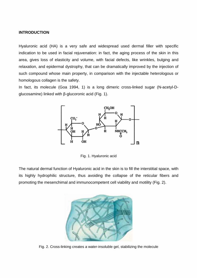
In fact, its molecule (Goa 1994, 1) is a long dimeric cross-linked sugar (N-acetyl-D-
glucosamine) linked with β-glucoronic acid (Fig. 1).
Fig. 1. Hyaluronic acid
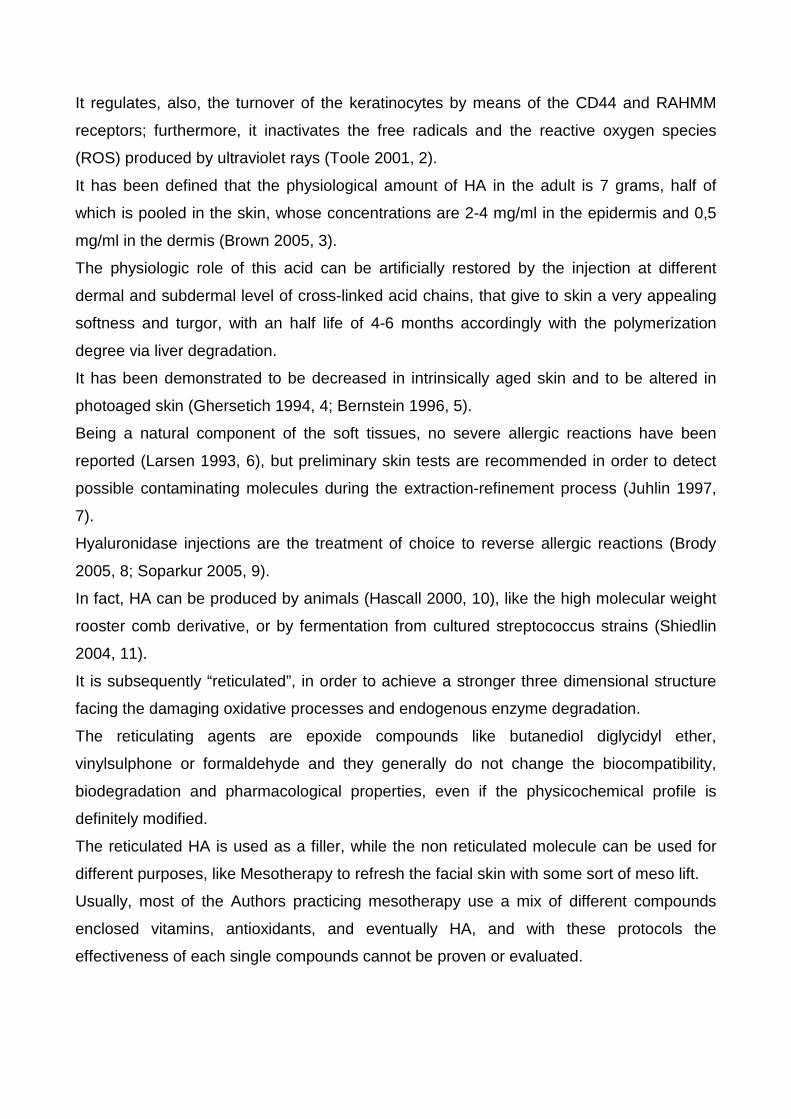
The natural dermal function of Hyaluronic acid in the skin is to fill the interstitial space, with
its highly hydrophilic structure, thus avoiding the collapse of the reticular fibers and
promoting the mesenchimal and immunocompetent cell viability and motility (Fig. 2).
Fig. 2. Cross-linking creates a water-insoluble gel, stabilizing the molecule
It regulates, also, the turnover of the keratinocytes by means of the CD44 and RAHMM
receptors; furthermore, it inactivates the free radicals and the reactive oxygen species
(ROS) produced by ultraviolet rays (Toole 2001, 2).
It has been defined that the physiological amount of HA in the adult is 7 grams, half of
which is pooled in the skin, whose concentrations are 2-4 mg/ml in the epidermis and 0,5
mg/ml in the dermis (Brown 2005, 3).
The physiologic role of this acid can be artificially restored by the injection at different
dermal and subdermal level of cross-linked acid chains, that give to skin a very appealing
softness and turgor, with an half life of 4-6 months accordingly with the polymerization
degree via liver degradation.
It has been demonstrated to be decreased in intrinsically aged skin and to be altered in
photoaged skin (Ghersetich 1994, 4; Bernstein 1996, 5).
Being a natural component of the soft tissues, no severe allergic reactions have been
reported (Larsen 1993, 6), but preliminary skin tests are recommended in order to detect
possible contaminating molecules during the extraction-refinement process (Juhlin 1997,
7).
Hyaluronidase injections are the treatment of choice to reverse allergic reactions (Brody
2005, 8; Soparkur 2005, 9).
In fact, HA can be produced by animals (Hascall 2000, 10), like the high molecular weight
rooster comb derivative, or by fermentation from cultured streptococcus strains (Shiedlin
2004, 11).
It is subsequently “reticulated”, in order to achieve a stronger three dimensional structure
facing the damaging oxidative processes and endogenous enzyme degradation.
The reticulating agents are epoxide compounds like butanediol diglycidyl ether,
vinylsulphone or formaldehyde and they generally do not change the biocompatibility,
biodegradation and pharmacological properties, even if the physicochemical profile is
definitely modified.
The reticulated HA is used as a filler, while the non reticulated molecule can be used for
different purposes, like Mesotherapy to refresh the facial skin with some sort of meso lift.
Usually, most of the Authors practicing mesotherapy use a mix of different compounds
enclosed vitamins, antioxidants, and eventually HA, and with these protocols the
effectiveness of each single compounds cannot be proven or evaluated.
MATERIALS
We choose to use Viscoderm®, a native Hyaluronic acid sodium salt Medical Device, with
the concentrations of 0.8%-1.6%-2.0% in pre-filled syringes, for intradermal use.
The compound is a native HA produced by bio-fermentation from Streptococcus Equi
(Chong 2005, 12), with an average M.W. of 1.2 MD. The finished product in pre-filled
syringes is almost iso-osmotic (Osmolarity 0.25-0.4 Osm/kg) and with a dynamic viscosity
proportional to the concentration, of 300-950-1500 mPa*s for the concentrations 0.8%-
1.6%-2.0% respectively.
Pre-clinical safety studies have been carried out as part of the Technical File of the
product, before CE marking and Two Phase IV clinical studies have been completed to
date.
In order to set up a protocol useful in the cosmetic practice, suitable to guarantee the best
patient compliance by reducing pain and local side effects, we also used:
• PRYLOCAINE 25% plus LIDOCAINE 25% (Okada 1998, 13), OINTMENT (EMLA-
Astra);
• 1% carbocaine saline solution (Astra);
• Dexhametazone 0,1 mg.
METHODS
In order to evaluate the biological activity of HA we chose to inject the product in a simple,
open trial, from February to April 2007, to a group of 40 patients with face skin aging
wishing to achieve some improvement of their cosmetic appearance.
The patients were women between 19 and 85 y.o. (average 52) who had been submitted
to several cosmetic treatments such as peeling, fillers, creams and ointments, and were
still complaining of severe facial relaxation and aging.
Preliminarily, the main parameters of skin physiology were investigated with
DERMOBIOTEST, SOFT 5.5 (Callegari S.p.A.–Parma, Italy). This instrument (Fig. 3)
encloses different measurement principles to give an exhaustive evaluation of the skin
properties in health and diseases (Table 1 and 2):
• Capacitance (water contents of the skin);
• Photometric reading absorbent strip (sebum levels);
• Measurement of the deformation of the skin by suction application (elasticity levels);
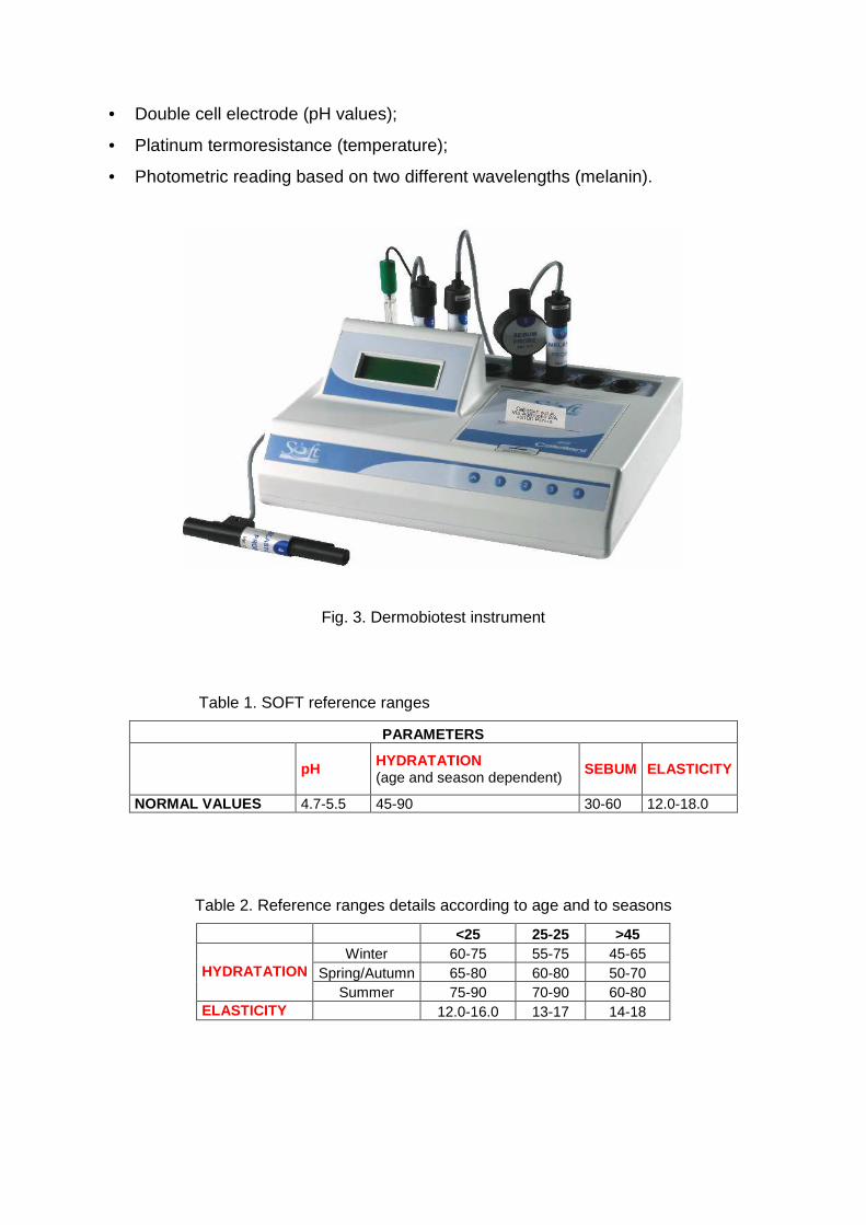
• Double cell electrode (pH values);
• Platinum termoresistance (temperature);
• Photometric reading based on two different wavelengths (melanin).
Fig. 3. Dermobiotest instrument
Table 1. SOFT reference ranges
PARAMETERS
pH HYDRATATION (age and season dependent)
SEBUM ELASTICITY
NORMAL VALUES 4.7-5.5 45-90 30-60 12.0-18.0
Table 2. Reference ranges details according to age and to seasons
<25 25-25 >45 Winter 60-75 55-75 45-65
Spring/Autumn 65-80 60-80 50-70 HYDRATATION Summer 75-90 70-90 60-80
ELASTICITY 12.0-16.0 13-17 14-18
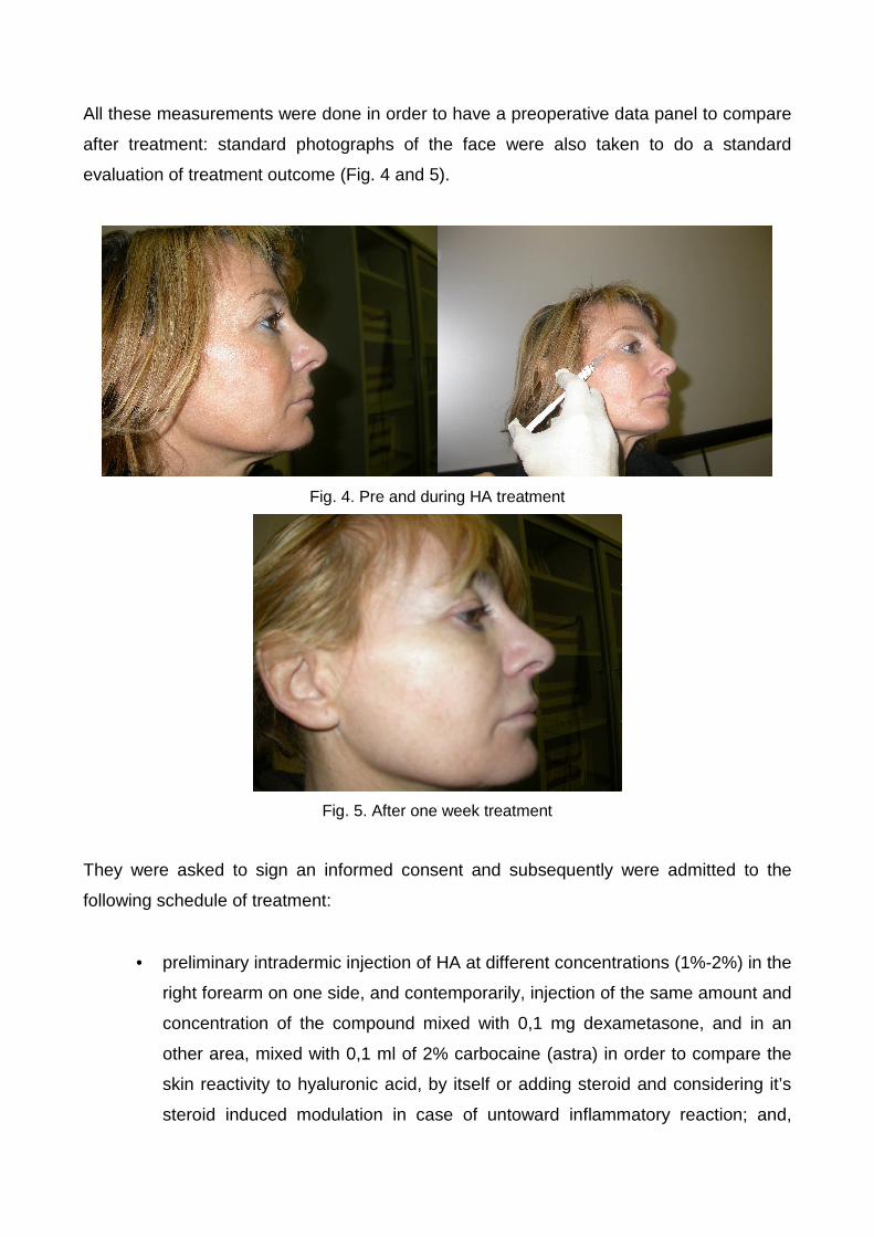
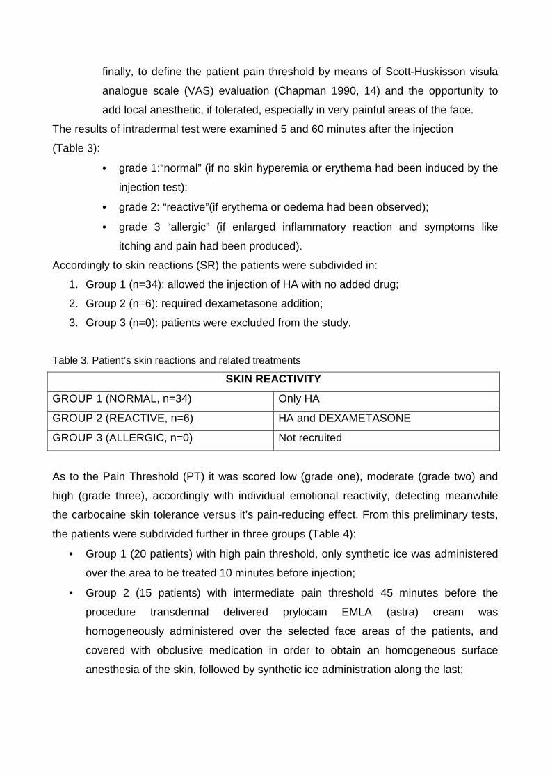
All these measurements were done in order to have a preoperative data panel to compare
after treatment: standard photographs of the face were also taken to do a standard
evaluation of treatment outcome (Fig. 4 and 5).
Fig. 4. Pre and during HA treatment
Fig. 5. After one week treatment
They were asked to sign an informed consent and subsequently were admitted to the
following schedule of treatment:
• preliminary intradermic injection of HA at different concentrations (1%-2%) in the
right forearm on one side, and contemporarily, injection of the same amount and
concentration of the compound mixed with 0,1 mg dexametasone, and in an
other area, mixed with 0,1 ml of 2% carbocaine (astra) in order to compare the
skin reactivity to hyaluronic acid, by itself or adding steroid and considering it’s
steroid induced modulation in case of untoward inflammatory reaction; and,
finally, to define the patient pain threshold by means of Scott-Huskisson visula
analogue scale (VAS) evaluation (Chapman 1990, 14) and the opportunity to
add local anesthetic, if tolerated, especially in very painful areas of the face.
The results of intradermal test were examined 5 and 60 minutes after the injection
(Table 3):
• grade 1:“normal” (if no skin hyperemia or erythema had been induced by the
injection test);
• grade 2: “reactive”(if erythema or oedema had been observed);
• grade 3 “allergic” (if enlarged inflammatory reaction and symptoms like
itching and pain had been produced).
Accordingly to skin reactions (SR) the patients were subdivided in:
1. Group 1 (n=34): allowed the injection of HA with no added drug;
2. Group 2 (n=6): required dexametasone addition;
3. Group 3 (n=0): patients were excluded from the study.
Table 3. Patient’s skin reactions and related treatments
SKIN REACTIVITY
GROUP 1 (NORMAL, n=34) Only HA
GROUP 2 (REACTIVE, n=6) HA and DEXAMETASONE
GROUP 3 (ALLERGIC, n=0) Not recruited
As to the Pain Threshold (PT) it was scored low (grade one), moderate (grade two) and
high (grade three), accordingly with individual emotional reactivity, detecting meanwhile
the carbocaine skin tolerance versus it’s pain-reducing effect. From this preliminary tests,
the patients were subdivided further in three groups (Table 4):
• Group 1 (20 patients) with high pain threshold, only synthetic ice was administered
over the area to be treated 10 minutes before injection;
• Group 2 (15 patients) with intermediate pain threshold 45 minutes before the
procedure transdermal delivered prylocain EMLA (astra) cream was
homogeneously administered over the selected face areas of the patients, and
covered with obclusive medication in order to obtain an homogeneous surface
anesthesia of the skin, followed by synthetic ice administration along the last;
• Group 3: (5 patients) with very low pain threshold, same protocol as above (EMLA+
ice) followed by carbocaine (2%) 0,1 ml pre treatment in the area to be infiltrated
with HA.
Table 4. Patient’s pain threshold and associated treatments
PAIN THRESHOLD
GROUP 1 (grade 1, n=20)) ICE 10 min before injection
GROUP 2 (grade 2, n=15) EMLA 45 min before treatment and ICE
GROUP 3 (grade 3, n=5) EMLA+ICE+0.1 ml CARBOCAINE
A weekly intradermal injection of Hyaluronic Acid 2ml (%) with single mesotherapy needle
(281 gauge, 3 mm length) in the following face areas:
Forehead (front), eyebrow , cheek, chin.
4 overall sessions were performed within a one month period.
Multiple injections 0,1 ml volume (two to ten) were performed on the selected areas and
followed by a light manual massage to homogeneously spread the injected compound; the
schedule enclosed weekly treatment plan along 8 weeks.
Measurements of the skin parameters were obtained, and pre-post treatment data were
plotted on a table 5:
0
10
20
30
40
50
60
70
80
Hydr Elas pH Hydr Elas pH Hydr Elas pH Hydr Elas pH
Front Eyebrow Cheek Chin
Pre-treatment
Post treatment
p<0,001
p<0,001
p<0,01
p<0,001
p<0,01
p<0,001
p<0,01p<0,01
Table 5: Skin parameters and pre/post treatment data
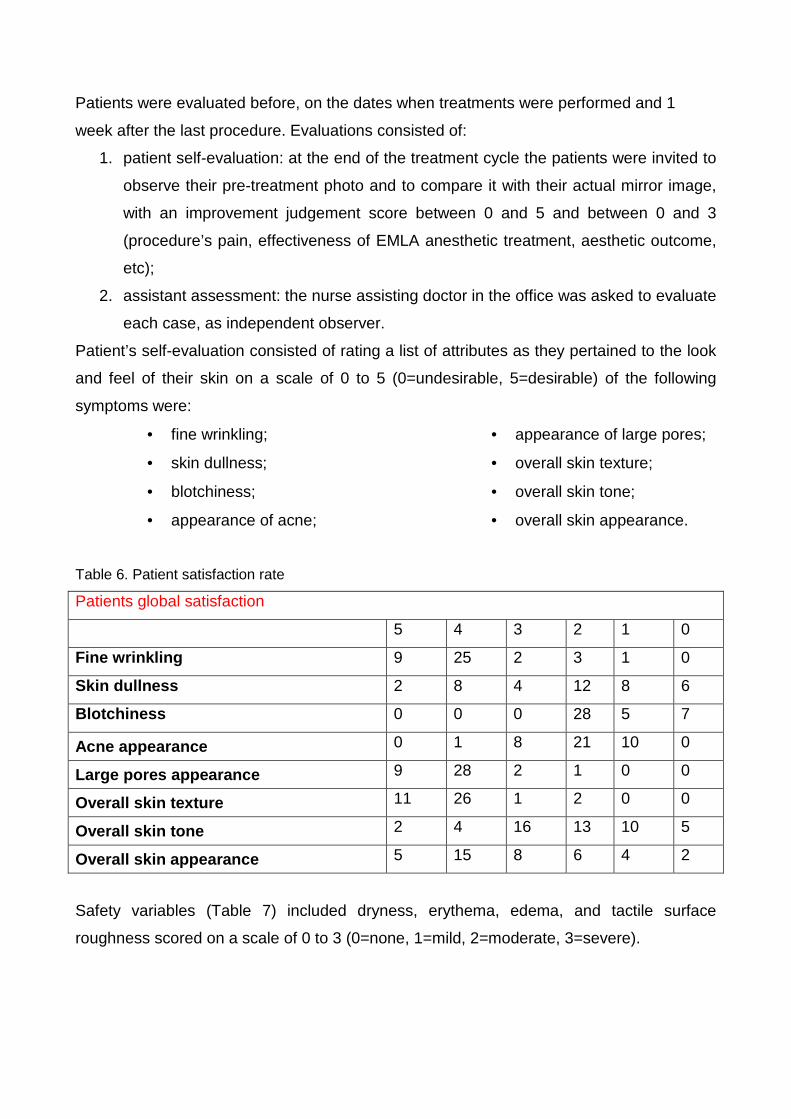
Patients were evaluated before, on the dates when treatments were performed and 1
week after the last procedure. Evaluations consisted of:
1. patient self-evaluation: at the end of the treatment cycle the patients were invited to
observe their pre-treatment photo and to compare it with their actual mirror image,
with an improvement judgement score between 0 and 5 and between 0 and 3
(procedure’s pain, effectiveness of EMLA anesthetic treatment, aesthetic outcome,
etc);
2. assistant assessment: the nurse assisting doctor in the office was asked to evaluate
each case, as independent observer.
Patient’s self-evaluation consisted of rating a list of attributes as they pertained to the look
and feel of their skin on a scale of 0 to 5 (0=undesirable, 5=desirable) of the following
symptoms were:
• fine wrinkling;
• skin dullness;
• blotchiness;
• appearance of acne;
• appearance of large pores;
• overall skin texture;
• overall skin tone;
• overall skin appearance.
Table 6. Patient satisfaction rate
Patients global satisfaction
5 4 3 2 1 0
Fine wrinkling 9 25 2 3 1 0
Skin dullness 2 8 4 12 8 6
Blotchiness 0 0 0 28 5 7
Acne appearance 0 1 8 21 10 0
Large pores appearance 9 28 2 1 0 0
Overall skin texture 11 26 1 2 0 0
Overall skin tone 2 4 16 13 10 5
Overall skin appearance 5 15 8 6 4 2
Safety variables (Table 7) included dryness, erythema, edema, and tactile surface
roughness scored on a scale of 0 to 3 (0=none, 1=mild, 2=moderate, 3=severe).
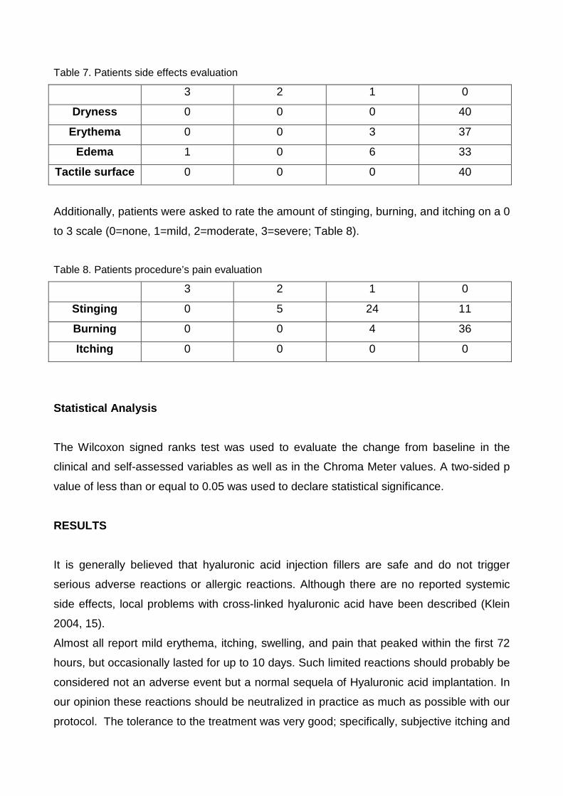
Table 7. Patients side effects evaluation
3 2 1 0
Dryness 0 0 0 40
Erythema 0 0 3 37
Edema 1 0 6 33
Tactile surface 0 0 0 40
Additionally, patients were asked to rate the amount of stinging, burning, and itching on a 0
to 3 scale (0=none, 1=mild, 2=moderate, 3=severe; Table 8).
Table 8. Patients procedure’s pain evaluation
3 2 1 0
Stinging 0 5 24 11
Burning 0 0 4 36
Itching 0 0 0 0
Statistical Analysis
The Wilcoxon signed ranks test was used to evaluate the change from baseline in the
clinical and self-assessed variables as well as in the Chroma Meter values. A two-sided p
value of less than or equal to 0.05 was used to declare statistical significance.
RESULTS
It is generally believed that hyaluronic acid injection fillers are safe and do not trigger
serious adverse reactions or allergic reactions. Although there are no reported systemic
side effects, local problems with cross-linked hyaluronic acid have been described (Klein
2004, 15).
Almost all report mild erythema, itching, swelling, and pain that peaked within the first 72
hours, but occasionally lasted for up to 10 days. Such limited reactions should probably be
considered not an adverse event but a normal sequela of Hyaluronic acid implantation. In
our opinion these reactions should be neutralized in practice as much as possible with our
protocol. The tolerance to the treatment was very good; specifically, subjective itching and
burning didn’t change statistically along the protocols performances (2 % of the cases);
objectively, erythema, dryness, and edema were very well controlled by adding low dose
steroid (1%).
The injection pain was effectively reduced by synthetic ice administration as a very basic
step of the protocol, integrated, in cases of hyperalgesia with EMLA CREAM, and in the
more painful areas by xylocaine pre-injection in order to achieve complete control of the
area to be treated.
No vasoconstriction was required, and only one case of subdermal haematoma was
observed that, however, remitted without pigmentation after 5 days.
Tactile surface roughness improved significantly by week 2.
Efficacy variables had a significant improvement from baseline, including the acne scars
and lesions which improved due to hyaluronic acid.
The parameters that improved definitively were:
• fine wrinkles (p=0.0005),
• dullness (p=0.0002),
• appearance of large pores (p=0.002),
• blotchiness (p=0.03),
• global assessment related to overall texture (p=0.0005),
• overall tone (p=0.001),
• and overall appearance (p=0.0005).
At the end of the study, showed improvement by one or two grades 75% of patients.
Patient Self-Evaluation
Patients noted a significant improvement, by the end of the treatment in:
• facial skin texture (p=0.01),
• tone (p=0.001),
• fine lines (p=0.001),
• age spots (p=0.001),
• smoothness (p=0.012),
• and softness (p=0.045).
Self-assessed pore size, skin hydration, and blotchiness instead were not perceived to
have changed significantly.
As to the patients expectations satisfaction was met as follows :
� Marked improvement : (+5) was acknowledged by 25 women
(+ 4) 3 cases
� Fair improvement (+3) 8 cases
� Slight improvement (+2) 3 cases
� No effect (+0) 1 case
The nurse’s judgement matched adequately the women score, except for the “No effect”
case which was included instead in the “small improvement” group.
The physico-chemical data showed a marked improvement of hydration and elasticity of
the skin areas treated, and no change in sebometry. The pH turned to more acid level.
DISCUSSION
Our experience confirms that the aging face can be restored to youthful contour and
harmony with adequate amounts of injected HA, whose revitalizing properties on dermal
collagen have not yet been extensively investigated, being that the literature frequently
focuses on the study of cross-linked “fillers”, rather than the rejuvenation effects of the
native product.
As a matter of fact, from the biochemical point of view, HA is able to interact with skin
collagen increasing the softness and hydrophilic properties of the dermis and, to a lesser
extent also the elasticity.
The study of Cantor (1998, 16) on the neutrophil elastase in a lung injury model suggests
a protection of the HA on the elastic fibers through a specific binding and an inhibiting
effect on the granulocytes enzyme. The pH of the skin remained unchanged immediately
after the injection, and definitely later in the long term, excluding modification of hydrogen
ions on the epidermal surface, suggesting a deeper balancing action mechanism on
dermal mesenchyma.
Subjective and objective judgements in this investigation have been very concordant, and
the compliance to the treatment, based on a pain-reducing protocol very high; the
anesthetic procedures used in our study can be easily standardized, and it’s very useful in
the clinical practice to recruit large cohorts of patients without fear of pain.
In our study we used HA as a simple skin rejuvenation compound, by means of a non
cross-linked, protein free chemical structure (Lupton 2000, 17; Lowe 2001, 18; Friedman
2002, 19; Andre 2004, 20); notwithstanding the association of small amount of steroids
into the HA containing syringe, completely eliminated adverse reactions due to skin
reactivity in cases where preliminary forearm injection had given signs of inflammation: this
drug-integrated procedure is very important, especially if multiinjection is required, on an
office-outpatient extended face treatment.
No local or general adverse effect has ever been observed by dexhametazone associated
injection and specifically, testing the injection of the low-dose single compound, we
reached the conclusion that it doesn’t adversely effect skin elasticity or hydration in the
short run.
As to the optimal injection technique, a thin (31 G), short (0,3 mm) needle should be used
to reduce injection trauma, homogeneously spreading the HA around the infiltrated area
with a delicate 3 minute finger massage. To prevent the diffusion of the compound out of
severely atrophic skin surfaces the needle track should be at a 30% inclination and not
perpendicular to avoid spreading of the injected volume in the subcutaneous area.
In previous unpublished studies we tried a multineedle mesotherapy approach, which is
also very effective, but that requires truncal anesthesia due to the cumulative increased
stimulation of the pain threshold of the contemporary needle infixion; with the single needle
technique, on the contrary, each injection can selectively be performed in specific skin
spots, following the face lines accordingly with the individual anatomy, and with a naturally,
refined final outcome.
Great care is required to avoid transfixing the dermal vascular network, potentially leading
to haematoma or in case of Hyaluronic Acid embolization into some terminal vessel, to
skin ischemia or, more rarely, to necrosis.
The safety of the product is confirmed by our investigation, and we strongly recommend
it’s use when a non surgical rejuvenation effect by subinvasive, surgery-sparing approach
is required; the injection can be repeated safely every 6-9 months as no allergenicity of the
compound has so far been detected even after 6-8 cycles of repeated procedure that have
been performed on patients in the last 5 years.
The use of a skin parameters measuring device has been proven quite useful to define the
background of each case to be treated, and to monitor the results in the follow-up making
the doctors and patients aware of the improvement achieved and better defining the
schedule of periodical injections.
REFERENCES
1. Goa KL, Benfield P. Hyaluronic acid. A review of its pharmacology and use as a surgical
aid in ophthalmology, and its therapeutic potential in joint disease and wound healing.
Drugs 1994;47(3):536-66.
2. Toole BP. Hyaluronan in morphogenesis. Semin Cell Dev Biol 2001;12(2):79-97.
3. Brown MB, Jones SA. Hyaluronic acid: a unique topical vehicle for the localized
delivery of drugs to the skin. J Eur Acad Dermatol Venereol 2005;19(3):303-18.
4. Ghersetich I, Lotti T, Campanile G, et al. Hyaluronic acid in cutaneous intrinsic
aging. Int J Dermatol 1994;33(2):119-22.
5. Bernstein EF, Underhill CB, Hahn PJ, et al. Chronic sun exposure alters both the
content and distribution of dermal glycosaminoglycans. Br J Dermatol
1996;135(2):255-62.
6. Larsen NE, Pollak CT, Reiner K, et al. Hylan gel biomaterial: dermal and
immunologic compatibility. J Biomed Mater Res 1993;27(9):1129-34.
7. Juhlin L. Hyaluronan in skin. J Intern Med 1997;242(1):61-6.
8. brody HJ. Use of hyaluronidase in the treatment of granulomatous hyaluronic acid
reactions or unwanted hyaluronic acid misplacement. Dermatol Surg 2005;31:893-
7.
9. Soparkar CN, Patrinely JR. Managing inflammatory reaction to restylane. Ophthal
Plast Reconstr Surg 2005;21(2):151-3.
10. Hascall VC. Hyaluronan, a common thread. Glycoconj J 2000;17(7-9):607-16.
11. Shiedlin A, Bigelow R, Christopher W, et al. Evaluation of hyaluronan from different
sources: Streptococcus zooepidemicus, rooster comb, bovine vitreous, and human
umbilical cord. Biomacromolecules 2004;5(6):2122-7.
12. Chong BF, Blank LM, Mclaughlin R, Nielsen Lk. Microbial hyaluronic acid
production. Appl Microbiol Biotechnol 2005;66(4):341-51.
13. Okada S, Hagan JB, Kato M, et al. Lidocaine and its analogues inhibit IL-5-
mediated survival and activation of human eosinophils. J Immunol
1998;160(8):4010-7.
14. Chapman CR, Syrjala KR. Measurement of pain. In: Bonica JJ editor. The
Managment of Pain. 2nd ed. Malvern, PA: Lea & Febiger;1990.p. 580-94.
15. Klein AW. Granulomatous foreign body reaction against hyaluronic acid. Dermatol
Surg 2004;30(7):1070.
16. Cantor JO, Cerreta JM, Armand G, et al. Aerosolized hyaluronic acid decreases
alveolar injury induced by human neutrophil elastase. Proc Soc Exp Biol Med
1998;217(4):471-5.
17. Lupton JR, Alster TS. Cutaneous hypersensitivity reaction to injectable hyaluronic
acid gel. Dermatol Surg 2000;26(2):135-7.
18. Lowe NJ, Maxwell CA, Lowe P, et al. Hyaluronic acid skin fillers: adverse reactions
and skin testing. J Am Acad Dermatol 2001;45(6):930-3.
19. Friedman PM, Mafong EA, Kauvar AN, et al. Safety data of injectable nonanimal
stabilized hyaluronic acid gel for soft tissue augmentation. Dermatol Surg
2002;28(6):491-4.
20. Andre P. Evaluation of the safety of a non-animal stabilized hyaluronic acid
(NASHA – Q-Medical, Sweden) in European countries: a retrospective study from
1997 to 2001. J Eur Acad Dermatol Venereol 2004;18(4):422-5.