human viral gastroenteritis - mmbr.asm.org · virology, epidemiology, and pathogenesis have been...
TRANSCRIPT
Vol. 48, No. 2MICROBIOLOGICAL REVIEWS, June 1984, p. 157-1790146-0749/84/020157-23$02.00/0Copyright C 1984, American Society for Microbiology
Human Viral GastroenteritisGEORGE CUKORt* AND NEIL R. BLACKLOW
Division of Infectious Disease, University of Massachusetts Medical School, Worcester, Massachusetts 01605
INTRODUCTION ......................................... 157
ROTAVIRUS ......................................... 158
Mapping of the Rotavirus Genome......................................... 158
Morphogenesis of Rotavirus ......................................... 158Classification of Rotavirus ......................................... 160Cultivation of HRV ................................................ 161Clinical Immunity to HRV......................................... 161
Pathogenesis and Pathophysiology ......................................... 163Epidemiology ......................................... 163
Control, Treatment, and Prevention of HRV Infection......................................... 164
Diagnosis......................................... 165
Pararotaviruses ......................................... 166
NORWALK VIRUS ............................................ 166Biological Characteristics ......................................... 166Diagnosis......................................... 166
Epidemiology ......................................... 167
Pathogenesis ......................................... 167
Immune Response and Clinical Immunity......................................... 167
Prevention and Control ......................................... 168ENTERIC ADENOVIRUSES ......................................... 168CALICIVIRUSES ......................................... 168
ASTROVIRUSES ......................................... 169
CORONAVIRUSES ......................................... 169
SMALL ROUND VIRUS PARTICLES......................................... 170
ACKNOWLEDGMENTS......................................... 170
LITERATURE CITED......................................... 170
INTRODUCTIONAcute viral gastroenteritis is a very common illness which
occurs in both epidemic and endemic forms. It affects all agegroups worldwide and also includes some of the commonlyencountered travelers diarrhea. This syndrome is recognizedas being second in frequency only to the common coldamong illnesses affecting U.S. families under epidemiologi-cal surveillance. The clinical presentation of the illness isvariable, but in general it is selflimited, has an explosiveonset, and is manifested by varying combinations of diar-rhea, nausea, vomiting, low-grade fever, abdominal cramps,headache, anorexia, myalgia, and malaise. It is not onlyresponsible for a great deal of misery and time lost fromschool and work, but can be severe, indeed fatal, in theinfant, elderly, or debilitated patient. Due to associatedmalabsorption, viral gastroenteritis may trigger or enhancethe morbidity associated with malnutrition in marginallynourished populations.
Identification and characterization of gastroenteritis virus-es and a clear understanding of their epidemiology andimmunology are necessary to be able to prevent theirtransmission and to develop appropriate vaccines. This hasproven to be an especially formidable task since all currentlyrecognized or suspected agents of human viral gastroenteri-tis share the property of being initially non-cultivatable byconventional in vitro techniques or in laboratory animalhosts. It has been long recognized that the commonly
* Corresponding author.t Present address: New England Nuclear/DuPont Infectious Dis-
ease Group, 331 Treble Cove Rd., North Billerica, MA 01862.
isolated cytopathic enteroviruses, including polio-, coxsack-ie-, and echoviruses, do not produce significant amounts ofdiarrheal illness in humans. The viral etiology of acuteinfectious nonbacterial gastroenteritis was only uncovered inthe past 12 years by electron microscopic examination ofhuman clinical specimens. Further progress in the study ofsome electron microscopically observed agents has beenaccomplished by the use of such techniques as immunoas-says, human volunteer studies, and the persistent search forcomplete or partial viral replication by a variety of unusualculture techniques.At this time, only two viruses, rotavirus and Norwalk
virus, are recognized as medically important etiologicalagents of human gastroenteritis. Rotavirus has been seriallypropagated in culture. Its place in the viral classificationscheme and many aspects of its antigenic nature, basicvirology, epidemiology, and pathogenesis have been eluci-dated. Although our understanding of the immunology ofrotaviral illness is incomplete, the need for disease preven-tion is recognized as being clear and urgent enough towarrant vaccine trials. Much information is available on theepidemiology and pathogenesis of Norwalk virus, but itscharacterization is likely to await its cultivation in vitro.Enteric or fastidious adenoviruses have recently emerged asstrong candidates for an important etiological role in humandiarrheal illness. These viruses have now been seriallypropagated and are classified as new serotypes of adenovi-rus. A host of other viruses or virus-like particles have beenobserved by electron microscopic examination of humandiarrheal stool specimens. Establishment of their roles inhuman disease requires further research.
157
on January 9, 2020 by guesthttp://m
mbr.asm
.org/D
ownloaded from
158 CUKOR AND BLACKLOW
A number of reviews have dealt with aspects of viralgastroenteritis (23, 24, 102, 106, 126, 142, 161, 283, 308, 323,326). Our aim in this article is a comprehensive presentationof the topic which will serve to summarize and place intoperspective many recent developments in this field; most ofwhich were reported by September 1983.
ROTAVIRUSHuman rotavirus (HRV) was first detected in 1973 in
Australia by thin-section electron microscopic examinationof duodenal biopsies obtained from children with acutediarrhea (19) and was observed by electron microscopy soonafterwards in diarrheal stool specimens from various parts ofthe world. The virus is 70 nm in size, contains segmenteddouble-stranded (ds) RNA as its genome, and has an innerand outer capsid but no envelope. It is classified as aseparate genus of the Reoviridae family. The name of thevirus is derived from the Latin word "rota," meaning wheel,which it resembles in appearance. Initially the virus was alsocalled orbivirus, duovirus, reovirus-like agent, and infantilegastroenteritis virus. HRVs are morphologically similar toand share certain common antigens with animal rotaviruses.Special techniques are required for the cultivation of HRV,but many animal rotaviruses are readily propagated. Avariety of rotaviruses have been isolated from mammalianand avian hosts in which they cause neonatal diarrhea. HRVis a major cause of infantile gastroenteritis.
Mapping of the Rotavirus Genome
An understanding of the molecular biology of rotaviruseshas become important in the planning of strategies fordisease prevention and control. This is because severalserotypes of rotavirus are now recognized with a possiblelack of cross-protection among serotypes, and there is alsosome indication that variant or recombinant viruses mayoccur in nature. We are currently in the process of under-standing the role that each of the rotavirus genes plays indetermining viral growth, infectivity, and induction of hostimmunity.
Rotaviruses possess a genome consisting of 11 segmentsof dsRNA. To devise gene assignment maps for rotaviruses,biochemical, gene cloning, genetic, and immunological ap-proaches have been taken by groups of investigators work-ing independently in several countries including the UnitedStates, Mexico, Australia, New Zealand, and England. Inone biochemical approach, genomic dsRNA was isolatedfrom purified virions and fractionated by gel electrophoresis.The fractionated RNA was heat denatured and translated invitro, and the translation products were compared withpolypeptides isolated from virions or infected cells (7, 80,207, 276). In an alternative biochemical approach, viral RNAtranscripts were synthesized in vitro by using the endoge-nous viral RNA polymerase. The transcripts were fractionat-ed by gel electrophoresis, and the gene product of eachtranscript was identified by in vitro translation. The genomicRNA segment coding for each transcript was determined byRNA-RNA hybridization (108, 201). In other studies clonedDNA copies of dsRNA genomic segments were used todetermine gene coding assignments and complete nucleotidesequences for several rotavirus genes (31, 32, 78, 147, 207).The genetic approach has taken advantage of the highfrequency of reassortment which occurs during mixed infec-tion of two rotaviral strains. Analysis of the phenotypicproperties of the reassortants obtained and identification ofparental origin of the viral genes have permitted the assign-ment of some properties to specific genes (107, 119, 150,
152). Monospecific antibodies have also been used to probethe functions of specific viral proteins (12, 121, 124).The major findings with regard to rotavirus gene coding
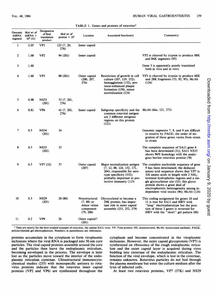
assignments and viral proteins are summarized in Table 1.VP7, the product of gene 9 of simian rotavirus, is an outercapsid glycoprotein which has been identified as the majorrotavirus neutralization antigen. (Genomic segments 7, 8,and 9 are difficult to resolve by gel electrophoresis and theorder of migration of these genes varies from strain tostrain.) A lesser neutralization function is associated withanother outer capsid protein, VP3, the product of gene 4.This protein, which is cleaved by trypsin, is responsible forrestriction of growth in cell culture and protease-enhancedplaque formation and also serves as the viral hemagglutinin.VP6, the product of gene 6, is an inner capsid protein whichhas distinct epitopes for subgroup specificity and a commonrotaviral antigen.
Extensive study of the genetics and function of the pro-teins of the taxonomically related reoviruses has yieldedmuch information with regard to viral virulence and tissuetropism in mice (99, 138). It is hoped that similar studies withrotavirus will result in a better understanding of the patho-genesis and immunity of this pathogen.
Morphogenesis of RotavirusRotaviruses are unusual among nonenveloped viruses in
that they contain at least two glycoproteins, bud through themembranes of the endoplasmic reticulum, and pass througha temporary enveloped phase during their assembly process.The viral envelope is lost before release of the progeny virusby cell lysis.Samples of rotavirus isolated from either feces or tissue
culture fluid have been noted to contain a mixture of twodifferent sized particles which can be separated by CsCldensity gradient centrifugation. The class of larger particlesbands at a density of 1.36 g/ml. They appear in negative-contrast electron microscopic preparations to be 70 to 75nm in diameter and are double shelled with a smooth outercapsid. The class of smaller particles bands at a density of1.38 g/ml and are 65 nm in diameter. They have a roughsurface and are single shelled. Only the smooth, doubleshelled particle has been found to be infectious (38, 86, 244).Various rotavirus particle types have also been observed
in thin-section electron microscopic studies of intestinaltissue from infected animals and of infected cell cultures (1,40, 47, 143, 187, 232). Petrie and co-workers (234) have beenable to clear up some previous confusion in the literature bydemonstrating that the enveloped particles seen within simi-an SAl1 virus-infected cells are not the same as the doubleshelled particles recovered from CsCl gradients. This findingis consistent with the observation that rotavirus infectivity isresistant to lipid solvents (97). Petrie et al. (234) describedfour rotavirus particle types: first, a subviral particle whichis the altered (uncoated) input virion seen within the lyso-somes, and second, an enveloped particle which is a tran-sient form seen in the endoplasmic reticulum. There are, inaddition, the two different sized particle types of releasedvirus, the double-shelled particle which is the mature virionand the single-shelled particle which may result from thebreakdown of double-shelled particles.The scheme for SAl1 virus maturation proposed by these
authors is consistent with morphological studies of HRV(143). It states that infectious virus enters the host cell byendocytosis and is sequestered into lysosomes. Uncoating ofthe input virions in the lysosome results in the production ofa 50-nm subviral particle. Newly synthesized viral RNA and
MIICROBIOL. REV.
on January 9, 2020 by guesthttp://m
mbr.asm
.org/D
ownloaded from
HUMAN VIRAL GASTROENTERITIS 159
TABLE 1. Genes and proteins of rotavirusa
Genomic Mol wt of DesignationdsRNA dsRNA x of final Mol wt of Lcto soitdfnto()Cmetssegment 106 (31) translation protein x 10 Location Associated function(s) Comment(s)
product1 2.05 VP1 125 (7, 20, Inner capsid
276)
2 1.68 VP2 94 (201) Inner capsid
3 1.60
VP2 is cleaved by trypsin to produce 88Kand 84K segments (95)
Gene 3 is apparently poorly translatedboth in vivo and in vitro
4 1.60 VP3 88 (201) Outer capsid(200, 207,276)
Restriction of growth in cellculture (107, 119, 152);hemagglutinin (152), pro-tease-enhanced plaqueformation (150), minorneutralization (124)
VP3 is cleaved by trypsin to produce 60Kand 28K fragments (53, 92, 95); McAb(124)
5 0.98 NS53 53 (7, 201,(201) 276)
6 0.81 VP6
7 0.5 NS34(201)
8 0.5 NS35(201)
41 (7, 201,276)
34
Inner capsid Subgroup specificity and thecommon rotaviral antigenare 2 different antigenicregions on this protein(121)
McAb (66a, 121, 272)
Genomic segments 7, 8, and 9 are difficultto resolve by PAGE; the order of mi-gration of these genes varies from strainto strain
35
9 0.5 VP7 (32) 37
10 0.3 NS29(201)
11 0.2 VP9
Outer capsid Major neutralization antigen(207) (7, 12, 88, 124, 152, 171,
204); responsible for sero-type specificity (332);probably important in pro-tective immunity (115)
20 (80)
26
Nonstructural(7, 88) orminor virioncomponent(79, 206)
Outer capsid?(201)
Glycosylated to become a29K protein; has impor-tant role in outer capsidassembly (233, 252, 279)
The complete sequence of SAl1 gene 8has been determined (31); SAl1 NS35shows 96% homology witfi the analo-gous bovine rotavirus protein (78)
The complete nucleotide sequence of gene9 has been determined; the deducedamino acid sequence shows that VP7 is326 amino acids in length with 2 NH2-terminal hydrophobic regions and a sin-gle glycosylation site (32); this glyco-protein shows a great deal ofelectrophoretic heterogeneity among in-dependent viral stocks (96); McAb (124)
This coding assignment for genes 10 and11 is true for SAl1 and HRV with"long" electropherotype but the posi-tion of these 2 genes is reversed forHRV with the "short" gel pattern (80)
a Data are mostly for the best-studied example of rotavirus, the simian SAll virus. VP, Viral protein; NS, nonstructural; McAb, monoclonal antibody; PAGE,polyacrylamide gel electrophoresis. Numbers in parentheses are references.
proteins accumulate in the cytoplasm to form viroplasmicinclusions where the viral RNA is packaged into 50-nm coreparticles. The viral capsid proteins assemble around the coreand the particles then leave the endoplasmic reticulum,becoming enveloped in the process. The envelope is laterlost as the particles move toward the interior of the endo-plasmic reticulum cisternae. Ultrastructural immunocyto-chemical studies (235) with monospecific antisera to rota-virus proteins indicate that the rotavirus inner capsidproteins (VP2 and VP6) are synthesized throughout the
cytoplasm and become concentrated in the viroplasmicinclusions. However, the outer capsid glycoprotein (VP7) issynthesized on ribosomes of the rough endoplasmic reticu-lum and the outer capsid layer is acquired during virusbudding into cistemae of the endoplasmic reticulum. Thefunction of the viral envelope, which is lost in the cisternae,remains unknown. Rotavirus particles do not bud throughthe plasma membrane but enter the culture medium after thelysis of infected cells.At least two rotavirus proteins, VP7 (37K) and NS29
VOL. 48, 1984
on January 9, 2020 by guesthttp://m
mbr.asm
.org/D
ownloaded from
160 CUKOR AND BLACKLOW
(29K) (see Table 1), are glycosylated. Data from studies withtunicamycin (233, 252), a specific inhibitor of N-linkedglycosylation, show that in the presence of the antibiotic,production of infectious progeny virus is reduced by as muchas 99.9%. Observation (233) of the morphogenesis of SAl1 intunicamycin-treated cells revealed that whereas formation ofviroplasmic inclusions and budding of particles into theendoplasmic reticulum occurred normally, 80 to 100% of theparticles were enveloped, whereas only about 10% of theparticles are enveloped in the absence of tunicamycin. Useof a mutant clone of SAl1, clone 28 (96), which is incapableof glycosylating VP7 but glycosylates NS29 in the usual way,has shown that the accumulation of enveloped virus intunicamycin-treated cells resulted from failure to glycosylateNS29 and was independent of VP7 glycosylation (233). Thisobservation, taken together with the presence of VP7 andNS29 in the portion of the endoplasmic reticulum throughwhich particles bud (235, 279), suggests that the viral enve-lope plays an important role in assembly of the outer capsid.A proposed function of the 29K protein is that of a scaffold-ing protein, structurally important during outer capsid as-sembly but later lost along with the lipid bilayer (233).
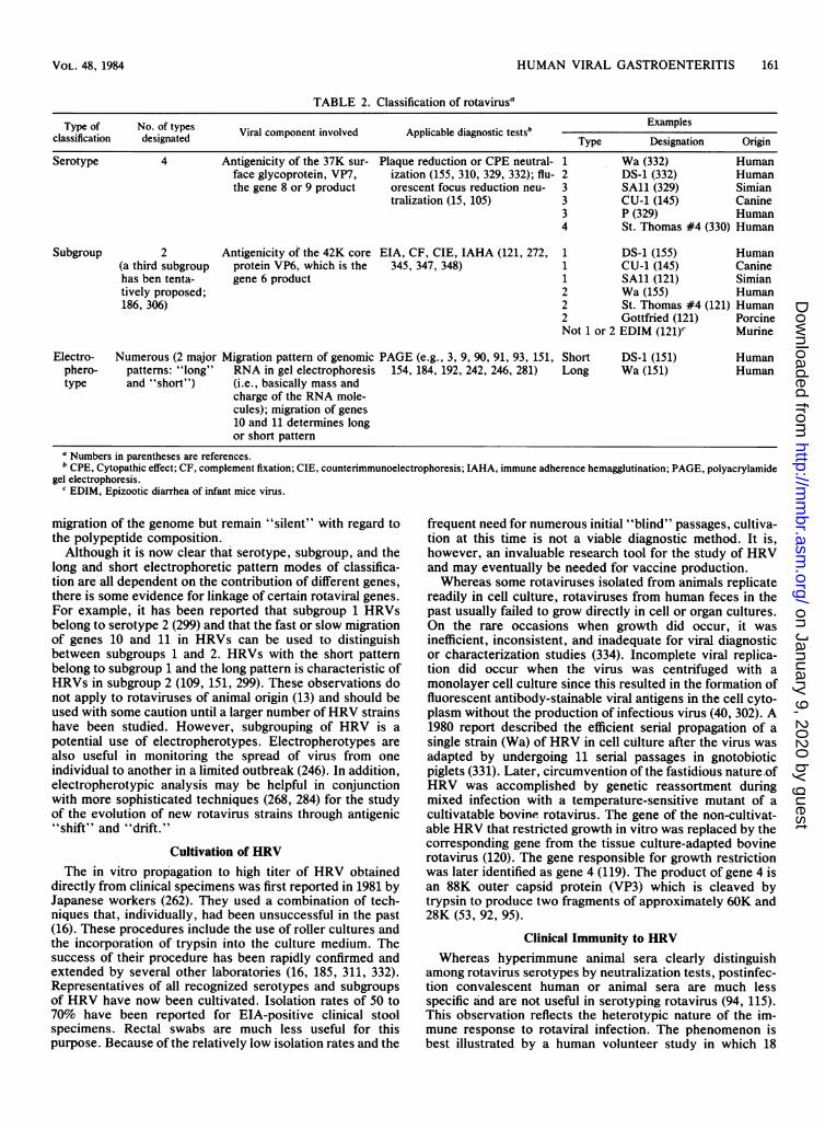
Classification of Rotavirus
Until recently confusion has abounded in the literaturewith regard to the serotypic classification of rotavirus.Before HRV could be cultivated, it was studied primarily byimmunological techniques such as solid-phase immunoas-says (enzyme-linked immunoassay [EIA] and (radio-immunoassay [RIA]), complement fixation, counterimmun-oelectrophoresis, and immune adherence hemagglutination.In addition, a fluorescent focus assay was also used whichtook advantage of a technique involving the partial replica-tion of HRV in cell culture. In 1978 Belgian investigatorsdescribed two distinct "serotypes" of HRV which weredistinguished first by immune electron microscopy (IEM)and complement fixation (348) and later by EIA (345, 347).At the same time investigators in England defined serotypesof rotavirus by serum neutralization of fluorescent focusformation in cell culture (15, 105). The relationship of therotavirus types defined by these different tests remainedunclear (15, 349) until 1981, when the technique for rescue ofnon-cultivatable HRV by gene reassortment during mixedinfection with temperature-sensitive mutants of cultivatablebovine rotavirus was developed by investigators in Bethes-da, Md. (120). Kapikian and co-workers (155), using reassor-tant HRV in a plaque reduction assay, determined that thecell culture infectivity neutralization antigen and the bindingassay (EIA, immune adherence hemagglutination) antigenwere completely distinct. These authors suggested that theterm serotype be reserved to identify the antigen that reactswith neutralizing antibodies, as is customary for other virus-es, and that the term subgroup be used to designate the typespecificity previously established by complement fixation,IEM, EIA, and immune adherence hemagglutination. Laterwork (152) has demonstrated that subgroup specificity isdetermined by the major inner capsid protein, the product ofgene 6, whereas serotype specificity is governed by an outercapsid glycoprotein coded for by gene 8 or 9 (see Table 1 andabove, "Mapping of the Rotavirus Genome"). The serotype-specific antigen may be important in protective immunity(115).Both reassortant and directly cultivated HRVs have been
used in the plaque reduction or cytopathic effect neutraliza-tion assay to define at least four rotavirus serotypes (128,
261, 310, 329, 330, 332). By the most conservative definitionused (329), serotypes are considered to be distinct based on a20-fold or greater difference between homologous and heter-ologous antibody titers. According to this scheme the classi-fication of Wa virus, for example, would be serotype 1,subgroup 2, whereas St. Thomas no. 4 virus would beserotype 4, subgroup 2 (Table 2). Although serotyping iscurrently accomplished only in cell culture, it is likely thatmonospecific reagents (124, 280, 299) with serotype specific-ity will be useful in a more convenient EIA type of assay(299). The relationship between serotypes established byplaque neutralization and those established by fluorescentfocus neturalization has been reconciled (332).The original two subgroups defined by complement fixa-
tion and EIA (345, 347, 348) have been retained, andmonoclonal subgroup-specific antibodies have now beendescribed (121, 272). The existence of a third subgroup hasrecently been proposed (186, 306).Human and animal rotaviruses have been reported to be in
the same realm of the classification scheme for both sub-group and serotype. Almost all mammalian animal rotavir-uses studied thus far have been typed as subgroup 1 (121,155). The exceptions are a porcine and an equine strainwhich belong to subgroup 2 and murine rotavirus (epizooticdiarrhea of infant mice virus), which cannot be classified intoeither subgroup (121). Two simian rotaviruses (strains SAlland MMV18006) and a canine rotavirus (CU-1), which are alldesignated as subgroup 1, have been classified as belongingto serotype 3 (145, 329). In addition, an independent serotyp-ing scheme has been proposed for bovine rotaviruses (219).
Parallel to the development of the subgroup and serotypeclassification for HRV, there has been an effort to classifythese viruses by analysis of the mobility of their virion RNAsegments during polyacrylamide gel electrophoresis (91,154). This technique offered the advantage of circumventingthe need for either cultivation of the virus (since dsRNA wasextracted directly from clinical specimens) or the develop-ment of discriminating standardized typing serum reagents.Electrophoresis also permitted a direct evaluation of allrotavirus genes rather than just a few selected antigens.However, after several years of experience with electropho-retic analysis of the rotavirus genome (9, 31, 90, 184, 192,242, 246, 284), it is appropriate to ask whether it is possibleto classify HRVs by electropherotypes. A recent editorial onthis subject (46) concludes that it is unlikely that electropho-retyping will be useful as a form of general classification, butmay serve a purpose in some limited situations. A majorproblem in electropherotype classification is the high vari-ability of electropherotypes. In one report (281) 32 electro-pherotypes were detected from 149 cases of rotaviral illness.Whereas electrophoretic mobility of genomic RNA reflectsmany differences among rotatvirus strains, they may notalways reflect the changes we are most interested in from anepidemiological point of view. For example, HRVs withidentical electropherotypes but different RNA homology byhybridization techniques have been reported (109). In anoth-er report, two HRVs of different serotypes had identicalelectropherotypes (13). It is clear that comigration of equiva-lent segments of different viruses does not guarantee theiridentity, nor does the lack of comigration always indicatesignificant variation. We may speculate that some minornucleotide changes which do not alter RNA migration mayresult in either a frame shift or an alteration of the tertiarystructure of the protein product, resulting in profound anti-genic differences. Conversely, rather large changes in cer-tain areas of the RNA molecule may result in an altered
MICROBIOL. REV.
on January 9, 2020 by guesthttp://m
mbr.asm
.org/D
ownloaded from
HUMAN VIRAL GASTROENTERITIS 161
TABLE 2. Classification of rotavirusa
Type of No. of types Viral component involved Applicable diagnostic testsb Examplesclassification designated Via opnn novd Apcbedansi eType Designation OriginSerotype 4 Antigenicity of the 37K sur- Plaque reduction or CPE neutral- 1 Wa (332) Human
face glycoprotein, VP7, ization (155, 310, 329, 332); flu- 2 DS-1 (332) Humanthe gene 8 or 9 product orescent focus reduction neu- 3 SAl1 (329) Simian
tralization (15, 105) 3 CU-1 (145) Canine3 P (329) Human4 St. Thomas #4 (330) Human
Subgroup 2 Antigenicity of the 42K core EIA, CF, CIE, IAHA (121, 272, 1 DS-1 (155) Human(a third subgroup protein VP6, which is the 345, 347, 348) 1 CU-1 (145) Caninehas ben tenta- gene 6 product 1 SAl1 (121) Simiantively proposed; 2 Wa (155) Human186, 306) 2 St. Thomas #4 (121) Human
2 Gottfried (121) PorcineNot 1 or 2 EDIM (121)c Murine
Electro- Numerous (2 major Migration pattern of genomic PAGE (e.g., 3, 9, 90, 91, 93, 151, Short DS-1 (151) Humanphero- patterns: "long" RNA in gel electrophoresis 154, 184, 192, 242, 246, 281) Long Wa (151) Humantype and "short") (i.e., basically mass and
charge of the RNA mole-cules); migration of genes10 and 11 determines longor short pattern
a Numbers in parentheses are references.b CPE, Cytopathic effect; CF, complement fixation; CIE, counterimmunoelectrophoresis; IAHA, immune adherence hemagglutination; PAGE, polyactylamide
gel electrophoresis.c EDIM, Epizootic diarrhea of infant mice virus.
migration of the genome but remain "silent" with regard tothe polypeptide composition.Although it is now clear thai serotype, subgroup, and the
long and short electrophoretic pattern modes of classifica-tion are all dependent on the contribution of different genes,there is some evidence for linkage of certain rotaviral genes.For example, it has been reported that subgroup 1 HRVsbelong to serotype 2 (299) and that the fast or slow migrationof genes 10 and 11 in HRVs can be used to distinguishbetween subgroups 1 and 2. HRVs with the short patternbelong to subgroup 1 and the long pattern is characteristic ofHRVs in subgroup 2 (109, 151, 299). These observations donot apply to rotaviruses of animal origin (13) and should beused with some caution until a larger number ofHRV strainshave been studied. However, subgrouping of HRV is apotential use of electropherotypes. Electropherotypes arealso useful in monitoring the spread of virus from oneindividual to another in a limited outbreak (246). In addition,electropherotypic analysis may be helpful in conjunctionwith more sophisticated techniques (268, 284) for the studyof the evolution of new rotavirus strains through antigenic"shift" and "drift."
Cultivation of HRVThe in vitro propagation to high titer of HRV obtained
directly from clinical specimens was first reported in 1981 byJapanese workers (262). They used a combination of tech-niques that, individually, had been unsuccessful in the past(16). These procedures include the use of roller cultures andthe incorporation of trypsin into the culture medium. Thesuccess of their procedure has been rapidly confirmed andextended by several other laboratories (16, 185, 311, 332).Representatives of all recognized serotypes and subgroupsof HRV have now been cultivated. Isolation rates of 50 to70% have been reported for EIA-positive clinical stoolspecimens. Rectal swabs are much less useful for thispurpose. Because of the relatively low isolation rates and the
frequent need for numerous initial "blind" passages, cultiva-tion at this time is not a viable diagnostic method. It is,however, an invaluable research tool for the study of HRVand may eventually be needed for vaccine production.Whereas some rotaviruses isolated from animals replicate
readily in cell culture, rotaviruses from human feces in thepast usually failed to grow directly in cell or organ cultures.On the rare occasions when growth did occur, it wasinefficient, inconsistent, and inadequate for viral diagnosticor characterization studies (334). Incomplete viral replica-tion did occur when the virus was centrifuged with amonolayer cell culture since this resulted in the formation offluorescent antibody-stainable viral antigens in the cell cyto-plasm without the production of infectious virus (40, 302). A1980 report described the efficient serial propagation of asingle strain (Wa) of HRV in cell culture after the virus wasadapted by undergoing 11 serial passages in gnotobioticpiglets (331). Later, circumvention of the fastidious nature.ofHRV was accomplished by genetic reassortment duringmixed infection with a temperature-sensitive mutant of acultivatable bovine rotavirus. The gene of the non-cultivat-able HRV that restricted growth in vitro was replaced by thecorresponding gene from the tissue culture-adapted bovinerotavirus (120). The gene responsible for growth restrictionwas later identified as gene 4 (119). The product of gene 4 isan 88K outer capsid protein (VP3) which is cleaved bytrypsin to produce two fragments of approximately 60K and28K (53, 92, 95).
Clinical Immunity to HRVWhereas hyperimmune animal sera clearly distinguish
among rotavirus serotypes by neutralization tests, postinfec-tion convalescent human or animal sera are much lessspecific and are not useful in serotyping rotavirus (94, 115).This observation reflects the heterotypic nature of the im-mune response to rotaviral infection. The phenomenon isbest illustrated by a human volunteer study in which 18
VOL. 48, 1984
on January 9, 2020 by guesthttp://m
mbr.asm
.org/D
ownloaded from
162 CUKOR AND BLACKLOW
adults were orally administered serotype 1 subgroup 2 HRV(162) and their immune responses evaluated by a number ofserological tests. Four individuals experienced diarrhealillness and an additional non-ill volunteer shed rotavirus.Twelve of the 18 subjects (including the 5 who shed rota-virus) developed a fourfold or greater rise in serum antibody(seroconversion) to rotavirus by one or all of the followingassays: complement fixation, neutralization, immune adher-ence hemagglutination, and EIA. Eight volunteers (includingthe four ill subjects) seroconverted by neutralization assay tothe homologous serotype 1 rotavirus. Five of these individ-uals plus one other subject seroconverted by neutralizationassay to the heterotypic serotype 2 rotavirus. Nine volun-teers (including the four ill subjects) also showed a fourfoldor greater rise in neutralizing serum antibody to the serotypi-cally distinct bovine rotavirus, Nebraska calf diarrhea virus.Seven subjects, including the five who shed virus, serocon-verted to both subgroup 1 and 2 rotavirus by subgroup-specific EIA or immune adherence hemagglutination orboth. Based on the presence of prechallenge rotavirus serumantibody, all of these volunteers showed previous experi-ence with rotavirus. However, similar heterotypic andheterosubgroup serological responses have also been ob-served in infants and young children apparently undergoingprimary infection with rotavirus (162, 333).
In the volunteer study, the presence of preinoculationserum immunofluorescent antibody to the challenge strain orhigh levels of neutralizing antibody to either serotype 1 or 2HRV correlated with resistance to diarrheal illness. Howev-er, the relationship of preexisting local neutralizing activityin the intestinal fluid of volunteers could not be associatedwith protection. Development of both homotypic and het-erotypic intestinal fluid neutralizing activity was observed ininoculated volunteers. There is an increase after naturallyoccurring infection in rotavirus-specific immunoglobulin A(IgA) in both the serum (269) and the gut (69), but thesespecimens have not been analyzed by type-specific tests.Two volunteers who had developed diarrhea after initial
inoculation were rechallenged with the same inoculum 19months later. Neither subject developed diarrhea, althoughone experienced constitutional and gastrointestinal symp-toms (162). In a prospective longitudinal study of youngAustralian children (17), neonates were monitored for thefirst 14 days of life for rotavirus excretion and were then keptunder serological surveillance for 3 years. It was found thatsymptoms associated with the first postneonatal rotavirus.infection were significantly less frequent and less severe inthe children who had experienced neonatal infection than inthose who had not. Whereas neonatal rotavirus infection byone strain of rotavirus did not confer immunity againstreinfection by other strains, it did provide protection againstthe development of clinically severe disease during reinfec-tion. By contrast, in a study of an isolated tribe of SouthAmerican Indians, when a rotavirus of a serotype to whichnone of the tribe had been exposed was introduced, peopleof all ages developed symptoms (190).
Heterotypic serological responses and cross-protectionhave also been observed in animal studies. Calves that wereexperimentally infected with bovine rotavirus developedheterotypic neutralizing antibody to serotype 1 HRV andwere later protected during challenge with serotype 1 HRV(333, 335). In one study, however, where gnotobiotic pigletswere "vaccinated" by oral administration of culture-adaptedrepresentatives of four rotavirus serotypes, only the homo-serotypic vaccines afforded protection from either symp-toms or virus shedding after challenge with a virulent
porcine strain (serotype 3) of rotavirus (115). Another animalstudy in which the findings apparently differ from the humanvolunteers demonstrated that in lambs local antibody torotavirus in the lumen of the gut rather than serum antibodyis of primary importance in protection from rotavirus illness(278). Children rarely have severe rotavirus gastroenteritismore than once, but sequential infections by different sub-groups or serotypes of HRV have been observed (17, 112,247, 271, 345).There is a general clinical impression that breast-fed
babies have less diarrhea than formula-fed infants. Howev-er, as reviewed by Cushing and Anderson (67), specificstudies to investigate this phenomenon have yielded contra-dictory results. Many such studies are hard to interpretbecause of difficulties encountered by the investigators incontrolling for sanitary and socioeconomic conditions, poordefinition of the amount and duration of breast feeding, andthe use of supplementation, as well as the retrospectivenature of some studies. Both breast- and bottle-fed infantsdevelop rotavirus diarrhea (18); however, in some studiesbreast-fed babies were observed to excrete rotavirus signifi-cantly less frequently and in lower titers than formula-fedinfants (52, 209). In a recent longitudinal study in northernCanada, breast feeding was not observed to provide anyprotection from either rotavirus infection or symptomaticillness (132). Although in vitro studies suggest that severalfactors in breast milk may be important in protecting infantsfrom rotavirus infections, it has been difficult to demonstratethe efficacy of these factors in vivo. Virus-specific secretoryIgA antibody is commonly detected in breast milk by EIA(344) or neutralization (300) and may persist in high titer for 6to 9 months postpartum, with occasional significant titerrises which are likely associated with subclinical rotavirusinfection of the mother during lactation (63). However, thepresence of rotavirus-specific EIA and neutralizing anti-body in ingested breast milk could not be associated withprotection by one group of investigators (303, 304) but wasfound to correlate with protection in another study (209).Serotype specificity of milk antibody has not been studied todate, but lacteal antibody has been shown to clump rotavirusin stool specimens (301). Trypsin-inhibitory activity of milkwas associated with protection in one study (209) but not inanother (304). Evidence from animal studies indicates thatprotection depends on the level of the challenge dose, andinfection occurs when the rotavirus infectious titer exceedsthe colostral neutralizing antibody capacity (56, 98, 188,277); this may be an explanation for the discrepancy amongthe studies involving humans (304).
Besides its role in protection, the humoral immune re-sponse apparently also plays a role in recovery from rota-virus infection. In two immunodeficient patients, one withX-linked agammaglobulinemia and the other with severecombined immunodeficiency, chronic symptomatic rota-virus infection with viral excretion lasting for more than 6weeks has been observed (263). In another study, the titersof IgG rotavirus antibody in convalescent serum specimenswere found to be significantly lower in patients with severeor prolonged rotavirus infection than in specimens fromsubjects with mild or moderate disease (239). In a study ofrotavirus-infected adults (275), both symptomatic andasymptomatic subjects had similar rotavirus-specific EIAantibody rises after infection, but asymptomatic infectionwas associated with development of simian rotavirus neu-tralizing antibody which was not seen in ill individuals.The role of cell-mediated immunity in either protection or
recovery from rotavirus needs to be investigated. It seems to
M ICROBIOL . REV
on January 9, 2020 by guesthttp://m
mbr.asm
.org/D
ownloaded from
HUMAN VIRAL GASTROENTERITIS 163
us that the murine system would be an ideal one with whichto begin investigation of cell-mediated immunity since agreat deal of knowledge about mouse immunity and appro-priate test reagents already exist. The parameters of murinerotavirus infection, pathogenesis, and the pathology in-volved have been well studied, and suitable assay proce-dures have been described (55, 191, 240, 248, 270, 322). Afurther advantage of the murine system is that rotavirusdisease in mice has a high morbidity and low mortality and a
large number of inbred animals can be used. With the recentapparent success in the propagation in cell culture of theepizootic diarrhea of infant mice virus, the murine rotavirus(121), the murine model should become available for cell-mediated immunity studies.
Pathogenesis and PathophysiologyThe average incubation period for rotavirus infection in
children as well as in experimentally inoculated adults is 1 to3 days (68, 162, 213). The onset of illness is characterized bysevere watery diarrhea, vomiting, and low-grade fever.Vomiting is a particularly common feature of rotavirusdiarrhea in children compared with the occurrence of thissymptom in gastroenteritis due to other causes (245). Themean duration of rotavirus illness is 5 to 8 days (174). Theconcentration of rotavirus in feces reaches its maximum titershortly after the onset of illness and diminishes graduallyuntil day 9 or 10 (222, 314). The disease is usually self-limited, but symptomatic relapses can occur (73, 291).Common clinical features of rotaviral illness are isotonic
dehydration and compensated metabolic acidosis. Dehydra-tion occurs in 40 to 83% of cases. The level of dehydration isusually <5% but can be greater in severe cases (73, 140,245). In fatal cases of rotavirus disease many of the childrenhave been 10 to 20% dehydrated (42). Fatal rotavirus infec-tion with prominent dehydration has also been reported ingeriatric patients with severe underlying disease (199). Anincreased concentration of sodium and chloride but lowconcentrations of sugar are evident in stools of rotaviruspatients (291). The stools are usually liquid and withoutblood, fecal leukocytes are uncommon, and mucus is some-times seen (140, 245). Pale fatty stools are significantlyassociated with rotaviral diarrhea, suggesting that the virusinfection can result in impedence of both the digestion of fatand the pigmentation of feces (296). In Japan, rotavirusinfection is often associated with white stools (174). Themajor pathophysiological mechanism for diarrhea in rota-virus patients appears to be decreased absorption of salt andwater related to selective infection of the absorptive intesti-nal villus cells, resulting in net fluid secretion. There is a
secondary contribution of carbohydrate malabsorption, re-
sulting in osmotic diarrhea (due to the presence of undigest-ed carbohydrates in the gut), and metabolic acidosis is due tothe bacterial fermentation of these malabsorbed carbohy-drates (253).Symptomatic illness occurs most commonly in children 6
months to 3 years in age. Neonates may excrete virus but areusually asymptomatic (18, 52). Adults typically tend to haveasymptomatic or mild disease (318), but severe symptoms inadults have also been reported (81). Four of 18 adultvolunteers challenged orally with HRV developed diarrhealillness, whereas 12 developed serological evidence of infec-tion (162).Symptoms of upper respiratory tract infection have been
reported to be associated with rotavirus illness (116, 189,245). However, since the seasonalities of respiratory tract
infections and rotavirus overlap, and since rotavirus itselfhas never been demonstrated to infect the respiratorytract (162, 189), the association may be coincidental. Severalstudies, however, have reported a rapid person-to-personspread of rotavirus infection, which in the absence of acommon source may be more consistent with a respiratorythan a fecal-oral route of spread (113, 118, 133).
Rotavirus was proposed as an etiological agent of intus-susception based on the detection of the virus by electronmicroscopy in the stools of 11 of 30 such patients (177). Nocontrols were run in this study as it was assumed thatrotavirus shedding in nondiarrheal children is rare. A recentstudy indicates that this assumption may not be valid (17). Inany case, two later prospective studies and one retrospectivestudy could not associate rotavirus with intussusception(218, 226). Two case reports present patients with centralnervous system disease (one with Reye's syndrome and theother with encephalitis) with concurrent rotavirus infections,but the etiological role of rotavirus in these cases was notestablished (256). Rotavirus was associated with 5 of 11cases during an outbreak of sudden infant death syndromeoccurring in Baltimore, Md. However, stool specimens from11 cases of sudden infant death syndrome in Boston, Mass.,were rotavirus negative (340). The possibility that rotavirusmay trigger some cases of sudden infant death syndromeneeds to be further investigated. A review of the literature onan etiological role of rotavirus in inflammatory bowel diseaseindicates a lack of an association (25). In one report,neonatal necrotizing enterocolitis was associated with rota-virus based upon the positivity of stools by the commercialRotazyme (Abbott Laboratories, North Chicago, Ill.)EIA test (249). This report must be interpreted with caution,however, since Rotazyme has been reported to producemany false-positive results in stool specimens collected fromneonates (180).
EpidemiologyPrevalence studies indicate rapid acquisition by young
children of binding (EIA-type) serum antibodies to rota-virus worldwide during the ages of 6 to 24 months. The greatmajority of adults and children older than 2 years possesssuch antibodies (20, 28, 83, 159, 229). Thus, serologicalsurveys have served to indicate the ubiquitous nature ofrotavirus. Serum antibody may be acquired after symptom-atic or asymptomatic infection (20, 162), and it is unknownhow long detectable antibody persists after infection orwhether it has a protective role. The highest incidence ofrotavirus illness is found in patients 6 to 24 months of age(34, 159), with the virus being responsible for about half ofthe hospitalized cases of acute diarrheal illness in this agegroup (34, 159).
Rotavirus infection can occur in neonates, but as a rulenewborn babies experience asymptomatic infection or verymild illness (17, 41, 52, 220). However, in one study at aspecial care baby unit in London (71) neonatal rotavirusinfection was associated with severe gastrointestinal symp-toms. Of 76 babies found to excrete rotavirus in this unit, 32developed diarrheal illness, of which 12 were severely ill. Ahigh incidence of neonates experiencing gastroenteritis withfecal excretion of rotavirus was also observed at a Texashospital (305). These two reports of symptomatic neonatalrotavirus infection (71, 305) should, however, be interpretedwith caution since both groups of investigators used thecommercial EIA test, Rotazyme, which, as noted above,has been reported to be unreliable for neonatal stool speci-mens (180).
VOL. 48, 1984
on January 9, 2020 by guesthttp://m
mbr.asm
.org/D
ownloaded from
164 CUKOR AND BLACKLOW
Adults are less likely to become infected with rotavirusthan children and infected adults are also more frequentlyasymptomatic than children. In a large prospective familystudy (318) the attack rate in children was 32% comparedwith 17% for adults. Seventy percent of infected childrenand 40% of infected adults were symptomatic. When 18selected adult volunteers were experimentally inoculatedwith rotavirus, 67% became infected but only 22% were ill(162). In a major rotavirus epidemic in an isolated Pacificisland group involving almost 3,500 cases, the illness attackrate was 40% in infants, 62% in young children, and 12% inadults (113).
Rotavirus infections often occur in adults who are in closecontact with young children (130, 159, 172); however, theyhave also been reported in the absence of any obviouscontact with children among adult travelers, military person-nel, patients on a cardiology ward, and the institutionalizedelderly (57, 82, 84, 85, 136, 144, 168, 199, 251, 315, 316).Whereas symptoms of rotavirus infection in adults tend to bemild (130, 172), severe symnptoms were reported in geriatricpatients (199) and in apparently normal young adults inThailand (81).
In general, however, children are the major source ofrotavirus in the community (87, 130). The virus is usuallyspread from person to person by the fecal-oral route (326).Increased family size is reportedly a significant risk factorfor acquiring rotavirus infection (132). This is also consistentwith the strict seasonality observed for rotavirus infection inthe winter in temperate climates (34, 132, 159, 176). In-creased indoor crowding may facilitate an airborne mode oftransmission which is probably not strictly respiratory butmay be due instead to environmental contamination byfecally shed rotavirus (132). Low indoor relative humidityhas also been proposed as being important in enhancementof rotavirus transmission (33). Indoor climate conditions areof particular significance in temperate climates since veryyoung infants spend most of their time indoors and show aseasonal pattern of infection (34). The seasonal waves ofrotavirus infection in the Washington, D.C., area wereobserved over an 8-year period to be more regular andpredictable than infections with any other pediatric pathogenstudied (34). The seasonal effect on rotavirus infection is notfully explained at this time, and there seem to be exceptionsto this effect. Rotavirus detection was extremely low in thesummer months in Washington, D.C. (34), and northernJapan (176), but the virus was found during the summer inabout 20% of patients with diarrhea in London, England(39), and Melbourne, Australia (68). Reports from tropicalareas are inconsistent. No rotavirus seasonal variation wasobserved in Venezuela (74) and Ecuador (287). HRV detec-tion increased during the dry season in Costa Rica (140),India (198), and Bangladesh (21). In Nigeria (231) lowrelative humidity was associated with increased rotavirusdetection, but relative humidity was found to be irrelevant innorthern Japan (176).Nosocomial spread of rotavirus may be facilitated by
asymptomatic or mildly ill infected staff, many of whom maybe young parents (102); rotavirus can also spread betweenpatients by contact with the hands of uninfected attendants(257). Community-wide epidemics of rotavirus diarrhea havebeen associated with fecally contaminated drinking water(144a, 193, 285) but common source outbreaks of infection,thus far, appear to be the exception, not the rule.
Epidemiological studies of rotavirus subgroups indicatethat infections with the two major subgroups occur simulta-neously in a population and that the proportion of each
subgroup appears to remain constant over a period of years(111, 306). In most geographic locations, illness due tosubgroup 2 has been found to be about four times moreprevalent than that due to subgroup 1 (306, 345). In addition,a small fraction of rotaviruses that have been detected haverecently been classified into a third subgroup (186, 306). In1978, Yolken et al. reported that most children living in theWashington, D.C., area acquire antibody to both subgroupsby the time they are 2 years old. The significance of thisfinding must now be reinterpreted in light of the observationof heterosubgroup serological responses in infants andyoung children (162, 333; see above). The heterosubgroupserum antibody response represents an alternative to theoriginally proposed explanation that infections with bothsubgroups occur with equal frequency but that subgroup 2produces more illness. The clinical symptoms associatedwith both subgroups appear to be similar (345).The epidemiology of rotavirus serotypes has not been
studied in detail, but it is already clear that rotaviruses fromaround the world fall into a limited number of serotypes (atthis time four or five) (299, 332).
Control, Treatment, and Prevention of HRV Infection
Young children are the major source of rotavirus in thecommunity. Avoidance of close contact with them greatlyreduces the risk of acquiring infection (87, 130), especially ina crowded family setting. Children excrete rotavirus in veryhigh titers, with 1011 virus particles per g of feces not beingunusual (102). Rotavirus in feces may survive for days orweeks on environmental surfaces (215) at ordinary tempera-tures and at high or low relative humidity (but not atintermediate levels of humidity). Some disinfectants, inparticular 95% ethanol and chlorinated phenolic compounds,are effective against rotavirus in feces, but skin disinfectantssuch as sodium hypochlorite, chlorhexidine gluconate, andprovidone-iodine are ineffective. Formaldehyde is somewhatuseful in inactivating rotavirus (292). However, as Flewetthas recently pointed out (102), wiping a surface with disin-fectant may reduce infectious virus by a millonfold but is oflittle use if 10,000 million particles are present.
Nosocomial outbreaks of rotavirus infection have beencontrolled successfully only if the affected ward was closedand staff movement restricted for 10 days (57, 102). Lessdrastic control measures have not proven effective (71).
Fluid replacement is an important supportive measure inthe treatment of rotavirus diarrhea. Patients with severedehydration, particularly very young, elderly, or debilitatedindividuals, require either parenteral fluid replacement ororal rehydration with a glucose-electrolytes formula (100,259). Avoidance for 1 or 2 days of lactose-containing milk orformulas in non-breast-fed infants with moderate to severediarrhea has also been recommended (100). Specific treat-ment orally with human gammaglobulin containing rotavirusantibodies was found to modify the course of infection anddisease caused by rotavirus in low-birth-weight babies bydelaying excretion of rotavirus and reducing the durationand quantity of virus excretion (10). Diarrhea was found tobe clinically less severe in babies receiving gammaglobulincompared with a placebo group, but any effect of thetreatment on the risk of infection could not be assessed inthis study. In animal experiments several antiviral drugshave shown some initial promise for the specific treatment ofrotavirus infection (274).
MICROBIOL. REV.
on January 9, 2020 by guesthttp://m
mbr.asm
.org/D
ownloaded from
HUMAN VIRAL GASTROENTERITIS 165
It is clear at the present time that indications for rotavirusvaccination exist in both developed and developing coun-tries. In developed nations rotavirus is responsible for about50% of pediatric hospitalizations for acute diarrheal disease.In developing regions, an even greater need exists since inthese areas the estimated annual death toll attributable torotavirus may be 1 million or more (312). Although youngchildren are the obvious candidates for vaccination, it maybe desirable to vaccinate adults in special circumstances.These may include the institutionalized elderly, travelers,and medical personnel in pediatric units to decrease rota-virus transmission, and perhaps pregnant or nursing mothersto induce immunity in their offspring.
It is, however, likely that before vaccines can be devel-oped we need to know more about the nature of rotavirusprotective immunity in humans; i.e., we must decide whattype of immune responses we need to induce and to whichviral antigens these immune responses should be directed toachieve protection. Initial vaccines will probably be cellculture adapted, attenuated HRV, or attenuated animalrotaviruses (101, 161) or reassortants between human andanimal rotaviruses. An attenuated, cell culture-adapted bo-vine rotavirus "vaccine" was recently administered toadults as well as young children and was found to be safe andto possess some immunogenic properties. This live, oralvaccine administered to 2 year olds did not produce gastroin-testinal or constitutional symptoms nor did it result in virusshedding. One oral dose induced seroconversion to thehomologous virus in 88% of children seronegative by EIAand in 68% of children seronegative by the neutralization test(312).
DiagnosisA rapid diagnosis of rotavirus illness provides the physi-
cian with useful information for formulating a prognosis andmay prevent the unnecessary use of antibiotic therapy andprolonged hospitalization. It also allows for the timelyinstitution of appropriate infection control measures and forthe gathering of epidemiological and clinical informationwhich is vital to vaccine development.Although cultivation of HRV (see above) has now been
achieved, the relatively low isolation rates and the frequentneed for initial "blind" passages preclude cultivation as aviable diagnostic method at the present time. The character-istic morphology of rotavirus is readily recognized in nega-tively stained stool preparations under the electron micro-scope. Indeed, electron microscopic studies resulted in theoriginal identification of rotavirus (19). This technique hasbeen used extensively for rotavirus diagnosis and remainsthe standard by which other assays must be judged. Howev-er, the use of electron microscopy is limited by the require-ment for highly specialized equipment and personnel as wellas by difficulties in handling large numbers of specimens.The diagnostic test of choice for most situations is the
solid-phase immunoassay. It offers the possibility of same-day or next-day diagnosis and can be performed on a largescale with routinely available equipment and personnel. Inthis type of assay the stool specimen to be tested is exposedto a solid phase which has been coated with antirotavirusantibody. After an absorption period and washing, in thedirect test a labeled virus-specific detection antibody is usedto demonstrate the presence of rotavirus bound to the solidphase. In the indirect test, an unlabeled antirotavirus serum(prepared in a species other than the origin of the serum inthe first step) is added followed by a labeled antiglobulin.Details of the test procedures and the preparations of
diagnostic reagents have been reviewed (22, 65, 163, 337,342).
In the original design of the solid-phase immunoassay forrotavirus (153, 338), the reactivity of the test specimens wascompared with the reactivity of a panel of known negativestool samples. A confirmatory test was recommended fordetermining the status of borderline or weakly positivespecimens. The performance of an extra confirmatory test,consisting of exogenous blocking (neutralization) of samplereactivity with high-titered antirotavirus serum, was thoughtto be unnecessary for routine use with each specimen tested.However, it became apparent after several years of experi-ence with the test that the frequency of nonspecific reactionswas a serious problem (35, 180, 341, 343). Nonspecificreactions to goat serum were first recognized in stools frompatients living in certain geographic areas, especially the FarEast, and were postulated to be related to patient diets.Other stools contained nonspecific antiglobulin activity re-lated to copral IgM antibody present in the specimen. Tocircumvent problems of non-specificity, investigators haveused various additions to stool specimens, including normalserum, reducing agent, chelating agent, protease inhibitors,and pH neutralization (137, 141, 146, 212, 341). Somenonspecific reactions could be eliminated by these mea-sures. Experience at Children's Hospital in Washington,D.C., showed (35) that, of 5,626 fecal specimens tested,1,344 gave positive results with the indirect EIA test, whichcould not be confirmed by electron microscopy, blockingEIA, confirmatory EIA, or a combination of these methods.Thus, 73% of 1,834 presumptively positive EIA tests were infact not positive. False-positive reactions were especiallycommon with specimens from hospitalized infants in atertiary care nursery. It was suggested that nonspecificbinding of antibody to intestinal bacteria or their productssuch as staphylococcal protein A may have been the basis ofsome of the false-positive reactions.Based on this extensive study it is apparent that measures
such as the addition of reducing agent are inadequate be-cause they avert only some kinds of nonspecific reactions.Reliance on a panel of known negative stools as controls isalso inadequate. Furthermore, all potential positive reac-tions must be confirmed, not just borderline specimens. Asin the system originally used for Norwalk virus antigendetection (129), the reaction of each stool specimen in themicrotiter plate well coated with rotavirus-positive (post-immunization) serum is compared with the reaction of thesame specimen in a control well coated with rotavirus-negative (preimmunization) serum (163, 342). When a confir-matory test of this type was included in the indirect EIA atChildren's Hospital in Washington, D.C. (35), false-positiveresults were not encountered in 400 consecutive tests.The dangers of not using a confirmatory test have again
been pointed out more recently with regard to the unreliabi-lity of the commercial Rotazyme EIA test for testingneonatal specimens (180). A large number of specimens havebeen tested by the Rotazyme assay in several laboratories(14, 48, 137, 146, 250) with favorable results as to thesensitivity of the test. The Rotazyme test, which is based onthe use of anti-simian rotavirus serum (62), appears to besomewhat less sensitive in detecting HRV than most elec-tron microscopic or indirect EIA assays. Despite the factthat the manufacturer (Abbott Laboratories) has not provid-ed a confirmatory test, problems of non-specificity have onlybeen reported with neonatal specimens. Apparently, thedirect test design and special formulations of sample diluentand wash solutions are sufficient to eliminate many false-
VOL. 48, 1984
on January 9, 2020 by guesthttp://m
mbr.asm
.org/D
ownloaded from
166 CUKOR AND BLACKLOW
positive results. However, with neonatal specimens, only 4(7%) of 61 Rotazyme-positive stools were also positive byconfirmatory tests such as electron microscopy, confirma-tory EIA, and RNA dot hybridization (180). Laboratoriesmay elect to use an exogenous confirmatory blocking testwith any high-titered antirotavirus serum in conjunction withthe Rotazyme test. Recent experience in our laboratoryindicates that replacement of the polyclonal coating antibodywith rotavirus-specific monoclonal antibody improves thesensitivity and specificity of the rotavirus immunoassay(66a). We have, however, not yet tested neonatal specimens.Monoclonal antibodies should be useful for development ofserotype- and subgroup-specific FIA tests (see above).Other techniques that have been used to detect rotavirus
antigen have mostly proven to be less sensitive than theEIA. They include counterimmunoelectrophoresis (307),complement fixation (156), immune adherence hemaggluti-nation (155), and other agglutination techniques (134, 258,273). Direct detection of rotaviral nucleic acid in silver-stained polyacrylamide gels has also been utilized (139). Thedot hybridization assay is a more specific test for rotavirusRNA. This assay is based on the in situ hybridization oflabeled single-stranded RNA probes (obtained by in vitrotranscription of rotavirus particles) to heat-denatured rota-virus RNA derived from test stools and immobilized onnitrocellulose membranes (110). This recently described testappears to be highly sensitive and specific and will be usefulfor epidemiological studies of rotavirus, particularly ifprobes for specific viral genes are used.
PararotavirusesRecently, a new viral agent termed pararotavirus has been
observed in stool specimens from an Australian (243), aFrench (225), and two Bulgarian (75) infants. Pararotavi-ruses are morphologically indistinguishable from conven-tional rotaviruses, but they are antigenically distinct in thatthey lack the group-specific common antigen shared by allpreviously recognized rotaviruses from both mammalian andavian hosts. Pararotaviruses contain 11 segments of dsRNAwhich, however, yield a unique and characteristic electro-phoretic migration pattern. Their lack of the group-specificantigen explains the negative result produced by pararota-virus in currently used rotavirus immunoassays. Based onavailable reports, human shedding of pararotavirus appearsto be a rare event. However, we must keep in mind that, fora specimen to be recognized as containing pararotavirus, itmust have been analyzed by a combination of tests includingelectron microscopy, immunoassay, and RNA gel electro-phoresis. It is important to include the last test becauseantigenically distinct rotaviruses that produce conventionalRNA patterns may exist (75). The medical importance ofpararotaviruses as a cause of diarrheal illness cannot beassessed at this time. Antigenically distinct rotaviruses havealso been observed in the feces of piglets and chickens (30,37, 210, 254). The alternative term, "group B rotaviruses,"has also been proposed to describe these agents (75).
NORWALK VIRUSIn 1972, a 27-nm particle was visualized (160) by IEM
examination of an infectious stool filtrate derived from anoutbreak of gastroenteritis which had occurred 4 yearsearlier in Norwalk, Ohio. This was the discovery of the firstrecognized human gastroenteritis virus of medical impor-tance, the Norwalk agent. Its infectivity and pathogenicity
were demonstrated by serial passage of the virus in adultvolunteers (76, 327). The Norwalk particle is the prototypeand best-studied member of a group of morphologicallysimilar but immunologically mostly unrelated small roundviruses (see "Small Round Virus Particles") which arecommonly seen in human stool by electron microscopy. Themedical importance and etiological roles of these Norwalk-like viruses are currently not established.Norwalk virus has not been propagated in vitro in any cell
or organ culture system. Administration of the virus tolaboratory animals including a number of primate specieshas not resulted in the production of illness (326). Inoculatedchimpanzees were, however, found to shed virus and dem-onstrate a Norwalk-specific immune response (328).When Norwalk virus-containing fecal filtrate is adminis-
tered orally to human volunteers, about 50% of inoculatedindividuals become ill (27). Diarrhea or vomiting or both maybe seen among symptomatic volunteers who receive identi-cal Norwalk inocula. No prolonged illness or long-termsequelae have been observed in Norwalk-inoculated sub-jects (27, 327). Much of what is known today about Norwalkvirus has depended upon reagents gathered as a result ofvolunteer studies.
Biological CharacteristicsThe Norwalk virus is a 27-nm-diameter, nonenveloped,
round particle of unclear substructure. It has a buoyantdensity in cesium chloride of 1.36 to 1.41 g/cm3. Its infectiv-ity remains stable after exposure to ether, acid, and heat.Based on similarities of stability, size, and density with theDNA-containing parvoviruses, Norwalk virus has beentermed "parvovirus-like" (27, 76, 157). However, one report(122) indicates that the Norwalk virus particle, concentratedfrom feces, contains a single 66,000-molecular-weight pro-tein which is characteristic of the RNA-containing calicivi-ruses (see "Caliciviruses"). A definitive classification ofNorwalk virus cannot be made without determination of itsnucleic acid. This, however, is likely to require the labora-tory propagation of the virus, since it is shed in human stoolin relatively low titer (126).
DiagnosisDue to the small size and amorphous surface of Norwalk
virus, IEM is required to directly visualize and identify theagent in stool samples. This procedure involves mixing astool specimen with a drop of serum containing virus-specific antibody, concentrating the mixture, and observingthe clumped virus-antibody complexes with an electronmicroscope. The degree of clumping can also be used to ratethe amount of antibody present in an unknown serum. IEMis cumbersome and impractical to perform on large numbersof specimens and requires specialized equipment and per-sonnel.The development of an RIA for the detection of Norwalk
virus in stool as well as for the measurement of serumantibody to the virus resulted in major advances in the studyof this agent (22, 129). The availability of the RIA permittedthe rapid testing of large numbers of specimens required forepidemiological studies. The RIA test procedure, however,requires the use of carefully defined fecal and serum samplesobtained from experimentally infected volunteers and istherefore limited to a few research laboratories that possesssmall amounts of these valuable reagents of human origin.Norwalk virus has not been purified sufficiently to permitpreparation of hyperimmune animal serum reagents usablefor diagnostic purposes. Development of diagnostic reagents
MICROBIOL. REV.
on January 9, 2020 by guesthttp://m
mbr.asm
.org/D
ownloaded from
HUMAN VIRAL GASTROENTERITIS 167
that would circumvent the current reliance on volunteermaterials is highly desirable. The resulting wider availabilityof Norwalk virus testing would permit a greatly expandedepidemiological surveillance and evaluation of disease out-breaks by public health authorities and would allow for thestudy of Norwalk virus by additional research laboratories.In addition, RIAs similar to that developed for Norwalkvirus are greatly needed for the other Norwalk-like viruses.
Epidemiology
Seroepidemiological studies indicate that infection withNorwalk virus occurs worldwide (20, 26, 64, 123, 129, 158).In the United States approximately two-thirds of adultspossess serum antibody to Norwalk virus. Serum antibody isuncommon during childhood but is acquired rapidly duringlate adolescence (26, 158). These findings correlate with thelack of association of Norwalk virus with severe gastroenter-itis of infancy in the United States (36, 236). However, inless-developed nations serum antibody to Norwalk viruscommonly appears during childhood (64, 123) and mayproduce some mild illness in the younger age group (20).Norwalk virus, in contradistinction to rotavirus, is currentlyregarded as primarily a pathogen of older children andadults.
Forty-two percent of 74 outbreaks of acute nonbacterialgastroenteritis investigated by the Centers for Disease Con-trol from 1976 to 1980 were attributed to Norwalk virus(164). This high percentage suggests that, even though a
large variety of viruses are observed in stool, only a few viralserotypes cause most outbreaks of gastroenteritis. Out-breaks associated with Norwalk virus have occurred in thefollowing settings: in recreational camps, on cruise ships, inregions with contaminated drinking or swimming water, inschools and nursing homes, and in association with ingestionof raw shellfish or other uncooked foods such as salads orcake frosting handled in an unsanitary manner (117, 125, 131,164-167, 221, 295, 321; J. N. Kuritsky, M. T. Osterholm,H. B. Greenberg, J. A. Korlath, J. R. Godes, C. W.Hedberg, J. C. Forfang, and A. Z. Kapikian, Program Abstr.Intersci. Conf. Antimicrob. Agents Chemother. 23rd, abstr.no. 419, p. 159, 1983; K. E. White, M. T. Osterholm, J. A.Korlath, J. A. Mariotti, J. N. Kuritsky, D. Lawrence, andInvestigative Team, Program Abstr. Intersci. Conf. Antimi-crob. Agents. Chemother. 23rd, abstr. no. 361, p. 149, 1983).Some of the outbreaks associated with contaminated food or
municipal drinking water have involved thousands of indi-viduals (165, 221; Kuritsky et al., 23rd ICAAC, abstr. no.
419). A small percentage (3 to 15%) of travelers diarrhea hasalso been associated with Norwalk virus (82, 168, 251).
It is clear from volunteer studies (27, 327), as well as fromepidemiological reports, that Norwalk virus is transmittedby the fecal-oral route. About half of ill volunteers sheddetectable virus in the feces during the first 72 h after theonset of illness (297). Respiratory symptoms have not beenassociated with Norwalk illness, and throat washings from illvolunteers were noninfectious when administered to a fewsubjects (76). The strikingly rapid secondary spread ofNorwalk virus infection may involve airborne transmissionvia aerosolized virus-containing vomitus (127).
PathogenesisA characteristic though not unique histological lesion of
the mucosa of the proximal small intestine has been ob-served during the first 2 weeks after inoculation of volun-teers with Norwalk virus (267). This transient lesion hasoccurred in all ill subjects examined and in a few asympto-
matic volunteers who developed seroconversion to the vi-rus. The virus has not been detected within involved muco-sal cells, perhaps because of its small size and patchydistribution. The gastric and colonic mucosa remain histo-logically normal during Norwalk illness (2, 211, 319). Malab-sorption of fat and xylose occur during illness and maypersist for at least 1 week even though clinical symptoms lastfor only 1 or 2 days (27).
Immune Response and Clinical ImmunityOur understanding of the immune response and clinical
immunity associated with Norwalk virus infection is derivedprimarily from human volunteer studies. The striking patternof clinical immunity seen in these volunteers fails to fitimmunological concepts usually associated with commonviral illness (26, 230). Only about half of unselected subjectsinoculated with Norwalk virus become ill. The rise in serumantibody seen after Norwalk illness appears to be a markerfor infection in susceptible individuals but lacks a protectiverole. Norwalk illness commonly occurs in the presence ofserum antibody. By contrast, volunteers who resist illnessusually have low to absent levels of serum antibody bothbefore and after exposure to the virus. Intestinal antibodylevels appear to follow the same pattern as serum antibody(26, 126). Paradoxically, then, the presence of preexistingNorwalk virus-specific antibody and the ability to generate itconstitute risk factors for this illness.When a group of 12 volunteers was inoculated with
Norwalk virus and then rechallenged 27 to 42 months later,precisely the same 6 volunteers who became ill on the initialchallenge became ill again on rechallenge (230). Significantantibody rises, usually from base lines of preexisting anti-body, occurred with each illness. Those six volunteers whowere clinically well on the initial challenge remained unaf-fected on rechallenge. Antibody responses and prechallengeantibody were for the most part absent in these non-illindividuals. Interestingly, short-term resistance to Norwalkillness was observed in that most previously ill volunteersremained well when rechallenged 4 to 14 weeks later (26, 27,126, 230).The reason for the pattern of clinical immunity to Norwalk
virus is unknown. Speculation has centered on the geneticcontrol of Norwalk virus resistance or susceptibility, per-haps on the level of intestinal receptor sites. A limitednumber of inoculated volunteers have been studied for thehistocompatibility loci A, B, and D, but no correlation withresistance or susceptibility to Norwalk illness has beenfound. An alternative explanation for the pattern of clinicalimmunity is that repetitive exposures to the virus are neededto generate eventual immune response as well as concomi-tant illness. According to this hypothesis, the resistant adultvolunteers are not "primed" due to fewer naturally occur-ring previous exposures to the virus than susceptible sub-jects. The volunteer studies indicate that immunity to Nor-walk virus is not long lasting and that bouts of illnessthroughout life would seem possible.The serum IgM response to Norwalk virus of inoculated
volunteers has been investigated (66). An IgM response hasoccurred in subjects who became ill, whether or not prechal-lenge total serum antibody was present. The peak IgMresponse occurred at about 2 weeks after illness but IgM wasdetectable in lower titers for up to 21 weeks after infection.On long-term rechallenge, volunteers who were previouslyill and had produced IgM antibody again became ill, and asecondary IgM response, greater than the first, was seen.Non-ill, challenged volunteers, as well as previously ill
VOL. 48, 1984
on January 9, 2020 by guesthttp://m
mbr.asm
.org/D
ownloaded from
168 CUKOR AND BLACKLOW
volunteers on short-term rechallenge, usually failed to gener-ate an IgM response, whether or not they had an IgGresponse. It seems clear that virus-specific IgM is notnecessarily indicative of primary infection with Norwalkvirus inasmuch as reinfection produces an enhancement ofthe IgM response. Furthermnore, Norwalk-specific IgM re-sponses appear not to be associated with subclinical illness.IgM testing has yet to be reported for naturally occurringNorwalk virus disease outbreaks.
Prevention and ControlInasmuch as Norwalk infections are often associated with
contaminated water, a useful step in infection control wouldbe a readily available test which was sensitive enough todetect the virus in environmental specimens. The currentlyavailable RIA test is not sufficiently sensitive for this pur-pose, and, as discussed above, it is restricted to a fewlaboratories.Based on our knowledge of the clinical immunity to
Norwalk virus, prospects for immunoprophylaxis are notencouraging at this time. Since ill individuals can redevelopdisease 2.5 to 3.5 years later upon rechallenge with identicalinocula, the use of a vaccine for producing long-lastingimmunity seems improbable. Perhaps short-term immunitycould be produced by vaccination of individuals at high riskof short-term exposure, such as travelers. However, thetechnology for producing a vaccine for Norwalk virus isundeveloped, because the agent has yet to be cultivated invitro or even to be purified extensively from feces.
ENTERIC ADENOVIRUSESA unique subgroup of adenoviruses, termed enteric or
fastidious adenoviruses, is emerging as a cause of diarrhealillness in infants and young children, although the epidemio-logical importance of these viruses has yet to be fullydefined. The enteric adenoviruses differ from the well-characterized 39 conventional serotypes of adenoviruseswhich are readily propagated in standard tissue cultures suchas human embryonic kidney cells; many of these cultivatableserotypes are well known as etiological agents in a widerange of human diseases, including respiratory tract infec-tions and conjunctivitis. Large-scale epidemiological studiesfailed to establish conclusively an etiological role for thesecultivatable adenoviruses in infantile gastroenteritis becausethese viral strains were also present in the feces of manyasymptomatic children (149, 216, 336, 346). During the1970s, non-cultivatable adenoviruses were demonstrated byelectron microscopy in the stools of children with diarrhea(103, 238, 266). A potential medical importance of theseadenoviruses in diarrheal illness began to emerge whenseveral investigators pointed out that strains which werevisualized by electron microscopy but which could not becultivated in human embryonic kidney cells were morehighly associated with gastroenteritis than were adenovi-ruses which could be readily grown in vitro (36, 197, 237). Itwas also recognized that the fastidious adenoviruses purifiedfrom the stools of children with acute gastroenteritis makeup distinct types of adenovirus determined both by antigenicand DNA restriction endonuclease banding techniques (114,148). Based on these techniques, two new serotypes ofadenovirus have been characterized, designated types 40and 41, also representing two new subgroups, F and G,which have been isolated from cases of infantile gastroenter-itis (72, 309, 320).The enteric adenoviruses have been recognized as being,
after rotavirus, the second most commonly identified agent
in stools of infants and young children with viral gastroenter-itis (34, 169, 309, 313, 339) in various parts of the world.Enteric adenoviruses may also be found in asymptomaticchildren, but less frequently. In a prospective study of 410Swedish children with acute gastroenteritis a viral agent wasidentified in 57% (309). Rotavirus was the major agent foundin 43% of patients, whereas adenovirus was found in 13% ofill children and 1.5% of controls. (In a U.S. study, 3.9% of illchildren versus 0.6% of controls shed enteric adenovirus[36].) Seventy percent of adenovirus strains were identifiedas enteric adenoviruses in the Swedish study. Whereaspatients with enteric adenovirus infections tended (71%) toseroconvert by the hemagglutination inhibition test, sero-conversion was not seen in patients shedding conventionaladenoviruses. Infection with enteric adenoviruses occurredearly in life, with 85% of patients being less than 3 years old.Diarrhea was the main symptom, lasting for an average of 9days. Fever and vomiting was mild, with a mean duration of2 days. Respiratory symptoms occurred in 20% of the casesin the Swedish study and were seen even more frequently inthe United States (339). Infection with enteric adenovirusesin Sweden showed two small seasonal peaks in summer andlate winter but no seasonality was noted in the United States(34) or Finland (313).Both group-specific (anti-adenovirus type 2 hexon anti-
gen) (4, 135) and type-specific (types 40 and 41) (148, 309)solid-phase immunoassays have been developed for thedetection of enteric adenoviruses in stool specimens. Whenhuman embryonic kidney cells are infected with entericadenoviruses, a transient cytopathic effect which cannot bepassaged may sometimes be seen (72, 197), and detection ofintracellular adenovirus antigens by immunofluorescencehas been reported (114, 237, 238). Some strains of fastidiousadenoviruses have reportedly produced cytopathic effectover a small number of passages in Chang conjunctiva cells(72, 170). The enteric adenoviruses, however, have beengrown efficiently in Graham 293 cells, an adenovirus type 5-transformed human embryonic kidney cell line (290). Theabortive infection of untransformed cells by enteric adeno-viruses is due to an early replicative block restricting viralDNA synthesis (289). Early enteric adenovirus functionsare, however, expressed in untransformed cells as evidencedby fluorescent antibody-stainable viral antigen and comple-mentation of adeno-associated virus (289). It has been postu-lated that the adenovirus-transformed cell line enablesgrowth of enteric adenoviruses by providing early viralfunctions. According to these authors (289) enteric adenovi-ruses can be functionally defined as those adenoviruses thatgrow in transformed 293 cells but not in untransformedparental human embryonic kidney cells. It is interesting tospeculate whether perhaps the enteric adenoviruses are ableto grow to high titer in a child's gastrointestinal tract due toeither a concurrent infection with conventional adenoviruses(possibly responsible for the observed respiratory symp-toms) or the presence of latent adenoviruses which may beshed in stool for months after a respiratory infection. It hasalso been suggested that some normal human cells mayexpress early adenovirus sequences (289).
CALICIVIRUSESThe name calicivirus is derived from the characteristic
cup-shaped (chalice) indentations observed by electron mi-croscopy on the surface of negatively stained virions. Thecaliciviruses, whose recognized members include the vesicu-lar exanthema of swine virus, feline calicivirus, and San
MICROBIOL. REV.
on January 9, 2020 by guesthttp://m
mbr.asm
.org/D
ownloaded from
HUMAN VIRAL GASTROENTERITIS 169
Miguel sea lion virus, are about 31 to 35 nm in size, containsingle-stranded RNA as their genome, and were formerlyclassified as a genus of the family Picornaviridae. With therecognition of their distinct morphology (sometimes de-scribed as a Star of David appearance), a single majorpolypeptide composition, and differing genome strategy,they have been reclassified as a separate family, Caliciviri-dae (264).
Caliciviruses of human origin were first reported to beobserved by electron microscopy in stools of children withgastroenteritis in 1976 (104, 195). Since that time continuallyaccumulating evidence has suggested the etiological role ofthis virus in human diarrheal illness. The agents of humanorigin are termed calicivirus based upon their observedmorphology, size, and buoyant density, although the lattertwo properties are shared by several groups of viruses.These viruses, like several other agents of viral gastroenteri-tis, have not been cultivated in vitro. They are not known tocross-react immunologically with animal caliciviruses. Al-though the classical caliciviruses of animals are readilypropagated in culture and do not produce gastrointestinaldisease, non-cultivatable calicivirus-like particles have beenobserved in association with diarrhea in calves and pigs (254,324).Community and nosocomial outbreaks, as well as sporadic
cases of gastroenteritis associated with human calicivirus,have been reported from Japan (49, 50, 227, 255, 288),England (58-61), Canada (282), and Scandinavia (173). Sero-conversions and rises in calicivirus-specific IgM antibodyhave been demonstrated in ill subjects by IEM (50). Eventhough caliciviruses are sometimes seen in asymptomaticindividuals, this finding is not inconsistent with subclinicalinfection. Calicivirus particles were not found in any ofseven stool specimens obtained by chance 2 to 10 daysbefore a diarrheal outbreak in a Japanese orphanage. How-ever, the virus was detected in 18 (95%) of 19 fecal samplescollected within 4 days after the outbreak. These includedthree samples from asymptomatic infants who nonethelessseroconverted (50).Laboratory studies with human calicivirus have relied
until recently on electron microscopy. With the recentdevelopment of a microtiter solid-phase RIA for the detec-tion of human calicivirus in stools (223), additional informa-tion with regard to the significance of this agent in humandisease should be more readily forthcoming.The epidemiological and clinical features reported for
human calicivirus appear to be similar to those of rotavirus.Antibody to calicivirus is rapidly acquired between 6 and 24months of age, and 90% of older children and adults haveserum antibody (255). Symptomatic illness most often oc-curs in younger children but not in neonates. One outbreakof illness has also been reported among residents of a homefor the elderly (61). Duration of calicivirus illness is similarto that of rotavirus as are the reported symptoms; however,symptoms are usually somewhat milder with calicivirus (58).
It has been suggested that Norwalk virus may be acalicivirus, based on its polypeptide composition and smallsize (122). No studies have been reported, however, on anypotential antigenic relatedness between human calicivirusand Norwalk virus. The reported sizes of these two virusesdiffer slightly, with calicivirus being approximately 31 to 35nm in diameter and Norwalk virus being 27 nm. Thesemeasurements have been made by different investigators,and careful comparative electron microscopic studies areneeded. It should also be noted that antibody prevalencelevels rise at a much earlier age for calicivirus than for
Norwalk virus in populations residing in developed, non-tropical areas.
ASTROVIRUSESSmall round virus-like particles, 29 to 30 nm in size, have
been observed in the feces of infants with mild gastroenteri-tis and are named astroviruses based on their characteristicsix-pointed-star morphology (196). Experimental inoculation(182) of eight adult volunteers with an astrovirus-containingfecal filtrate obtained from a child with mild gastroenteritisresulted in development of vomiting and diarrhea by onesubject, who concurrently shed large amounts of virus. Nineother volunteers were given a fecal filtrate derived from thevolunteer with gastroenteritis, and astrovirus shedding sub-sequently occurred in two of them, without, however,diarrhea or vomiting. Virus-specific antibody rises werenoted in 13 of 16 inoculated volunteers. It appears that theastrovirus strain studied causes a transmissible infection thatis of low pathogenicity for adults.A seroepidemiological study indicates that in a British
population over 70% of individuals have acquired antibodyto astrovirus by 3 to 4 years of age (181). Several pediatricoutbreaks of gastroenteritis have been associated with astro-virus (8, 175, 183). In a recent report from Japan (175), 43(54.2%) of 84 children in a kindergarten were ill during anoutbreak of gastroenteritis. Three acute-phase stool speci-mens were examined by electron microscopy and astroviruswas found in two of them. In addition, an IEM-determinedvirus-specific antibody rise was demonstrated in all of sixpaired sera obtained from ill children. Paired sera fromindividuals infected with Norwalk virus, calicivirus, andOtofuke agent (see "Small Round Virus Particles") failed toshow a serological response to astrovirus by IEM (175).Although astrovirus has not been propagated serially in
vitro, immunofluorescence-stainable viral antigens havebeen noted in inoculated primary human embryonic kidneycells (182).
CORONAVIRUSESCoronaviruses (208) are medium-sized, lipid-containing,
enveloped viruses with an RNA genome which are recog-nized as a distinct genus. They possess a distinctive mor-phology in that they appear as round, oval, or moderatelypleomorphic particles with widely spaced club-shaped pro-jections in negatively stained electron microscopic prepara-tions. The overall diameter of the particles, including projec-tions, is 80 to 180 nm, and the projections themselves are 18to 20 nm long. Coronaviruses are recognized pathogens inmany animal species and include agents which cause hepati-tis and encephalitis in mice, bronchitis in chickens, andsevere gastroenteritis in neonatal calves, piglets, and dogs.Human coronaviruses, which have been cultivated mostsuccessfully in tracheal organ culture, produce a mild upperrespiratory infection with characteristics of the commoncold.
In the past 8 years a number of investigators have ob-served coronavirus-like particles (CVLPs) in stool speci-mens from adults and children with gastroenteritis as well asfrom healthy subjects (reviewed in references 142 and 194).Fecal CVLPs have been reported from various parts of theworld, including Europe (45, 54, 205, 286), Australia (265),India (203), and South America (202). The association ofCVLPs with gastroenteritis cannot be made at this time sincein most reports virus was observed in a similar proportion ofill and non-ill individuals. For example, in one British study(54) fecal CVLPs were seen in specimens from 15 (4.2%) of
VOL. 48, 1984
on January 9, 2020 by guesthttp://m
mbr.asm
.org/D
ownloaded from
170 CUKOR AND BLACKLOW
355 adults with diarrhea and from 5 (5.2%) of 96 adultswithout diarrhea. Similar results are reported from Germany(205) and India (203), but in Australia (265) CVLPs wereseen more frequently in neonates with gastroenteritis than incontrols. Further confounding the association of CVLPswith disease is the frequent observation of individuals whochronically excrete the particles over a period of months oryears (54, 203, 217). One group of workers has reported thatCVLPs were shed in the feces of a high proportion of illbabies during an outbreak of severe necrotizing enterocolitisin a maternity unit in France (286). This agent, in contrast toother enteric CVLPs of human origin, was readily propagat-ed in cell culture. However, this unique observation awaitsconfirmation, since no structural details of the original agentor immunological or biophysical characterization of thecultivated isolate have been published and since the possibil-ity of culture contamination by a coronavirus of bovineorigin has been raised (194). In another study (44), 1 of 15CVLP-containing fecal specimens showed some evidence ofviral replication upon inoculation of primary human embry-onic kidney monolayers and human fetal intestinal organcultures, but the virus could not be further passaged.
In summary, the presence of CVLPs in human feces hasnot been conclusively associated with gastroenteritis, andcharacterization of the particles as coronavirus-like dependsentirely on morphological criteria.
SMALL ROUND VIRUS PARTICLES
In recent years, electron microscopic examination ofhuman diarrheal stool specimens has revealed a variety ofnoncytopathic small round virus-like particles (SRVs). Thefirst recognized SRV, described in 1972 (160), was the 27-nmNorwalk virus (see above). The etiological role of Norwalkvirus in human gastroenteritis is well established. OtherSRVs of similar size, i.e., 25 to 35 nm, have been observed,some of which possess a distinctive morphology and othersof which lack this distinctiveness. The astroviruses andcalicivirus-like particles are examples of agents with a char-acteristic definable structure and have already been dis-cussed above.The SRVs in the 25- to 35-nm size range without distinc-
tive morphology have been referred to as Norwalk-like or
parvovirus-like viruses. Although an attempt has been madeto subdivide this group based on electron microscopic sub-structure (43), there is no general agreement on this. Thisgroup of agents, in general, shares properties of morphology,density, and derivation from epidemics or family outbreaksof gastroenteritis. They are immunologically distinguishablefrom hepatitis A virus. Agents belonging to this group haveusually been named by the geographic location of theoutbreak or the presumed source of contagion. Examples ofNorwalk-like viruses include Hawaii (298), W-Ditchling (5),Cockle (6), Parramatta (51), Marin County (228), and SnowMountain (77). Other unnamed amorphous SRVs in this sizeclass have also been frequently observed in association withgastroenteritis (e.g., 179, 214, 241). By IEM testing, theNorwalk, Hawaii, and W-Ditchling agents are recognized as
being serologicaily distinct, and the Marin County and SnowMountain agents show antigenic differences from one anoth-er and from Hawaii agent and Norwalk virus. Thus, based on
IEM testing, there is some evidence to suggest four or fiveantigenically distinct agents: Norwalk, Hawaii, Marin Coun-ty, Snow Mountain, and perhaps W-Ditchling. However,this evidence is based mostly on IEM studies, using conva-lescent human sera, and definitive antigenic analysis iscurrently lacking. The antigenic relationships of the other
agents in the Norwalk-like virus group are not known. Thetransmissible infectious and pathogenic nature of Norwalk,Hawaii, W-Ditchling, and Snow Mountain agents has beendemonstrated by human volunteer studies, some of whichhave also shown a lack of cross-protection betweenNorwalkand Hawaii agents in short-term rechallenge studies (327).The medical importance of the Norwalk-like viruses, withthe exception of Norwalk virus itself, remains to be estab-lished.The suggestion has been made (241) that a group of slightly
larger SRVs (33 to 40 nm), which have been associated withdiarrhea in several areas of the world, may actually repre-sent a single group. They have been variously termedminireovirus (214), minirotavirus (282), Otofuke (294), andSapporo (178) agents. As a group these agents share similarsize range and some common morphological features. TheOtofuke agent has produced naturally occurring disease inseveral outbreaks affecting children and adults. By IEMtesting, it is distinct from Norwalk virus, W-Ditchling, andcalicivirus-like particles (293). SRVs of all classes were
recently observed in fecal samples from infants with diarrheawho were admitted to a New York State hospital over a 2-year period (241).Within a single outbreak SRVs are usually described as
being consistent in size and appearance, but there is oftenvariation among outbreaks. Some of this variation may bedue to extraneous factors such as specimen storage, electronmicroscopic technique including microscope calibration,presence of copral antibody (224), and, of course, theinterpretation of the investigator. Development of immuno-assays for SRVs, continued search for ways to propagatethem in vitro, and frequent exchange of materials amonglaboratories would be helpful in delineating the role inhuman disease of these frequently observed but poorlyunderstood agents.
ACKNOWLEDGMENTSWe are grateful to Cheryl Rich for her aid in the preparation of this
manuscript.Work in our laboratory was supported by contract DAMD 17-83-
C3087 from the U.S. Army Medical Research and DevelopmentCommand, by Cooperative Agreement CR8-10803-01 from the U.S.Enviromental Protection Agency, and by a grant from the WorldHealth Organization.
LITERATURE CITED
1. Adams, W. R., and L. Kraft. 1967. Electron microscopic studyof the intestinal epithelium of mice infected with an agent ofepizootic diarrhea of infant mice. Am. J. Pathol. 51:39-60.
2. Agus, S. G., R. Dolin, R. G. Wyatt, A. J. Tousinis, and R. S.Northrop. 1973. Acute infectious nonbacterial gastroenteritis:intestinal histopathology: histologic and enzymatic alterationsduring illness produced by the Norwalk agent in man. Ann.Intern. Med. 79:18-25.
3. Albert, M. J., R. F. Bishop, and F. A. Shann. 1983. Epidemiol-ogy of rotavirus diarrhea in the highlands of Papua, NewGuinea, in 1979, as revealed by electrophoresis of genomeRNA. J. Clin. Microbiol. 17:162-164.
4. Anderson, L. J., E. Godfrey, K. McIntosh, and J. C. Hier-holzer. 1983. Comparison of a monoclonal antibody with a
polyclonal serum in an enzyme-linked immunosorbent assayfor detecting adenovirus. J. Clin. Microbiol. 18:463-468.
5. Appleton, H., M. Buckley, B. T. Thorn, J. L. Cotton, and S.Henderson. 1977. Virus-like particles in winter vomiting dis-ease. Lancet i:409-411.
6. Appleton, H., and M. S. Pereira. 1977. A possible virusaetiology in outbreaks of food poisoning from cockles. Lanceti:780-781.
MICROBIOL. REV.
on January 9, 2020 by guesthttp://m
mbr.asm
.org/D
ownloaded from
HUMAN VIRAL GASTROENTERITIS 171
7. Arias, C. F., S. Lopez, and R. T. Espejo. 1982. Gene proteinproducts of SAl1 simian rotavirus genome. J. Virol. 41:42-50.
8. Ashley, C. R., E. 0. Caul, and W. K. Paver. 1978. Astrovirus-associated gastroenteritis in children. J. Clin. Pathol. 31:939-943.
9. Avendano, L. F., A. Calderon, J. Macaya, I. Prenzel, and E.Duarte. 1982. Rotavirus viral RNA electrophoresis in hospital-ized infants with diarrhea in Santiago, Chile. Pediatr. Res.16:329-330.
10. Barnes, G. L., P. H. Hewson, J. A. McLellan, L. W. Doyle,A. M. L. Knoches, W. H. Kitchen, and R. F. Bishop. 1982. Arandomized trial of oral gammaglobulin in low-birth-weightinfants infected with rotavirus. Lancet ii:1371-1373.
11. Barnett, B. B., R. S. Spendlove, and M. L. Clark. 1979. Effectof enzymes on rotavirus infectivity. J. Clin. Microbiol. 10:111-113.
12. Bastardo, J. W., J. L. McKimm-Breschkin, S. Sonza, L. D.Mercer, and I. H. Holmes. 1981. Preparation and characteriza-tion of antisera to electrophoretically purified SAl1 viruspolypeptides. Infect. Immun. 34:641-647.
13. Beards, G. M. 1982. Polymorphism of genomic RNAs withinrotavirus serotypes and subgroups. Arch. Virol. 74:65-70.
14. Beards, G. M., and A. S. Bryden. 1981. Evaluation of a newenzyme-linked immunosorbent assay test for rotavirus antigenin faeces. J. Clin. Pathol. 34:1388-1391.
15. Beards, G. M., J. N. Pilfold, M. E. Thouless, and T. H. Flewett.1980. Rotavirus serotypes by serum neutralization. J. Med.Virol. 5:231-237.
16. Birch, C. J., S. M. Rodger, J. A. Marshall, and I. D. Gust.1983. Replication of human rotavirus in cell culture. J. Med.Virol. 11:241-250.
17. Bishop, R. F., G. L. Barnes, E. Cipriani, and J. S. Lund. 1983.Clinical immunity after neonatal rotavirus infection. A pro-spective longitudinal study in young children. N. Engl. J. Med.309:72-76.
18. Bishop, R. F., D. J. Cameron, A. A. Veenstra, and G. L.Barnes. 1979. Diarrhea and rotavirus infection associated withdiffering regimens for postnatal care of newborn babies. J.Clin. Microbiol. 9:525-529.
19. Bishop, R. F., G. P. Davidson, I. H. Holmes, and B. J. Ruck.1973. Virus particles in epithelial cells of duodenal mucosafrom children with acute nonbacterial gastroenteritis. Lancetii:1281-1283.
20. Black, R. E., H. B. Greenberg, A. Z. Kapikian, K. H. Brown,and S. Becker. 1982. Acquisition of serum antibody to Norwalkvirus and rotavirus and relation to diarrhea in a longitudinalstudy of young children in rural Bangladesh. J. Infect. Dis.145:483-489.
21. Black, R. E., M. H. Merson, A. S. M. M. Rahman, M. Yunus,A. R. M. A. Alim, I. Huq, R. H. Yolken, and G. T. Curlin.1980. A two-year study of bacterial, viral and parasitic agentsassociated with diarrhea in rural Bangladesh. J. Infect. Dis.142:660-664.
22. Blacklow, N. R., and G. Cukor. 1980. Viral gastroenteritisagents, p. 891-898. In E. H. Lennette, A. Balows, W. J.Hausler, and J. P. Truant, (ed.), Manual of clinical microbiolo-gy, 3rd ed. American Society for Microbiology, Washington,D.C.
23. Blacklow, N. R., and G. Cukor. 1981. Viral gastroenteritis. N.Engl. J. Med. 304:397-406.
24. Blacklow, N. R., and G. Cukor. 1982. Norwalk virus: a majorcause of epidemic gastroenteritis. Am. J. Public Health72:1321-1323.
25. Blacklow, N. R., and G. Cukor. 1982. Viruses and gastrointesti-nal disease, p. 75-87. In D. A. J. Tyrrell and A. Z. Kapikian(ed.), Virus infections of the gastrointestinal tract. MarcelDekker, Inc., New York.
26. Blacklow, N. R., G. Cukor, M. K. Bedigian, P. Echeverria,H. B. Greenberg, D. S. Schreiber, and J. S. Trier. 1979.Immune response and prevalence of antibody to Norwalkenteritis virus as determined by radioimmunoassay. J. ClinMicrobiol. 10:903-909.
27. Blacklow, N. R., R. Dolin, D. S. Fedson, H. DuPont, R. S.
Northrop, R. B. Hornick, and R. M. Chanock. 1972. Acuteinfectious nonbacterial gastroenteritis: etiology and pathogene-sis. Ann. Intern. Med. 76:993-1008.
28. Blacklow, N. R., P. Echeverria, and D. H. Smith. 1976.Serological studies with reovirus-like agent. Infect. Immun.13:1563-1566.
29. Blaskovic, P. J., R. M. Freitag, and B. McLaughlin. 1982.Detection of adenovirus in stools of children with nonbacterialgastroenteritis. Can. Med. Assoc. J. 127:16.
30. Bohl, E. H., L. J. Saif, K. W. Theil, A. G. Agnes, and R. F.Cross. 1982. Porcine pararotavirus: detection, differentiationfrom rotavirus, and pathogenesis in gnotobiotic pigs. J. Clin.Microbiol. 15:312-319.
31. Both, G. W., A. R. Bellamy, J. E. Street, and L. J. Siegman.1982. A general strategy for cloning double-stranded RNA;nucleotide sequence of the simian-11 rotavirus gene 8. NucleicAcids Res. 10:7075-7088.
32. Both, G. W., J. S. Mattick, and A. R. Bellamy. 1983. Serotype-specific glycoprotein of simian 11 rotavirus: coding assignmentand gene sequence. Proc. Natl. Acad. Sci. U.S.A. 80:3091-3095.
33. Brandt, C. D., H. W. Kim, W. J. Rodriguez, J. D. Arrobio,B. C. Jeffries, and R. H. Parrott. 1982. Rotavirus gastroenteri-tis and weather. J. Clin. Microbiol. 16:478-482.
34. Brandt, C. D., H. W. Kim, W. J. Rodriguez, J. 0. Arrobio,B. C. Jeffries, E. P. Stallings, C. Lewis, A. J. Miles, R. M.Chanock, A. Z. Kapikian, and R. H. Parrott. 1983. Pediatricviral gastroenteritis during eight years of study. J. Clin. Micro-biol. 18:71-78.
35. Brandt, C. D., H. W. Kim, W. J. Rodriguez, L. Thomas, R. H.Yolken, J. 0. Arrobio, A. Z. Kapikian, R. H. Parrott, andR. M. Chanock. 1981. Comparison of direct electron microsco-py, immune electron microscopy, and rotavirus enzyme-linkedimmunosorbent assay for detection of gastroenteritis viruses inchildren. J. Clin. Microbiol. 13:976-981.
36. Brandt, C. D., H. W. Kim, R. H. Yolken, A. Z. Kapikian, J. 0.Arrobio, W. J. Rodriguez, R. G. Wyatt, R. M. Chanock, andR. H. Parrott. 1979. Comparative epidemiology of two rota-virus serotypes and other viral agents associated with pediatricgastroenteritis. Am. J. Epidemiol. 110:243-254.
37. Bridger, J. C., I. N. Clarke, and M. A. McCrae. 1982.Characterization of an antigenically distinct porcine rotavirus.Infect. Immun. 35:1058-1062.
38. Bridger, J. C., and G. N. Woode. 1976. Characterization of twoparticle types of calf rotavirus. J. Gen. Virol. 31:245-250.
39. Bryden, A. S., H. A. Davies, R. E. Hadley, T. H. Flewett, C. A.Morris, and P. Oliver. 1975. Rotavirus enteritis in the WestMidlands during 1974. Lancet ii:241-243.
40. Bryden, A. S., H. A. Davies, M. E. Thouless, and T. H. Flewett.1977. Diagnosis of rotavirus infection by cell culture. J. Med.Microbiol. 10:121-125.
41. Bryden, A. S., M. E. Thouless, C. J. Hall, T. H. Flewett, B. A.Wharton, P. M. Mathew, and I. Craig. 1982. Rotavirus infec-tions in a special-care baby unit. J. Infection 4:43-48.
42. Carlson, J. A. K., P. J. Middleton, M. T. Szymanski, J. Huber,and M. Petric. 1978. Fatal rotavirus gastroenteritis: an analysisof 21 cases. Am. J. Dis. Child. 132:477-479.
43. Caul, E. O., and H. Appleton. 1982. The electron microscopicaland physical characteristics of small round human fecal virus-es: an interim scheme for classification. J. Med. Virol. 9:257-265.
44. Caul, E. O., and S. I. Egglestone. 1977. Further studies onhuman enteric coronaviruses. Arch. Virol. 54:107-117.
45. Caul, E. O., W. K. Paver, and S. K. R. Clarke. 1975.Coronavirus particles in faeces from patients with gastroen-teritis. Lancet i:1192.
46. Chanock, S. J., E. A. Wenske, and B. N. Fields. 1983. Humanrotaviruses and genome RNA. J. Infect. Dis. 148:49-50.
47. Chasey, D. 1977. Different particle types in tissue culture andintestinal epithelium infected with rotavirus. J. Gen. Virol.37:443-451.
48. Cheung, E. Y., S. I. Hnatko, H. Gunning, and J. Wilson. 1982.Comparison of rotazyme and direct electron microscopy for
VOL. 48, 1984
on January 9, 2020 by guesthttp://m
mbr.asm
.org/D
ownloaded from
172 CUKOR AND BLACKLOW
detection of rotavirus in human stools. J. Clin. Microbiol.16:562-563.
49. Chiba, S., Y. Sakuma, R. Kogasaka, M. Akihara, K. Horino, T.Nakao, and S. Fukui. 1979. An outbreak of gastroenteritisassociated with calicivirus in an infant home. J. Med. Virol.4:249-254.
50. Chiba, S., Y. Sakuma, R. Kogasaka, M. Akihara, H. Tera-shima, M. Horino, and T. Nakao. 1980. Fecal shedding of virusin relation to the days of illness in infantile gastroenteritis dueto calicivirus. J. Infect. Dis. 142:247-249.
51. Christopher, P. J., G. S. Grohmann, R. H. Millson, and A. M.Murphy. 1978. Parvovirus gastroenteritis-a new entity forAustralia. Med. J. Aust. 1:121-124.
52. Chrystie, I. L., B. M. Totterdell, and J. E. Banatvala. 1978.Asymptomatic endemic rotavirus infections in the newborn.Lancet i:1176-1178.
53. Clark, S. M., J. R. Roth, M. L. Clark, B. B. Barnett, and R. S.Spendlove. 1981. Trypsin enhancement of rotavirus infectivity:mechanism of enhancement. J. Virol. 39:816-822.
54. Clarke, S. K. R., E. 0. Caul, and S. I. Egglestone. 1979. Thehuman enteric coronaviruses. Postgrad. Med. J. 55:135-142.
55. Coelho, K. I. R., A. S. Bryden, C. Hall, and T. H. Flewett. 1981.Pathology of rotavirus infection in suckling mice: a study byconventional histology, immunofluorescence, ultrathin sec-tion, and scanning electron microscopy. Ultrastruct. Pathol.2:59-81.
56. Conner, M. E., and R. W. Darlington. 1980. Rotavirus infec-tion in foals. Am. J. Vet. Rec. 41:1699-1703.
57. Cubitt, W. D., and H. Holzel. 1980. Hospital acquired rotavirusinfection in adults. Who is at risk? J. Hosp. Infect. 1:327-331.
58. Cubitt, W. D., and D. A. McSwiggan. 1981. Calicivirus gastro-enteritis in North West London. Lancet ii:975-977.
59. Cubitt, W. D., D. A. McSwiggan, and S. Arstall. 1980. Anoutbreak of calicivirus infection in a mother and baby unit. J.Clin. Pathol. 33:1095-1098.
60. Cubitt, W. D., D. A. McSwiggan, and W. Moore. 1979. Wintervomiting disease caused by calicivirus. J. Clin. Pathol. 32:786-793.
61. Cubitt, W. D., P. J. Pend, and A. A. Saeed. 1981. A newserotype of calicivirus associated with an outbreak of gastroen-teritis in a residential home for the elderly. J. Clin. Pathol.34:924-926.
62. Cukor, G., M. K. Berry, and N. R. Blacklow. 1978. Simplifiedradioimmunoassay for detection of human rotavirus in stools.J. Infect. Dis. 138:906-910.
63. Cukor, G., N. R. Blacklow, F. E. Capozza, Z. F. Panjvani, andF. Bednarek. 1979. Persistence of antibodies to rotavirus inhuman milk. J. Clin. Microbiol. 9:93-96.
64. Cukor, G., N. R. Blacklow, P. Echeverria, M. K. Bedigian, H.Puruggan, and V. Basaca-Sevilla. 1980. Comparative study ofthe acquisition of antibody to Norwalk virus in pediatricpopulations. Infect. Immun. 29:822-823.
65. Cukor, G., S. H. Cheeseman, and N. R. Blacklow. 1983.Immunoassays for the diagnosis of rotavirus and Norwalkvirus infections In: Coonrod, J. D., Kunz, L. Ferraro, M. J.(eds.) "The direct detection of microorganisms in clinicalsamples." (Academic Press, N.Y.) p. 267-278.
66. Cukor, G., N. A. Nowak, and N. R. Blacklow. 1982. Immuno-globulin M responses to the Norwalk virus of gastroenteritis.Infect. Immun. 37:463-468.
66a.Cukor, G., D. M. Perron, R. Hudson, and N. R. Blacklow.1984. Detection of rotavirus in human stools by using monoclo-nal antibody. J. Clin. Microbiol. 19:888-892.
67. Cushing, A. H., and L. Anderson. 1982. Diarrhea in breast-fedand non-breast-fed infants. Pediatrics 70:921-925.
68. Davidson, G. P., R. F. Bishop, R. R. Townley, I. H. Holmes,and B. J. Ruck. 1975. Importance of a new virus in acutesporadic enteritis in children. Lancet i:242-246.
69. Davidson, G. P., R. J. Hogg, and C. P. Kirubakaran. 1983.Serum and intestinal immune response to rotavirus enteritis inchildren. Infect. Immun. 40:447-452.
70. Dean, A. G., D. K. Bowden, D. Easa, S. H. Waxman, P.Courtney, and K. A. Poon. 1980. Rotavirus in newborn nurser-
ies: negative results from Honolulu and the New Hebrides.Hawaii Med. J. 39:170-171.
71. Dearlove, J., P. Latham, B. Dearlove, K. Pearl, A. Thomson,and I. G. Lewis. 1983. Clinical range of neonatal rotavirusgastroenteritis. Br. Med. J. 286:1473-1475.
72. dejong, J. C., R. Wigand, A. H. Kidd, G. Wadell, J. G.Kapsenberg, C. J. Muzerie, A. G. Wermenbol, and R. G.Firtzlaff. 1983. Candidate adenoviruses 40 and 41: fastidiousadenoviruses from human infant stool. J. Med. Virol. 11:215-231.
73. Delage, G., B. McLaughlin, and L. Berthiaume. 1978. A clinicalstudy of rotavirus gastroenteritis. J. Pediatr. 93:455.
74. deTorres, B. V., R. M. de lija, and J. Esparza. 1978. Epidemio-logical aspects of rotavirus infection in hospitalized Venezue-lan children with gastroenteritis. Am. J. Trop. Med. Hyg.27:567-572.
75. Dimitrov, D. H., M. K. Estes, S. M. Rangelova, L. M. Shin-darov, J. L. Melnick, and D. Y. Graham. 1983. Detection ofantigenically distinct rotaviruses from infants. Infect. Immun.41:523-526.
76. Dolin, R., N. R. Blacklow, H. DuPont, R. F. Buscho, R. G.Wyatt, J. A. Kasel, R. Hornick, and R. M. Chanock. 1972.Biological properties of Norwalk agent of acute infectiousnonbacterial gastroenteritis. Proc. Soc. Exp. Biol. Med.140:578-583.
77. Dolin, R., C. Reichman, K. D. Roessner, T. S. Tralka, R. T.Schooley, W. Gary, and D. Morens. 1982. Detection by immuneelectron microscopy of the Snow Mountain Agent of acuteviral gastroenteritis. J. Infect. Dis. 146:184-189.
78. Dyall-Smith, D. L., T. C. Elleman, P. A. Hoyne, I. H. Holmes,and A. A. Azad. 1983. Cloning and sequence of UK bovinerotavirus gene segment 7: marked sequence homology withsimian rotavirus gene segment 8. Nucleic Acids Res. 11:3351-3362.
79. Dyall-Smith, M. L., and I. H. Holmes. 1981. Comparisons ofrotavirus polypeptides by limited proteolysis: close similarityof certain polypeptides of different strains. J. Virol. 40:720-728.
80. Dyall-Smith, M. L., and I. H. Holmes. 1981. Gene-codingassignments of rotavirus double-stranded RNA segments 10and 11. J. Virol. 38:1099-1103.
81. Echeverria, P., N. R. Blacklow, G. Cukor, S. Vibulbandhitkit,S. Changchawalit, and P. Boonthai. 1983. Rotavirus as a causeof severe gastroenteritis in adults. J. Clin. Microbiol. 18:663-667.
82. Echeverria, P., N. R. Blacklow, L. B. Sanford, and G. Cukor.1981. A study of travelers' diarrhea among American PeaceCorps volunteers in rural Thailand. J. Infect. Dis. 143:767-771.
83. Echeverria, P., D. S. Burke, N. R. Blacklow, G. Cukor, C.Charoenkul, and S. Yanggratoke. 1983. Age-specific preva-lence of antibody to rotavirus, Escherichia coli heat-labileenterotoxin, Norwalk virus, and hepatitis A virus in a ruralcommunity in Thailand. J. Clin. Microbiol. 17:923-925.
84. Echeverria, P., F. A. Hodge, N. R. Blacklow, J. A. Vollet, G.Cukor, H. L. DuPont, and J. H. Cross. 1978. Travelers'diarrhea among United States Marines in South Korea. Am. J.Epidemiol. 108:68-73.
85. Echeverria, P., G. Ramirez, N. R. Blacklow, T. Ksiazek, G.Cukor, and J. H. Cross. 1979. Travelers' diarrhea among U.S.Army Troops in South Korea. J. Infect. Dis. 139:215-219.
86. Elias, M. M. 1977. Separation of infectivity of two particletypes of human rotavirus. J. Gen. Virol. 37:191-194.
87. Engleberg, N. C., E. N. Holburt, T. J. Barrett, G. W. Gary, Jr.,M. H. Trujillo, R. A. Feldman, and J. M. Hughes. 1982.Epidemiology of diarrhea due to rotavirus on an Indian reser-vation: risk factors in the home environment. J. Infect. Dis.145:894-898.
88. Ericson, B. L., D. Y. Graham, B. B. Mason, and M. K. Estes.1982. Identification, synthesis, and modifications of simianrotavirus SAl polypeptides in infected cells. J. Virol. 42:825-839.
89. Esparza, J., M. Gorziglia, F. Gil, and H. Romer. 1980. Multipli-cation of human rotavirus in cultured cells: an electron micro-
MICROBIOL. REV.
on January 9, 2020 by guesthttp://m
mbr.asm
.org/D
ownloaded from
HUMAN VIRAL GASTROENTERITIS 173
scopic study. J. Gen. Virol. 47:461-472.90. Espejo, R. T., L. F. Avendano, 0. Munoz, P. Romero, J. G.
Eternod, S. Lopez, and J. Moncaya. 1980. Comparison ofhuman rotaviruses isolated in Mexico City and in Santiago,Chile, by electrophoretic migration of their double-strandedribonucleic acid genome segments. Infect. Immun. 30:342-348.
91. Espejo, R. T., E. Calderon, N. Gonzalez, A. Salomon, A.Martuscelli, and P. Romero. 1979. Presence of two distincttypes of rotavirus in infants and young children hospitalizedwith acute gastroenteritis in Mexico City. J. Infect. Dis.139:474-477.
92. Espejo, R. T., S. Lopez, and C. Arias. 1981. Structural polypep-tides of simian rotavirus SA1l and the effect of trypsin. J.Virol. 37:156-160.
93. Espejo, R. T., 0. Munoz, F. Serafin, and P. Romero. 1980. Shiftin the prevalent human rotavirus detected by ribonucleic acidsegment differences. Infect. Immun. 27:351-354.
94. Estes, M. K., and D. Y. Graham. 1980. Identification ofrotaviruses of different origins by the plaque reduction test.Am. J. Vet. Res. 41:151-152.
95. Estes, M. K., D. Y. Graham, and B. B. Mason. 1981. Proteolyt-ic enhancement of rotavirus infectivity: molecular mecha-nisms. J. Virol. 39:879-888.
96. Estes, M. K., D. Y. Graham, R. F. Ramig, and B. L. Ericson.1982. Heterogeneity in the structural glycoprotein (VP7) ofsimian rotavirus SAl1. Virology 122:8-14.
97. Estes, M. K., D. Y. Graham, E. M. Smith, and C. P. Gerba.1979. Rotavirus stability and inactivation. J. Gen. Virol.43:403-409.
98. Fahey, F. J., D. R. Snodgrass, I. Campbell, A. Dawson, and C.Burrells. 1981. IgGl antibody in milk protects lambs againstrotavirus diarrhoea. Vet. Immunol. Immunopathol. 2:27-33.
99. Fields, B. N. 1982. Molecular basis of reovirus virulence. Arch.Virol. 71:95-107.
100. Finberg, L., P. A. Harper, H. E. Harrison, and R. B. Sack.1982. Oral rehydration for diarrhea. J. Pediatr. 101:497-499.
101. Flewett, T. H. 1982. New prospects for control of virusdiarrhoea in children (editorial). J. R. Soc. Med. 75:493-494.
102. Flewett, T. H. 1983. Rotavirus in the home and hospitalnursery. Br. Med. J. 287:568-569.
103. Flewett, T. H., A. S. Bryden, H. Davies, and C. A. Morris.1975. Epidemic viral enteritis in a long-stay children's ward.Lancet i:4-5.
104. Flewett, T. H., and H. Davies. 1976. Caliciviruses in man.Lancet i:311.
105. Flewett, T. H., M. E. Thouless, J. N. Pilford, A. S. Bryden, andJ. Candeias. 1978. More serotypes of human rotaviruses.Lancet ii:632.
106. Flewett, T. H., and G. N. Woode. 1978. The rotaviruses. Arch.Virol. 57:1-23.
107. Flores, J., H. B. Greenberg, J. Myslinski, A. R. Kalica, R. G.Wyatt, A. Z. Kapikian, and R. M. Chanock. 1982. Use oftranscription probes for genotyping rotavirus reassortants.Virology 121:288-295.
108. Flores, J., J. Myslinski, A. R. Kalica, H. B. Greenberg, R. G.Wyatt, A. Z. Kapikian, and R. M. Chanock. 1982. In vitrotranscription of two human rotaviruses. J. Virol. 43:1032-1037.
109. Flores, J., I. Perez, L. White, M. Perez, A. R. Kalica, R.Marquina, R. G. Wyatt, A. Z. Kapikian, and R. M. Chanock.1982. Genetic relatedness among human rotaviruses as deter-mined by RNA hybridization. Infect. Immun. 37:648-655.
110. Flores, J., R. H. Purcell, I. Perez, R. G. Wyatt, E. Boeggman,M. Sereno, L. White, R. M. Chanock, and A. Z. Kapikian.1983. A dot hybridization assay for detection of rotavirus.Lancet i:555-559.
111. Follet, E. A. C., and U. Desselberger. 1983. Cocirculation ofdifferent rotavirus strains in a local outbreak of infantilegastroenteritis: monitoring by rapid and sensitive nucleic acidanalysis. J. Med. Virol. 11:39-52.
112. Fonteyne, J., G. Zissis, and J. P. Lambert. 1978. Recurrentrotavirus gastroenteritis. Lancet i:983.
113. Foster, S. O., E. L. Palmer, G. W. Gary, Jr., M. L. Maron,K. L. Herrmann, P. Beasley, and J. Sampson. 1980. Gastroen-
teritis due to rotavirus in an isolated Pacific Island Group: anepidemic of 3,439 cases. J. Infect. Dis. 141:32-39.
114. Gary, G. W., J. C. Hierholzer, and R. E. Black. 1979. Charac-teristics of noncultivatable adenoviruses associated with diar-rhea in infants: a new subgroup of human adenoviruses. J.Clin. Microbiol. 10:96-103.
115. Gaul, S. K., T. F. Simpson, G. N. Woode, and R. W. Fulton.1982. Antigenic relationships among some animal rotaviruses:virus neutralization in vitro and cross-protection in piglets. J.Clin. Microbiol. 16:495-503.
116. Goldwater, P. N., I. L. Chrystie, and J. E. Banatvala. 1979.Rotaviruses and the respiratory tract. Br. Med. J. 2:1551.
117. Goodman, R. A., J. W. Buehler, H. B. Greenberg, T. W.McKinley, and J. D. Smith. 1982. Norwalk gastroenteritisassociated with a water system in a rural Georgia Community.Arch. Environ. Health 37:358-360.
118. Gordon, A. G. 1982. Rotavirus infections and the SompeSyndrome. J. Infect. Dis. 146:117-118.
119. Greenberg, H. B., J. Flores, A. R. Kalica, R. G. Wyatt, and R.Jones. 1983. Gene coding assignments for growth restriction,neutralization and subgroup specificities of the W and Ds-1strains of human rotavirus. J. Gen. Virol. 64:313-320.
120. Greenberg, H. B., A. R. Kalica, R. G. Wyatt, R. W. Jones,A. Z. Kapikian, and R. M. Chanock. 1981. Rescue of nonculti-vatable human rotavirus by gene reassortment during mixedinfection with ts mutants of a cultivatable bovine rotavirus.Proc. Natl. Acad. Sci. U.S.A. 78:420-424.
121. Greenberg, H., V. McAuliffe, J. Valdesuso, R. Wyatt, J. Flores,A. Kalica, Y. Hoshino, and N. Singh. 1983. Serological analysisof the subgroup protein of rotavirus, using monoclonal anti-bodies. Infect. Immun. 39:91-99.
122. Greenberg, H. B., J. R. Valdesuso, A. R. Kalica, R. G. Wyatt,V. J. McAuliffe, A. Z. Kapikian, and R. M. Chanock. 1981.Proteins of Norwalk virus. J. Virol. 37:994-999.
123. Greenberg, H. B., J. Valdesuso, A. Z. Kapikian, R. M. Chan-ock, R. G. Wyatt, W. Szmuness, J. Larrick, J. Kaplan, R. H.Gilman, and D. A. Sack. 1979. Prevalence of antibody to theNorwalk virus in various countries. Infect. Immun. 26:270-273.
124. Greenberg, H. B., J. Valdesuso, K. vanWyke, K. Midthun, M.Walsh, V. McAuliffe, R. C. Wyatt, A. R. Kalica, J. Flores, andY. Hoshino. 1983. Production and preliminary characterizationof monoclonal antibodies directed at two surface proteins ofrhesus rotavirus. J. Virol. 47:267-275.
125. Greenberg, H. B., J. Valdesuso, R. H. Yolken, E. Gangarosa,W. Gary, R. G. Wyatt, J. Konno, H. Suzuki, R. M. Chanock,and A. Z. Kapikian. 1979. Role of Norwalk virus in outbreaksof nonbacterial gastroenteritis. J. Infect. Dis. 139:564-568.
126. Greenberg, H. B., R. G. Wyatt, A. R. Kalica, R. H. Yolken, R.Black, A. Z. Kapikian, and R. M. Chanock. 1981. New insightsin viral gastroenteritis. Perspect. Virol. 11:163-184.
127. Greenberg, H. B., R. G. Wyatt, and A. Z. Kapikian. 1979.Norwalk virus in vomitus. Lancet i:55.
128. Greenberg, H. B., R. G. Wyatt, A. Z. Kapikian, A. R. Kalica, J.Flores, and R. Jones. 1982. Rescue and serotypic characteriza-tion of noncultivatable human rotavirus by gene reassortment.Infect. Immun. 37:104-109.
129. Greenberg, H. B., R. G. Wyatt, J. Valdesuso, A. R. Kalica,W. T. London, R. M. Chanock, and A. Z. Kapikian. 1978.Solid-phase microtiter radioimmunoassay for detection of theNorwalk strain of acute nonbacterial epidemic gastroenteritisvirus and its antibodies. J. Med. Virol. 2:97-108.
130. Grimwood, K., G. D. Abbott, D. M. Fergusson, L. C. Jennings,and J. M. Allan. 1983. Spread of rotavirus within families: acommunity based study. Br. Med. J. 287:575-577.
131. Gunn, R. A., H. T. Janowski, S. Lieb, E. C. Prather, and H.Greenberg. 1982. Norwalk virus gastroenteritis following rawoyster consumption. Am. J. Epidemiol. 115:348-351.
132. Gurwith, M., W. Wenman, D. Gurwith, J. Brunton, S. Fel-tham, and H. Greenberg. 1983. Diarrhea among infants andyoung children in Canada: a longitudinal study in three north-em communities. J. Infect. Dis. 147:685-692.
133. Gurwith, M., W. Wenman, D. Hinde, S. Feltham, and H.
VOL. 48, 1984
on January 9, 2020 by guesthttp://m
mbr.asm
.org/D
ownloaded from

174 CUKOR AND BLACKLOW
Greenberg. 1981. A prospective study of rotavirus infection ininfants and young children. J. Infect. Dis. 144:218-224.
134. Haikala, 0. J., J. 0. Kokkonen, M. K. Leinonen, T. Nurmi, R.Mantyajarvi, and H. K. Sarkkinen. 1983. Rapid detection ofrotavirus in stool by latex agglutination: comparison withradioimmunoassay and electron microscopy and clinical evalu-ation of the test. J. Med. Virol. 11:91-97.
135. Halonen, P., H. Sarkkinen, P. Arstila, E. Hjertsson, and E.Torfason. 1980. Four-layer radioimmunoassay for detection ofadenovirus in stool. J. Clin. Microbiol. 11:614-617.
136. Halvorsrud, J., and I. Orstavik. 1980. An epidemic of rota-virus-associated gastroenteritis in a nursing home for theelderly. Scand. J. Infect. Dis. 12:161-164.
137. Hammond, G. W., G. S. Ahluwalia, F. G. Barker, G. Horsman,and P. R. Hazelton. 1982. Comparison of direct and indirectenzyme immunoassays with direct ultracentrifugation beforeelectron microscopy for detection of rotaviruses. J. Clin.Microbiol. 16:53-59.
138. Hayes, E. C., P. W. K. Lee, S. E. Miller, and W. K. Joklik.1981. The interaction of a series of hybridoma IgGs withreovirus particles. Virology 108:147-155.
139. Herring, A. J., N. F. Inglis, C. K. Ojeh, D. R. Snodgrass, andJ. D. Menzies. 1982. Rapid diagnosis of rotavirus infection bydirect detection of viral nucleic acid in silver-stained polyacryl-amide gels. J. Clin. Microbiol. 16:473-477.
140. Hieber, J. P., S. Shelton, J. D. Nelson, J. Leon, and E. Mohs.1978. Comparison of human rotavirus disease in tropical andtemperate settings. Am. J. Dis. Child. 132:853-858.
141. Hogg, R. J., and G. P. Davidson. 1982. Improved specificity ofELISA for rotavirus. Aust. Paediatr. J. 18:184-185.
142. Holmes, I. H. 1979. Viral gastroenteritis. Prog. Med. Virol.25:1-36.
143. Holmes, I. H., B. J. Ruck, R. F. Bishop, and G. P. Davidson.1975. Infantile enteritis viruses: morphogenesis and morpholo-gy. J. Virol. 16:937-943.
144. Holzel, H., D. W. Cubitt, D. A. McSwiggan, P. J. Sanderson,and J. Church. 1980. An outbreak of rotavirus infection amongadults in a cardiology ward. J. Infection 2:33-37.
144a.Hopkins, R. S., G. B. Gaspard, F. P. Williams, R. J. Karlin, G.Cukor, and N. R. BLacklow. 1984. A community waterbornegastroenteritis outbreak: evidence for rotavirus as the agent. Am.J. Pub. Health 74:263-265.
145. Hoshino, Y., R. G. Wyatt, H. B. Greenberg, A. R. Kalica, J.Flores, and A. Z. Kapikian. 1983. Serological comparison ofcanine rotavirus with various simian and human rotaviruses byplaque reduction neutralization and hemagglutination tests.Infect. Immun. 41:169-173.
146. Hovi, T., V. Vaisanen, P. Ukkonen, and C. H. VonBonsdorff.1982. Solid-phase enzyme immunoassay for rotavirus antigen:faecal protease activity as a reason for false-negative results. J.Virol. Methods 5:45-53.
147. Imai, M., M. A. Richardson, N. Igegomi, A. J. Shatkin, and Y.Furvichi. 1983. Molecular cloning of double-stranded RNAvirus genomes. Proc. Natl. Acad. Sci. U.S.A. 80:373-377.
148. Johansson, M. E., J. Uhnoo, A. H. Kidd, C. R. Madeley, and G.Wadell. 1980. Direct identification of enteric adenovirus, acandidate new serotype, associated with infant gastroenteritis.J. Clin. Microbiol. 12:95-100.
149. Joncus, J., and V. Pavilanis. 1960. Diarrhoea and vomiting ininfancy and childhood. Viral studies. Can. Med. Assoc. J.83:1108-1113.
150. Kalica, A. R., J. Flores, and H. Greenberg. 1983. Identificationof the rotaviral gene that codes for hemagglutination andprotease-enhanced plaque formation. Virology 125:194-205.
151. Kalica, A. R., H. B. Greenberg, R. T. Espejo, J. Flores, R. G.Wyatt, A. Z. Kapikian, and R. M. Chanock. 1981. Distinctiveribonucleic acid patterns of human rotavirus subgroups 1 and2. Infect. Immun. 33:958-61.
152. Kalica, A. R., H. B. Greenberg, R. G. Wyatt, J. Flores, M. M.Sereno, A. Z. Kapikian, and R. M. Chanock. 1981. Genes ofhuman (strain Wa) and bovine (strain UK) rotaviruses thatcode for neutralization and subgroup antigens. Virology112:385-390.
153. Kalica, A. R., R. H. Purcell, M. M. Seneno, R. G. Wyatt, H. W.
Kim, R. M. Chanock, and A. Z. Kapikian. 1977. A microtitersolid phase radioimmunoassay for detection of the humanreovirus-like agent in stools. J. Immunol. 118:1275-1279.
154. Kalica, A. R., M. M. Sereno, R. G. Wyatt, C. A. Mebus, R. M.Chanock, and A. Z. Kapikian. 1978. Comparison of human andanimal rotavirus strains by gel electrophoresis of viral RNA.Virology 87:247-255.
155. Kapikian, A. Z., W. L. Cline, H. B. Greenberg, R. G. Wyatt,A. R. Kalica, C. E. Banks, H. D. James, Jr., J. Flores, andR. M. Chanock. 1981. Antigenic characterization of human andanimal rotaviruses by immune adherence hemagglutinationassay (IAHA): evidence for distinctness of IAHA and neutral-ization antigens. Infect. Immun. 33:415-425.
156. Kapikian, A. Z., W. L. Cline, C. A. Mebus, R. G. Wyatt, A. R.Kalica, H. D. James, Jr., D. Vankirk, and R. M. Chanock.1975. New complement-fixation test for the human reovirus-like agent of infantile gastroenteritis. Nebraska calf diarrheavirus used as antigen. Lancet i:1056-1061.
157. Kapikian, A. Z., J. L. Gerin, R. G. Wyatt, T. S. Thornhill, andR. M. Chanock. 1973. Density in cesium chloride of the 27nm"8FIIa" particle associated with acute infectious nonbacterialgastroenteritis: determination by ultracentrifugation and im-mune electron microscopy. Proc. Soc. Exp. Biol. Med.142:874-877.
158. Kapikian, A. Z., H. B. Greenberg, W. L. Cline, A. R. Kalica,R. G. Wyatt, H. D. James, Jr., N. L. Lloyd, R. M. Chanock,R. W. Ryder, and H. W. Kim. 1978. Prevalence of antibody tothe Norwalk agent by a newly developed immune adherencehemagglutination assay. J. Med. Virol. 2:281-294.
159. Kapikian, A. Z., H. W. Kim, R. G. Wyatt, W. L. Cline, J. 0.Arrobio, C. D. Brandt, W. J. Rodriguez, S. A. Sack, R. M.Chanock, and R. H. Parrott. 1976. Human reovirus-like agentas the major pathogen associated with winter gastroenteritis inhospitalized infants and young children. N. Engl. J. Med.294:965-972.
160. Kapikian, A. Z., R. G. Wyatt, R. Dolin, T. S. Thornhill, A. R.Kalica, and R. M. Chanock. 1972. Visualization by immuneelectron microscopy of a 27-nm particle associated with acuteinfectious nonbacterial gastroenteritis. J. Virol. 10:1075-1081.
161. Kapikian, A. Z., R. G. Wyatt, H. B. Greenberg, A. R. Kalica,H. W. Kim, C. D. Brandt, W. J. Rodriguez, R. H. Parrott, andR. M. Chanock. 1980. Approaches to immunization of infantsand young children against gastroenteritis due to rotaviruses.Rev. Infect. Dis. 2:459-469.
162. Kapikian, A. Z., R. G. Wyatt, M. M. Levine, R. H. Yolken,D. H. Vankirk, R. Dolin, H. B. Greenberg, and R. M. Chanock.1983. Oral administration of human rotavirus to volunteers:induction of illness and correlates of resistance. J. Infect. Dis.147:95-106.
163. Kapikian, A. Z., R. H. Yolken, H. B. Greenberg, R. G. Wyatt,A. R. Kalica, R. M. Chanock, and H. W. Kim. 1979. Gastroen-teritis viruses, p. 927-995. In E. H. Lennette and N. J. Schmidt(ed.), Diagnostic procedures for viral, rickettsial and chlamy-dial infections. American Public Health Association, Washing-ton, D.C.
164. Kaplan, J. E., G. W. Gary, R. C. Baron, N. Singh, L. B.Schonberger, R. Feldman, and H. B. Greenberg. 1982. Epide-miology of Norwalk gastroenteritis and the role of Norwalkvirus in outbreaks of acute nonbacterial gastroenteritis. Ann.Intern. Med. 96:756-761.
165. Kaplan, J. E., R. A. Goodman, L. B. Schonberger, E. C. Lippy,and G. W. Gary. 1982. Gastroenteritis due to Norwalk virus:an outbreak associated with a municipal water system. J.Infect. Dis. 146:190-197.
166. Kaplan, J. E., L. B. Schonberger, G. Varano, N. Jackman, J.Bied, and G. W. Gary. 1982. An outbreak of acute nonbacterialgastroenteritis in a nursing home. Am. J. Epidemiol. 116:940-948.
167. Kappus, K. D., J. S. Marks, R. C. Holman, J. K. Bryant, C.Baker, G. W. Gary, and H. B. Greenberg. 1982. An outbreak ofNorwalk gastroenteritis associated with swimming in a pooland secondary person-to-person transmission. Am. J. Epide-miol. 116:834-839.
168. Keswick, B. H., N. R. Blacklow, G. Cukor, H. L. DuPont, and
MICROBIOL. REV.
on January 9, 2020 by guesthttp://m
mbr.asm
.org/D
ownloaded from
HUMAN VIRAL GASTROENTERITIS 175
J. Vollet. 1982. Norwalk virus and rotavirus in travellers'diarrhoea in Mexico. Lancet i:109-110.
169. Kidd, A. H., B. P. Cosgrove, R. A. Brown, and C. R. Madeley.1982. Faecal adenoviruses from Glasgow babies. J. Hyg.88:463-474.
170. Kidd, A. H., and C. R. Madeley. 1981. In vitro growth of somefastidious adenoviruses from stool specimens. J. Clin. Pathol.34:213-216.
171. Killen, H. M., and N. J. Dimmock. 1982. Identification of aneutralization-specific antigen of a calf rotavirus. J. Gen.Virol. 62:297-311.
172. Kim, H. W., C. D. Brandt, A. Z. Kapikian, R. G. Wyatt, J. P.Arrobio, W. J. Rodriguez, R. M. Chanock, and R. H. Parrott.1977. Human reovirus-like agent (HRVLA) infection: occur-rence in adult contacts of pediatric patients with gastroenteri-tis. J. Am. Med. Assoc. 238:404-407.
173. Kjeldsberg, E. 1977. Small spherical viruses in feces fromgastroenteritis patients. Acta Pathol. Microbiol. Scand.85:351-354.
174. Konno, T., H. Suzuki, A. Imai, and N. Ishida. 1977. Reovirus-like agent in acute epidemic gastroenteritis in Japanese infants:fecal shedding and serologic response. J. Infect. Dis. 135:259-266.
175. Konno, T., H. Suzuki, N. Ishida, R. Chiba, R. Mochizuki, andA. Tsunoda. 1982. Astrovirus-associated epidemic gastroenter-itis in Japan. J. Med. Virol. 9:11-17.
176. Konno, T., H. Suzuki, N. Katsushima, A. Imai, F. Tazawa, T.Kutsuzawa, S. Kitaoka, M. Sakamoto, N. Yazaki, and N.Ishida. 1983. Influence of temperature and relative humidity onhuman rotavirus infection in Japan. J. Infect. Dis. 147:125-128.
177. Konno, T., H. Suzuki, T. Kutsuzawa, A. Imai, N. Katsushima,M. Sakamoto, S. Kitaoka, R. Tsuboi, and M. Adachi. 1978.Human rotavirus infection in infants and young children withintussusception. J. Med. Virol. 2:265-269.
178. Kogasaka, R., S. Nakamura, S. Chiba, Y. Sakuma, H. Tera-shima, T. Yokoyama, and T. Nakao. 1981. The 33- to 39-nmvirus-like particles, tentatively designated as Sapporo agent,associated with an outbreak of acute gastroenteritis. J. Med.Virol. 8:187-193.
179. Kogasaka, R., Y. Sakuma, S. Chiba, M. Akihara, K. Horine,and T. Nakao. 1980. Small round virus-like particles associatedwith acute gastroenteritis in Japanese children. J. Med. Virol.5:151-160.
180. Krause, P. J., J. S. Hyams, P. J. Middleton, V. C. Herson, andJ. Flores. 1983. Unreliability of rotazyme ELISA test inneonates. J. Pediatr. 10:259-262.
181. Kurtz, J., and T. Lee. 1978. Astrovirus gastroenteritis agedistribution of antibody. Med. Microbiol. Immunol. 166:227-230.
182. Kurtz, J. B., T. W. Lee, J. W. Craig, and S. E. Reed. 1979.Astrovirus infection in volunteers. J. Med. Virol. 3:221-230.
183. Kurtz, J. B., T. W. Lee, and D. Pickering. 1977. Astrovirusassociated gastroenteritis in a children's ward. J. Clin. Pathol.30:948-952.
184. Kutsuzawa, T., T. Konno, H. Suzuki, T. Ebina, and N. Ishida.1982. Two distinct electrophoretic migration patterns of RNAsegments of human rotaviruses prevalent in Japan in relation totheir serotypes. Microbiol. Immunol. 26:271-273.
185. Kutsuzawa, T., T. Konno, H. Suzuki, A. Z. Kapikian, T. Ebina,and N. Ishida. 1982. Isolation of human rotavirus subgroups 1and 2 in cell culture. J. Clin. Microbiol. 16:727-730.
186. Lambert, J. P., D. Marissens, P. Marbehant, and G. Zissis.1983. Prevalence of subgroup 1, 2, and 3 rotaviruses in Belgianchildren suffering from acute diarrhea (1978-1981). J. Med.Virol. 11:31-38.
187. Lecatsas, G. 1972. Electron microscopic and serological studieson simian viruses SA-11 and the related 0 agent. Onderste-poort J. Vet. Res. 39:133-138.
188. Lecce, J. G., M. W. King, and W. E. Dorsey. 1978. Rearingregimen producing piglet diarrhea (rotavirus) and its relevanceto acute infantile diarrhea. Science 199:776-778.
189. Lewis, H. M., J. V. Parry, H. A. Davies, R. P. Parry, A. Mott,R. R. Dourmashkin, P. J. Sanderson, D. A. J. Tyrrell, and H. B.Valman. 1979. A year's experience of the rotavirus syndrome
and its association with respiratory illness. Arch. Dis. Child.54-339-346.
190. Linhares, A. C., F. P. Pinheiro, R. B. Freitas, Y. B. Gabbay,J. A. Shirley, and G. M. Beards. 1981. An outbreak of rotavirusdiarrhea among a nonimmune, isolated South American IndianCommunity. Am. J. Epidemiol. 113:703-710.
191. Little, L. M., and J. A. Shadduck. 1982. Pathogenesis ofrotavirus infection in mice. Infect. Immun. 38:755-763.
192. Lourenco, M. H., J. C. Nicolas, J. Cohen, R. Scherrer, and F.Bricout. 1981. Study of human rotavirus genome by electro-phoresis: attempt of classification among strains isolated inFrance. Ann. Virol. 132:161-173.
193. Lycke, E., J. Blomberg, G. Berg, A. Ericksson, and L. Madsen.1978. Epidemic acute diarrhoea in adults associated withinfantile gastroenteritis virus. Lancet ii:1056-1057.
194. MacNaughtoh, M. R., and H. A. Davies. 1981. Human entericcoronaviruses. Arch. Virol. 70:301-313.
195. Madeley, C. R., and B. P. Cosgrove. 1976. Caliciviruses in man.Lancet i:199-200.
196. Madeley, C. R., and B. P. Cosgrove. 1975. 27nm particles infaeces in infantile gastroenteritis. Lancet ii:451.
197. Madeley, C. R., B. P. Cosgrove, E. J. Bell, and R. J. Fallon.1977. Stool viruses in babies in Glasgow. I. Hospital admis-sions with diarrhoea. J. Hyg. 78:261-273.
198. Maiya, P. P., S. M. Pereira, M. Mathan, P. Bhat, M. J. Albert,and S. J. Baker. 1977. Aetiology of acute gastroenteritis ininfancy and childhood in southern India. Arch. Dis. Child.52:482-485.
199. Marrie, T. J., S. H. S. Lee, R. S. Faulkner, J. Ethier, and C. H.Young. 1982. Rotavirus infection in a geriatric population.Arch. Intern. Med. 142:313-316.
200. Mason, B. B., D. Y. Graham, and M. K. Estes. 1980. In vitrotranscription and translation of simian rotavirus SA1l geneproducts. J. Virol. 33:1111-1121.
201. Mason, B. B., D. Y. Graham, and M. K. Estes. 1983. Biochemi-cal mapping of the simian rotavirus SA1l genome. J. Virol.46:413-423.
202. Mata, L., J. J. Urrutia, G. Serrato, E. Mohs, and T. Chin. 1977.Viral infections during pregnancy and in early life. Am. J. Clin.Nutr. 30:1834-1842.
203. Mathan, M., V. I. Mathan, S. P. Swaminathan, S. Yesudoss,and S. J. Baker. 1975. Pleomorphic virus-like particles inhuman faeces. Lancet i:1068-1069.
204. Matsuno, S., and S. Inovye. 1983. Purification of an outercapsid glycoprotein of neonatal calf diarrhea virus and prepara-tion of its antisera. Infect. Immun. 39:155-158.
205. Mauss, G., and H. G. Baumeister. 1982. Coronavirus-likeparticles as aetiological agents of acute non-bacterial gastroen-teritis in humans. Dev. Biol. Stand. 53:319-324.
206. McCrae, M. A., and G. P. Faulkner-Valle. 1981. Molecularbiology of rotaviruses. I. Characterization of basic growthparameters and pattern of macromolecular synthesis. J. Virol.39:490-496.
207. McCrae, M. A., and J. G. McCorquodale. 1982. The molecularbiology of rotaviruses. II. Identification of the protein-codingassignments of calf rotavirus genome RNA species. Virology117:435-443.
208. McIntosh, K. 1978. Coronaviridae, p. 313-321. In D. Seligson,(ed.), CRC handbook series in clinical laboratory science, sect.H. vol. 1, part 1. CRC Press, West Palm Beach, Fla.
209. McLean, B. S., and I. H. Holmes. 1981. Effects of antibodies,trypsin, and trypsin inhibitors on susceptibility of neonates torotavirus infection. J. Clin. Microbiol. 13:22-29.
210. McNulty, M. S., G. M. Allan, D. Todd, J. B. McFerran, andR. M. McCracken. 1981. Isolation from chickens of a rotaviruslacking the rotavirus group antigen. J. Gen. Virol. 55:405-413.
211. Meeroff, J. C., D. S. Schreiber, J. S. Trier, and N. R. Blacklow.1980. Abnormal gastric motor function in viral gastroenteritis.Ann. Intern. Med. 92:370-373.
212. Middleton, P. J., M. D. Holdaway, M. Petric, M. T. Szymanski,and J. S. Tam. 1977. Solid-phase radioimmunoassay for thedetection of rotavirus. Infect. Immun. 16:439-444.
213. Middleton, P. J., M. T. Szymanski, G. D. Abbott, R. Bortolussi,and J. R. Hamilton. 1974. Orbivirus gastroenteritis of infancy.
VOL. 48, 1984
on January 9, 2020 by guesthttp://m
mbr.asm
.org/D
ownloaded from
176 CUKOR AND BLACKLOW
Lancet i:1241-1244.214. Middleton, P. J., M. T. Szymanski, and M. Petric. 1977.
Viruses associated with acute gastroenteritis in young chil-dren. Am. J. Dis. Child. 131:733-737.
215. Moe, K., and J. A. Shirley. 1982. The effects of relativehumidity and temperature on the survival of human rotavirusin faeces. Arch. Virol. 72:179-186.
216. Moffett, H. L., H. K. Shulenberger, and E. R. Burkholder.1968. Epidemiology and etiology of severe infantile diarrhoea.J. Pediatr. 72:1-14.
217. Moore, B., P. H. Lee, M. Hewish, B. Dixon, and T. Mukherjee.1977. Coronaviruses in training centre for intellectually retard-ed. Lancet i:261.
218. Mulcahy, D. L., K. R. Kamath, L. M. DeSilva, S. Hodges, I. W.Carter, and M. J. Cloonan. 1982. A two-part study of theaetiological role of rotavirus in intussusception. J. Med. Virol.9:51-55.
219. Murakami, Y., N. Nishioka, Y. Hashiguchi, and C. Kuniyasu.1983. Serotypes of bovine rotaviruses distinguished by serumneutralization. Infect. Immun. 40:851-855.
220. Murphy, A., M. Albrey, and E. Crewe. 1977. Rotavirus infec-tion in neonates. Lancet ii:1149-1150.
221. Murphy, A. M., G. S. Grohmann, P. J. Christopher, W. A.Lopez, G. R. Davey, and R. H. Millsom. 1979. An Australia-wide outbreak of gastroenteritis from oysters caused by Nor-walk virus. Med. J. Aust. 2:329-333.
222. Nagayoshi, S., H. Yamaguchi, T. Ichikawa, M. Miyazu, T.Morishima, T. Ozaki, S. Isomura, S. Suzuki, and M. Hoshino.1980. Changes of the rotavirus concentration in faeces duringthe course of acute gastroenteritis as determined by theimmune adherence hemagglutination test. Eur. J. Pediatr.134:99-102.
223. Nakata, S., S. Chiba, M. Terashima, Y. Sakuma, R. Kogasaka,and T. Nakao. 1983. Microtiter solid-phase radioimmunoassayfor detection of human calicivirus in stools. J. Clin. Microbiol.17:198-201.
224. Narang, H. K., and A. A. Codd. 1981. Frequency of pre-clumped virus in routine fecal specimens from patients withacute nonbacterial gastroenteritis. J. Clin. Microbiol. 13:982-988.
225. Nicolas, J. C., J. Cohen, B. Fortier, M. H. Lourenco, and F.Bricout. 1983. Isolation of a human pararotavirus. Virology124:181-184.
226. Nicolas, J. C., D. Ingrand, B. Fortier, and F. Bricout. 1982. Aone-year virological survey of acute intussusception in child-hood. J. Med. Virol. 9:267-271.
227. Oishi, I., A. Maeda, K. Yamazaka, Y. Yninekawa, H. Nishi-mura, and T. Kitaura. 1980. Calicivirus detected in outbreaksof acute gastroenteritis in school children. Biken J. 23:163-168.
228. Oshiro, L. S., C. E. Haley, R. R. Roberto, J. L. Riggs, M.Groughan, H. Greenberg, and A. Z. Kapikian. 1981. A 27nmvirus isolated during an outbreak of acute infectious nonbac-terial gastroenteritis in a convalescent hospital: a possible newserotype. J. Infect. Dis. 143:791-796.
229. Ostravik, I., K. J. Figenschau, K. W. Haug, and J. C. Ulstrup.1976. A reovirus-like agent (rotavirus) in gastroenteritis ofchildren: virus detection and serologic studies. Scand. J.Infect. Dis. 8:1-5.
230. Parrino, T. A., D. S. Schreiber, J. S. Trier, A. Z. Kapikian, andN. R. Blacklow. 1977. Clinical immunity in acute gastroenteritiscaused by Norwalk agent. N. Engl. J. Med. 297:86-89.
231. Paul, M. O., and E. A. Erinle. 1982. Influence of humidity onrotavirus prevalence among Nigerian infants and young chil-dren with gastroenteritis. J. Clin. Microbiol. 15:212-215.
232. Pearson, G. R., and M. S. NcNulty. 1979. Ultrastructuralchanges in small intestinal epithelium of neonatal pigs infectedwith pig rotavirus. Arch. Virol. 59:127-136.
233. Petrie, B. L., M. K. Estes, and D. Y. Graham. 1983. Effects oftunicamycin on rotavirus morphogenesis and infectivity. J.Virol. 46:270-274.
234. Petrie, B. L., D. Y. Graham, and M. K. Estes. 1981. Identifica-tion of rotavirus particle types. Intervirology 16:20-28.
235. Petrie, B. L., D. Y. Graham, H. Hanssen, and M. K. Estes.1982. Localization of rotavirus antigens in infected cells by
ultrastructural immunocytochemistry. J. Gen. Virol. 63:457-467.
236. Pickering, L. K., H. L. DuPont, N. R. Blacklow, and G. Cukor.1982. Diarrhea due to Norwalk virus in families. J. Infect. Dis.146:116-117.
237. Retter, M., P. J. Middleton, J. S. Tam, and M. Petric. 1979.Enteric adenoviruses: detection, replication, and significance.J. Clin. Microbiol. 10:574-578.
238. Richmond, S. J., E. 0. Caul, S. M. Dunn, C. R. Ashley,S. K. R. Clarke, and N. R. Seymour. 1979. An outbreak ofgastroenteritis in young children caused by adenoviruses.Lancet i:1178-1180.
239. Riepenhoff-Talty, M., S. Bogger-Goren, P. Li, P. J. Carmody,H. J. Barrett, and P. L. Ogra. 1981. Development of serum andintestinal antibody response to rotavirus after naturally ac-quired rotavirus infection in man. J. Med. Virol. 8:215-222.
240. Riepenhoff-Talty, M., P. C. Lee, P. J. Carmody, H. J. Barrett,and P. L. Ogra. 1982. Age-dependent rotavirus-enterocyteinteractions. Proc. Soc. Exp. Biol. Med. 170:146-154.
241. Riepenhoff-Talty, M., L. J. Saif, H. J. Barrett, H. Suzuki, andP. L. Ogra. 1983. Potential spectrum of etiological agents ofviral enteritis in hospitalized infants. J. Clin. Microbiol. 17:352-356.
242. Rodger, S. M., R. F. Bishop, C. Birch, B. McLean, and I. H.Holmes. 1981. Molecular epidemiology of human rotaviruses inMelbourne, Australia, from 1973 to 1979, as determined byelectrophoresis of genome ribonucleic acid. J. Clin. Microbiol.13:272-278.
243. Rodgers, S. M., R. F. Bishop, and I. H. Holmes. 1982. Detec-tion of a rotavirus-like agent associated with diarrhea in aninfant. J. Clin. Microbiol. 16:724-726.
244. Rodger, S. M., R. D. Schnagl, and I. H. Holmes. 1977. Furtherbiochemical characterization, including the detection of sur-face glycoproteins, of human, calf, and simian rotaviruses. J.Virol. 24:91-98.
245. Rodriguez, W. J., H. W. Kim, J. 0. Arrobio, C. D. Brandt,R. M. Chanock, A. Z. Kapikian, R. G. Wyatt, and R. H.Parrott. 1977. Clinical features of acute gastroenteritis associ-ated with human reovirus-like agent in infants and youngchildren. J. Pediatr. 91:188-193.
246. Rodriguez, W. J., H. W. Kim, C. D. Brandt, M. K. Gardner,and R. H. Parrott. 1983. Use of electrophoresis of RNA fromhuman rotavirus to establish the identity of strains involved inoutbreaks in a tertiary care nursery. J. Infect. Dis. 148:34-40.
247. Rodriguez, W. J., H. W. Kim, C. D. Brandt, R. H. Yolken,J. 0. Arrobio, A. Z. Kapikian, R. M. Chanock, and R. H.Parrott. 1978. Sequential enteric illness associated with differ-ent rotavirus serotypes. Lancet ii:37.
248. Rodriguez-Toro, G. 1980. Natural epizootic diarrhea of infantmice (EDIM), a light and electron microscope study. Exp.Mol. Pathol. 32:241-252.
249. Rotbart, H. A., M. J. Levin, R. H. Yolken, D. K. Manchester,and J. Jantzen. 1983. An outbreak of rotavirus-associatedneonatal necrotizing enterocolitis. J. Pediatr. 103:454-459.
250. Rubenstein, A. S., and M. F. Miller. 1982. Comparison of anenzyme immunoassay with electron microscopic proceduresfor detecting rotavirus. J. Clin. Microbiol. 15:938-944.
251. Ryder, R. W., C. A. Oquist, H. Greenberg, D. N. Taylor, F.Orskov, I. Orskov, A. Z. Kapikian, and R. B. Sack. 1981.Travelers' diarrhea in Panamanian tourists in Mexico. J.Infect. Dis. 144:442-448.
252. Sabara, M., L. A. Babiuk, J. Gilchrist, and V. Misra. 1982.Effect of tunicamycin on rotavirus assembly and infectivity. J.Virol. 43:1082-1090.
253. Sack, D. A., M. Rhoads, A. Molla, A. M. Molla, and M. A.Wahed. 1982. Carbohydrate malabsorption in infants withrotavirus diarrhea. Am. J. Clin. Nutr. 36:1112-1118.
254. Saif, L. J., E. H. Bohl, K. W. Theil, R. F. Cross, and J. A.House. 1980. Rotavirus-like, calicivirus-like, and 23-nm virus-like particles associated with diarrhea in young pigs. J. Clin.Microbiol. 12:105-111.
255. Sakuma, Y., S. Chiba, R. Kogasaka, H. Terashima, S. Naka-mura, K. Horino, and T. Nakao. 1981. Prevalence of antibodyto human calicivirus in general population of Northern Japan.
MICROBIOL. REV.
on January 9, 2020 by guesthttp://m
mbr.asm
.org/D
ownloaded from
HUMAN VIRAL GASTROENTERITIS 177
J. Med. Virol. 7:221-225.256. Salmi, T. T., P. Arstila, and A. Koivikko. 1978. Central nervous
system involvement in patients with rotavirus gastroenteritis.Scand. J. Infect. Dis. 10:29-31.
257. Samadi, A. R., M. H. Huq, and Q. S. Ahmed. 1983. Detectionof rotavirus in handwashings of attendants of children withdiarrhea. Br. Med J. 286:188.
258. Sanekata, T., and H. Okada. 1983. Human rotavirus detectionby agglutination of antibody coated erythrocytes. J. Clin.Microbiol. 17:1141-1147.
259. Santosham, M., R. S. Daum, L. Diliman, J. L. Rodriguez, S.Luque, R. Russell, M. Kourany, R. W. Ryder, A. V. Bartlett, A.Rosenberg, A. S. Benenson, and R. B. Sack. 1982. Oral rehydra-tion therapy of infantile diarrhea. N. Engl. J. Med. 306:1070-1076.
260. Saroso, J. S., I. Koiman, and Suharyono. 1982. Rotavirusgastroenteritis studies in Indonesia, p. 445. In J. S. MacKenzie(ed.), Viral diseases in South-east Asia and the WesternPacific. Academic Press, Inc., New York.
261. Sato, K., Y. Inaba, Y. Miura, S. Tokuhisa, and M. Matumoto.1982. Antigenic relationships between rotaviruses from differ-ent species as studied by neutralization and immunofluores-cence. Arch. Virol. 73:45-50.
262. Sato, K., Y. Inaba, T. Shinozaki, R. Fujii, and M. Matumoto.1981. Isolation of human rotavirus in cell cultures: brief report.Arch. Virol. 69:155-160.
263. Saulsbury, F. T., J. A. Winkelstein, and R. H. Yolken. 1980.Chronic rotavirus infection in immunodeficiency. J. Pediatr.97:61-65.
264. Schaffer, F. L. 1979. Calciviruses. Comp. Virol. 14:249-284.265. Schnagl, R. D., F. Morey, and I. H. Holmes. 1979. Rotavirus
and coronavirus-like particles in aboriginal and non-aboriginalneonates in Kalgoorlie and Alice springs. Med. J. Aust. 2:178-179.
266. Schoub, B. D., H. J. Koornhof, G. Lecastas, 0. W. Prozeskey,I. Freiman, E. Hartman, and H. Kassel. 1975. Viruses in acutesummer gastroenteritis in infants. Lancet i:1093-1094.
267. Schreiber, D. S., N. R. Blacklow, and J. S. Trier. 1973. Themucosal lesion of the proximal small intestine in acute infec-tious non-bacterial gastroenteritis. N. Engl. J. Med. 288:1318-1323.
268. Schroeder, B. A., J. E. Street, J. Kalmakoff, and A. R. Bellamy.1982. Sequence relationships between the genome segments ofhuman and animal rotavirus strains. J. Virol. 43:379-385.
269. Sheridan, J. F., L. Aurelian, G. Barbour, M. Santosham, R. B.Sack, and R. W. Ryder. 1981. Traveler's diarrhea associatedwith rotavirus infection: analysis of virus-specific immuno-globulin classes. Infect. Immun. 31:419-429.
270. Sheridan, J. F., R. S. Eydelloth, S. L. Vonderfecht, and L.Aurelian. 1983. Virus-specific immunity in neonatal and adultmouse rotavirus infection. Infect. Immun. 39:917-927.
271. Simhon, A., I. L. Chrystie, B. M. Totterdell, J. E. Banatvala,S. J. Rice, and J. A. Walker-Smith. 1981. Sequential rotavirusdiarrhoea caused by virus of same subgroup (letter). Lancetii:1174.
272. Singh, N., M. Sereno, J. Flores, and A. Z. Kapikian. 1983.Monoclonal antibodies to subgroup 1 rotavirus. Infect. Immun.42:835-837.
273. Skaug, K., K. J. Figenschau, and I. Ostravik. 1983. A rotavirusstaphylococcal co-agglutination test. Acta Pathol. Microbiol.Immunol. Scand. Sect. B 91:175-178.
274. Smee, D. F., R. W. Sidwell, S. M. Clark, B. B. Barnett, andR. S. Spendlove. 1982. Inhibition of rotaviruses by selectedantiviral substances: mechanisms of viral inhibition and in vivoactivity. Antimicrob. Agents Chemother. 21:66-73.
275. Smith, C. C., L. Aurelian, M. Santosham, and R. B. Sack. 1983.Rotavirus-associated traveler's diarrhea: neutralizing antibodyin asymptomatic infections. Infect. Immun. 41:829-833.
276. Smith, M. L., I. Lazdins, and I. H. Holmes. 1980. Codingassignments of double-stranded RNA segments of SAl rota-virus established by in vitro translation. J. Virol. 33:976-981.
277. Snodgrass, D. R., K. J. Fahey, P. W. Well, I. Campbell, and A.Whitelaw. 1980. Passive immunity in calf rotavirus infections:maternal vaccination increases and prolongs immunoglobulin
G, antibody secretion in milk. Infect. Immun. 28:344-349.278. Snodgrass, D. R., and P. W. Wells. 1978. Passive immunity in
rotaviral infections. J. Am. Vet. Med. Assoc. 173:565-568.279. Soler, C., C. Musalem, M. Lorono, and R. T. Espejo. 1982.
Association of viral particles and viral proteins with mem-branes in SA1l-infected cells. J. Virol. 44:983-992.
280. Sonza, S., A. M. Breschkin, andI. H. Holmes. 1983. Derivationof neutralizing monoclonal antibodies against rotavirus. J.Virol. 45:1143-1146.
281. Spencer, E., F. Avendano, and M. Araya. 1983. Characteristicsand analysis of electropherotypes of human rotavirus isolatedin Chile. J. Infect. Dis. 148:41-48.
282. Spratt, H. C., M. I. Marks, M. Gomersall, P. Gill, and C. H.Pai. 1978. Nosocomial infantile gastroenteritis associated withminirotavirus and calicivirus. J. Pediatr. 93:922-926.
283. Steinhoff, M. C. 1980. Rotavirus: the first five years. J. Pediatr.96:611-622.
284. Street, J. E., M. C. Croxson, W. F. Chadderton, and A. R.Bellamy. 1982. Sequence diversity of human rotavirus strainsinvestigated by northern blot hybridization analysis. J. Virol.43:369-378.
285. Sutmoller, F., R. S. Azeredo, M. D. Lacerda, 0. M. Barth,H. G. Pereira, E. Hoffer, and H. G. Schatzmayr. 1982. Anoutbreak of gastroenteritis caused by both rotavirus and Shi-gella sonnei in a private school in Rio de Janeiro. J. Hyg.88:285.
286. Sureau, C., C. Amiel-Tison, 0. Moscoici, C. Lebon, J. Laporte,and C. Chany. 1980. Une epidemie d'enterocolitis ulceronecro-santes en maternite. Arguments en faveur de son origine virale.Bull. Acad. Natl. Med. (Paris) 164:286-293.
287. Suzuki, H., Y. Amano, H. Kinebuchi, E. G. Vera, A. Davila, J.Lopez, R. Gustabo, T. Konno, and N. Ishida. 1981. Rotavirusinfection in children with acute gastroenteritis in Ecuador. Am.J. Trop. Med. Hyg. 30:293-294.
288. Suzuki, H., T. Konno, T. Kutsuzawa, A. Imai, F. Tazawa, N.Ishida, N. Katsushima, and M. Sakamoto. 1979. The occur-rence of calicivirus in infants with acute gastroenteritis. J.Med. Virol. 4:321-326.
289. Takiff, H. E., and S. E. Straus. 1982. Early replicative blockprevents the efficient growth of fastidious diarrhea-associatedadenovirus in cell culture. J. Med. Virol. 9:93-100.
290. Takiff, H. E., S. E. Straus, and C. F. Garon. 1981. Propagationand in vitro studies of previously noncultivatable enteraladenoviruses in 293 cells. Lancet ii:832-834.
291. Tallett, S., C. MacKenzie, P. Middleton, B. Kerzner, and R.Hamilton. 1977. Clinical, laboratory and epidemiological fea-tures of viral gastroenteritis in infants and children. Pediatrics60:217-222.
292. Tan, J. A., and R. D. Schnagl. 1981. Inactivation of a rotavirusby disinfectants. Med. J. Aust. 1:19-23.
293. Taniguchi, K., S. Urasawa, and T. Urasawa. 1979. Virus-likeparticles 35 to 40 mm, associated with an institutional outbreakof acute gastroenteritis in adults. J. Clin. Microbiol. 10:730-736.
294. Taniguchi, K., S. Urasawa, and T. Urasawa. 1981. Furtherstudies of 35-40 mm virus-like particles associated with out-breaks of acute gastroenteritis. J. Med. Microbiol. 14:107-118.
295. Taylor, J. W., G. W. Gary, Jr., and H. B. Greenberg. 1981.Norwalk-related viral gastroenteritis due to contaminateddrinking water. Am. J. Epidemiol. 114:584-592.
2%. Thomas, M. E. M., P. Luton, and J. Y. Matimer. 1981. Virusdiarrhoea associated with pale fatty faeces. J. Hyg. 87:313-319.
297. Thornhill, T. S., A. R. Kalica, R. G. Wyatt, A. Z. Kapikian,and R. M. Chanock. 1975. Pattern of shedding of the Norwalkparticle in stools during experimentally induced gastroenteritisin volunteers as determined by immune electron microscopy.J. Infect. Dis. 132:28-34.
298. Thorahill, T. S., R. G. Wyatt, A. R. Kalica, R. Dolin, R. M.Chanock, and A. Z. Kapikian. 1977. Detection by immuneelectron microscopy of 26 to 27 nm viruslike particles associat-ed with two family outbreaks of gastroenteritis. J. Infect. Dis.135:20-27.
299. Thouless, M. E., G. M. Beards, and T. H. Flewett. 1982.
VOL. 48, 1984
on January 9, 2020 by guesthttp://m
mbr.asm
.org/D
ownloaded from
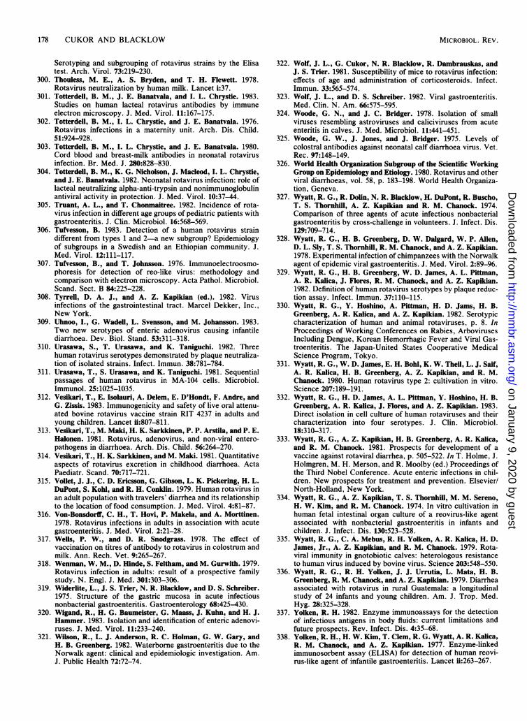
178 CUKOR AND BLACKLOW
Serotyping and subgrouping of rotavirus strains by the Elisatest. Arch. Virol. 73:219-230.
300. Thouless, M. E., A. S. Bryden, and T. H. Flewett. 1978.Rotavirus neutralization by human milk. Lancet i:37.
301. Totterdell, B. M., J. E. Banatvala, and I. L. Chrystie. 1983.Studies on human lacteal rotavirus antibodies by immuneelectron microscopy. J. Med. Virol. 11:167-175.
302. Totterdell, B. M., I. L. Chrystie, and J. E. Banatvala. 1976.Rotavirus infections in a maternity unit. Arch. Dis. Child.51:924-928.
303. Totterdell, B. M., I. L. Chrystie, and J. E. Banatvala. 1980.Cord blood and breast-milk antibodies in neonatal rotavirusinfection. Br. Med. J. 280:828-830.
304. Totterdell, B. M., K. G. Nicholson, J. Macleod, I. L. Chrystie,and J. E. Banatvala. 1982. Neonatal rotavirus infection: role oflacteal neutralizing alpha-anti-trypsin and nonimmunoglobulinantiviral activity in protection. J. Med. Virol. 10:37-44.
305. Truant, A. L., and T. Chonmaitree. 1982. Incidence of rota-virus infection in different age groups of pediatric patients withgastroenteritis. J. Clin. Microbiol. 16:568-569.
306. Tufvesson, B. 1983. Detection of a human rotavirus straindifferent from types 1 and 2-a new subgroup? Epidemiologyof subgroups in a Swedish and an Ethiopian community. J.Med. Virol. 12:111-117.
307. Tufvesson, B., and T. Johnsson. 1976. Immunoelectroosmo-phoresis for detection of reo-like virus: methodology andcomparison with electron microscopy. Acta Pathol. Microbiol.Scand. Sect. B 84:225-228.
308. Tyrrell, D. A. J., and A. Z. Kapikian (ed.). 1982. Virusinfections of the gastrointestinal tract. Marcel Dekker, Inc.,New York.
309. Uhnoo, I., G. Wadell, L. Svensson, and M. Johansson. 1983.Two new serotypes of enteric adenovirus causing infantilediarrhoea. Dev. Biol. Stand. 53:311-318.
310. Urasawa, S., T. Urasawa, and K. Taniguchi. 1982. Threehuman rotavirus serotypes demonstrated by plaque neutraliza-tion of isolated strains. Infect. Immun. 38:781-784.
311. Urasawa, T., S. Urasawa, and K. Taniguchi. 1981. Sequentialpassages of human rotavirus in MA-104 cells. Microbiol.Immunol. 25:1025-1035.
312. Vesikari, T., E. Isolauri, A. Delem, E. D'Hondt, F. Andre, andG. Zissis. 1983. Immunogenicity and safety of live oral attenu-ated bovine rotavirus vaccine strain RIT 4237 in adults andyoung children. Lancet ii:807-811.
313. Vesikari, T., M. Maki, H. K. Sarkkinen, P. P. Arstila, and P. E.Halonen. 1981. Rotavirus, adenovirus, and non-viral entero-pathogens in diarrhoea. Arch. Dis. Child. 56:264-270.
314. Vesikari, T., H. K. Sarkkinen, and M. Maki. 1981. Quantitativeaspects of rotavirus excretion in childhood diarrhoea. ActaPaediatr. Scand. 70:717-721.
315. Vollet, J. J., C. D. Ericsson, G. Gibson, L. K. Pickering, H. L.DuPont, S. Kohl, and R. H. Conklin. 1979. Human rotavirus inan adult population with travelers' diarrhea and its relationshipto the location of food consumption. J. Med. Virol. 4:81-87.
316. Von-Bonsdorff, C. H., T. Hovi, P. Makela, and A. Morttinen.1978. Rotavirus infections in adults in association with acutegastroenteritis. J. Med. Virol. 2:21-28.
317. Wells, P. W., and D. R. Snodgrass. 1978. The effect ofvaccination on titres of antibody to rotavirus in colostrum andmilk. Ann. Rech. Vet. 9:265-267.
318. Wenman, W. M., D. Hinde, S. Feltham, and M. Gurwith. 1979.Rotavirus infection in adults: result of a prospective familystudy. N. Engl. J. Med. 301:303-306.
319. Widerlite, L., J. S. Trier, N. R. Blacklow, and D. S. Schreiber.1975. Structure of the gastric mucosa in acute infectiousnonbacterial gastroenteritis. Gastroenterology 68:425-430.
320. Wigand, R., H. G. Baumeister, G. Maass, J. Kuhn, and H. J.Hammer. 1983. Isolation and identification of enteric adenovi-ruses. J. Med. Virol. 11:233-240.
321. Wilson, R., L. J. Anderson, R. C. Holman, G. W. Gary, andH. B. Greenberg. 1982. Waterborne gastroenteritis due to theNorwalk agent: clinical and epidemiologic investigation. Am.J. Public Health 72:72-74.
322. Wolf, J. L., G. Cukor, N. R. Blacklow, R. Dambrauskas, andJ. S. Trier. 1981. Susceptibility of mice to rotavirus infection:effects of age and administration of corticosteroids. Infect.Immun. 33:565-574.
323. Wolf, J. L., and D. S. Schreiber. 1982. Viral gastroenteritis.Med. Clin. N. Am. 66:575-595.
324. Woode, G. N., and J. C. Bridger. 1978. Isolation of smallviruses resembling astroviruses and caliciviruses from acuteenteritis in calves. J. Med. Microbiol. 11:441-451.
325. Woode, G. W., J. Jones, and J. Bridger. 1975. Levels ofcolostral antibodies against neonatal calf diarrhoea virus. Vet.Rec. 97:148-149.
326. World Health Organization Subgroup of the Scientific WorkingGroup on Epidemiology and Etiology. 1980. Rotavirus and otherviral diarrhoeas, vol. 58, p. 183-198. World Health Organiza-tion, Geneva.
327. Wyatt, R. G., R. Dolin, N. R. Blacklow, H. DuPont, R. Buscho,T. S. Thornhill, A. Z. Kapikian and R. M. Chanock. 1974.Comparison of three agents of acute infectious nonbacterialgastroenteritis by cross-challenge in volunteers. J. Infect. Dis.129:709-714.
328. Wyatt, R. G., H. B. Greenberg, D. W. Dalgard, W. P. Allen,D. L. Sly, T. S. Thornhill, R. M. Chanock, and A. Z. Kapikian.1978. Experimental infection of chimpanzees with the Norwalkagent of epidemic viral gastroenteritis. J. Med. Virol. 2:89-96.
329. Wyatt, R. G., H. B. Greenberg, W. D. James, A. L. Pittman,A. R. Kalica, J. Flores, R. M. Chanock, and A. Z. Kapikian.1982. Definition of human rotavirus serotypes by plaque reduc-tion assay. Infect. Immun. 37:110-115.
330. Wyatt, R. G., Y. Hoshino, A. Pittman, H. D. Jams, H. B.Greenberg, A. R. Kalica, and A. Z. Kapikian. 1982. Serotypiccharacterization of human and animal rotaviruses, p. 8. InProceedings of Working Conferences on Rabies, ArbovirusesIncluding Dengue, Korean Hemorrhagic Fever and Viral Gas-troenteritis. The Japan-United States Cooperative MedicalScience Program, Tokyo.
331. Wyatt, R. G., W. D. James, E. H. Bohl, K. W. Theil, L. J. Saif,A. R. Kalica, H. B. Greenberg, A. Z. Kapikian, and R. M.Chanock. 1980. Human rotavirus type 2: cultivation in vitro.Science 207:189-191.
332. Wyatt, R. G., H. D. James, A. L. Pittman, Y. Hoshino, H. B.Greenberg, A. R. Kalica, J. Flores, and A. Z. Kapikian. 1983.Direct isolation in cell culture of human rotaviruses and theircharacterization into four serotypes. J. Clin. Microbiol.18:310-317.
333. Wyatt, R. G., A. Z. Kapikian, H. B. Greenberg, A. R. Kalica,and R. M. Chanock. 1981. Prospects for development of avaccine against rotaviral diarrhea, p. 505-522. In T. Holme, J.Holmgren, M. H. Merson, and R. Moolby (ed.) Proceedings ofthe Third Nobel Conference. Acute enteric infections in chil-dren. New prospects for treatment and prevention. Elsevier/North-Holland, New York.
334. Wyatt, R. G., A. Z. Kapikian, T. S. Thornhill, M. M. Sereno,H. W. Kim, and R. M. Chanock. 1974. In vitro cultivation inhuman fetal intestinal organ culture of a reovirus-like agentassociated with nonbacterial gastroenteritis in infants andchildren. J. Infect. Dis. 130:523-528.
335. Wyatt, R. G., C. A. Mebus, R. H. Yolken, A. R. Kalica, H. D.James, Jr., A. Z. Kapikian, and R. M. Chanock. 1979. Rota-viral immunity in gnotobiotic calves: heterologous resistanceto human virus induced by bovine virus. Science 203:548-550.
336. Wyatt, R. G., R. H. Yolken, J. J. Urrutia, L. Mata, H. B.Greenberg, R. M. Chanock, and A. Z. Kapikian. 1979. Diarrheaassociated with rotavirus in rural Guatemala: a longitudinalstudy of 24 infants and young children. Am. J. Trop. Med.Hyg. 28:325-328.
337. Yolken, R. H. 1982. Enzyme immunoassays for the detectionof infectious antigens in body fluids: current limitations andfuture prospects. Rev. Infect. Dis. 4:35-68.
338. Yolken, R. H., H. W. Kim, T. Clem, R. G. Wyatt, A. R. Kalica,R. M. Chanock, and A. Z. Kapikian. 1977. Enzyme-linkedimmunosorbent assay (ELISA) for detection of human reovi-rus-like agent of infantile gastroenteritis. Lancet ii:263-267.
MICROBIOL. REV.
on January 9, 2020 by guesthttp://m
mbr.asm
.org/D
ownloaded from
HUMAN VIRAL GASTROENTERITIS 179
339. Yolken, R. H., F. Lawrence, F. Leister, H. E. Takiff, and S. E.Strauss. 1982. Gastroenteritis associated with enteric typeadenovirus in hospitalized infants. J. Pediatr. 101:21-26.
340. Yolken, R., and M. Murphy. 1983. Sudden infant death syn-drome associated with rotavirus infection. J. Med. Virol.10:291-296.
341. Yolken, R. H., and P. J. Stopa. 1979. Analysis of nonspecificreactions in enzyme-linked immunosorbent assay testing forhuman rotavirus. J. Clin. Microbiol. 10:703-707.
342. Yolken, R. H., P. J. Stopa, and C. C. Harris. 1980. Enzymeimmunoassay for the detection of rotavirus antigen and anti-body, p. 692-699. In N. Rose and H. Friedman (ed.), Manualof clinical immunology, 2nd ed. American Society for Microbi-ology, Washington, D.C.
343. Yolken, R., R. G. Wyatt, and A. Z. Kapikian. 1977. Elisa forrotavirus (letter). Lancet ii:819.
344. Yolken, R. H., R. G. Wyatt, L. Mata, J. J. Urrutia, B. Garcia,R. M. Chanock, and A. Z. Kapikian. 1978. Secretory antibodydirected against rotavirus in human milk-measurement by
means of enzyme-linked immunosorbent assay. J. Pediatr.93:916-921.
345. Yolken, R. H., R. G. Wyatt, G. Zissis, C. D. Brandt, W. J.Rodriguez, H. W. Kim, R. H. Parrott, J. J. Urrutia, L. Mata,H. B. Greenberg, A. Z. Kapikian, and R. M. Chanock. 1978.Epidemiology of human rotavirus types 1 and 2 studied byenzyme-linked immunosorbent assay. N. Engl. J. Med.299:1156-1161.
346. Yow, M. D., J. L. Melnick, R. J. Blattner, W. B. Stephenson,N. M. Robinson, and M. A. Burkhardt. 1970. The associationof viruses and bacteria with infantile diarrhoea. Am. J. Epide-miol. 92:33-39.
347. Zissis, G., and J. P. Lambert. 1980. Enzyme-linked immuno-sorbent assays adapted for serotyping of human rotavirusstrains. J. Clin. Microbiol. 11:1-5.
348. Zissis, G., and J. P. Lambert. 1978. Different serotypes ofhuman rotaviruses (letter). Lancet i:38-39.
349. Zissis, G., J. P. Lambert, J. G. Kapsenberg, G. Enders, andL. N. Mutanda. Human rotavirus serotypes (letter). Lanceti:944-945.
VOL. 48, 1984
on January 9, 2020 by guesthttp://m
mbr.asm
.org/D
ownloaded from