human genetics of urinary tract malformation ali gharavi, md division of nephrology columbia...
TRANSCRIPT
Human Genetics of Urinary Tract Human Genetics of Urinary Tract Malformation Malformation
Ali Gharavi, MD Ali Gharavi, MD Division of NephrologyDivision of Nephrology
Columbia UniversityColumbia UniversityNew York, NYNew York, NY
[email protected]@columbia.edu
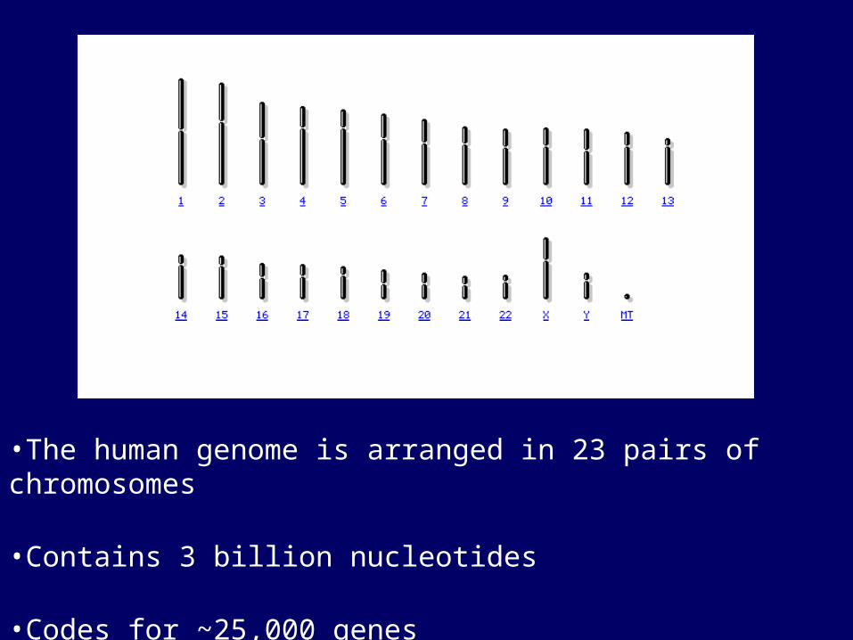
•The human genome is arranged in 23 pairs of chromosomes
•Contains 3 billion nucleotides
•Codes for ~25,000 genes
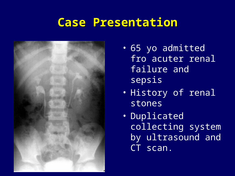
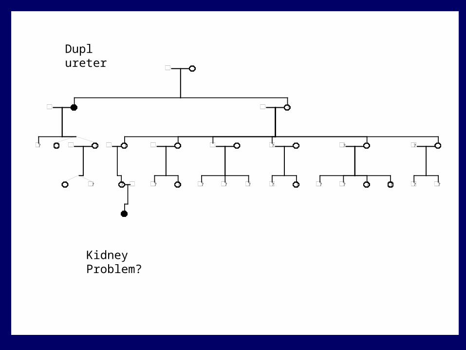
Case PresentationCase Presentation
• 65 yo admitted fro acuter renal failure and sepsis
• History of renal stones
• Duplicated collecting system by ultrasound and CT scan.
?
?
?
?
?
? ? ?
?
? ? ? ? ?
?
? ? ? ?
?
? ?
Kidney Problem?
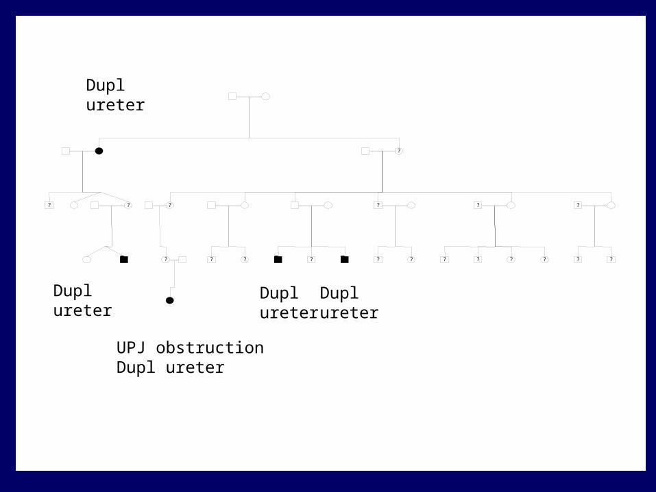
Duplureter
?
?
?
?
?
? ? ?
?
? ? ? ? ?
?
? ? ? ?
?
? ?
Duplureter
Duplureter
Duplureter
Duplureter
UPJ obstructionDupl ureter
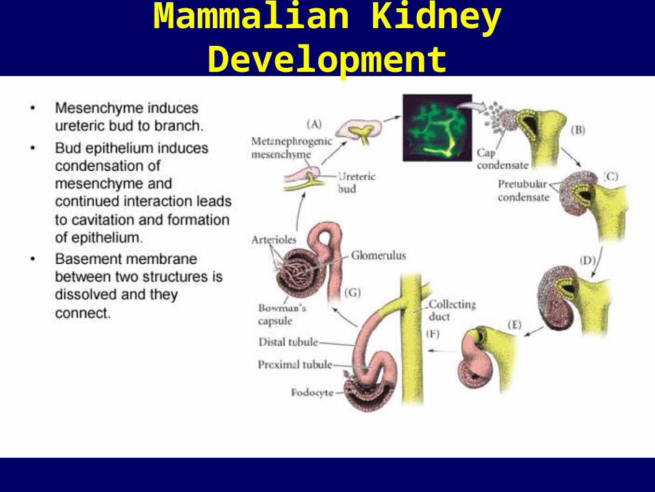
Mammalian Kidney Development
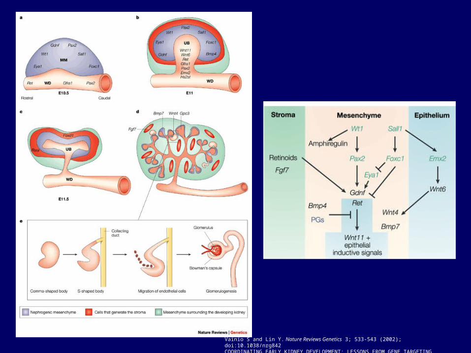
Vainio S and Lin Y. Nature Reviews Genetics 3; 533-543 (2002); doi:10.1038/nrg842COORDINATING EARLY KIDNEY DEVELOPMENT: LESSONS FROM GENE TARGETING
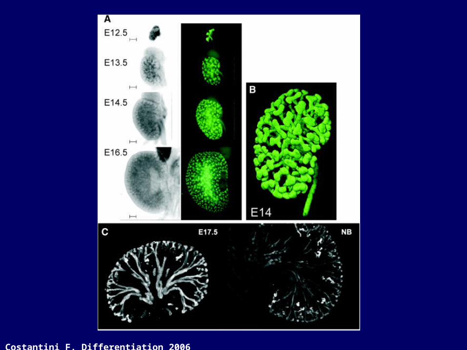
Costantini F. Differentiation 2006
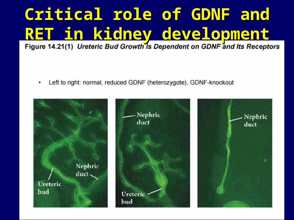
Critical role of GDNF and RET in Critical role of GDNF and RET in kidney developmentkidney development
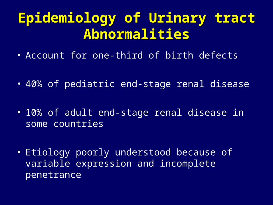
Epidemiology of Urinary tract Epidemiology of Urinary tract AbnormalitiesAbnormalities
• Account for one-third of birth defects
• 40% of pediatric end-stage renal disease
• 10% of adult end-stage renal disease in some countries
• Etiology poorly understood because of variable expression and incomplete penetrance
Clinical FeaturesClinical Features
• Due to overlap between developmental pathways, phenotypes are complex, involving anatomic defects in both upper and lower urinary tract
• Often asymmetric
• Severe phenotypes result in perinatal death due to pulmonary hypoplasia
• The majority of cases are nonsyndromic
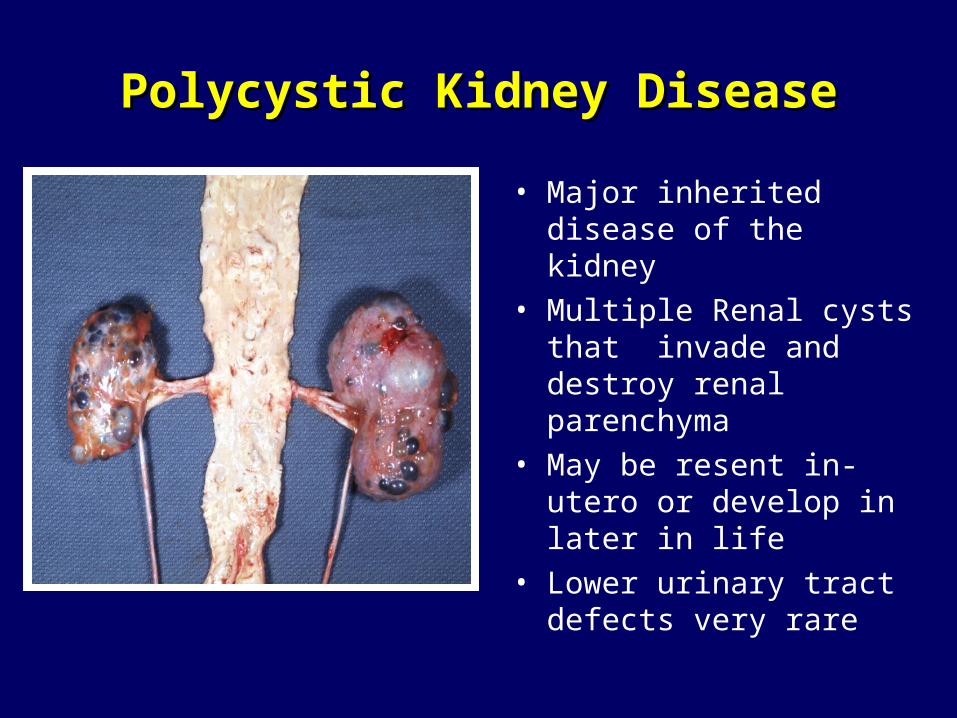
Polycystic Kidney DiseasePolycystic Kidney Disease
• Major inherited disease of the kidney
• Multiple Renal cysts that invade and destroy renal parenchyma
• May be resent in-utero or develop in later in life
• Lower urinary tract defects very rare
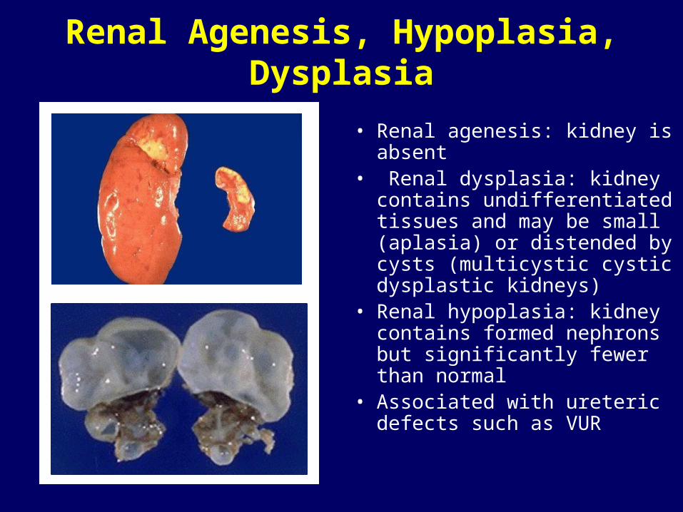
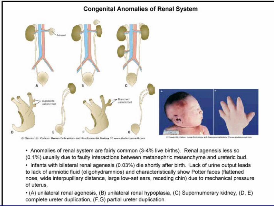
Renal Agenesis, Hypoplasia, Dysplasia
• Renal agenesis: kidney is absent
• Renal dysplasia: kidney contains undifferentiated tissues and may be small (aplasia) or distended by cysts (multicystic cystic dysplastic kidneys)
• Renal hypoplasia: kidney contains formed nephrons but significantly fewer than normal
• Associated with ureteric defects such as VUR
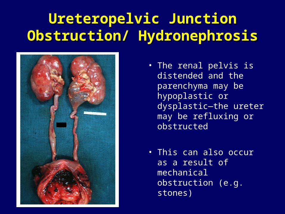
Ureteropelvic Junction Obstruction/ Ureteropelvic Junction Obstruction/ HydronephrosisHydronephrosis
• The renal pelvis is distended and the parenchyma may be hypoplastic or dysplastic—the ureter may be refluxing or obstructed
• This can also occur as a result of mechanical obstruction (e.g. stones)
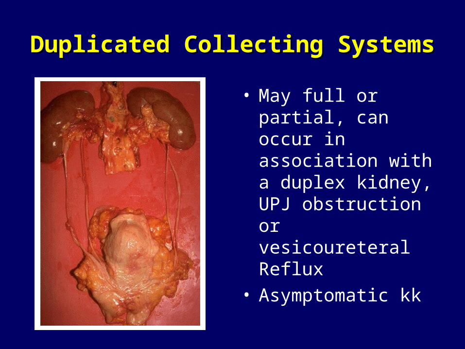
Duplicated Collecting SystemsDuplicated Collecting Systems
• May full or partial, can occur in association with a duplex kidney, UPJ obstruction or vesicoureteral Reflux
• Asymptomatic kk
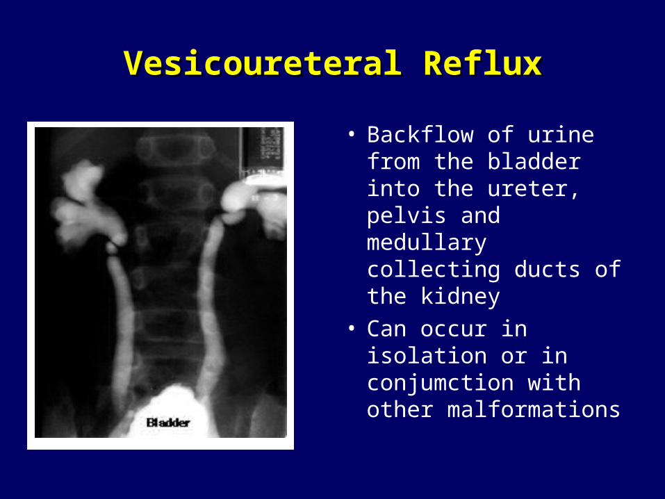
Vesicoureteral RefluxVesicoureteral Reflux
• Backflow of urine from the bladder into the ureter, pelvis and medullary collecting ducts of the kidney
• Can occur in isolation or in conjumction with other malformations
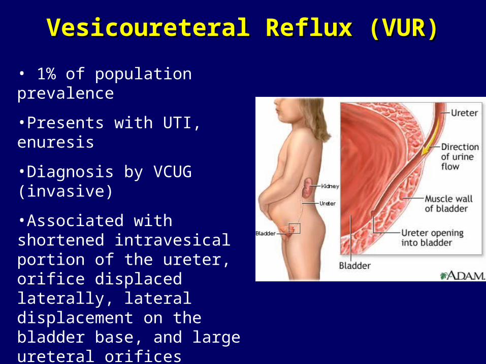
Vesicoureteral Reflux (VUR)Vesicoureteral Reflux (VUR)
• 1% of population prevalence
•Presents with UTI, enuresis
•Diagnosis by VCUG (invasive)
•Associated with shortened intravesical portion of the ureter, orifice displaced laterally, lateral displacement on the bladder base, and large ureteral orifices
• Histologically, attenuation of the trigonal and ureteral musculature.
•25% of pediatric ESRD
Inheritance of VURInheritance of VUR
• Prospective screening of 354 siblings of 275 index patients with VUR revealed reflux in 119 (34%) cases
• Spontaneous resolution of VUR in patients maintained on antibiotic prophylaxis over 10 years (49-69%)
• Most urologists screen sibs, particularly age<5• Complex inheritance
Noe J Urology, 1992, Greenfeld et al J urology J , Scott et al , Lancet 1997
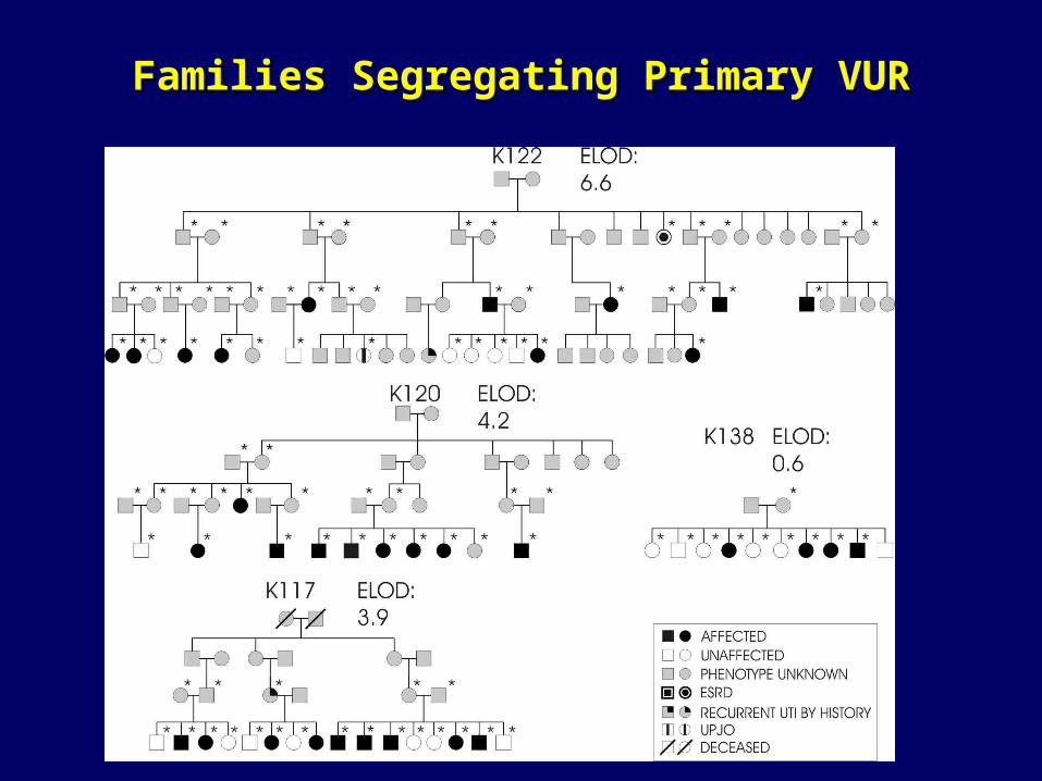
Families Segregating Primary VURFamilies Segregating Primary VUR
Chromosomal abnormalitiesChromosomal abnormalities
Syndromic forms Syndromic forms
• Associated with certain chromosomal abnormalities
– Deletion 4q, 18q
– Duplication 3q, 10q
• Implicate defects in multiple genes in the development of the trait
• Associated with multiple organ defects
10q deletion syndrome10q deletion syndrome
• Cardiac, urogenital, and respiratory complications, orofacial dysmorphism, and psychomotor retardation which vary with different karyotypes.
• Urogenital system: Cryptorchidism, genital hypoplasia, and streaked ovaries. Urinary anomalies include kidney aplasia or hypoplasia, hydronephrosis, hydroureter, and cystic disease.
• Systematic analysis suggest that deletion of 10q26 segment results in this phenotype
Ogata et al. Kidney Int , 2000
Single Gene Disorders in HumansSingle Gene Disorders in Humans
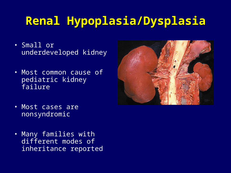
Renal Hypoplasia/DysplasiaRenal Hypoplasia/Dysplasia
• Small or underdeveloped kidney
• Most common cause of pediatric kidney failure
• Most cases are nonsyndromic
• Many families with different modes of inheritance reported
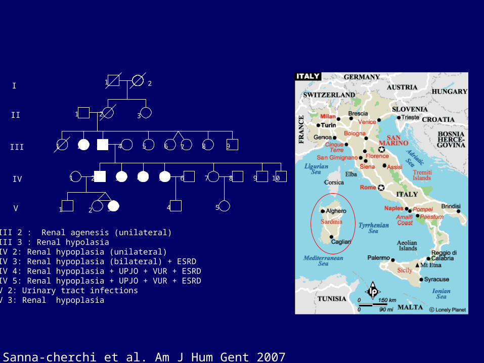
21
1
1 2 3 4 5 6 7 8 9 10
2 3 4 5
III
II
I
IV
V
III 2 : Renal agenesis (unilateral)III 3 : Renal hypolasiaIV 2: Renal hypoplasia (unilateral)IV 3: Renal hypoplasia (bilateral) + ESRDIV 4: Renal hypoplasia + UPJO + VUR + ESRDIV 5: Renal hypoplasia + UPJO + VUR + ESRDV 2: Urinary tract infectionsV 3: Renal hypoplasia
1 2 3
1 2 3 4 5 6 7 8 9
Sanna-cherchi et al. Am J Hum Gent 2007
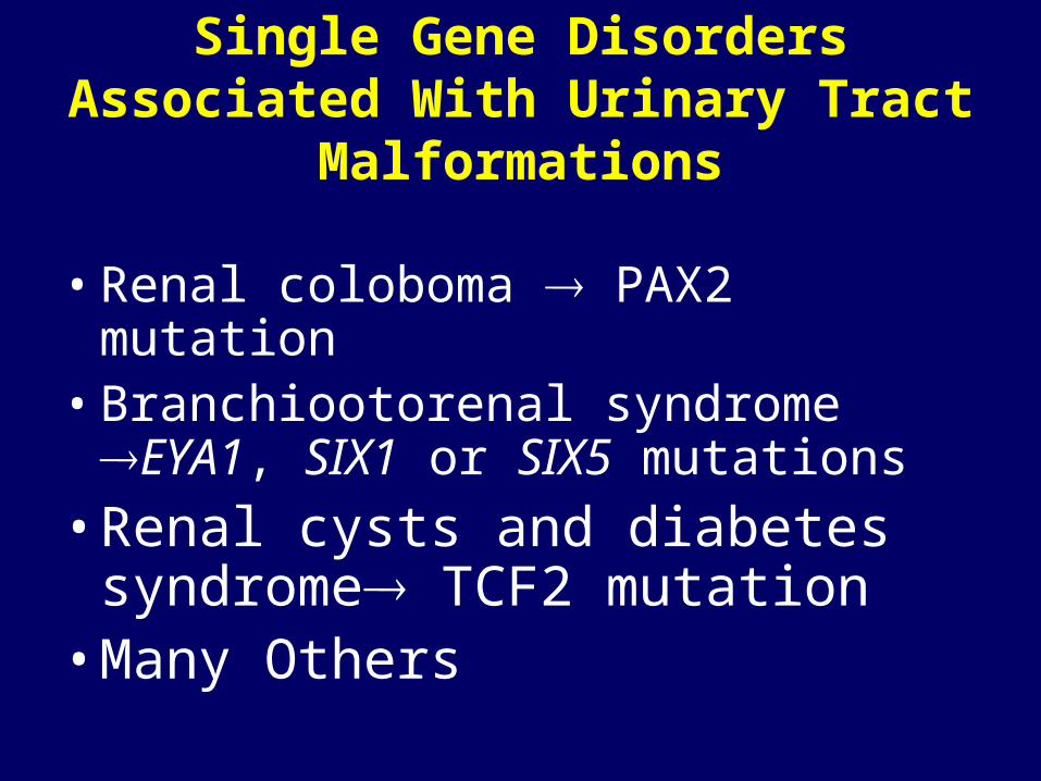
Single Gene Disorders Associated With Urinary Tract Malformations
• Renal coloboma PAX2 mutation• Branchiootorenal syndrome EYA1,
SIX1 or SIX5 mutations
• Renal cysts and diabetes syndrome TCF2 mutation
• Many Others
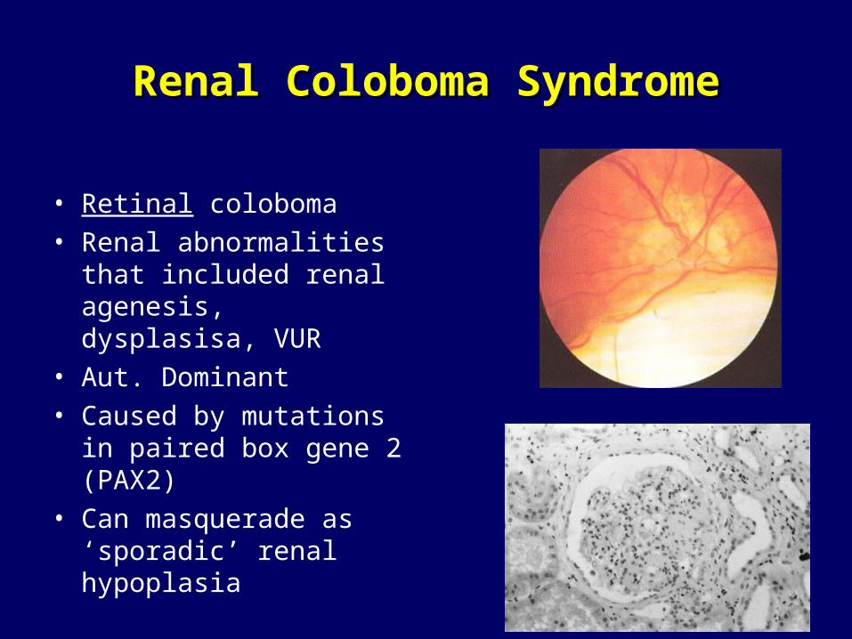
Renal Coloboma SyndromeRenal Coloboma Syndrome
• Retinal coloboma• Renal abnormalities that
included renal agenesis, dysplasisa, VUR
• Aut. Dominant• Caused by mutations in
paired box gene 2 (PAX2)• Can masquerade as
‘sporadic’ renal hypoplasia
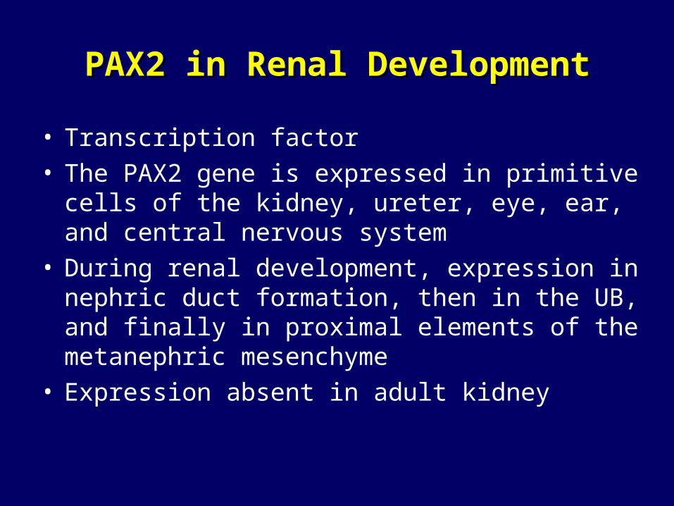
PAX2 in Renal DevelopmentPAX2 in Renal Development
• Transcription factor• The PAX2 gene is expressed in primitive cells of
the kidney, ureter, eye, ear, and central nervous system
• During renal development, expression in nephric duct formation, then in the UB, and finally in proximal elements of the metanephric mesenchyme
• Expression absent in adult kidney
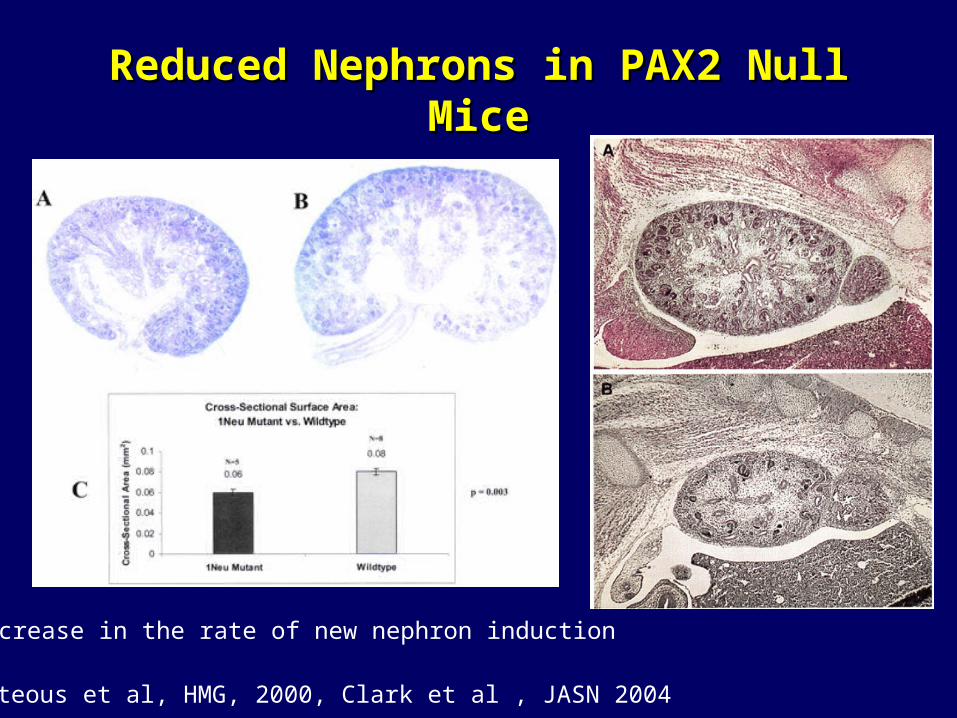
Reduced Nephrons in PAX2 Null MiceReduced Nephrons in PAX2 Null Mice
Porteous et al, HMG, 2000, Clark et al , JASN 2004
Decrease in the rate of new nephron induction
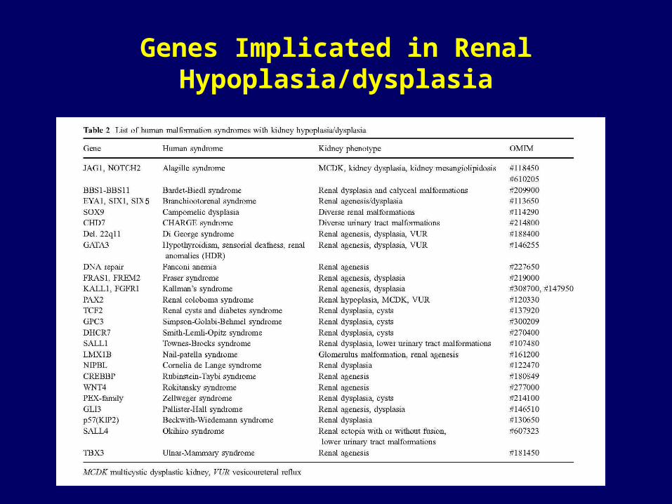
Genes Implicated in Renal Hypoplasia/dysplasia
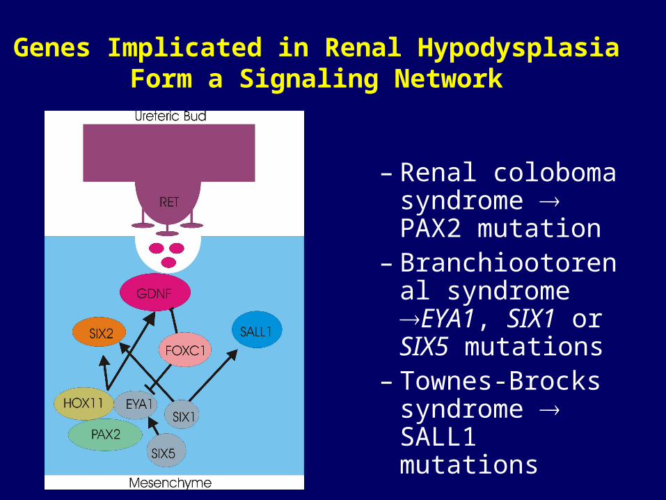
Genes Implicated in Renal Hypodysplasia Form a Signaling Network
– Renal coloboma syndrome PAX2 mutation
– Branchiootorenal syndrome EYA1, SIX1 or SIX5 mutations
– Townes-Brocks syndrome SALL1 mutations
Cystic Kidney DiseaseCystic Kidney Disease
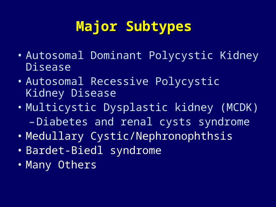
Major Subtypes Major Subtypes
• Autosomal Dominant Polycystic Kidney Disease
• Autosomal Recessive Polycystic Kidney Disease
• Multicystic Dysplastic kidney (MCDK)– Diabetes and renal cysts syndrome
• Medullary Cystic/Nephronophthsis• Bardet-Biedl syndrome• Many Others
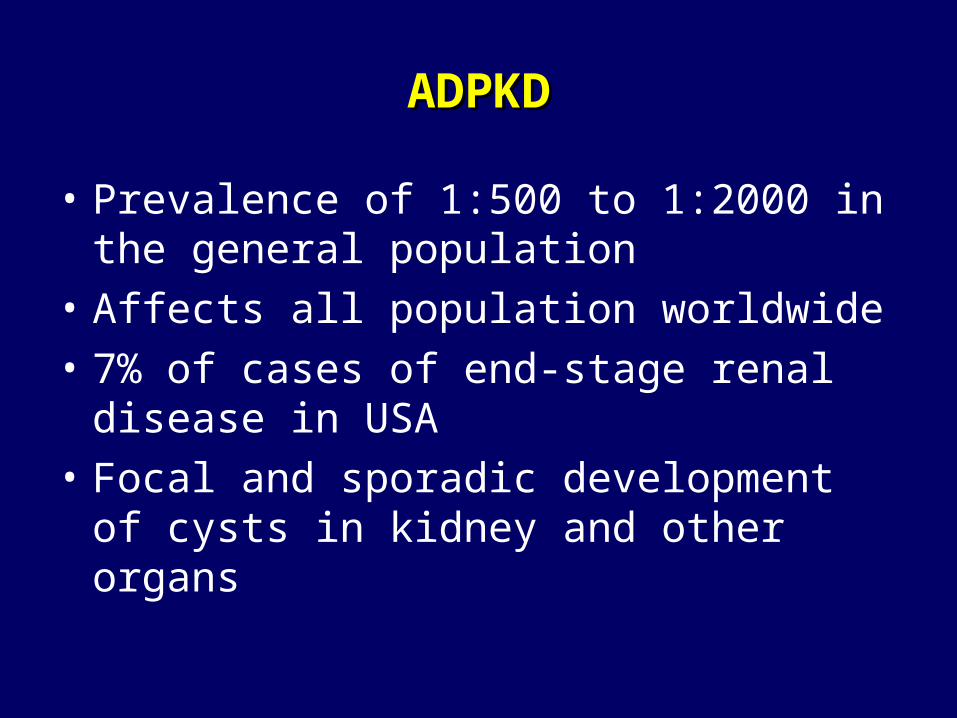
ADPKDADPKD
• Prevalence of 1:500 to 1:2000 in the general population
• Affects all population worldwide
• 7% of cases of end-stage renal disease in USA
• Focal and sporadic development of cysts in kidney and other organs
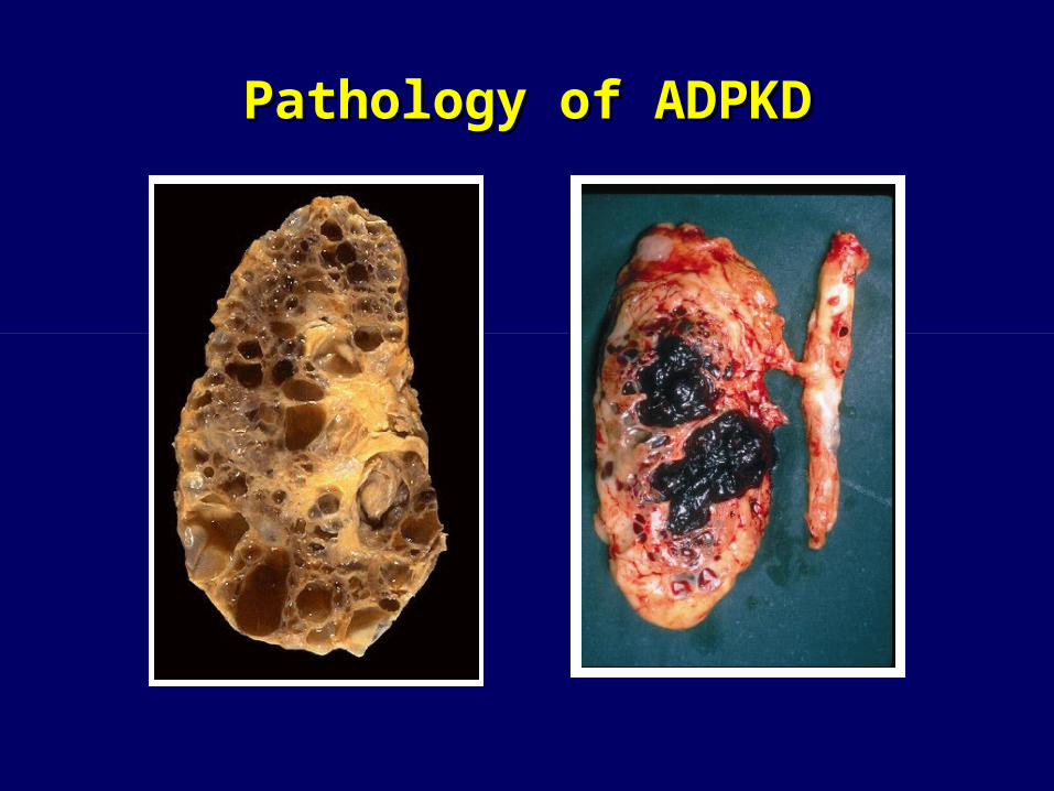
Pathology of ADPKDPathology of ADPKD
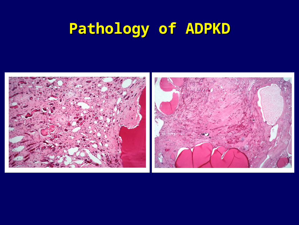
Pathology of ADPKDPathology of ADPKD
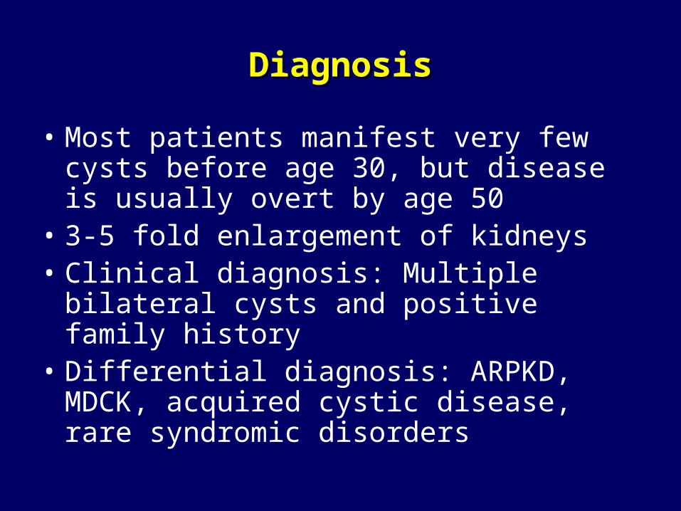
DiagnosisDiagnosis
• Most patients manifest very few cysts before age 30, but disease is usually overt by age 50
• 3-5 fold enlargement of kidneys• Clinical diagnosis: Multiple bilateral cysts
and positive family history• Differential diagnosis: ARPKD, MDCK,
acquired cystic disease, rare syndromic disorders
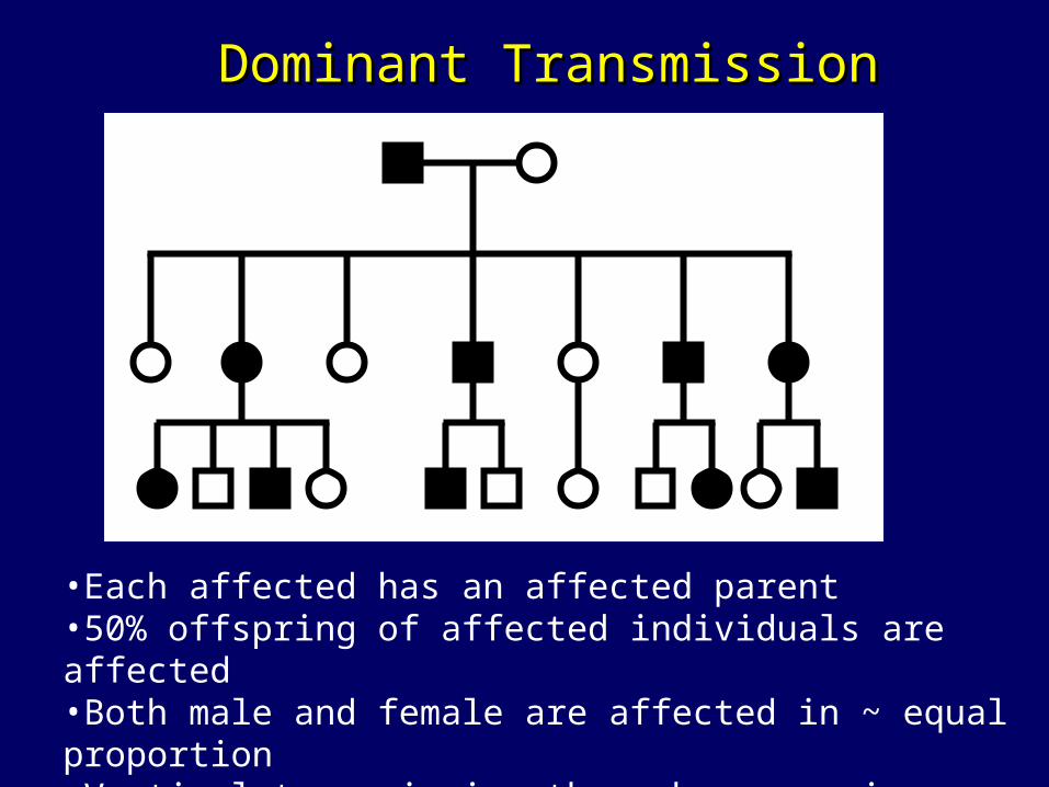
•Each affected has an affected parent•50% offspring of affected individuals are affected •Both male and female are affected in ~ equal proportion•Vertical transmission through successive generation
Dominant TransmissionDominant Transmission
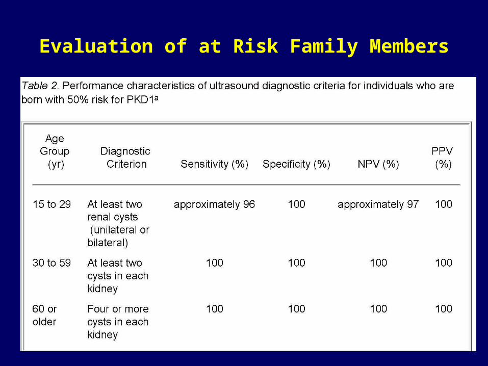
Evaluation of at Risk Family Members
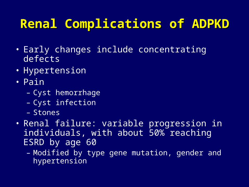
Renal Complications of ADPKDRenal Complications of ADPKD
• Early changes include concentrating defects• Hypertension• Pain
– Cyst hemorrhage– Cyst infection– Stones
• Renal failure: variable progression in individuals, with about 50% reaching ESRD by age 60– Modified by type gene mutation, gender and
hypertension
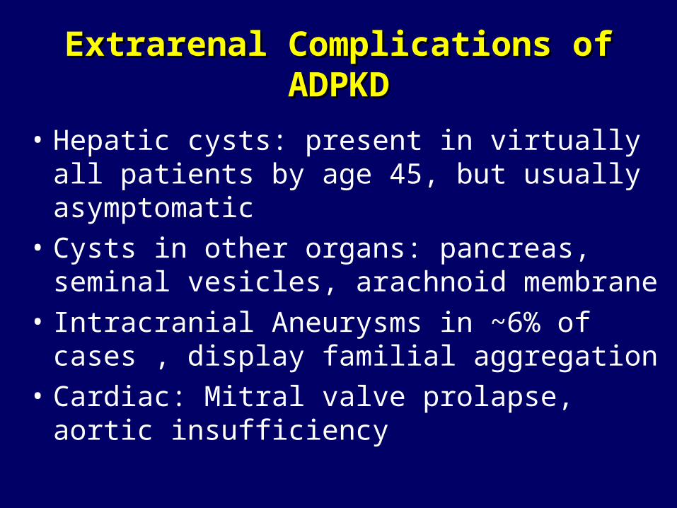
• Hepatic cysts: present in virtually all patients by age 45, but usually asymptomatic
• Cysts in other organs: pancreas, seminal vesicles, arachnoid membrane
• Intracranial Aneurysms in ~6% of cases , display familial aggregation
• Cardiac: Mitral valve prolapse, aortic insufficiency
Extrarenal Complications of ADPKDExtrarenal Complications of ADPKD
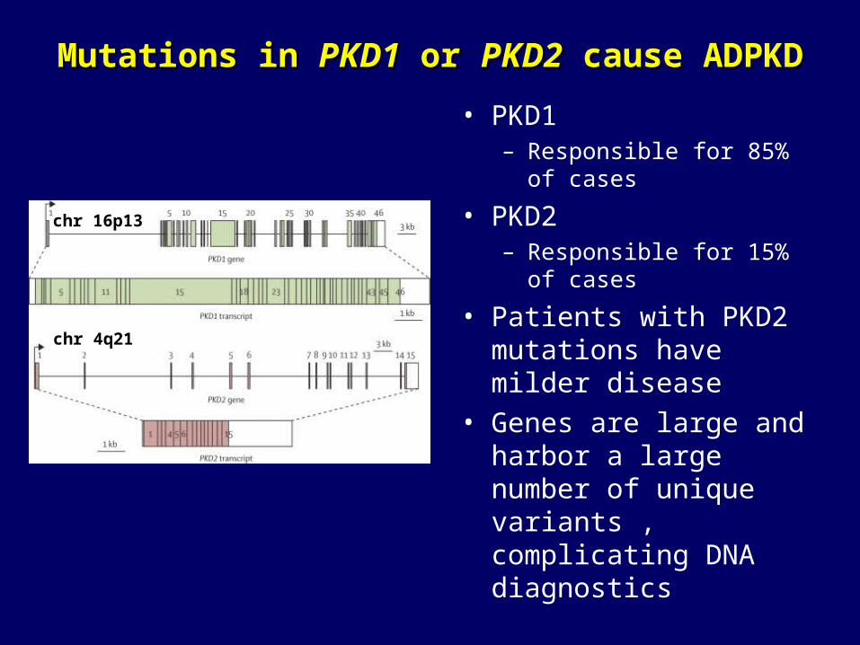
Mutations in Mutations in PKD1PKD1 or or PKD2PKD2 cause ADPKD cause ADPKD
• PKD1 – Responsible for 85% of
cases
• PKD2– Responsible for 15% of
cases
• Patients with PKD2 mutations have milder disease
• Genes are large and harbor a large number of unique variants , complicating DNA diagnostics
chr 16p13
chr 4q21
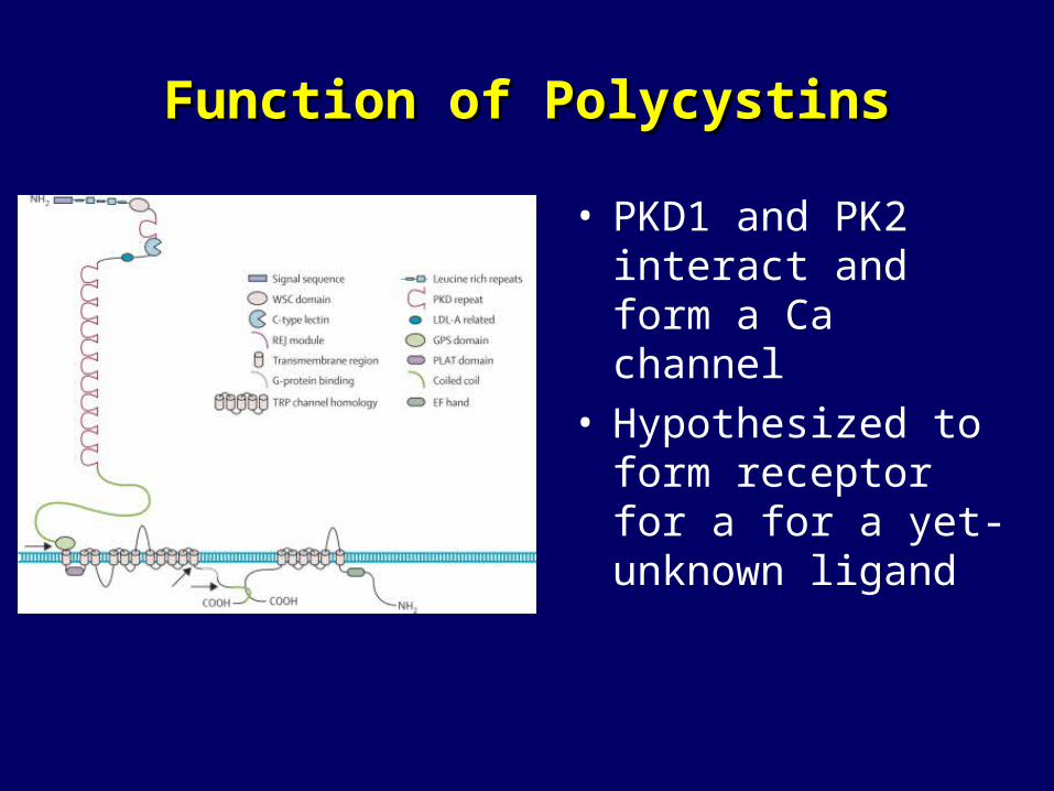
Function of PolycystinsFunction of Polycystins
• PKD1 and PK2 interact and form a Ca channel
• Hypothesized to form receptor for a for a yet-unknown ligand
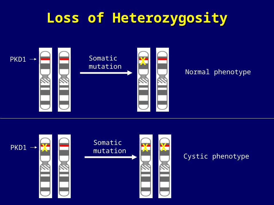
Loss of HeterozygosityLoss of Heterozygosity
PKD1 X
X
Somaticmutation
Normal phenotype
PKD1Somaticmutation X X
Cystic phenotype
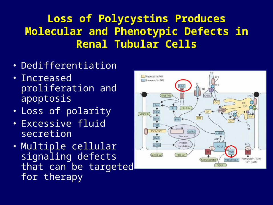
Loss of Polycystins Produces Molecular and Loss of Polycystins Produces Molecular and Phenotypic Defects in Renal Tubular CellsPhenotypic Defects in Renal Tubular Cells
• Dedifferentiation• Increased proliferation
and apoptosis• Loss of polarity• Excessive fluid
secretion• Multiple cellular
signaling defects that can be targeted for therapy
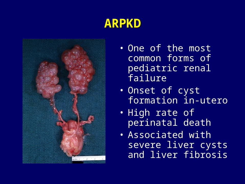
ARPKDARPKD
• One of the most common forms of pediatric renal failure
• Onset of cyst formation in-utero
• High rate of perinatal death
• Associated with severe liver cysts and liver fibrosis
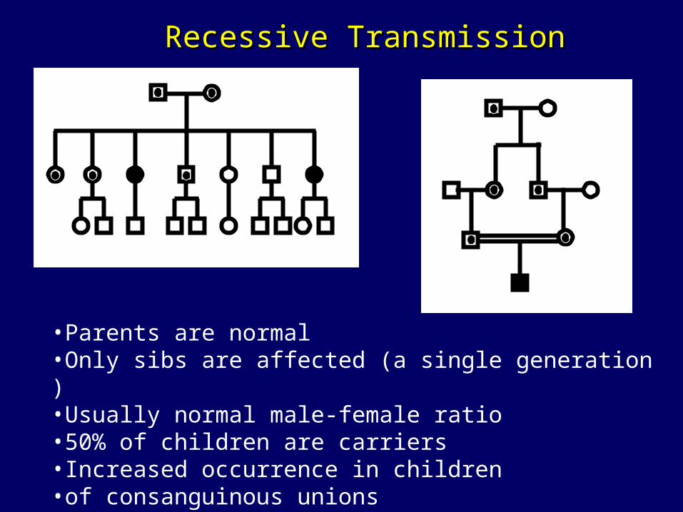
•Parents are normal•Only sibs are affected (a single generation )•Usually normal male-female ratio•50% of children are carriers•Increased occurrence in children •of consanguinous unions
Recessive TransmissionRecessive Transmission
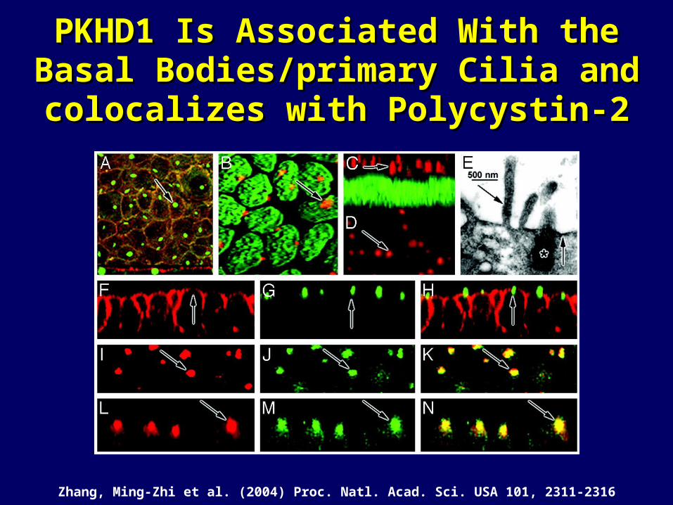
Zhang, Ming-Zhi et al. (2004) Proc. Natl. Acad. Sci. USA 101, 2311-2316
PKHD1 Is Associated With the Basal PKHD1 Is Associated With the Basal Bodies/primary Cilia and colocalizes Bodies/primary Cilia and colocalizes
with Polycystin-2with Polycystin-2
Diabetes and Renal Cysts SyndromeDiabetes and Renal Cysts Syndrome
• Type II diabetes in individuals <25 yrs (MODY)• Cystic renal disease, including unilateral agenesis,
horseshoe kidney, and hyperuricemic nephropathy• Some individuals have genital malformations (e.g.
vaginal aplasia, bicornuate uterus, epididymal cysts)
• Autosomal dominant transmission• Caused by mutations in the Hepatocyte Nuclear
Factor 1 (HNF1B)• Can masquerade as ‘sporadic’ renal hypoplasia
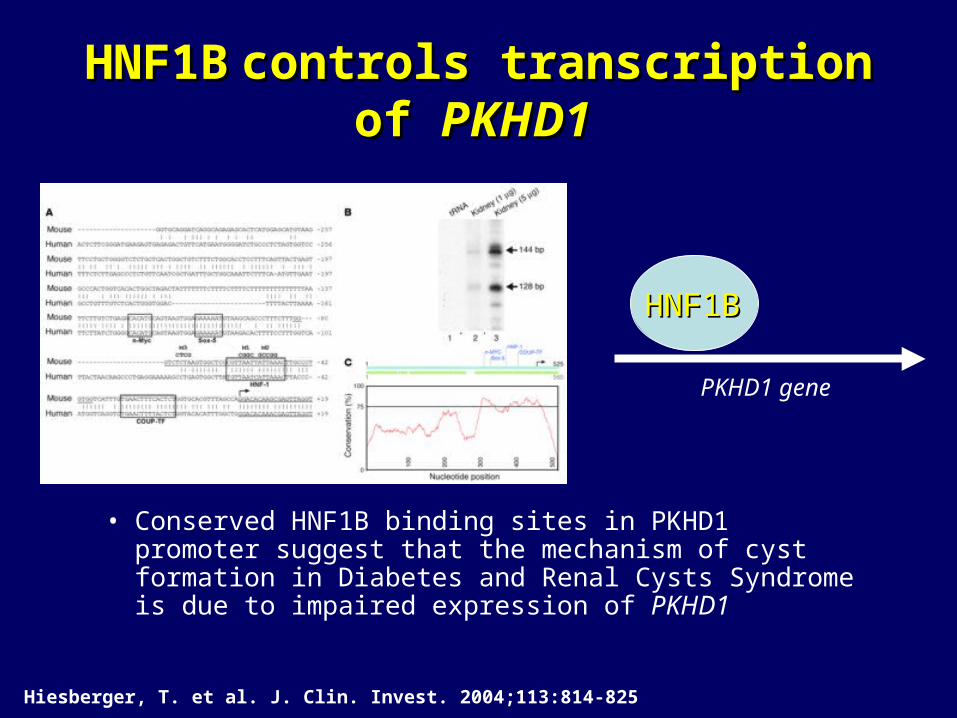
HNF1BHNF1B controls transcription of controls transcription of PKHD1 PKHD1
•
• Conserved HNF1B binding sites in PKHD1 promoter suggest that the mechanism of cyst formation in Diabetes and Renal Cysts Syndrome is due to impaired expression of PKHD1
PKHD1 gene
HNF1BHNF1B
Hiesberger, T. et al. J. Clin. Invest. 2004;113:814-825
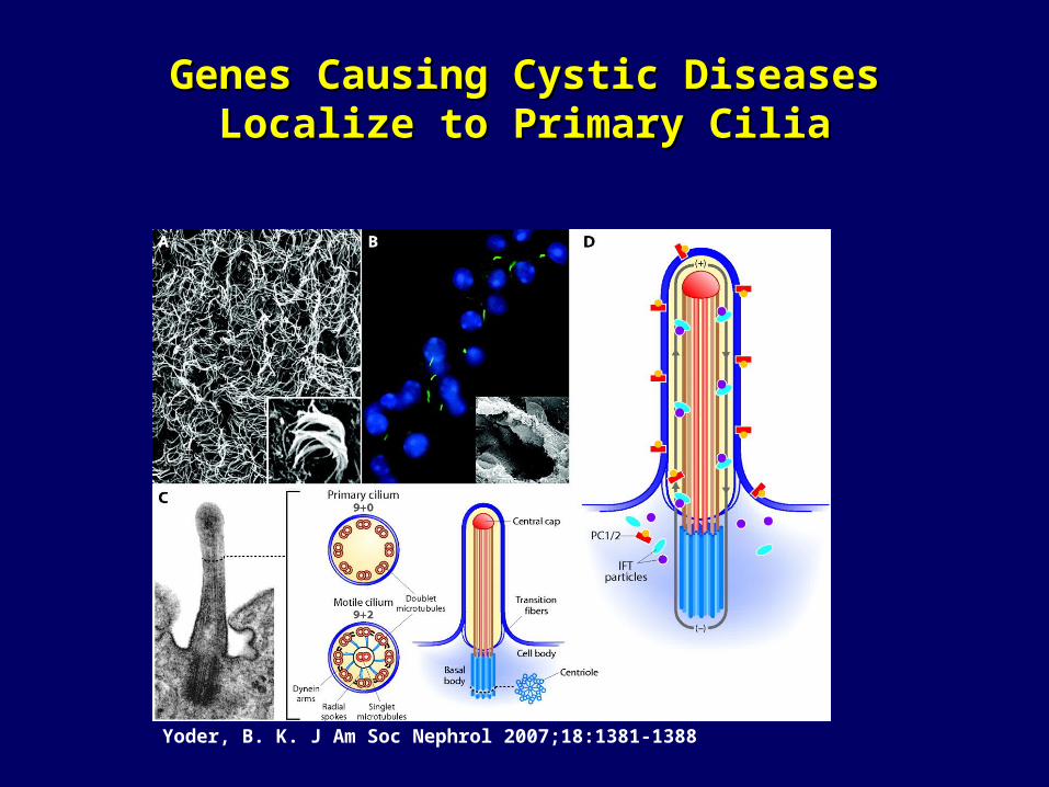
Yoder, B. K. J Am Soc Nephrol 2007;18:1381-1388
Genes Causing Cystic Diseases Localize to Genes Causing Cystic Diseases Localize to Primary CiliaPrimary Cilia
Genes Causing Cystic Diseases Localize to Genes Causing Cystic Diseases Localize to Primary CiliaPrimary Cilia
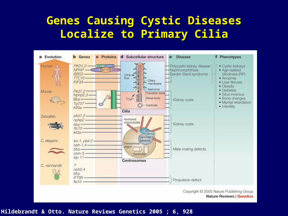
•Hildebrandt & Otto. Nature Reviews Genetics 2005 ; 6, 928
Web ReferencesWeb References
• Pathology Pictures– Columbia Pathology:
http://cpmcnet.columbia.edu/dept/curric-pathology/pathology/pathology/pathoatlas/index.html
– Pathology Education Instructional resources (PEIR) http://peir.net/
• Human Genetics– OMIMTM - Online Mendelian Inheritance in ManTM
http://www.ncbi.nlm.nih.gov/sites/entrez?db=OMIM
Further ReadingFurther Reading
• Woolf AS. et al. Evolving concepts in human renal dysplasia. J Am Soc Nephrol. 2004 : 998
• Genetic approaches to human renal agenesis/hypoplasia and dysplasia. Pediatr Nephrol. 2007 :1675
• Torres et al. Autosomal Dominant Polycystic Kidney disease. Lancet 2007; 369:1287
• Hildebrandt & Otto. Cilia and centrosomes: a unifying pathogenic concept for cystic kidney disease? Nature Reviews Genetics 2005 ; 6, 928