human babesiosis: pathogens, prevalence, diagnosis, and … · human babesiosis: pathogens,...
TRANSCRIPT
PARASITOLOGY (AVAIDYA, SECTION EDITOR)
Human Babesiosis: Pathogens, Prevalence,Diagnosis, and Treatment
Rosalynn Louise Ord1& Cheryl A. Lobo1
Published online: 28 September 2015# Springer International Publishing AG 2015
Abstract Human babesiosis is a zoonotic disease caused byprotozoan parasites of the Babesia genus, primarily in theNortheastern and Midwest USA due to Babesia microti andWestern Europe due to Babesia divergens. Parasites are trans-mitted by the bite of the ixodid tick when the vector takes ablood meal from the vertebrate host, and the economic impor-tance of bovine babesiosis is well understood. The pathologyof human disease is a direct result of the parasite’s ability toinvade host’s red blood cells. The current understanding ofhuman babesiosis epidemiology is that many infections re-main asymptomatic, especially in younger or immune compe-tent individuals, and the burden of severe pathology resideswithin older or immunocompromised individuals. However,transfusion-transmitted babesiosis is an emerging threat topublic health as asymptomatic carriers donate blood, and thereare as yet no licensed or regulated tests to screen blood prod-ucts for this pathogen. Reports of tick-borne cases within newgeographical regions such as the Pacific Northwest of theUSA, through Eastern Europe, and into China are also onthe rise. Further, new Babesia spp. have been identified glob-ally as agents of severe human babesiosis, suggesting that theepidemiology of this disease is rapidly changing, and it is clearthat human babesiosis is a serious public health concern thatrequires close monitoring and effective intervention measure.
Keywords Babesia .Malaria .B. microti . B. divergens .
Zoonosis . Transfusion-transmitted babesiosis
Introduction
Human babesiosis is a zoonotic infection caused by Babesiaparasites transmitted by the bite of ixodid ticks that have dis-tinct geographical distributions based on the presence of theircompetent natural animal hosts, which include rodents, cattle,and deer. The genus Babesia comprises many species of par-asites [1], which are transmitted when the ticks takes a bloodmeal from the vertebrate host [1, 2]. While parasite infectionof these natural hosts, such as in cattle, which results in bovinebabesiosis causing significant economic cost has long beenestablished [3, 4], the severity of human infection is rapidlybecoming apparent, whether the disease has been primarilytransmitted from a tick bite or secondarily transmitted via ablood transfusion with infected blood [5–7], or even congen-itally during pregnancy [8–11]. The four identified Babesiaspecies definitively confirmed that infect humans so far areBabesia microti [12], Babesia divergens [13], Babesiaduncani [14, 15], and Babesia venatorum [16–19]. As sam-pling has become expansive and techniques have becomemore sensitive, there is evidence that more B. microti-likeand B. divergens-like spp. are able to cause human infection(as detailed in [20]). However, the general life cycle withinhumans remains the same; Babesia parasites are intracellularobligates that target red blood cells, and the parasite’s ability tofirst recognize and then invade host RBCs is central to thedisease pathology.
Vector Life Cycle and Transmission
The only confirmed vectors of Babesia parasites are membersof the Ixodidae family. Not all life cycles for known parasite–vector–host groups are fully understood yet [20]. The 2-yearlife cycle of Ixodes scapularis, which is endemic across mostof the eastern states of America and Canada [21–23], and its
This article is part of the Topical Collection on Parasitology
* Cheryl A. [email protected]
1 Department of Blood-Borne Parasites, Lindsley Kimball ResearchInstitute, New York Blood Center, New York, NY 10065, USA
Curr Clin Micro Rpt (2015) 2:173–181DOI 10.1007/s40588-015-0025-z
role in B. microti transmission has been well documented [1,24, 25]. In brief, the larva and nymph stages both need to takea blood meal from their rodent hosts to mature to the nextstages, and adults feed primarily on deer as a permanent foodsource [1, 22, 25]. Reports show that up to 60 % of theserodents may be infected with B. microti [26]. Newly hatchedlarval stages take a blood meal from their vertebrate host at theend of summer, usually August and September, which is whenthey first acquire parasites if the host is infected. During thewinter when they are dormant and molt into nymph stages, theparasites cross the tick gut epithelium and travel to the salivaryglands. It has been shown that these parasites require someactivation from exposure to warm-blooded hosts to generateactive sporozoites once the ticks feed again [27]. The follow-ing summer, the nymph stages are required to feed again inpreparation for development into adults later the same year;they now are able to transmit parasites into the vertebrate host.It is at this stage of tick development that zoonotic infectioninto the human host occurs [1]. The adult stages ofI. scapularis feed primarily on the white-tailed deer(Odocoileus virginianus), which are not reservoirs forB. microti but may be a direct contributor to the expansionof ixodes ticks and babesiosis in general. B. divergens is trans-mitted by the Ixodes ricinus tick, whose life cycle is 3 years, asthe larva, nymph, and adults each mature in a consecutiveyear. Most tick-borne infections are reported between Apriland October, which coincides not only with the warmerweather when ticks are more active but also when individualsspend more timewithin tick-infested areas. Although vaccinesare not available, prophylactic antibiotic therapy is not recom-mended. However, preventative measures, such as suitableclothing, insect repellants containing DEET, and prompt re-moval of attached ticks when noticed are the best ways oflimiting exposure to bites [28].
Pathogens of Human Babesiosis
In the Americas
B. microti was first identified in the USA in 1966 [29] and in2011; babesiosis became a nationally notifiable disease in 18states, as its emergence, and the potential for transfusion-associated cases were recognized [30], but the parasite itselfis currently endemic within the Midwest states of Minnesotaand Wisconsin and the northeastern corridor of New Jersey,New York, Connecticut, Massachusetts, and Rhode Island[25], where its main host, the white-footed mouse(Peromyscus leucopus) is prevalent [20]. The increase in hu-man babesiosis in the northeast corridor is highlighted in theincidence of cases from New Jersey, where babesiosis casereporting began in 1985. During the 1993–2001, only 8 of21 counties reported babesiosis cases [31], but during the fol-lowing 6 years from 2006 to 2011, the incidence of reported
cases increased 260 % with a total of 568 cases reported, andall counties reporting at least 1 case within that time period[32]. Further, a recent incident of B. microti infection in Can-ada [33] and cases reported further east into Pennsylvania [34]shows that the boundaries of transmission are clearlyexpanding. On the Northwestern Coast of the USA, there havebeen limited reported of babesiosis caused by B. duncani orB. duncani-type organisms in healthy individuals [14, 15, 35,36]. Unlike I. scapularis in the east, Ixodes pacificus is theprimary candidate, but this has not been confirmed [37], andthe natural host remains unclear [20]. Isolated and severecases of B. divergens-like infections have been reported inasplenic individuals from Missouri [38], Kentucky [39], andWashington State [40]. In South America, symptomatic hu-man babesiosis infections have been acquired in Brazil [41],Colombia [42], as well as asymptomatic cases of Babesiabovis in Mexico [36].
In Europe
B. divergens, a natural pathogen of cattle, is the main pathogenof human babesiosis in Europe [41, 43, 44], with the majorityof cases being reported in the British Isles and France [45••],along with some cases attributed B. microti and B. venatorum,a pathogen of roe deer (Capreolus capreolus), and all aretransmitted by the I. ricinus tick [20]. However, a case ofB. divergens in Norway [46], a case of B. microti in Germany[47], coupled with the detection of B. microti in two asymp-tomatic individuals in Poland [48], and B. venatorum infec-tions reported in Germany [18], Austria, and Italy [19] showagain that these pathogens are not absolutely segregated geo-graphically and are becoming increasingly important as path-ogens of human disease.
In Africa and Asia
B. divergens-like infections have been reported in on the Ca-nary Islands [22], and other, as yet uncharacterized babesiaspecies, have been reported in Egypt and Mozambique, SouthAfrica [22, 40, 49]. B. microti-like organisms have been re-ported in Taiwan [50, 51], Japan [52], and South Korea [53],and a definitive case of B. microti was identified in Australia[54–57]. However, it is clear that in recent years, there hasbeen a steady and significant increase of new tick-borne in-fections in People’s Republic of China, and the incidence rateof these infections is rising with certain regions [58]. Ixodespersulcatus is considered the main vector throughout People’sRepublic of China, and the historical cases of babesiosis havebeen attributed to either B. microti or B. divergens but havebeen sporadic [17, 59–61]. Yet two recent reports demonstratethe seriousness of this emerging zoonosis in this region. First-ly, the China–Myanmar border is highly endemic area formalaria. Reports of human babesiosis due to B. microti from
174 Curr Clin Micro Rpt (2015) 2:173–181
this region [58, 61, 62] show that in areas where co-infectionswith other tick-borne infections and malaria occur, differentialdiagnoses are essential to determine whether the causativeagents of disease are Babesia or Plasmodium spp. This isespecially important as the therapeutic regimes for these par-asites are different, and Babesia parasites are not known torespond to anti-malarials. If babesiosis infection ismisdiagnosed as malaria and treated with standard anti-malarials, it would appear the parasites were drug-resistant.Secondly, a systematic review of 2912 participants from pa-tients attending a hospital in Mudanjiang City, HeilongjiangProvince, in northeastern China, identified 32 confirmed casesof B. venatorum, presenting with fever, anemia, thrombocyto-penia, chills, sweats, headache, myalgia, or arthralgia, and 16probable or asymptomatic cases, suggesting an asymptomaticinfection rate of ~30 % [16]. B. venatorum is phylogeneticallyrelated to B. divergens. The disease profile among these 48individuals significantly differs from those of the Europeancases in that all European cases were asplenic individualspresenting with severe disease, yet the cohort in the People’sRepublic of China presented a broad range of disease severity,where none of the 48 cases had a splenectomy. This suggeststhat pathogen virulence may differ greatly between endemicregions and intense, broad range investigations into theseemerging pathogens in differing endemic regions are neces-sary to understand the full scope of disease and implementappropriate treatment regimens at the local level.
Pathogen Life Cycle in Humans
When Babesia spp. sporozoites are first injected into the hu-man host, they target the host RBCs immediately, unlikePlasmodium spp. which are required to undergo an exoeryth-rocytic phase in hepatic cells. Further, infected RBCs re-main circulating in peripheral blood stream, includingregularly passing through the hosts’ spleen, and do notsequester to the fine capillaries of the bone marrow ororgans. It is the parasite’s ability to first recognize andthen invade host RBCs that is central to human babesi-osis and the parasites invade RBCs using multiple com-plex interactions between parasite proteins and the hostcell surface, which are not fully elucidated yet [63–70].Once inside the RBC, the parasite begins a cycle ofmaturation and growth. The early stages of the cycleare morphologically indistinguishable from Plasmodiumspp., with both appearing as ring-like parasites. Replica-tion occurs by budding, where one ring forms dividesinto two, often referred to as Bfigure eight^ form. Bud-ding may occur again, giving ride to the tetrad formknow as a BMaltese Cross^ [65]. Both these morpholog-ical forms are unique to Babesia spp. and are the basis ofdefinitive diagnosis by microscopy, especially ifPlasmodium spp. are also suspected. Once the parasites
have concluded division, the resulting merozoites egressfrom the RBCs, destroying it in the process and seeknew, uninfected RBCs to invade, perpetuating the intra-cellular cycle of infection.
Clinical Disease
Infections vary greatly in their presentation and are dependenton a multiple of factors, such as parasite species, age, andimmune competency of the host. Although individuals ofany age can harbor parasites, the burden of disease pathologyis associated with age, with severe symptoms presenting inneonates, usually due to congenitally transmitted infection[8–11] or, to older adults, possibly due to depressed cellularimmunity inhibiting the ability to prevent mild infections de-veloping into severe disease [45••, 71, 72]. Further, individ-uals of any age that are immunocompromised, particularlythose which are asplenic, are greater risk from presenting withsevere, acute disease, compared to healthy immune competentindividuals.
B. microti infections in healthy individuals are usually si-lent and asymptomatic, with very low or undetectable levelsof parasites [45••, 73], especially in younger age groups, asshown by sero-prevalence data from endemic regions, rangingfrom about 6 [74] to 16 % [75], and the numbers of actualreported cases and as such the disease prevalence is probablyunderestimated. However, in 2011, babesiosis became a re-portable disease in 18 of the USA, and in that year alone,1124 cases were reported [30]. Although the long-term effectsof circulating parasites are not well understood, the greatestrisk of asymptomatic infection is the ability to donate bloodand contribute to transfusion-transmitted babesiosis. Mild dis-ease caused by B. microti usually presents with intermittentfever, general malaise, and weakness, often accompanied withchills, sweats, headache, anorexia, and myalgia and upon ex-amination, patients often have splenomegaly and hepatomeg-aly. In patients that were hospitalized with severe B. microtiinfection, death occurred in about 10 % of cases [76, 77]. Butif patients are splenectomized or are immune-compromised inother ways, such as with HIV infection, or are receiving im-munosuppressive therapy, or are elderly, then the severesymptoms, usually high fever (40–41 °C), chills, night sweats,myalgia, hemolytic anemia, and hemoglobinuria are morelikely to develop.
Although comparative animal studies have indicatedB. duncani may be more virulent than B. microti [78], it re-mains unclear from the clinical data what the true virulence ofB. duncani truly is as the few reported cases range in severityfrom asymptomatic to fatal, the same disease spectrum asB. microti [15, 44]. The few B. venatorum cases in Europe[19, 32] would indicate this parasite causes a moderate tosevere infection; however, the larger studies of this speciesperformed in China show that approximately a third of the
Curr Clin Micro Rpt (2015) 2:173–181 175
confirmed B. venatorum cases were asymptomatic, nearly halfof which were children, and the median age of individualsinfected was 45 [40], a prevalence profile very similar toB. microti infection.
B. divergens infections are much rarer than B. microti, withless than 50 cases having been reported so far throughoutEurope [4, 20, 25, 43, 45••], but cases present with muchgreater severity pathology, usually with hemoglobinuria asthe presenting symptom, but jaundice due to hemolysis,vomiting, and diarrhea are often present, and the toxins andanoxia, resulting from the hemolysis and the host immunolog-ical response, may cause respiratory, cardiac, renal, or hepaticfailure [4, 41]. As such, they require immediate treatment andare treated as medical emergencies.
Transfusion-Transmitted Babesiosis
Almost 5 million individuals receive a blood transfusion eachyear in the USA [7]. The current blood banking safeguards toprevent transmission of babesia through the blood supplywhich relies on a blood donor questionnaire to self-identifyany previous history of babesiosis [5]. Although individualsthat answer affirmatively to such queries are barred immedi-ately and indefinitely from donating, the effectiveness of self-identified screening measures are limiting due the fact thatindividuals may be asymptomatic for disease and thus remainparasitemic and infectious carriers. Further, as asymptomaticindividuals can harbor parasites for extended periods of time,they are able to contribute to the donor pool at any time, andnot during the seasons associated with tick-borne infections,TTB cases occur year round. And as recipients of blood prod-ucts are immunocompromised to some degree in the verynature of requiring a donated unit, they are at greater risk ofdeveloping severe disease. In areas of highest prevalence,studies suggest that there is a transmission risk of 1 per 601blood units [79]. Since 1980, there have been approximately162 reported cases of babesiosis, which included 12 fatalitiesfrom 2005–2008, making it the most frequent transfusion-transmitted infection [5, 7, 80], and the Food and Drug Ad-ministration (FDA) reporting that 3.6 % of all transfusionrelated fatalities from 2005 to 2010 were due to TTB [81].Unlike many other blood-borne pathogens such as HIV andhepatitis, there are no licensed screening technologies avail-able in order to detect Babesia spp. in the blood supply, andstudies have shown that B. divergens parasites can survive theroutine cold-storage all donated blood is subjected to for up to31 days and still yield high end point parasitemia [82]. Al-though there are no current pathogen reduction and/or inacti-vation technologies that are commercially available to useagainst Babesia, a promise has been made in this area. Thegreatest hurdle to overcome is inactivating the parasite insidethe red cell while ensuring the absolute competency of thecells themselves. These technologies, such as the Mirasol
system utilizing riboflavin and ultraviolet light to inactivateB. microti [83, 84], and the S-303 inactivation system [85]have become significantly more reliable in maintaining redcell integrity while making headway in preventing blood-borne pathogens from being transmitted through donatedblood. The significant advantage of these technologies is theyare being designed as Bin bag^ treatments that can be ubiqui-tously applied to all units, without significantly increasing theprocessing steps between donor and recipient, thus keepingprocessing costs to a minimumwhile maximizing the safety ofthe donor pool.
Diagnosis and Treatment of Babesiosis
Direct Detection
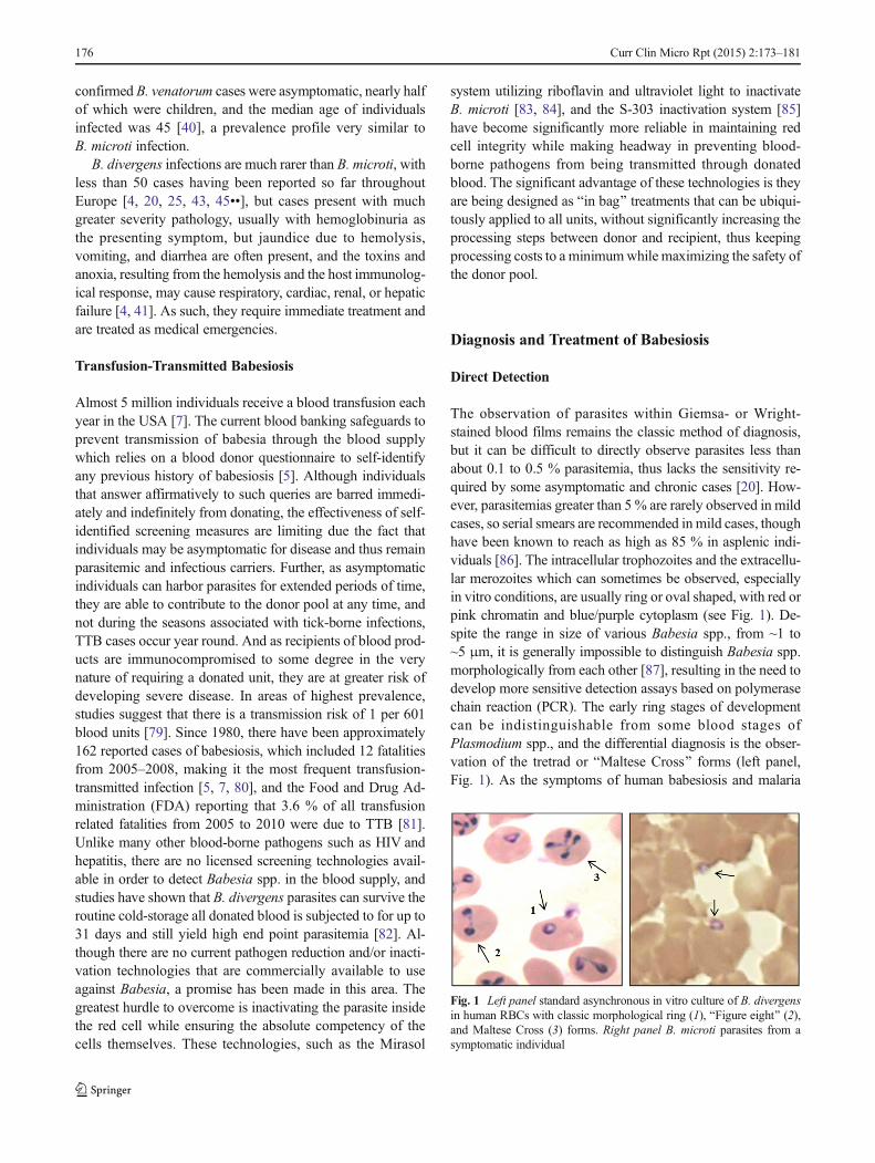
The observation of parasites within Giemsa- or Wright-stained blood films remains the classic method of diagnosis,but it can be difficult to directly observe parasites less thanabout 0.1 to 0.5 % parasitemia, thus lacks the sensitivity re-quired by some asymptomatic and chronic cases [20]. How-ever, parasitemias greater than 5 % are rarely observed in mildcases, so serial smears are recommended inmild cases, thoughhave been known to reach as high as 85 % in asplenic indi-viduals [86]. The intracellular trophozoites and the extracellu-lar merozoites which can sometimes be observed, especiallyin vitro conditions, are usually ring or oval shaped, with red orpink chromatin and blue/purple cytoplasm (see Fig. 1). De-spite the range in size of various Babesia spp., from ~1 to~5 μm, it is generally impossible to distinguish Babesia spp.morphologically from each other [87], resulting in the need todevelop more sensitive detection assays based on polymerasechain reaction (PCR). The early ring stages of developmentcan be indistinguishable from some blood stages ofPlasmodium spp., and the differential diagnosis is the obser-vation of the tretrad or BMaltese Cross^ forms (left panel,Fig. 1). As the symptoms of human babesiosis and malaria
Fig. 1 Left panel standard asynchronous in vitro culture of B. divergensin human RBCs with classic morphological ring (1), BFigure eight^ (2),and Maltese Cross (3) forms. Right panel B. microti parasites from asymptomatic individual
176 Curr Clin Micro Rpt (2015) 2:173–181
are often similar, this differential diagnosis is essential, espe-cially in regions where these pathogens are known to coexist,especially as the trophozoites, schizonts, and particularly ga-metocyte stages of Plasmodium spp. can be more easily rec-ognized if present in peripheral blood smear, meaning co-infections can be easily overlooked.
The PCR particularly real-time or quantitative (qPCR)based on the 18s rRNA gene is much more sensitivethan microscopy and are becoming common for the de-tection of B. microti, where the limits of pathogen de-tection are about ~100 gene copies, equivalent to about5–10 parasites/μL [88, 89], and PCR technology lendsitself to high-throughput screening more readily thanmicroscopy. One drawback of the qPCR method is theneed for a standard curve for each assay, and as yet, nomultiplex exists to detect multiple species per sample.Adaptations to the qPCR method include the dropletdigital PCR (ddPCR), where the amplification reactioncontaining the DNA sample, fluorescently labeled probe,is divided into many microscopic reaction droplets, eachcontaining one or less copies of the target DNA. Thistechnology has recently been applied to the detection ofB. microti and B. duncani and was shown to discrimi-nate B. microti from B. duncani at a limit of limits ofdetection of ~10 gene copies [90].
Indirect Detection
As chronic infections do not always present with ob-servable levels of parasites, serological testing, usuallyin the form of immunofluorescence assay, can be usedto support clinical diagnostics of human babesiosis.Nearly all asymptomatic chronic B. microti infectionsseroconvert in immunocompetent individuals, but thepresence of anti-B. microti antibodies merely suggestan infection at some point in time, as B. microti-specificIgG titers of 1:64 have been detected up 12 monthsafter the parasite infection has cleared. However, IgGtiters exceeding 1:1024 have been shown to correlatewell with acute B. microti infection and is an accurateindicator of recent infection, as well as the presence ofBabesia-specific IgM [91••, 92]. In severe cases, such asB. divergens-infected or immunocompromised individ-uals, serology is rarely performed as the sudden onsetof disease pathology and need for treatment prevents thehost from generating anti-Babesia antibodies, and directdetection methods are usually suitable for diagnosis. Al-though sera from individuals infected with B. microtiand B. duncani do not cross react with antigens of otherBabesia spp., it has been shown that sera fromB. venatorum and B. divergens-like infections does crossreact with B. divergens antigens, meaning, a sero-positive tests for B. divergens would also need further
investigation to determine which of these three specieswas the causative agent [25, 45••].
Treatment
Treatment for mild babesiosis is clindamycin for 7–10 days inB. divergens and B. venatorum [22, 45], with the addition ofquinidine or quinine for B. duncani. As the drug-related tox-icity for quinine can be significant [93], intravenously admin-istered quinidine is a recommended alternative [44, 45••]. Inmild B. microti infections, a 7–10-day course atovaquone plusazithromycin is the combination of choice after it was shownthis is was just as effective but with fewer side effects com-pared with the clindamycin/quinine combination [94]. Indeed,atovaquone plus azithromycin was successfully used to treat aB. venatorum infection in Germany [18], suggesting that in-vestigation into the most effective and most tolerable treat-ments are necessary. It is also suggested that chronic B. microtiparasite-positive but asymptomatic infection lastingmore than3 months are also treated with this same combination [95••].However, in immunocompromised individuals or any severecase with symptoms of severe hemolysis, renal or hepaticdistress, compromised respiratory function, or a parasitemiagreater than 10 %, regardless of species, a 10-day course ofintravenous clindamycin plus quinine coupled with exchangetransfusion is the recommended course of treatment [41, 44,96]. The additive measure of exchange transfusion swiftlyremoves the parasitized RBCs from the host circulation, re-solving any pathologies arising from anemia, such as lowhematocrit and circulating toxic metabolites which releasedinto the host’s circulatory system from the cyclical destructionof the RBCs.
Drug Resistance
Although the two main treatment regimens of clindamycin/quinine or atovaquone plus azithromycin appear to remaineffective in general, problems with speed of response to ther-apy and parasite persistence have been reported both in Eu-rope [18] and in the USA [97], though parasite drug resistanceitself is not suspected in these cases, and highlights the need tomonitor patient parasitemia throughout treatment. However,three incidents of drug resistance to atovaquone/azithromycin,as defined by parasite relapse after more than 28 days of anti-babesial treatment, in immunocompromised patients withB. microti has been reported [95••]. The authors note that theyusedmore stringent definitions of resistance than is commonlyused for other parasites such as malaria. The results highlightthe need to robustly and systematically assess anti-babesialdrugs and compounds where possible. Recent in vitro studieswith artemether and lumefantrine showed a positive synergis-tic interaction of these compounds against Babesia gibsoni[98] and compounds that directly target the pathways of
Curr Clin Micro Rpt (2015) 2:173–181 177
parasite DNA and RNA synthesis of B. gibsoni and B. bovis,such as mycophenolate mofetil, mizoribine, ribavirin, and 7-nitroindole, directly inhibit in the in vitro propagations ofthese parasites.
Conclusions
The clinical epidemiology of human babesiosis appears to bechanging, and this increased awareness will improve the man-agement of local incidence of disease. The pathology of ba-besiosis is a direct consequence of the cyclical replication ofparasites in RBCs. Like malaria, the parasite’s ability to firstrecognize and then invade host RBCs is central to the diseaseprocess, and thus the invasion step provides a vulnerable pointin the parasite’s life cycle. The recent publication of theB. divergens [99••] and B. microti [100••] genomes will great-ly assist the discovery of therapeutic agents that target thisstage in the parasite life cycle. Utilizing these tools to minefor agents that might prove effective against Babesia parasitesis especially important for two diverse reasons. Firstly, thepotential that human pathogens have develop and then swiftlyspread resistance, rendering therapeutic agents ineffective hasbeen shown many times before. Secondly, B. microti cannotcurrently be cultured in vitro and relies upon rodentmodels fortesting the effectiveness of potential anti-B. microti agents,often making such studies difficult to perform or translate intothe human system. The current treatment options for Babesiainfections are limited to clindamycin plus quinidine or quinineor atovaquone plus azithromycin, yet there is evidence thatdrug resistance to atovaquone plus azithromycin may alreadyhave been observed in immunocompromised individuals. Ad-vances in the treatment of donated blood products are a sig-nificant step forward in protecting the blood supply and lim-iting recipients to the risk of transfusion-transmitted babesio-sis, yet more assessment needs to be done to ensure the para-sites are completely inactivated without compromising theintegrity of the blood components. However, it is clear thatas clinical information becomes more readily available,there are significant gaps in our understanding of thebasic biology of these parasites which warrants immedi-ate and extensive investigation.
Compliance with Ethics Guidelines
Conflict of Interest The authors state that this work that has beencompleted in the authors’ laboratory is supported by grants from theNIH to CAL: HL105694 and HL129215 and a grant from the GeorgeLink Jr. Foundation.
Human and Animal Rights and Informed Consent This article con-tains no studies with human or animal subjects performed by the author.
References
Papers of particular interest, published recently, have beenhighlighted as:•• Of very impotance
1. SpielmanA,WilsonML, Levine JF, Piesman J. Ecology of Ixodesdammini-borne human babesiosis and Lyme disease. Annu RevEntomol. 1985;30:439–60.
2. Lantos PM, Krause PJ. Babesiosis: similar tomalaria but different.Pediatr Ann. 2002;31(3):192–7.
3. Schnittger L, Rodriguez AE, Florin-Christensen M, Morrison DA.Babesia: a world emerging. Infect Genet Evol. 2012;12(8):1788–809.
4. Zintl A, Mulcahy G, Skerrett HE, Taylor SM, Gray JS. Babesiadivergens, a bovine blood parasite of veterinary and zoonotic im-portance. Clin Microbiol Rev. 2003;16(4):622–36.
5. Leiby DA. Transfusion-associated babesiosis: shouldn’t we beticked off? Ann Intern Med. 2011;155(8):556–7.
6. Leiby DA. Transfusion-transmitted Babesia spp.: bull’s-eye onBabesia microti. Clin Microbiol Rev. 2011;24(1):14–28.
7. Gubernot DM, Nakhasi HL, Mied PA, Asher DM, Epstein JS,Kumar S. Transfusion-transmitted babesiosis in the UnitedStates: summary of a workshop. Transfusion. 2009;49(12):2759–71.
8. Yager PH, Luginbuhl LM, Dekker JP. Case records of theMassachusetts General Hospital. Case 6–2014. A 35-day-oldboy with fever, vomiting, mottled skin, and severe anemia. NEngl J Med. 2014;370(8):753–62.
9. Aderinboye O, Syed SS. Congenital babesiosis in a four-week-oldfemale infant. Pediatr Infect Dis J. 2010;29(2):188.
10. New DL, Quinn JB, Qureshi MZ, Sigler SJ. Vertically transmittedbabesiosis. J Pediatr. 1997;131(1 Pt 1):163–4.
11. Sethi S, Alcid D, Kesarwala H, Tolan Jr RW. Probable congenitalbabesiosis in infant, New Jersey. USA Emerg Infect Dis.2009;15(5):788–91.
12. Western KA, Benson GD, Gleason NN, Healy GR, Schultz MG.Babesiosis in a Massachusetts resident. N Engl J Med.1970;283(16):854–6.
13. Skrabalo Z, Deanovic Z. Piroplasmosis in man; report of a case.Doc Med Geogr Trop. 1957;9(1):11–6.
14. Bloch EM, Herwaldt BL, Leiby DA, Shaieb A, Herron RM,Chervenak M, et al. The third described case of transfusion-transmitted Babesia duncani. Transfusion. 2012;52(7):1517–22.
15. Conrad PA, Kjemtrup AM, Carreno RA, Thomford J,Wainwright K, Eberhard M, et al. Description of Babesiaduncani n.sp. (Apicomplexa: Babesiidae) from humans andits differentiation from other piroplasms. Int J Parasitol.2006;36(7):779–89.
16. Jiang JF, Zheng YC, Jiang RR, Li H, Huo QB, Jiang BG, et al.Epidemiological, clinical, and laboratory characteristics of 48cases of BBabesia venatorum^ infection in China: a descriptivestudy. Lancet Infect Dis. 2015;15(2):196–203.
17. Sun Y, Li SG, Jiang JF, Wang X, Zhang Y, Wang H, et al. Babesiavenatorum infection in child. China Emerg Infect Dis. 2014;20(5):896–7.
18. Haselbarth K, Tenter AM, Brade V, Krieger G, Hunfeld KP. Firstcase of human babesiosis in Germany—clinical presentation andmolecular characterisation of the pathogen. Int J Med Microbiol.2007;297(3):197–204.
19. Herwaldt BL, Caccio S, Gherlinzoni F, Aspock H, Slemenda SB,Piccaluga P, et al. Molecular characterization of a non-Babesia
•• Of very importance
178 Curr Clin Micro Rpt (2015) 2:173–181
divergens organism causing zoonotic babesiosis in Europe. EmergInfect Dis. 2003;9(8):942–8.
20. YabsleyMJ, ShockBC.Natural history of Zoonotic Babesia: role ofwildlife reservoirs. Int J Parasitol Parasites Wildl. 2013;2:18–31.
21. Spielman A. Human babesiosis on Nantucket Island: transmissionby nymphal Ixodes ticks. Am J TropMedHyg. 1976;25(6):784–7.
22. Vannier E, Krause PJ. Human babesiosis. N Engl J Med.2012;366(25):2397–407.
23. Hersh MH, Tibbetts M, Strauss M, Ostfeld RS, Keesing F.Reservoir competence of wildlife host species for Babesia microti.Emerg Infect Dis. 2012;18(12):1951–7.
24. Blaschitz M, Narodoslavsky-Gfoller M, Kanzler M, Stanek G,Walochnik J. Babesia species occurring in Austrian Ixodes ricinusticks. Appl Environ Microbiol. 2008;74(15):4841–6.
25. Vannier EG, Diuk-Wasser MA, Ben Mamoun C, Krause PJ.Babesiosis. Infect Dis Clin North Am. 2015;29(2):357–70.
26. Etkind P, Piesman J, Ruebush 2nd TK, Spielman A, Juranek DD.Methods for detecting Babesia microti infection in wild rodents. JParasitol. 1980;66(1):107–10.
27. Piesman J, Mather TN, Dammin GJ, Telford 3rd SR, LastavicaCC, Spielman A. Seasonal variation of transmission risk of Lymedisease and human babesiosis. Am J Epidemiol. 1987;126(6):1187–9.
28. Buckingham SC. Tick-borne diseases of the USA: ten things cli-nicians should know. J Infect. 2015;71 Suppl 1:S88–96.
29. Scholtens RG, Braff EH, Healey GA, Gleason N. A case of babe-siosis in man in the United States. Am J Trop Med Hyg.1968;17(6):810–3.
30. Herwaldt BL, Montgomery S, Woodhall D, Bosserman E.Babesiosis surveillance—18 states, 2011. MMWR Morb MortalWkly Rep. 2012;61(27):505–9.
31. Herwaldt BL, McGovern PC, Gerwel MP, Easton RM,MacGregor RR. Endemic babesiosis in another eastern state:New Jersey. Emerg Infect Dis. 2003;9(2):184–8.
32. Apostolou A, Sorhage F, Tan C. Babesiosis surveillance, NewJersey, USA, 2006–2011. Emerg Infect Dis. 2014;20(8):1407–9.
33. Bullard JM, Ahsanuddin AN, Perry AM, Lindsay LR, IranpourM, Dibernardo A, et al. The first case of locally acquired tick-borne Babesia microti infection in Canada. Can J Infect Dis MedMicrobiol. 2014;25(6):e87–9.
34. Acosta ME, Ender PT, Smith EM, Jahre JA. Babesia microti infec-tion, eastern Pennsylvania. USA Emerg Infect Dis. 2013;19(7):1105–7.
35. Persing DH, Herwaldt BL, Glaser C, Lane RS, Thomford JW,Mathiesen D, et al. Infection with a babesia-like organism innorthern California. N Engl J Med. 1995;332(5):298–303.
36. Kjemtrup AM, Conrad PA. Human babesiosis: an emerging tick-borne disease. Int J Parasitol. 2000;30(12–13):1323–37.
37. Kjemtrup AM, Wainwright K, Miller M, Penzhorn BL, CarrenoRA. Babesia conradae, sp. Nov., a small canine Babesia identifiedin California. Vet Parasitol. 2006;138(1–2):103–11.
38. Herwaldt B, Persing DH, Precigout EA, GoffWL, Mathiesen DA,Taylor PW, et al. A fatal case of babesiosis in Missouri: identifi-cation of another piroplasm that infects humans. Ann Intern Med.1996;124(7):643–50.
39. Beattie JF, Michelson ML, Holman PJ. Acute babesiosis causedby Babesia divergens in a resident of Kentucky. N Engl J Med.2002;347(9):697–8.
40. Herwaldt BL, de Bruyn G, Pieniazek NJ, Homer M, Lofy KH,Slemenda SB, et al. Babesia divergens-like infection, WashingtonState. Emerg Infect Dis. 2004;10(4):622–9.
41. Hunfeld KP, Hildebrandt A, Gray JS. Babesiosis: recent insightsinto an ancient disease. Int J Parasitol. 2008;38(11):1219–37.
42. Rios L, Alvarez G, Blair S. Serological and parasitological studyand report of the first case of human babesiosis in Colombia. RevSoc Bras Med Trop. 2003;36(4):493–8.
43. Gorenflot A,Moubri K, Precigout E, Carcy B, Schetters TP. Humanbabesiosis. Ann Trop Med Parasitol. 1998;92(4):489–501.
44. Gray J, Zintl A, Hildebrandt A, Hunfeld KP, Weiss L. Zoonoticbabesiosis: overview of the disease and novel aspects of pathogenidentity. Ticks Tick Borne Dis. 2010;1(1):3–10.
45.•• Hildebrandt A, Gray JS, Hunfeld KP. Human babesiosis inEurope: what clinicians need to know. Infection. 2013;41(6):1057–72. Focuses on the epidemiology, diagnostics and treat-ment of human babesiosis strictly within the European setting,where babesiosis mainly caused by B. divergens is much lessfrequent than other regions, but often presents as a more se-vere disease.
46. Morch K, Holmaas G, Frolander PS, Kristoffersen EK. Severe hu-man Babesia divergens infection in Norway. Int J Infect Dis.2015;33:37–8.
47. Hildebrandt A, Hunfeld KP, Baier M, Krumbholz A, Sachse S,Lorenzen T, et al. First confirmed autochthonous case of humanBabesia microti infection in Europe. Eur J Clin Microbiol InfectDis. 2007;26(8):595–601.
48. Welc-Faleciak R, Pawelczyk A, Radkowski M, Pancewicz SA,Zajkowska J, Sinski E. First report of two asymptomatic cases ofhuman infection with Babesia microti (Franca, 1910) in Poland.Ann Agric Environ Med. 2015;22(1):51–4.
49. El-Bahnasawy MM, Khalil HH, Morsy TA. Babesiosis in anEgyptian boy aquired from pet dog, and a general review. JEgypt Soc Parasitol. 2011;41(1):99–108.
50. Shih CM, Liu LP, Chung WC, Ong SJ, Wang CC. Human babesi-osis in Taiwan: asymptomatic infection with a Babesia microti-likeorganism in a Taiwanese woman. J Clin Microbiol. 1997;35(2):450–4.
51. Shaio MF, Lin PR. A case study of cytokine profiles in acutehuman babesiosis. Am J Trop Med Hyg. 1998;58(3):335–7.
52. Wei Q, Tsuji M, Zamoto A, Kohsaki M, Matsui T, Shiota T, et al.Human babesiosis in Japan: isolation of Babesia microti-like par-asites from an asymptomatic transfusion donor and from a rodentfrom an area where babesiosis is endemic. J Clin Microbiol.2001;39(6):2178–83.
53. Kim JY, Cho SH, Joo HN, Tsuji M, Cho SR, Park IJ, et al. Firstcase of human babesiosis in Korea: detection and characterizationof a novel type of Babesia sp. (KO1) similar to ovine babesia. JClin Microbiol. 2007;45(6):2084–7.
54. Paparini A, Senanayake SN, Ryan UM, Irwin PJ. Molecular con-firmation of the first autochthonous case of human babesiosis inAustralia using a novel primer set for the beta-tubulin gene. ExpParasitol. 2014;141:93–7.
55. Senanayake SN, Paparini A, Latimer M, Andriolo K, Dasilva AJ,Wilson H, et al. First report of human babesiosis in Australia. MedJ Aust. 2012;196(5):350–2.
56. Mayne PJ. Emerging incidence of Lyme borreliosis, babesiosis,bartonellosis, and granulocytic ehrlichiosis in Australia. Int J GenMed. 2011;4:845–52.
57. Mayne PJ. Clinical determinants of Lyme borreliosis, babesiosis,bartonellosis, anaplasmosis, and ehrlichiosis in an Australian co-hort. Int J Gen Med. 2015;8:15–26.
58. Zhou X, Xia S, Huang JL, Tambo E, Zhuge HX, Zhou XN.Human babesiosis, an emerging tick-borne disease in thePeople’s Republic of China. Parasit Vectors. 2014;7:509.
59. Zhou X, Li SG, Chen SB, Wang JZ, Xu B, Zhou HJ, et al. Co-infections with Babesia microti and Plasmodium parasites alongthe China-Myanmar border. Infect Dis Poverty. 2013;2(1):24.
60. Qi C, Zhou D, Liu J, Cheng Z, Zhang L, Wang L, et al.Detection of Babesia divergens using molecular methods inanemic patients in Shandong Province, China. ParasitolRes. 2011;109(1):241–5.
61. Wang H, Huang F. Babesia infection in the southwest of China, acase report. Jundishapur J Microbiol. 2014;7(11):e13504.
Curr Clin Micro Rpt (2015) 2:173–181 179
62. Zhou X, Li SG, Wang JZ, Huang JL, Zhou HJ, Chen JH, et al.Emergence of human babesiosis along the border of China withMyanmar: detection by PCR and confirmation by sequencing.Emerg Microbes Infect. 2014;3(8):e55.
63. Cursino-Santos JR, HalversonG, RodriguezM, NarlaM, Lobo CA.Identification of binding domains on red blood cell glycophorins forBabesia divergens. Transfusion. 2014;54(4):982–9.
64. Lobo CA. Babesia divergens and Plasmodium falciparum usecommon receptors, glycophorins A and B, to invade the humanred blood cell. Infect Immun. 2005;73(1):649–51.
65. Lobo CA, Rodriguez M, Cursino-Santos JR. Babesia and red cellinvasion. Curr Opin Hematol. 2012;19(3):170–5.
66. Montero E, Rafiq S, Heck S, Lobo CA. Inhibition of human eryth-rocyte invasion by Babesia divergens using serine protease inhib-itors. Mol Biochem Parasitol. 2007;153(1):80–4.
67. Montero E, Rodriguez M, Gonzalez LM, Lobo CA. Babesiadivergens: identification and characterization of BdHSP-20, asmall heat shock protein. Exp Parasitol. 2008;119(2):238–45.
68. Montero E, Rodriguez M, Oksov Y, Lobo CA. Babesia divergensapical membrane antigen 1 and its interaction with the human redblood cell. Infect Immun. 2009;77(11):4783–93.
69. Rodriguez M, Alhassan A, Ord RL, Cursino-Santos JR, Singh M,Gray J, et al. Identification and characterization of theRouenBd1987 Babesia divergens Rhopty-associated protein 1.PLoS One. 2014;9(9):e107727.
70. Repnik U, Gangopadhyay P, Bietz S, Przyborski JM, Griffiths G,Lingelbach K. The apicomplexan parasite Babesia divergens in-ternalizes band 3, glycophorin A and spectrin during invasion ofhuman red blood cells. Cell Microbiol. 2015.
71. Hunfeld KP, Brade V. Zoonotic Babesia: possibly emerging path-ogens to be considered for tick-infested humans in Central Europe.Int J Med Microbiol. 2004;293 Suppl 37:93–103.
72. Mylonakis E.When to suspect and how tomonitor babesiosis. AmFam Physician. 2001;63(10):1969–74.
73. Krause PJ, McKay K, Gadbaw J, Christianson D, Closter L,Lepore T, et al. Increasing health burden of human babesiosis inendemic sites. Am J Trop Med Hyg. 2003;68(4):431–6.
74. Filstein MR, Benach JL, White DJ, Brody BA, Goldman WD,Bakal CW, et al. Serosurvey for human babesiosis in New York.J Infect Dis. 1980;141(4):518–21.
75. Krause PJ, Telford 3rd SR, Ryan R, Hurta AB, Kwasnik I, LugerS, et al. Geographical and temporal distribution of babesial infec-tion in Connecticut. J Clin Microbiol. 1991;29(1):1–4.
76. White DJ, Talarico J, Chang HG, Birkhead GS, Heimberger T,Morse DL. Human babesiosis in New York State: review of 139hospitalized cases and analysis of prognostic factors. Arch InternMed. 1998;158(19):2149–54.
77. Bruning G. Localization of NADPH diaphorase, a histochemicalmarker for nitric oxide synthase, in the mouse spinal cord. ActaHistochem. 1992;93(2):397–401.
78. Wozniak EJ, Lowenstine LJ, Hemmer R, Robinson T, Conrad PA.Comparative pathogenesis of human WA1 and Babesia microtiisolates in a Syrian hamster model. Lab Anim Sci. 1996;46(5):507–15.
79. Gerber MA, Shapiro ED, Krause PJ, Cable RG, Badon SJ, RyanRW. The risk of acquiring Lyme disease or babesiosis from ablood transfusion. J Infect Dis. 1994;170(1):231–4.
80. Gubernot DM, Lucey CT, Lee KC, ConleyGB, Holness LG,WiseRP. Babesia infection through blood transfusions: reports receivedby the US Food and Drug Administration, 1997–2007. Clin InfectDis. 2009;48(1):25–30.
81. Cushing M, Shaz B. Transfusion-transmitted babesiosis: achiev-ing successful mitigation while balancing cost and donor loss.Transfusion. 2012;52(7):1404–7.
82. Cursino-Santos JR, Alhassan A, Singh M, Lobo CA. Babesia:impact of cold storage on the survival and the viability of parasitesin blood bags. Transfusion. 2014;54(3):585–91.
83. Tonnetti L, Eder AF, Dy B, Kennedy J, Pisciotto P, Benjamin RJ,et al. Transfusion-transmitted Babesia microti identified throughhemovigilance. Transfusion. 2009.
84. Tonnetti L, Thorp AM, Reddy HL, Keil SD, Goodrich RP, LeibyDA. Riboflavin and ultraviolet light reduce the infectivity ofBabesia microti in whole blood. Transfusion. 2012.
85. Winter KM, Johnson L, Kwok M, Vidovic D, Hyland RA, MuftiN, et al. Red blood cell in vitro quality and function is maintainedafter S-303 pathogen inactivation treatment. Transfusion.2014;54(7):1798–807.
86. Sun T, Tenenbaum MJ, Greenspan J, Teichberg S, Wang RT,Degnan T, et al. Morphologic and clinical observations in humaninfection with Babesia microti. J Infect Dis. 1983;148(2):239–48.
87. Demeter Z, Palade EA, Balogh E, Jakab C, Farkas R, Tanczos B,et al. Postmortem small babesia-like morphology of Babesiacanis—short communication. Acta Vet Hung. 2011;59(4):427–32.
88. Teal AE, Habura A, Ennis J, Keithly JS, Madison-Antenucci S. Anew real-time PCR assay for improved detection of the parasiteBabesia microti. J Clin Microbiol. 2012;50(3):903–8.
89. Bloch EM, Lee TH, Krause PJ, Telford SR, 3rd, Montalvo L,Chafets D, et al. Development of a real-time polymerase chainreaction assay for sensitive detection and quantitation of Babesiamicroti infection. Transfusion. 2013.
90. Wilson M, Glaser KC, Adams-Fish D, Boley M, Mayda M,Molestina RE. Development of droplet digital PCR for the detectionof Babesia microti and Babesia duncani. Exp Parasitol. 2015;149:24–31.
91.•• Vannier E, Krause PJ. Babesiosis in China, an emerging threat.Lancet Infect Dis. 2015;15(2):137–9. Discusses the recent andsudden emergence of human babesiosis from new geographi-cal regions, giving context to the emergence of B. venatorumexpansion within China.
92. Vannier E, Krause PJ. Update on babesiosis. Interdiscip PerspectInfect Dis. 2009;2009:984568.
93. Brasseur P, Lecoublet S, Kapel N, Favennec L, Ballet JJ. Quininein the treatment of Babesia divergens infections in humans. Eur JClin Microbiol Infect Dis. 1996;15(10):840–1.
94. Krause PJ, Lepore T, Sikand VK, Gadbaw Jr J, Burke G, Telford3rd SR, et al. Atovaquone and azithromycin for the treatment ofbabesiosis. N Engl J Med. 2000;343(20):1454–8.
95.•• Wormser GP, Prasad A, Neuhaus E, Joshi S, Nowakowski J,Nelson J, et al. Emergence of resistance to azithromycin-atovaquone in immunocompromised patients with Babesiamicroti infection. Clin Infect Dis. 2010;50(3):381–6. Currentlyavailable treatments for B.microti are limited. Infection eitherresolves spontaneously, or after of a course of azithromycinplus atovaquone or clindamycin plus quinine. Sub curativeresponses were reported in immunocompromised individualstreated with azithromycin-atovaquone, indicating that B.microti may become resistant to this regime, and highlightingthe need for research into drug regimens for immunocompro-mised individuals as well as research into alternative thera-peutics should full resistance emerge.
96. Nathavitharana RR, Mitty JA. Diseases from North America: fo-cus on tick-borne infections. Clin Med. 2015;15(1):74–7.
97. Krause PJ, Spielman A, Telford 3rd SR, Sikand VK, McKay K,Christianson D, et al. Persistent parasitemia after acute babesiosis.N Engl J Med. 1998;339(3):160–5.
98. Iguchi A, Matsuu A, Matsuyama K, Hikasa Y. The efficacy ofartemisinin, artemether, and lumefantrine against Babesia gibsoniin vitro. Parasitol Int. 2015;64(2):190–3.
99.•• Cuesta I, Gonzalez LM, Estrada K, Grande R, Zaballos A, LoboCA, et al. High-quality draft genome sequence of Babesia
180 Curr Clin Micro Rpt (2015) 2:173–181
divergens, the etiological agent of cattle and human babesiosis.Genome Announc. 2014;2(6). Large scaffolds were obtainedduring the sequencing of the B. divergens genome, indicatingreconstruction of almost complete chromosomes, and 80%completeness of the genomewith a degree of continuity obtain-ed in this study is shown to be superior to others. This providesessential data that can be used as a reference to study thegenome structure of B. divergens in order to understand theregulation of its genes and probe shared synteny with otherApicomplexan species, as well as an essential resource to in-vestigate novel therapeutics.
100.•• Cornillot E, Hadj-Kaddour K, Dassouli A, Noel B, Ranwez V,Vacherie B, et al. Sequencing of the smallest Apicomplexan ge-nome from the human pathogen Babesia microti. Nucleic AcidsRes. 2012;40(18):9102–14. Sequenced the genome of the B.microti, showing it has the smallest nuclear genome amongall Apicomplexan parasites sequenced to date and phylogenet-ic analyses indicate it is significantly distant, and defines a newclade in the phylum Apicomplexa. As B. microti cannot yet becultured in in vitro, these data provide an invaluable resourcefor comparative studies with other human babesia species forthe identification of novel diagnostics and interventions.
Curr Clin Micro Rpt (2015) 2:173–181 181