honors biology chapter 34: introduction to animals
TRANSCRIPT

Human Body SystemsChapters 35-40

Themes
How do systems work together to achieve a shared function?
How do systems maintain homeostasis?
What’s the relationship between a system’s structure and function?

Objectives – SWBAT:
Know that complex organisms are comprised of organ systems, which are comprised of organs, which are comprised of tissues, which are comprised of cells.
Describe how feedback control can act to maintain homeostasis.

Feedback Control Homeostasis is often
maintained through the use of Feedback systems (or loops).
A feedback system uses the consequences of the process (too much or too little produced) to regulate the rate at which the process occurs Consists of a sensor, a
control center, and an effector pathway

Maintaining homeostasis
high
low
hormone 1
lowersbody condition
hormone 2
gland
specific body condition
raisesbody condition
gland
Feedback

liver
pancreas
liver
Regulation of Blood Sugar
blood sugar level(90mg/100ml)
insulin
body cells takeup sugar
from blood
liver storessugar
reducesappetite
glucagon
pancreas
liver releases
sugartriggershunger
high
low
FeedbackEndocrine System Control


Organization in Multi-cellular Organisms
1. Cell –smallest functional unit of life
2. Tissue – group of similar cells that perform a specific function
3. Organ – group of tissues that perform a general function
4. Organ System – group of organs that achieve a major bodily function
5. Organism

Tissue Types
Four major types of tissues:• Muscle: controls the internal movement of materials
in the body and external movements of body parts• Nervous: receives and transmits messages• Epithelial: covers internal and external organ
surfaces • Connective: holds organs in place and binds body
parts together

Body Cavities The organs and systems are housed in
compartments called body cavities to protect them and allow them to contract or expand as needed
Four major human body cavities Cranial: holds the brain Spinal: holds the spine and spinal cord Thoracic: Upper compartment in trunk of body—holds
the heart, esophagus, and respiratory system Abdominal: Lower compartment in trunk of body—
holds the digestive, reproductive, and excretory systems
Thoracic and abdominal cavities are separated by the diaphragm muscle

11 Organ Systems

Objectives – SWBAT:
Identify the functions of 11 major human organ systems (nervous, endocrine, digestive, excretory/urinary, cardiovascular/circulatory, respiratory, muscular, skeletal, reproductive, lymphatic, integumentary).
Distinguish between the mechanical and chemical digestion of foodIdentify the organs of the digestive system, in the order that food passes through them, and their roles in processing food.
Explain the functions of the accessory organs of the
digestive system.

Digestive System
Structures: mouth, pharynx, esophagus, stomach, small intestine, large intestine
Accessory Structures (help digestion, but no food enters them): salivary glands, pancreas, liver, gallbladder
Functions: Acquiring nutrients
Converts food into simpler molecules that can be transported to body cells
Absorbs food molecules into bloodstream
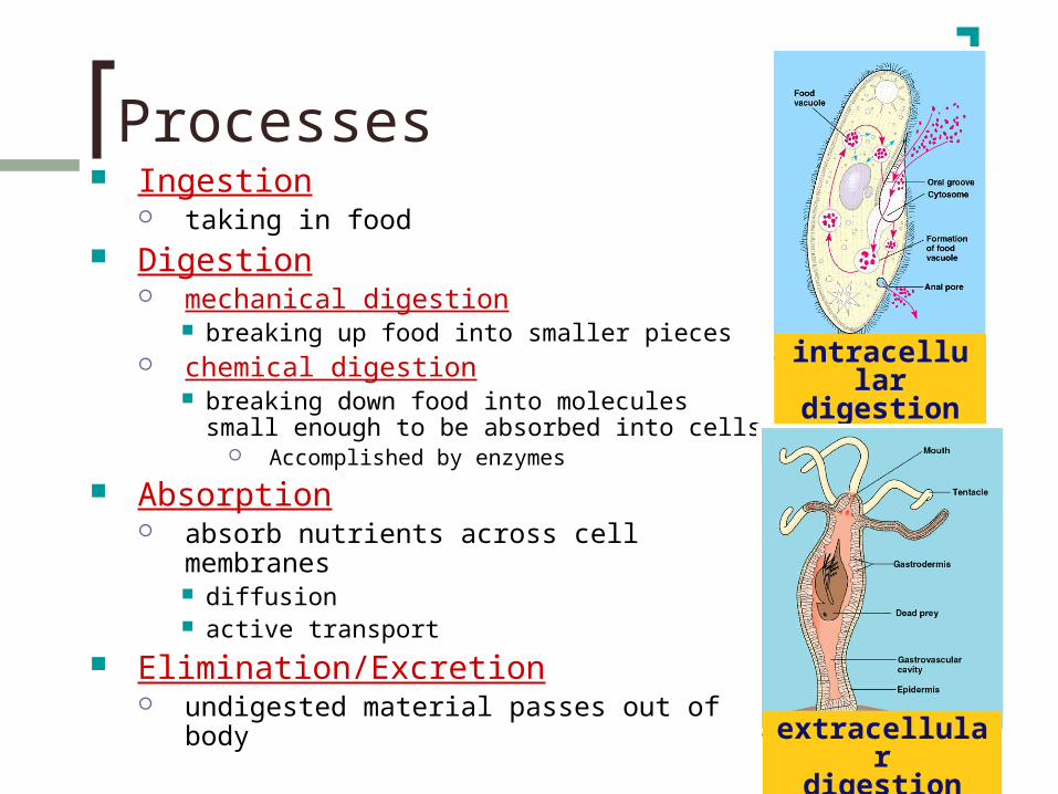
Processes Ingestion
taking in food Digestion
mechanical digestion breaking up food into smaller pieces
chemical digestion breaking down food into molecules small
enough to be absorbed into cells Accomplished by enzymes
Absorption absorb nutrients across cell membranes
diffusion active transport
Elimination/Excretion undigested material passes out of body
intracellulardigestion
extracellulardigestion

Human digestive system

Mouth
Functions mechanical digestion (teeth) chemical digestion (saliva from salivary
glands) amylase enzyme
digests the polysaccharide starch
mucus protects soft lining of digestive system lubricates food for easier swallowing
buffers neutralize acid to prevent tooth decay

Swallowing (& not choking)
Pharynx Uppermost part of throat Epiglottis
flap of tissue in pharynx, closes pathway to respiratory or digestive system to ensure proper pathway for food
Esophagus Peristalsis
involuntary muscle contractions push food to stomach

StomachFunctions
disinfect foodhydrochloric acid = pH 2
kills bacteria
food storagecan stretch to fit ~2L food
digests proteinpepsin enzyme
But the stomach is made out of protein!What stops the stomach from digesting itself?
mucus secreted by stomach cells protects stomach lining

Small intestine Functions: Digestion
Liver and pancreas secrete enzymes into small intestine
digest carbohydrates Enzymes from pancreas
digest proteins Enzymes from pancreas
digest lipids (fats) Bile from liver & enzymes from pancreas
Absorption Absorbs all three nutrient groups into bloodstream Nutrients move into body cells by:
Diffusion, active transport

Absorption in Small Intestines Absorption through villi & microvilli
finger-like projections of intestine wall, full of capillaries (blood vessels)
increases surface area for absorption
SMALL INTESTINES6 meters long,but can stretch
to cover a tennis court
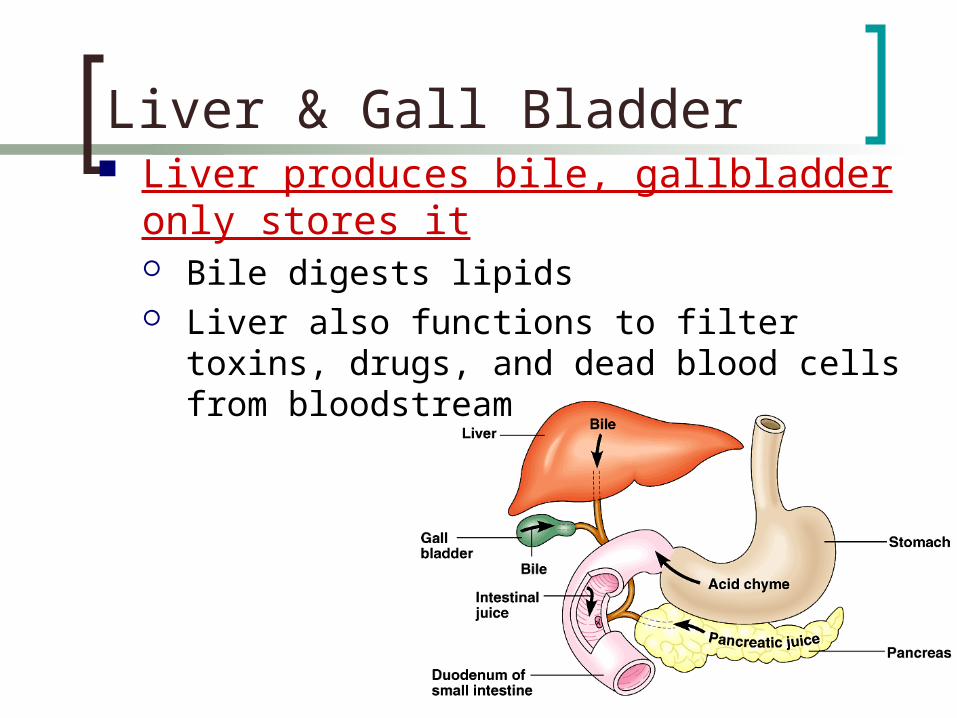
Liver & Gall Bladder Liver produces bile, gallbladder only stores
it Bile digests lipids Liver also functions to filter toxins, drugs, and
dead blood cells from bloodstream

Function Re-absorbs water
You use ~9 liters of water per day in digestive juices if you don’t reabsorb water, death by
dehydration
> 90% of water re-absorbed If not enough water re-absorbed
Diarrhea (can be fatal) If too much water re-absorbed
constipation
Large intestines (colon)

You’ve got company! Living in the large intestine is a
community of mutualistic bacteria E. coli
digest cellulose in fruits & vegetables
produce vitamins vitamin K & B vitamins
but generate gases byproducts of bacterial metabolism

stomachkills germs break up fooddigest proteinsstore food
mouthbreak up fooddigest starchmoisten food
small intestinesbreakdown food
- proteins- starch- fats
absorb nutrients
pancreasproduces enzymes to digest proteins & carbs
liverproduces bile
- stored in gall bladderbreak up fats
large intestinesabsorb water

Rectum
Last section of large intestines eliminate feces
undigested materials mainly cellulose from plants, called
roughage or fiber masses of bacteria dead intestinal cells

Objectives – SWBAT:
Identify the functions of 11 major human organ systems (nervous, endocrine, digestive, excretory/urinary, cardiovascular/circulatory, respiratory, muscular, skeletal, reproductive, lymphatic, integumentary).
Identify the organs of the respiratory system in the order that air passes through them.
Explain how the respiratory system conducts gas exchange.Identify the main components of the cardiovascular system (heart chambers, vessels) in the order that blood passes through them.
Distinguish between the pathways of oxygenated and deoxygenated blood.
Give examples of things the cardiovascular system transports. Explain the parts of blood and their functions, including the roles of
hemoglobin and bone marrow.

Respiratory System
Structures: nose, pharynx, larynx, trachea, bronchi, bronchioles, lungs (alveoli)
Functions: Gas exchange
Provides oxygen (cellular respiration reactant)
Removes excess carbon dioxide (cellular respiration waste product)

Negative pressure breathing Diaphragm moves down & expands chest
cavity, pulls air into lungs
inhale exhale

Structures
alveoli
Air passes through, in order:
nose/mouth pharynx larynx trachea bronchi bronchioles alveoli
capillaries(circulatory system)

Lungsspongy texture
high surface area for more absorption of O2
cilia move mucus and trapped particles
upward to clear out lungs
line most of respiratory tract from nose to bronchioles

Respiratory Surface Lung interior made of small air sacs called alveoli
– the site of gas exchange Each bronchiole ends in a cluster of them
Thin—for simple diffusion Moist—allow for gases to dissolve for diffusion Surrounded by tiny blood vessels (capillaries)

Moving gases into bloodstream Inhale
Bring air in, O2
diffuses from alveoli to blood
Exhale CO2 diffuses from
blood to alveoli, air pushed out
capillaries(circulatory system)

Breathing and Homeostasis
Homeostasis need to balance O2 in and CO2 out
For cellular respiration Exercise
breathe faster need more ATP, muscles conducting more
respiration
= bring in more O2 & remove more CO2
Disease poor lung or heart function = breathe faster
need to work harder to bring in enough O2 & remove CO2
O2
ATP
CO2

Circulatory System
Structures: heart, blood vessels, blood
Functions: Transport
Brings oxygen, nutrients, and hormones to cells
Carries waste away from cells Helps fight infection
Regulation of body temperature

Blood & blood cells Blood is a tissue of fluid & cells
plasma liquid part of blood dissolved macromolecule monomers (sugars,
amino acids), salts, and more cells
red blood cells (RBC) white blood cells (WBC)
defense & immunity
platelets blood clotting

Red blood cells Small round cells
produced in bone marrow full of hemoglobin, an iron-containing protein
which carries oxygen 5-6 million RBC in drop of human blood
last 3-4 months (120 days) Dead cells filtered out by liver
~3 million RBC destroyed each second
O2O2
O2O2

Blood vesselsarteries
arterioles
capillaries
venules
veins
artery
arteriolesvenules
veins
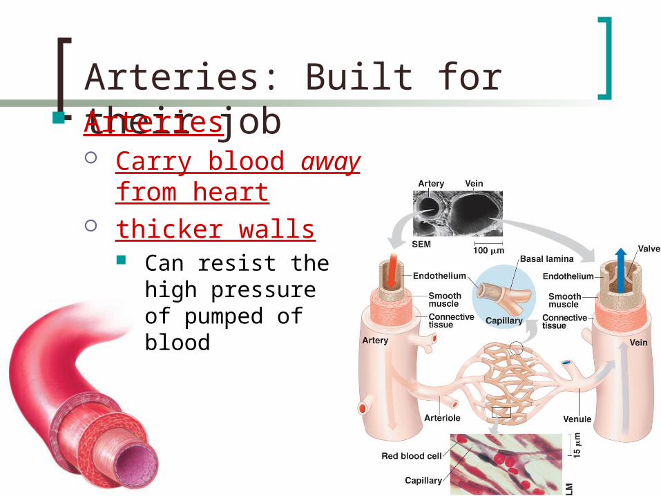
Arteries: Built for their job Arteries
Carry blood away from heart
thicker walls Can resist the high
pressure of pumped of blood

Veins: Built for their job Veins
carry blood back to heart thinner-walled
blood travels back to heart at low speed & pressure
valves in large veins in larger veins one-way valves
allow blood to flow only toward heart
Open valve
Blood flowstoward heart
Closed valve

Structure-function relationship Capillaries
very thin vessels with very thin walls
allows diffusion of materials across capillary O2, food diffuse out of
capillary blood and to nearby body cell
CO2, waste diffuse from body cell into capillary
body cell
O2
food
waste
CO2

Blood In describing circulation, blood is either:
Oxygenated (oxygen is bound to hemoglobin, not much CO2)
Deoxygenated (little oxygen, lots of CO2) (Deoxygenated blood is not blue!)

2 part system Circulation to lungs
blood gets O2, drops off CO2
brings oxygenated blood from lungs to heart
Circulation to body pumps oxygenated blood to
body picks up nutrients from
digestive system collects CO2 & cell wastes
Circulation of Blood
heart
lungs
body
Circulationto lungs
Circulationto body

Stops along the way…
Lungs gas exchange
Small Intestines pick up nutrients from digested food
Large Intestines pick up water from
digested food Liver
clean out toxins and deteriorated blood cells

More stops along the way… Kidneys
filters out cell wastes (urea) and extra water
Bone pick up new red
blood cells Spleen
pick up new white blood cells

Vertebrate Heart
Made of muscle, pumps blood
4 chambers 2 atria (atrium)
Small, thin-walled chambers that receive blood
2 ventricles Large, thick-walled
chambers that pump blood out
rightatrium
leftatrium
rightventricle
leftventricle

Circulatory System: Heart FLOW:
Right side: Deoxygenated blood Right Atrium: Blood comes in from the
body Right Ventricle: Pumps blood to lungs
Left side: Oxygenated blood Left Atrium: Blood returns from lungs Left Ventricle: Pumps blood out to
body
Pacemaker: bundle of tissue, coordinates contraction

AV
SL
AV
Lub-dub, lub-dub 4 valves in the heart
flaps of tissue prevent backflow of blood
Heart sounds caused by closing of valves
Heart murmur = leaking valve, causes a hissing sound

waste
food
Circulatory System & Homeostasis Homeostasis
need to balance food & O2 in, CO2 & waste out
Exercise heart beat faster
In response to muscles’ need for more cellular respiration for more ATP
Disease poor lung or heart function =
heart beat faster need to work harder to bring in O2 &
food & remove wastes Temperature
Blood is hot. More blood in surface capillaries = more body heat lost.
O2
ATP
CO2

Objectives – SWBAT:
Identify the functions of 11 major human organ systems (nervous, endocrine, digestive, excretory/urinary, cardiovascular/circulatory, respiratory, muscular, skeletal, reproductive, lymphatic, integumentary).
Diagram the basic anatomy of a neuron and how neurons communicate.
Distinguish between the central and peripheral nervous system, and sensory vs motor neurons.
Explain the locations and functions of the three types of muscle. Differentiate between connective tissues: cartilage, tendon, and
ligament

Nervous System
Structures: brain, spinal cord, peripheral nerves (sensory and motor)
Function: Communication and
control Coordinates functions
throughout the body in response to internal and external stimuli

Nervous system cells
dendrites
cell body
axon
synapse
Neuron = basic cell of nervous system
signal direction
signaldirection

Action potentials, electrical “messages,” travel down each neuron in one direction.
Neurons consist of 3 parts:• Dendrites – receive messages from other cells• Cell body – contains the nucleus and performs normal
cell metabolism to support cell• Axon – sends messages to other cells

Synapse
synapse
Synapse = junction between nerve cells 1st cell releases chemical
(neurotransmitter) to trigger action potential in the next cell
(FYI: psychoactive drugs affect nervous system by affecting neurotransmission at synapse)

Nervous System
Central nervous system brain & spinal chord “decision making”
Peripheral nervous system body nerves “detecting and action-taking”
cerebrum
cerebellum
spinal cord cervicalnerves
thoracicnerves
lumbarnerves
femoral nerve
sciatic nerve
tibialnerve


Sending and Receiving Messages
Information Collection Sensory neurons– gather information about
what is happening in and around your body and send this information to the CNS for processing. Receptors detect changes inside and outside the body
example, receptors in your eyes detect light.
Delivering Orders Motor neurons– send impulses from the brain
and spinal cord to other systems. When muscles get impulses from motor neurons,
they respond by contracting example, motor neurons signal muscles around your eyes to
contract

Types of neuronssensory neuron(from senses)
interneuron(brain & spinal chord)
motor neuron(to muscle)

Simplest Nerve Circuit Reflex, or automatic response
signal only goes to spinal cord no higher level
processing = rapid response
advantage Fast, don’t need to
think or make decisions about blinking balance pupil dilation startle

Muscular System
Structures: Skeletal muscle, smooth muscle, cardiac muscle
Functions: Movement
Works with skeletal system to produce voluntary movement
Helps circulate blood and move food through the digestive system

Three Types of Muscle: Skeletal: attached to bones by tendons;
responsible for voluntary bodily movement Smooth: controls digestion, breathing,
capillary width; involuntary Cardiac: heart muscle; involuntary

Muscles movement Muscles do work by
contracting contracting = shortening
move skeletal parts involves active transport, so
it requires ATP
tendons tough connective tissue,
connects bone to muscle

Voluntary muscles work in opposing pairs so when one muscle contracts, the other one relaxes
Example: When the bicep muscle (a flexor) contracts and the tricep muscle relaxes, the arm bends.
When the tricep muscle (an extensor) contracts and the bicep muscle relaxes, the arm straightens.
Voluntary Muscle Contractions


Skeletal SystemThe adult human body has approx. 206 bones organized into an internal framework called the skeleton
Structures: bones, cartilage, ligaments, tendons
Functions: Structural support
Supports the body Protects internal organs
Allows movementStores mineral reserves (calcium)Provides a site for blood cell formation
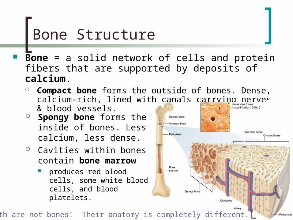
Bone Structure
Bone = a solid network of cells and protein fibers that are supported by deposits of calcium. Compact bone forms the outside of bones. Dense,
calcium-rich, lined with canals carrying nerves & blood vessels.
Spongy bone forms the inside of bones. Less calcium, less dense.
Cavities within bones contain bone marrow produces red blood cells,
some white blood cells, and blood platelets.
FYI, teeth are not bones! Their anatomy is completely different.

Joints
Joints are where bones are joined together. Bones in immovable joints are
fused (ex: skull) Bones in moveable joints are
held together by ligaments. Unlike tendons, which connect
muscle to bone.
Cartilage covers the area of the bones that rub together when joints move, prevent bones from grinding.

Objectives – SWBAT:
Identify the functions of 11 major human organ systems (nervous, endocrine, digestive, excretory/urinary, cardiovascular/circulatory, respiratory, muscular, skeletal, reproductive, lymphatic, integumentary).
Identify the organs involved in removing non-gaseous waste from the bloodstream.
Recognize that the kidneys remove nitrogenous waste and excess water, and the liver filters toxic compounds from the bloodstream.
Know what lymph is. Explain the three lines of defense against pathogens, distinguishing between
specific and non-specific defenses. Explain what a hormone is and how one can act to maintain homeostasis. Give examples of different functions that hormones can regulate. Identify the pathways to fertilization taken by male and female gametes, in the
order that they pass through each structure. Compare and contrast the functions of the male and female reproductive
systems.

Integumentary System
Structures: skin, hair, nails, sweat glands, oil glands
Functions: Serves as a barrier against infection
and injury Provides protection against ultraviolet
radiation from the sun Helps to regulate body temperature Helps remove some waste

Close-up of Integumentary System
Body temperature regulation = sweat evaporation cools skin
Waste function = sweat contains excess salt, water

Excretory System
Structures: kidneys, ureters, urinary bladder, urethra
Functions: Eliminates waste products of
metabolism from the body Maintains homeostasis by
regulating the water content of the blood which controls blood volume, blood pH, and waste

What liquid waste do we make?
Digesting protein makes ammonia nitrogen waste = ammonia = poison
H
CO2 + H2O
NH2 = ammonia
H
HN C—OH
|| O
H|
—C—|

Waste-laden blood enters the kidneys through the renal artery.
Urea, excess water, and other wastes are filtered out of the blood by the kidney and collected as urine.
Urine travels down the ureters, is stored in the bladder, and then is expelled from the body through the urethra.
How do the kidneys work?

Lymphatic SystemStructures: white blood cells, thymus,
spleen, lymph nodes, lymph vessels
Functions: Immune defense and fluid balance
Helps protect the body from disease Collects fluid lost from blood vessels
(lymph), cleans it, and returns it to the circulatory system

Lymph
Lymph = excess tissue fluid pale white, similar to blood
plasma, but with more fats and immune cells
seeps between cells, accumulating “junk” like bacteria, cell debris
Lymph vessel = carries lymph, like blood vessel
Lymph node = filters the “junk” from lymph

Why an immune system? Attack from the outside & inside
lots of organisms want you for lunch! we are a tasty vitamin-packed meal
cells are packages of proteins, carbohydrates & fats no cell wall
animals must defend themselves against invaders… viruses
HIV, flu, cold, measles, chicken pox, SARS bacteria
pneumonia, meningitis, tuberculosis fungi
yeast protists
amoeba, Lyme disease, malaria cancer cells
…but, must do so as efficiently as possible!
What’s forlunch?!

Immune System
Immune defenses may be non-specific or specific Non-specific = broad, defends against
many kinds of attackers Specific = targets one kind or a small
number of attackers
Three lines of defense…
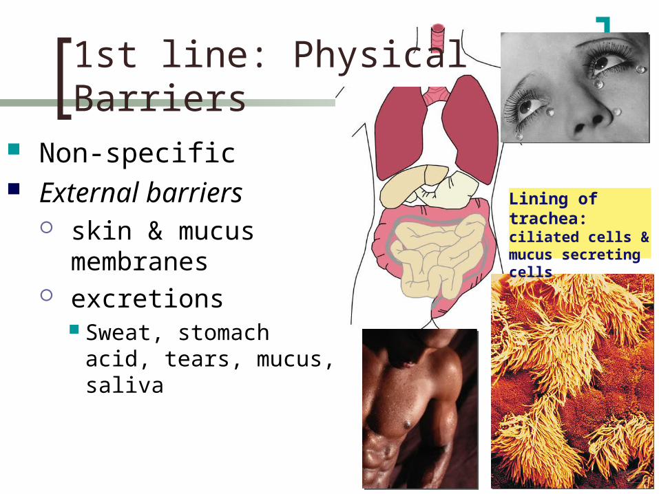
1st line: Physical Barriers
Non-specific External barriers
skin & mucus membranes
excretions Sweat, stomach acid,
tears, mucus, saliva
Lining of trachea:
ciliated cells & mucus secreting cells

2nd line: Generalist responses Non-specific, internal defenses,
including: Patrolling white blood cells
attack invaders that get through the skin phagocyte cells
Macrophages “eat” invaders
Macrophage “eating” bacteria

histamines increases blood
flow =more WBC to
fight, RBC and platelets to repair
2nd line: Generalist responses
Bacteria
Blood vessel
Chemicalalarm
signals
Pin or splinter Blood clot
Phagocytes
Swelling
Inflammation injured cells release chemical signals

2nd line: Generalist responses Fever = elevated body temperature
slows growth of germs (enzyme denaturation) helps macrophages speeds up repair of tissues

3rd line: Lymphocytes Specific defense
responds to specific invaders recognizes specific
foreign antigens white blood cells
B cells T cells
antibodies
B cell
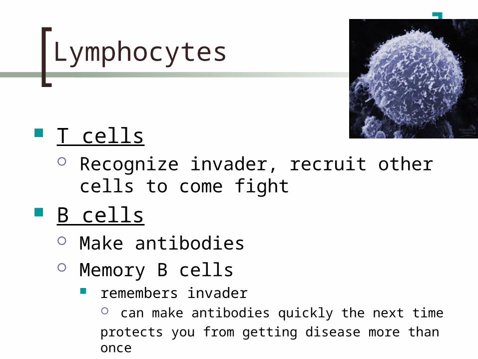
Lymphocytes
T cells Recognize invader, recruit other cells to come
fight B cells
Make antibodies Memory B cells
remembers invader can make antibodies quickly the next time
protects you from getting disease more than once

Proteins that tag invaders in the blood so macrophages can eat them tag says “this is an invader, sic ‘em!”
Antibodies
macrophageeating tagged invaders
invading germs tagged with antibodies Y
Y
YY
YY
Y
B cells releasing antibodies
Y
YY
Y
Y
Y
Y
Y Y
Y
Y
Y
Y
Y
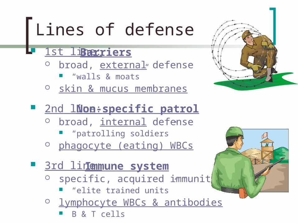
Lines of defense 1st line:
broad, external defense “walls & moats”
skin & mucus membranes
2nd line: broad, internal defense
“patrolling soldiers” phagocyte (eating) WBCs
3rd line: specific, acquired immunity
“elite trained units” lymphocyte WBCs & antibodies
B & T cells
Barriers
Non-specific patrol
Immune system

Endocrine SystemStructures: glands: hypothalamus,
pituitary gland, pineal gland, thyroid gland, parathyroid glands, thymus gland, adrenal glands. Also pancreas, ovaries and testes
Function: Control
Secretes hormones = chemicals that cause changes in other parts of body
growth hormones sex hormones response hormones metabolism hormones and more….

Responding to hormones Lock and key system
hormone fits receptor on “target” cell
targetcell
non-targetcells
secretingcell
can’tread
signal
can’tread
signal

Glands Pineal
melatonin Pituitary
many hormones: master gland
Thyroid thyroxine
Adrenal adrenaline
Pancreas insulin, glucagon
Ovary estrogen
Testes testosterone

Reproductive SystemMale Structures: testes,
epididymis, vas deferens, urethra, penis
Female Structures: ovaries, Fallopian tubes, uterus, vagina
Functions: Reproduction
Produce gametes Males: deliver gametes to females Females: support developing
embryo

Structures Testes: Produce sperm and testosterone. Kept outside of body (in
scrotum) = permits temperature control. Epididymis: Stores sperm as they develop Vas Deferens: Tube from epididymis to urethra Urethra/Penis: Tube within the penis that leads sperm to outside of
body. Semen: Fluid containing hundreds of millions of sperm per ejaculation. Also
contains sugars (energy for sperm), bases (protect sperm from acidic environment of vagina), other chemicals to enhance sperm motility.
Testes produce hormones More testosterone than estrogen
Produces secondary sex characteristics (facial & body hair, increases body size/muscles, deepens voice)
Stimulates sperm production
Reproductive System: Male
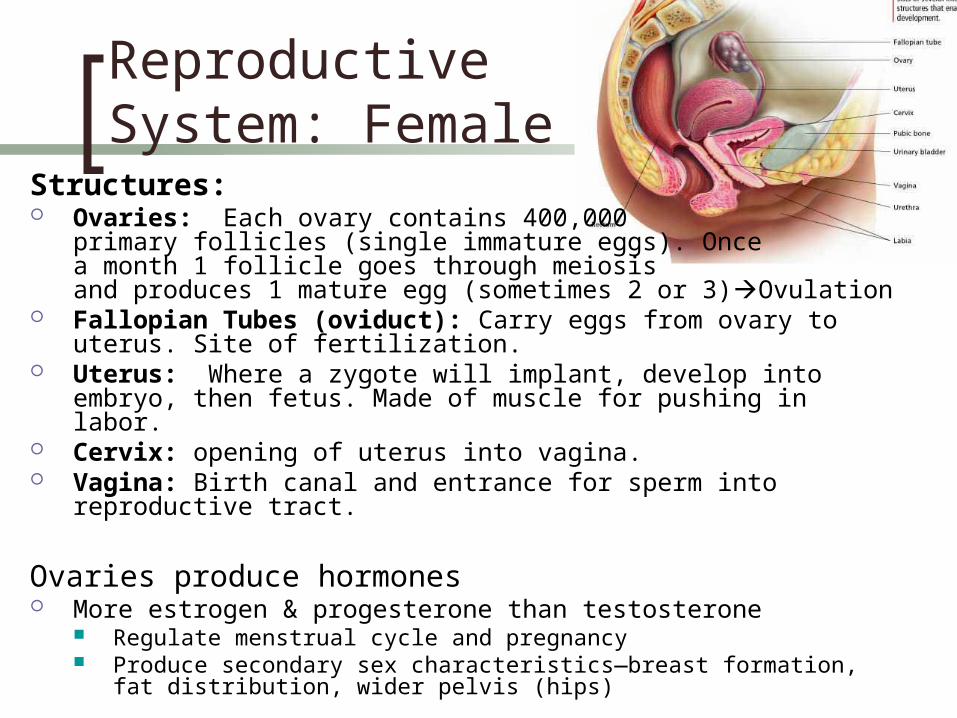
Reproductive System: Female
Structures: Ovaries: Each ovary contains 400,000
primary follicles (single immature eggs). Once a month 1 follicle goes through meiosis and produces 1 mature egg (sometimes 2 or 3)Ovulation
Fallopian Tubes (oviduct): Carry eggs from ovary to uterus. Site of fertilization.
Uterus: Where a zygote will implant, develop into embryo, then fetus. Made of muscle for pushing in labor.
Cervix: opening of uterus into vagina. Vagina: Birth canal and entrance for sperm into reproductive tract.
Ovaries produce hormones More estrogen & progesterone than testosterone
Regulate menstrual cycle and pregnancy Produce secondary sex characteristics—breast formation, fat distribution,
wider pelvis (hips)