hiv aids inhibition by nanoparticles---report
TRANSCRIPT

Modelling of Inhibition of HIV Entry by Silver Nanoparticles
SUMMER INTERNSHIP REPORT May 18
th 2015-23
th July 2015
Faculty In charge
Professor Narendra M Dixit
Associate Professor
Chemical Engineering Department
Indian Institute of Science
Submitted by
Ashwanth Subramanian
3rd Year Chemical Engineering Student
R V College of Engineering
Department of Chemical Engineering
Indian Institute of Science
C V Raman Avenue, Bengaluru – 560012
Table of Contents Page No
1. Introduction 4
2. The Viral Entry Process 6
3. Interaction of silver nanoparticles with HIV-1 8
4. Anti-viral Action modes 10
5. The Idea of Threshold 11
6. The Model 12
7. Conclusions 15
8. Discussions 15
9. Acknowledgement 15
References 15
List of Figures and Tables
Figures:
1. Figure 1: Prevalence of HIV in the adult age group 15-49 years as given by WHO by the end of 2013
2. Figure 2: (a-f): The steps for HIV entry into the host cell.
3. Figure 3: (a) HAADF image with silver nanoparticles.(b)HAADF image without silver nanoparticles
4. Figure 4: % fusion inhibition vs Silver nanoparticle concentration
5. Figure 5: Number of gp120 bound to CD4 vs silver nanoparticle concentration
6. Figure 6: F(Go) vs Go
7. Figure 7: The graph of percentage inhibition vs concentration of nanoparticles.
Tables:
1. Table 1: Antiviral metal nanoparticle
MODELLING OF INHIBITION OF HIV ENTRY BY SILVER NANOPARTICLES
4
HIV AIDS Inhibition by Nanoparticles
Abstract: HIV, the causative agent of a pandemic disease called AIDS, has infected more
than 78 million people till date. Research is going on to find a cure for the disease. Several
approaches have been attempted to find a cure for the disease, and one such promising area
that has emerged recently is using nanotechnology. Metal nanoparticles have been tested for
various medical applications and have shown promising results as antivirals. Elechiguerra et
al and Lara et al have shown that silver nanoparticles can interact with HIV and also the
possible modes of interaction have been hypothesized. In this report, based on the previous
work done, a model has been developed for the inhibition of HIV using silver nanoparticles.
We estimated the rate of disruption by the silver nanoparticles, and the model yields a very
good fit with experimental data. We have also proposed other plausible models with alternate
mechanisms, although scarcity of data prevented reliable prediction of the parameter
estimates used in these models. We conclude that further study must be carried out to better
understand the mechanism of interaction between the HIV and silver nanoparticles and to
estimate key parameters reliably, which would serve nanoparticle-based drug development.
1. INTRODUCTION
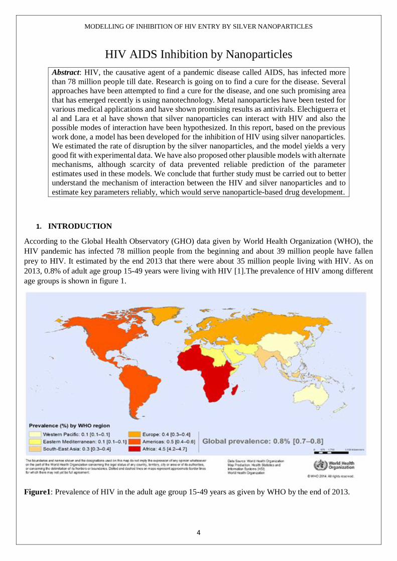
According to the Global Health Observatory (GHO) data given by World Health Organization (WHO), the
HIV pandemic has infected 78 million people from the beginning and about 39 million people have fallen
prey to HIV. It estimated by the end 2013 that there were about 35 million people living with HIV. As on
2013, 0.8% of adult age group 15-49 years were living with HIV [1].The prevalence of HIV among different
age groups is shown in figure 1.
Figure1: Prevalence of HIV in the adult age group 15-49 years as given by WHO by the end of 2013.
MODELLING OF INHIBITION OF HIV ENTRY BY SILVER NANOPARTICLES
5
Some Facts about HIV
The age group which gets affected by HIV is between 15-49 years.
By the end of 2013, 78 million people had been infected with HIV and 39 million had died.
In 2014, 36.9 million people were living with HIV and 2.6 million of those were children.
28 million were the number of people who were eligible for ART by 2013.
At the end of 2014, 13.5 million people had access to antiretroviral therapy in low and middle income
countries.
The outburst of HIV AIDS and the resistance which the HIV develops to the traditional antiretroviral drugs
has lead the researches to pursue search for new antiretroviral agents.
Nanotechnology is a transpiring field from todays’ context which uses the unusual physical and chemical
properties of nanomaterials to make nanoscale-sized materials. Among the nanomaterials, the metal based
nanomaterials have burgeoned to have good potential for application in a wide variety of fields. The use of
these materials in medical field are being extensively researched and is one of the fastest growing areas today.
The physicochemical properties of noble-metal nanomaterials like silver have made them a good candidate
for molecular labelling due to the Surface Plasmon Resonance and large effective scattering cross section of
individual silver nanoparticles [4-5]. Also the toxicity of silver nanoparticles on interaction with
microorganism in various forms is well understood [4,6-8]. Out of the metal based nanoparticles, silver and
gold nanoparticles have been shown to have antimicrobial activity and are extremely appealing as to treat HIV
as antiretroviral agents [2, 9].
Silver is nontoxic to humans if properly used and it has a long history of medical and public health use dating
back 6000 years. There are reports of silvers use in treating wounds by the ancient Egyptians and
Macedonians. Silver has been used to purify water and to treat infections. It has also been used to sterilize
surgical equipment and as plates in the surgical repair of bones. Silver has been used to treat hundreds of
ailments including syphilis, eczema, pneumonia, tuberculosis, pleurisy, gonorrhoea, leg ulcers and impetigo,
from around 1893 until antibiotics came into common use. Silver is classified as a multivalent metallic oxide
rather than a heavy metal.
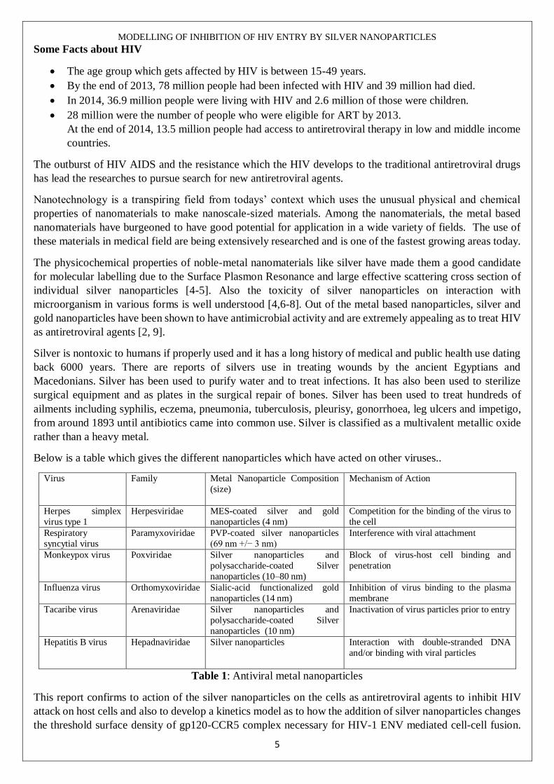
Below is a table which gives the different nanoparticles which have acted on other viruses..
Virus Family Metal Nanoparticle Composition
(size)
Mechanism of Action
Herpes simplex
virus type 1
Herpesviridae MES-coated silver and gold
nanoparticles (4 nm)
Competition for the binding of the virus to
the cell
Respiratory
syncytial virus
Paramyxoviridae PVP-coated silver nanoparticles
(69 nm +/− 3 nm)
Interference with viral attachment
Monkeypox virus Poxviridae Silver nanoparticles and
polysaccharide-coated Silver
nanoparticles (10–80 nm)
Block of virus-host cell binding and
penetration
Influenza virus Orthomyxoviridae Sialic-acid functionalized gold
nanoparticles (14 nm)
Inhibition of virus binding to the plasma
membrane
Tacaribe virus Arenaviridae Silver nanoparticles and
polysaccharide-coated Silver
nanoparticles (10 nm)
Inactivation of virus particles prior to entry
Hepatitis B virus Hepadnaviridae Silver nanoparticles Interaction with double-stranded DNA
and/or binding with viral particles
Table 1: Antiviral metal nanoparticles
This report confirms to action of the silver nanoparticles on the cells as antiretroviral agents to inhibit HIV
attack on host cells and also to develop a kinetics model as to how the addition of silver nanoparticles changes
the threshold surface density of gp120-CCR5 complex necessary for HIV-1 ENV mediated cell-cell fusion.

MODELLING OF INHIBITION OF HIV ENTRY BY SILVER NANOPARTICLES
6
Well understood method to determine the gp120-CCR5 threshold surface density has been published by
Mulampaka et al [3].
2. The Viral Entry Process
First, virions must bind to the target cell, with this being mediated either by the viral envelope (ENV) protein
or host cell membrane proteins incorporated into the virion with any one of a number of various cell attachment
factors.
The second step of virus entry and the first absolutely required for infection entails binding of ENV to its
primary receptor, the host protein CD4. The gp120 subunit is responsible for receptor binding and gp120
contains five relatively conserved domains (C1–C5) and five variable loops (V1– V5), named for their relative
genetic heterogeneity. The variable loops lie predominantly at the surface of gp120andplaycritical roles in
immune evasion and co-receptor binding, particularly the V3 loop. CD4 binding leads to formation of the
bridging sheet. The bridging sheet and repositioned V3 loop play critical roles in the next step of virus entry,
co-receptor engagement.
The third step of virus entry, co-receptor binding, is widely thought to be the trigger that activates the
membrane fusion potential of ENV.
NOTE: Viruses that use the chemokine receptor CCR5 are termed R5 HIV, those that use CXCR4 are termed
X4 HIV, and viruses that can use both co-receptors are called R5X4 HIV.
The fourth step of virus entry is movement of the virus particle to the site where productive membrane fusion
occurs. The virus entry process is shown below diagrammatically in figure 2.
a) Approach

MODELLING OF INHIBITION OF HIV ENTRY BY SILVER NANOPARTICLES
7
b) CD4 binding
c) Co-receptor binding
d) Membrane fusion

MODELLING OF INHIBITION OF HIV ENTRY BY SILVER NANOPARTICLES
8
e) Virus entry into the host cell
f) Multiplication of virus in host cell
Figure 2 (a-f): The steps for HIV entry into the host cell. After step f, the process continues.
3. Interaction of silver nanoparticles with HIV-1
Elechiguerra et al and group in 2005 published a paper which confirms the interaction of silver nanoparticles
with HIV-1. They used silver nanoparticles of different surface chemistries as interaction was affected by the
surface chemistry. The three types were Foamy carbon, PVP and Bovine serum albumin (BSA). The different
types of nanoparticles were characterized by various available techniques like TEM, IR spectroscopy, UV
visible spectroscopy, XPS analysis and HAADF images were taken to confirm the interaction between the
nanoparticles and HIV-1.
They used HAADF to confirm the interaction because it was an already proven technique for analysing
biological sample [13, 14]. The figures 3 a and b show the HAADF images obtained in the experiment they
performed.
They confirmed the silver’s presence with EDS (Energy Dispersive X-ray Spectroscopy) analysis. Also
reported that the nanoparticles which interacted were less than 10nm in size. This showed that the interaction
between HIV-1 and nanoparticles were size dependent.
Another observation they made was that the nanoparticles had a regular spatial arrangement among groups of
three particles. By using work by Leonard [15] which tells that three of the nine glycoproteins are in the
vicinity of CD4 to make a bond with the CD4 cell and also the site for the nanoparticles to attach as the sulphur
residues on gp120 could attract the nanoparticles.

MODELLING OF INHIBITION OF HIV ENTRY BY SILVER NANOPARTICLES
9
(a)
(b)
Figure 3: (a) HAADF image with silver nanoparticles.(b)HAADF image without silver nanoparticles
There are some darker spots which could indicate the low density regions which correspond to glycoprotein
knobs as they have membrane spanning gp41 [4].
By combining the two observations along with the work done by Nermut [11], they conclude that the spatial
arrangements of nanoparticles co-relate with position of the glycoprotein knobs.
Moreover, they had done some spatial analysis in which they found the centre to centre distance between
silver particles which co-relates between the spacing between the glycoprotein knobs as determined before
[12]. Finally, they concluded telling that nanoparticles could interact with HIV-1 by preferential binding.
The glycoprotein knob is roughly of 14 nm in diameter [12]. Hence it can be judged that the nanoparticles
which interact with the HIV-1 should be less than or equal to 14 nm as bigger particles will interact with HIV-

MODELLING OF INHIBITION OF HIV ENTRY BY SILVER NANOPARTICLES
10
1 by using only small fraction of their total area and hence the bond may be unstable. Thus nanoparticles of
size less than or equal to 14 nm would have only participated in bonding and this confirmed closely with their
experimental observation as stated earlier. Their aim was to find a new drug which could which could act as
a fusion inhibitor.
4. Anti-viral Action modes
Lara et al and group had done a study on inhibition of ENV/CD4-mediated membrane fusion [16]. They used
a cell based fusion assay to mimic the gp120-CD4 mediated fusion process. They were able to conclude that
the silver nanoparticles were efficacious in inhibiting the fusion based on concentration by blocking the viral
entry particularly the gp120-CD4 interaction. They were able to conclude that the action of silver nanoparticle
does not depend on the cell tropism. The experimental data they obtained is shown below in figures 4 and 5.
Figure 4: A cell-based fusion assay was used to mimic the gp120-CD4 mediated fusion of the viral and host
cell membranes. HL2/3 and HeLa-CD4-LTR-b-gal cells were incubated with a two-fold serial dilution of
silver nanoparticles and known antiretroviral. The assay was performed in triplicate; the data points represent
the mean ± S.E.M.
They drew important hypothesis which was useful for constructing the model proposed in the model section
of this report.
From the experiment they conducted, they were able to hypothesize that inhibition by silver nanoparticles is
not dependent on the V3 loop which has a net positive charge that contributes to its role in determining viral
co-receptor tropism. The surface of silver nanoparticle is positively charged and hence it was hypothesized
that V3 loop would not be their preferred site of interaction.
Moreover, from the experimental analysis they conducted, they found that nanoparticles may possibly act as
attachment inhibitors by impeding the gp120-CD4 interaction, rather than as co-receptor antagonists that
interfere with the gp120-CXCR4/CCR5 contact. Likewise, silver nanoparticles might interact with the two
disulphide bonds located in the carboxyl half of the HIV-1 gp120 glycoprotein, an area that has been
implicated in binding to the CD4 receptor.

MODELLING OF INHIBITION OF HIV ENTRY BY SILVER NANOPARTICLES
11
Figure 5: The degree of inhibition of the gp120-CD4 protein binding was assessed with a gp120/CD4 ELISA
capture in the presence or absence of silver nanoparticles. Gp120 protein was pre-treated for 10 min with a
two-fold serial dilution of silver nanoparticles, then added to a CD4-coated plate. The assay was done twice;
the error bars indicate the S.E.M.
Another hypothesis is drawn from the fact that Silver ions bind to sulfhydryl groups, which lead to protein
denaturation by the reduction of disulphide bonds. Thus, silver nanoparticles not only bind to gp120 but also
modify this viral protein by denaturing its disulphide-bonded domain located in the CD4 binding region.
Hence silver nanoparticles initially bind to gp120 knobs and then inhibit infection by irreversibly modifying
these viral structures.
5. The Idea of Threshold
The definition of threshold surface density from already published paper by Mulampaka et al says that
threshold surface density is the number of gp120-CCR complexes formed across a closely apposed target cell-
effector cell pair as a function of the CCR5 expression level on the target cell for the virus to enter into the
host cell.
The above was defined for a cell-cell fusion assay. A similar definition can be given as a function of the gp120
expression level on the target cell for the virus to enter into the host cell.
The distribution of gp120 on the effector cell is given by using the error function:
𝑓(𝐺𝑜) = √2
𝜋
𝑒(−(𝐺𝑜−𝐺
𝑜)2
2𝜎𝐺2 )
𝜎𝐺erfc(−𝐺𝑜
√2𝜎𝐺
) ---1
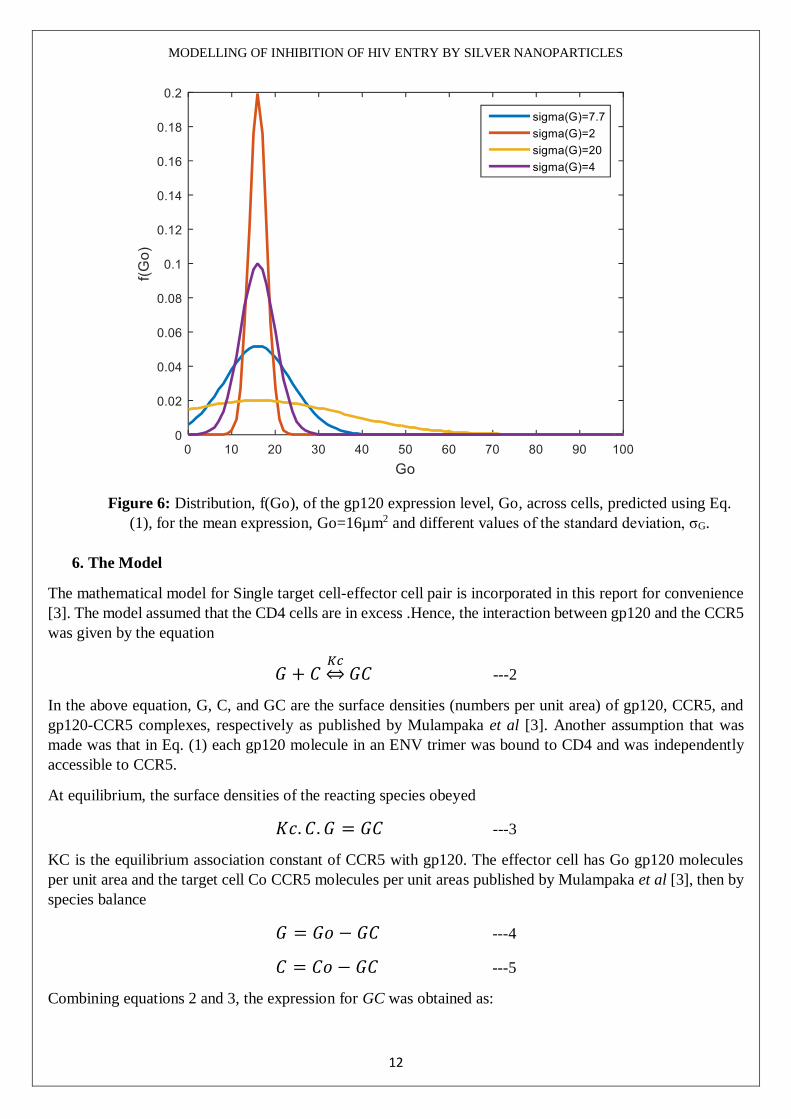
Plotting the distribution f (Go) vs Go we get the below graph shown in figure 6. The parameters were taken
similar to what has been taken in the Mulampaka et al paper.
MODELLING OF INHIBITION OF HIV ENTRY BY SILVER NANOPARTICLES
12
Figure 6: Distribution, f(Go), of the gp120 expression level, Go, across cells, predicted using Eq.
(1), for the mean expression, Go=16µm2 and different values of the standard deviation, σG.
6. The Model
The mathematical model for Single target cell-effector cell pair is incorporated in this report for convenience
[3]. The model assumed that the CD4 cells are in excess .Hence, the interaction between gp120 and the CCR5
was given by the equation
𝐺 + 𝐶𝐾𝑐⇔𝐺𝐶 ---2
In the above equation, G, C, and GC are the surface densities (numbers per unit area) of gp120, CCR5, and
gp120-CCR5 complexes, respectively as published by Mulampaka et al [3]. Another assumption that was
made was that in Eq. (1) each gp120 molecule in an ENV trimer was bound to CD4 and was independently
accessible to CCR5.
At equilibrium, the surface densities of the reacting species obeyed
𝐾𝑐. 𝐶. 𝐺 = 𝐺𝐶 ---3
KC is the equilibrium association constant of CCR5 with gp120. The effector cell has Go gp120 molecules
per unit area and the target cell Co CCR5 molecules per unit areas published by Mulampaka et al [3], then by
species balance
𝐺 = 𝐺𝑜 − 𝐺𝐶 ---4
𝐶 = 𝐶𝑜 − 𝐺𝐶 ---5
Combining equations 2 and 3, the expression for GC was obtained as:

MODELLING OF INHIBITION OF HIV ENTRY BY SILVER NANOPARTICLES
13
𝐺𝐶 =1
2[(
1
𝐾𝑐+ 𝐺𝑜 + 𝐶𝑜) ± √(
1
𝐾𝑐+ 𝐺𝑜 + 𝐶𝑜)
2
− 4𝐶𝑜𝐺𝑜] ---6
Eq. (6) gives the surface density of gp120 bound to a single target cell-effector cell pair as published by
Mulampaka et al [3].
Jose Luis Elechiguerra et al in 2005, demonstrated the size dependent interaction of HIV-1 with silver
nanoparticles. It was showed that the exposed sulphur bearing gp120 glycoprotein knobs were sites for
nanoparticles like silver to interact by preferential binding. Due to the binding, the gp120 are inhibited from
binding to the host cell (In Vitro studies).
When Nanoparticles are added, they attach to the gp120 and form an irreversible bond which can be
represented by the equation
𝐺 +𝑁
(4.19∗10−12)
𝐾𝑛→ 𝐺𝑁 ---7
By species balance for G, we get
𝐺0 = 𝐺 + 𝐺𝐶 + 𝐺𝑁 ---8
Similarly, species balance for N we get,
𝑁𝑜 = 𝑁 + {𝐺𝑁 ∗ (4.19 ∗ 10−12)} ---9
𝑑[𝐺]
𝑑𝑡= −𝐾𝑛. 𝐺.𝑁 ---10
Where,
G is the expression of the gp120 on the cells in µm-2.
Kn is the equilibrium association constant of the gp120 and the nanoparticle nM-1S-1.
N is the concentration of nanoparticles in nM.
𝑁 = 𝑁𝑜 −𝐴
𝑉[𝐺𝑜 − 𝐺 −
𝐾𝑐𝐺𝐶𝑜
1+𝐾𝑐𝐺] ---11
Where,
No is the initial nanoparticle concentration in nM
A is the surface are of the cells on which the nanoparticles act in m2.
V is the volume of in which the drug acts on the cells in m3.
Go is the initial gp120 concentration in µ𝑚-2.
Kc is the equilibrium association constant of gp120 and CCR5 in µm2.
Co is the initial CCR5/CD4 concentration µm-2.
On solving 7 and 9 along with 1 and 2 we can get new Gc in presence of nanoparticles.
NOTE: Other models with formation of intermediate GCN has been given in the supplementary document.
After the equations for all the variables were found in terms of constant, the threshold values of Go and Go(No)
were found to be GoT and Go
T(No)

MODELLING OF INHIBITION OF HIV ENTRY BY SILVER NANOPARTICLES
14
GoT is defined as the expression level of gp120 that would result in the formation of the threshold surface
density GCT of the complex. Likewise, GoT(No) can be defined in presence of the nanoparticles.
Now F, the fraction of cells with Go>GoT is given by
𝐹 =𝑒𝑟𝑓𝑐(
𝐺𝑜𝑇−𝐺𝑜
√2𝜎𝐺)
𝑒𝑟𝑓𝑐(−𝐺𝑜
√2𝜎𝐺) ---12
In presence of the nanoparticles, the fraction of cells with Go>GoT(No) is given by:
𝐹(𝑁𝑜) =𝑒𝑟𝑓𝑐(
𝐺𝑜𝑇(𝑁𝑜)−𝐺𝑜
√2𝜎𝐺)
𝑒𝑟𝑓𝑐(−𝐺𝑜
√2𝜎𝐺)
---13
The percentage inhibition of cell-cell fusion due to the addition of nanoparticles is then
𝐼(𝑁𝑜) = (1 −𝐹(𝑁𝑜)
𝐹) . 100 ---14
The above equation 14 was coded using Matlab to try and fit to the experimental data published by Lara et al.
The graph obtained was as shown below in figure 7.
Figure 7: The graph of percentage inhibition vs concentration of nanoparticles. The red solid line is the curve
obtained according to the model. The two dashed blue lines are the curves according to the model with 10%
change in the parameter value Kn.
Conclusions
1. Nanoparticles have good potential to inhibit the HIV entry into the host cell by inhibiting the target
cell effector cell fusion.
2. From the above graph, it can be seen that the model is a very good fit to the experimental data.

MODELLING OF INHIBITION OF HIV ENTRY BY SILVER NANOPARTICLES
15
3. Other possible mechanisms are also discussed however, due to insufficient data and reliable estimates,
these could not be confirmed.
4. Studies must be conducted to understand the mode of interaction of silver nanoparticles on HIV and
to estimate the parameters reliably and hence could serve as a base for development of nanoparticle
based drug.
Discussion
1. An in depth study must be conducted to find out where the nanoparticles attach on the gp120 to inhibit
the HIV entry into the host cell.
2. The nanoparticles combined with other drugs like AZT can be tried to inhibit the HIV entry. Also,
development of vaccine for the disease could be thought about using nanoparticles.
3. The effect of size of nanoparticles size must be studied.
4. In this project, cell-cell based fusion assay has been considered. Further studies can be conducted on
action of nanoparticles directly on viruses.
5. Effect of time of addition of nanoparticles on the anti HIV activity is also under scope for further study.
The data is provided in the supplementary document in figures S1 to S4.
Acknowledgement
I thank Professor Narendra M Dixit for giving me an opportunity to explore a new path in the research which
is being conducted in his laboratory.
I also thank the PhD scholar Pradeep Nagaraja for supervising my project work.
Finally, I thank the Chemical Engineering department of IISc for giving me an opportunity to work in one of
their labs and giving me a good exposure in doing research in the field of chemical engineering.
References:
[1.] World Health Organization http://www.who.int/gho/hiv/en/ Accessed on 11th June 2015 at 11:26 AM
IST.
[2.] Stefania Galdiero, Annarita Falanga , Mariateresa Vitiello , Marco Cantisani , Veronica Marra and
Massimiliano Galdiero, “Silver Nanoparticles as Potential Antiviral Agents” .Molecules 2011, ISSN 1420-
3049
[3.] Shiva Naresh Mulampaka, Narendra M. Dixit, Estimating the Threshold Surface Density of Gp120-CCR5
Complexes Necessary for HIV-1 Envelope-Mediated CellCell Fusion, PlosOne 2011,e19941.
[4.] Jose Luis Elechiguerra, Justin L Burt, Jose R Morones1, Alejandra Camacho-Bragado, Xiaoxia Gao,
Humberto H Lara and Miguel Jose Yacaman, Interaction of silver nanoparticles with HIV-1, Journal of
Nanobiotechnology 2005, doi:10.1186/1477-3155-3-6
[5.] Schultz S, Smith DR, Mock JJ, Schultz DA: Single-target molecule detection with nonbleaching
multicolor optical immunolabels. Proceedings of the National Academy of Sciences of the United States of
America FIELD Publication Date:2000 Feb 1 97:996-1001. FIELD Reference Number: FIELD Journal
Code:7505876 FIELD Call Number:.
[6.] Liau SY, Read DC, Pugh WJ, Furr JR, Russell AD: Interaction of silver nitrate with readily identifiable
groups: ralationship to the
antibacterial action of silver ions. Lett Appl Microbiol 1997, 25:279-283.
[7.] Gupta A, Silver S: Silver as biocide:Will resistance become a problem? Nat Biotechnol 1998, 16:888.

MODELLING OF INHIBITION OF HIV ENTRY BY SILVER NANOPARTICLES
16
[8.] Nomiya K, Yoshizawa A, Tsukagoshi K, Kasuga NC, Hirakawa S, Watanabe J: Synthesis and
structural characterization of silver (I), aluminium (III) and cobalt(II) complexes with 4-isopropyltropolone
(hinokitiol) showing noteworthy biological activities. Action of silver(I)-oxygen bonding complexes on the
antimicrobial activities. J Inorg Biochem 2004, 98:46-60.
[9.] Sondi I, Salopek-Sondi B: Silver nanoparticles as antimicrobial agent: A case study on E. coli as a
model for Gram-negative bacteria. J Colloid Interface Sci 2004, 275:177-182.
[10.] Leonard CK, Spellman MW, Riddle L, Harris RJ, Thomas JN, Gregory TJ: Assignment of intrachain
disulfide bonds and characterization of potential glycosylation sites of the type 1 recombinant human
immunodeficiency virus envelope glycoprotein (gp120) expressed in Chinese hamster ovary cells. J Biol
Chem 1990, 265:10373-10382
[11.] Forster MJ, Mulloy B, Nermut MV: Molecular modelling study of HIV p17gag (MA) protein shell
utilising data from electron microscopy and X-ray crystallography. J Mol Biol 2000, 298:841-857.
[12.] Gelderblom HR, Hausmann EHS, Ozel M, Pauli G, Koch MA: Fine structure of human
immunodeficiency virus (HIV) and immunolocalization of structural proteins. Virology 1987, 156:171-176.
[13.] Burt JL, Gutierrez-Wing C, Miki-Yoshida M, Jose-Yacaman MJ: Noble-Metal Nanoparticles Directly
Conjugated to Globular Proteins. Langmuir 2004, 20:11778-11783.
[14.] Sweeney RY, Mao C, Gao X, Burt JL, Belcher AM, Georgiou G, Iverson BL: Bacterial Biosynthesis
of Cadmium Sulphide Nanocrystals. Chemistry & Biology 2004, 11:1553-1559.
[15.] Leonard CK, Spellman MW, Riddle L, Harris RJ, Thomas JN, Gregory TJ: Assignment of intrachain
disulfide bonds and characterization of potential glycosylation sites of the type 1 recombinant human
immunodeficiency virus envelope glycoprotein (gp120) expressed in Chinese hamster ovary.
[16.] Humberto H Lara, Nilda V Ayala-Nuñez, Liliana Ixtepan-Turrent, Cristina Rodriguez-Padilla “Mode of
antiviral action of silver nanoparticles against HIV-1”, Journal of Nanobiotechnology 2010