histone deacetylase inhibitors in hodgkin lymphoma
TRANSCRIPT
SPECIAL ISSUE ARTICLE
Histone deacetylase inhibitors in Hodgkin lymphoma
Daniela Buglio & Anas Younes
Received: 27 October 2010 /Accepted: 28 October 2010 /Published online: 3 December 2010# The Author(s) 2010. This article is published with open access at Springerlink.com
Summary Although Hodgkin lymphoma (HL) is consid-ered one of the most curable human cancers, the treatmentof patients with relapsed and refractory disease, especiallythose who relapse after autologous stem cell transplanta-tion, remains challenging. Furthermore, because of theyoung age of these patients, the impact of early mortality onthe number of years lost from productive life is remarkable.Patients with relapsed HL post stem cell transplantationcurrently have no curative therapy, and are in need for newdrugs and novel treatment strategies. While no new drugshave been approved for the treatment of patients with HL inmore than three decades, several new agents are demon-strating promising results in early clinical trials. This reviewwill focus on the emerging role of histone deacetylaseinhibitors in patients with relapsed HL.
Keywords Hodgkin lymphoma . Role of histonedeacetylase inhibitors
Introduction
Epigenetics is a heritable process that alters gene expressionwithout changing the DNA sequence [1]. This processincludes DNA and chromatin modifications by methylation,acetylation, phosphorylation, ubiquitylation, and sumolya-tion. Epigenetic processes are natural and essential to manyorganism functions, but if they occur improperly, they mayresult in major adverse health effects. The best knownepigenetic process is DNA methylation, which involves the
addition of a methyl group (CH3), which primarily occurs atthe 5′ position of the pyrimidine ring of the cytosine. Theresulting methylcytosine is mainly found in cytosine-guanine(CpG) islands [2]. The presence of 5-methylcytosine in thepromoter of specific genes alters the binding of transcrip-tional factors and other proteins to DNA and recruits methyl-DNA-binding proteins and histone deacetylases that compactthe chromatin around the gene-transcription start site. Bothmechanisms block transcription and cause gene silencing.
Another significant epigenetic process is the post-transcriptional modification of histones. Chromatin is thecomplex of histone proteins and DNA that is tightly bundledto fit into the nucleus. The complex can be modified byhistone acetylation, which alters chromatin structure toinfluence gene expression [3]. In general, tightly foldedchromatin tends to be shut down, or not expressed, whilemore open chromatin is functional, or expressed. Among allthe epigenetic research conducted so far, the most extensivelystudied disease family is cancer, and the evidence linkingepigenetic processes with cancer continues to grow [1, 4, 5].
Histone deacetylases (HDACs)
Post-transcriptional histone modification plays an importantrole in regulating gene transcription, and is mediated by avariety of enzymes, including histone acetyltransferases(HATs) and histone deacetylation (HDACs) [3]. Theseenzymes mediate acetylation and deacetylation of specificlysine amino acid residues on histone tails. In general,histone H3 and H4 acetylation is associated with openchromatin status favoring gene transcription, whereashistone deacetylation is associated with closed chromatinstatus favoring transcription repression and gene silencing.The balance between HATs and HDACs is critical for
D. Buglio :A. Younes (*)Department of Lymphoma/Myeloma,1515 Holcombe Boulevard, Box # 429, Houston, TX 77030, USAe-mail: [email protected]
Invest New Drugs (2010) 28 (Suppl 1):S21–S27DOI 10.1007/s10637-010-9588-y
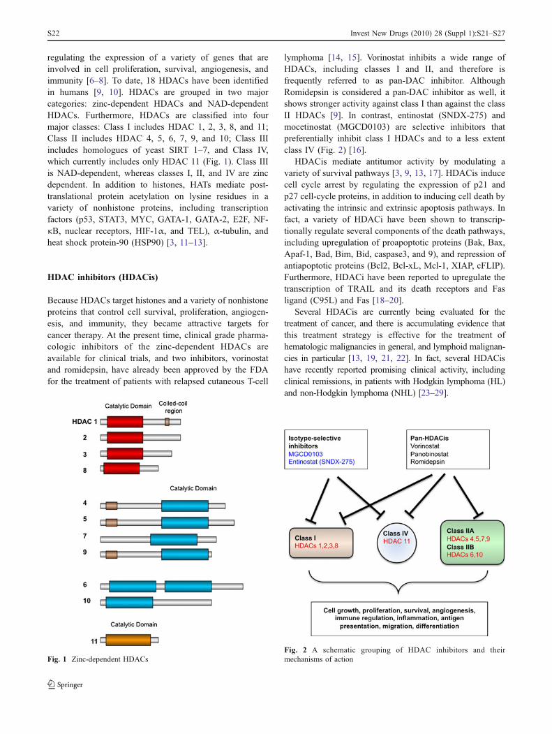
regulating the expression of a variety of genes that areinvolved in cell proliferation, survival, angiogenesis, andimmunity [6–8]. To date, 18 HDACs have been identifiedin humans [9, 10]. HDACs are grouped in two majorcategories: zinc-dependent HDACs and NAD-dependentHDACs. Furthermore, HDACs are classified into fourmajor classes: Class I includes HDAC 1, 2, 3, 8, and 11;Class II includes HDAC 4, 5, 6, 7, 9, and 10; Class IIIincludes homologues of yeast SIRT 1–7, and Class IV,which currently includes only HDAC 11 (Fig. 1). Class IIIis NAD-dependent, whereas classes I, II, and IV are zincdependent. In addition to histones, HATs mediate post-translational protein acetylation on lysine residues in avariety of nonhistone proteins, including transcriptionfactors (p53, STAT3, MYC, GATA-1, GATA-2, E2F, NF-κB, nuclear receptors, HIF-1α, and TEL), α-tubulin, andheat shock protein-90 (HSP90) [3, 11–13].
HDAC inhibitors (HDACis)
Because HDACs target histones and a variety of nonhistoneproteins that control cell survival, proliferation, angiogen-esis, and immunity, they became attractive targets forcancer therapy. At the present time, clinical grade pharma-cologic inhibitors of the zinc-dependent HDACs areavailable for clinical trials, and two inhibitors, vorinostatand romidepsin, have already been approved by the FDAfor the treatment of patients with relapsed cutaneous T-cell
lymphoma [14, 15]. Vorinostat inhibits a wide range ofHDACs, including classes I and II, and therefore isfrequently referred to as pan-DAC inhibitor. AlthoughRomidepsin is considered a pan-DAC inhibitor as well, itshows stronger activity against class I than against the classII HDACs [9]. In contrast, entinostat (SNDX-275) andmocetinostat (MGCD0103) are selective inhibitors thatpreferentially inhibit class I HDACs and to a less extentclass IV (Fig. 2) [16].
HDACis mediate antitumor activity by modulating avariety of survival pathways [3, 9, 13, 17]. HDACis inducecell cycle arrest by regulating the expression of p21 andp27 cell-cycle proteins, in addition to inducing cell death byactivating the intrinsic and extrinsic apoptosis pathways. Infact, a variety of HDACi have been shown to transcrip-tionally regulate several components of the death pathways,including upregulation of proapoptotic proteins (Bak, Bax,Apaf-1, Bad, Bim, Bid, caspase3, and 9), and repression ofantiapoptotic proteins (Bcl2, Bcl-xL, Mcl-1, XIAP, cFLIP).Furthermore, HDACi have been reported to upregulate thetranscription of TRAIL and its death receptors and Fasligand (C95L) and Fas [18–20].
Several HDACis are currently being evaluated for thetreatment of cancer, and there is accumulating evidence thatthis treatment strategy is effective for the treatment ofhematologic malignancies in general, and lymphoid malignan-cies in particular [13, 19, 21, 22]. In fact, several HDACishave recently reported promising clinical activity, includingclinical remissions, in patients with Hodgkin lymphoma (HL)and non-Hodgkin lymphoma (NHL) [23–29].
Fig. 1 Zinc-dependent HDACsFig. 2 A schematic grouping of HDAC inhibitors and theirmechanisms of action
S22 Invest New Drugs (2010) 28 (Suppl 1):S21–S27
Rationale for using epigenetic-based therapy in HL
HRS cells are B cells with epigentically-silenced B-cellgenes Molecular analysis demonstrated that the malignantHodgkin and Reed-Sternberg (HRS) cells of Hodgkinlymphoma (HL) are of B-cell origin. However, HRS cellsinfrequently express B-cell antigens. This loss of B-cellphenotype has been reported to be epigenetically regulatedand is thought to enable HRS cells to evade immunosur-veillance. For example, gene expression profiles of HRScells revealed decreased mRNA levels for nearly all B-celllineage-specific genes [30]. The decrease in mRNAexpression was associated with downregulation of proteinexpression in primary HL cases, as determined by immu-nohistochemistry, including CD20, CD19, Syk, Lck, Blk,Oct-2, and B-cell receptor [30]. Furthermore, microdis-sected primary HRS cells and HL cell lines are down-regulated in HL. Importantly, the expression of thesesilenced genes could be re-induced by the DNA hypome-thylating agent 5-aza-deoxycytidine [31, 32]. A variety ofHDAC inhibitors have demonstrated promising pre-clinicalactivity in HL cell lines by inducing cell cycle arrest andapoptosis [28]. AT the molecular level, HDACis upregu-lated p21, activated the caspase pathway, downregulatedSTAT6 expression, and altered cytokine and chemokinesecretion [33–35]. In fact, we have recently demonstratedthat vorinostat decreased the expression and secretion ofTh2-type cytokines and chemokines, including Thymus andActivation-Regulated Chemokine (TARC/CCL17) and in-terleukin (IL)-5, and increased Th1-type cytokines/chemo-kines, including a profound increase in IP-10 levels,suggesting that HDACi may alter the microenvironmentsurrounding HRS cells to favor cell death [35].
Epigenetic therapy induces the expression of Epstein-Barrvirus antigens and tumor-associated antigens in HRS cells,which are potential targets for cytotoxic T cells Epstein-Barr virus (EBV)-associated antigens are known to beexpressed by a proportion of HRS cells, however, only thesubdominant EBV antigens LMP1 and LMP2 are expressedand provide T-cell target antigens. EBV-specific T cells thatare enriched for LMP-specific T-cell clones have beenexpanded from patients and administered to patients withrelapsed disease [36, 37]. Unfortunately, T cells specific forthe viral latent nuclear antigens, EBNAs 3A, 3B and 3 Cand the immediate early lytic cycle antigen, BZLF1,BRLF1 and BMLF1 dominate the human immune responseto EBV. If these proteins could be reactivated in EBV-positive HRS cells, then their susceptibility to EBV-specificT-cell lines would be increased. Both HDACis andhypomethylating agents induce EBV gene expression.HDACs reactivate EBV from latency to replication, so thatthe immunogenic lytic cycle antigens are expressed [38–
40], while azacytidine can switch latently infected cellsfrom a minimal form of latency (LMP1,LMP2 andEBNA1) to an unrestricted latency in which all EBNAs(2, 3’s and-LP) and LMPs are expressed. Thus, these drugsmay transform poorly antigenic tumors into highly immu-nogenic tumors and increase the number of available targetantigens for T-cell recognition immunodominant viralproteins. In addition, combining different agents likechemotherapy and HDAC inhibitors results in synergisticeffects on activating lytic viral replication [40]. Since onlyabout 20% of patients with relapsed HL have EBV-positivetumors, it is also important to determine if other tumor-associated, T-cell target antigens are upregulated byepigenetic therapy. Cancer Testis Antigens (CTAs) areknown to comprise at least 44 families of genes orisoforms. Among these, 19 families are purely testis-restricted, while others are expressed in one or moresomatic tissues. Some CTAs are expressed in lymphoma[41, 42], and provide potential T-cell targets. CTA-specificT cells from melanoma patients have demonstrated thera-peutic benefits, including complete remissions. CTA-specific CTL could have similar beneficial effects inEBV-negative lymphomas. Recently, hypomethylatingagents have been shown to induce the expression of severalCTAs in cultured tumor cell lines (including lymphomas)[43]. Such upregulated expression has been reported forseveral MAGE members – LAGE-1, SSX-2, CAGE, andNY-ESO-1. Demethylation proved necessary and sufficientfor MAGE-A1 gene expression in melanoma cell lines,suggesting that methylation is a primary mechanism oftranscription control. Histone deacetylase (HDAC) inhib-itors, on their own, or in combination with hypomethylatingagents, can also induce CTA expression, including MAGE,SSX, and NY-ESO-1 family members [43]. Recent datasuggested that HDACi may upregulate the expression ofCTA in HRS, suggesting HDACis may enhance the activityof EBV-directed cellular therapy in HL [33]. Sinceepigenetically enhanced CTA expression may activateendogenous CTA-specific T cells in vivo, this may induceepitope spreading and facilitate the ex vivo activation andexpansion of these normally low frequency CTA-specific Tcells.
Clinical results with HDACi in patients with HL
A variety of HDACis are currently being evaluated inpatients with relapsed HL (Table 1). The following is abrief update on the current status of HDACis in patientswith relapsed HL. Interestingly, the expression pattern ofHDAC enzymes varied among different types of lymphoma[44]. Consistent with their ubiquitous distribution in normal
Invest New Drugs (2010) 28 (Suppl 1):S21–S27 S23
tissues, class I enzymes were highly expressed in allHodgkin and non-Hodgkin lymphoma cell lines andprimary tumors studied, including the non-malignantreactive cells in HL microenvironment. In contrast, theclass II enzyme HDAC6 was infrequently expressed,compared with HDACs 5, 8, and 10. These results suggestthat HDAC6 may not be an important therapeutic target inselected lymphoid malignancies [44].
Vorinostat (SAHA) Vorinostat, which inhibits HDAC clas-ses I and II, has a potent antiproliferative activity in HL celllines, as it induces cell cycle arrest and apoptosis, inaddition to synergizing with chemotherapy [45]. Furthe-more, Vorinostat inhibited STAT6 phosphorylation andtranscription in HL cell lines, an effect that was associatedwith decreased expression and secretion of Th2-typecytokines and chemokines (TARC and IL-5). This effectwas associated with and increases of in Th1-type cytokines/chemokines (IP-10) [45]. The in vivo activity of vorinostatwas recently evaluated in a phase II study that wasconducted by the Southwest Oncology Group (SWOG) inpatients with relapsed HL [16]. Twenty-five patients weretreated with 200 mg vorinostat given orally twice per dayfor 14 days every 21-day cycle. Vorinostat producedmodest clinical activity, as only 1 patient (4%) achieved apartial remission.
Mocetinostat (MGCD-0103) Mecetinostat is a novel oralHDAC inhibitor that selectively inhibits HDAC 1, 2, 3(class I) and 11 (class IV) isoforms, with no effect on class-II HDACs [46]. The safety and efficacy of MGCD-0103given orally 3 times per week (85 to 110 mg starting doses)has been evaluated in a phase II study in patients withrelapsed and refractory HL [26]. Patients were allowed tocontinue therapy up to 1 year in absence of heavy toxicityor disease progression. Of the 20 patients who were treatedwith 110-mg dose level, 7 (35%) patients achieved partialor complete remissions. However, this dose level waspoorly tolerated resulting in dose interruptions and reduc-tions, and discontinuation of therapy after a median of4.5 months. Subsequently, the study was revised to allow alower starting dose of 85 mg at the same schedule. Three ofthe 10 (30%) patients enrolled on the reduced dose
achieved partial remissions. Furthermore, grade 3 and 4toxicity (mainly fatigue, with no significant hematologictoxicity) was reduced to 20%. Overall, 80% of the 30evaluable patients had some decrease in their tumormeasurements. Although none of the patients developedsignificant EKG abnormalities, two patients developedsignificant pericardial effusions requiring discontinuationof therapy [47]. Importantly, Serum TARC levels weredetermined in 15 patients before and 1 week after initiationof therapy [26]. After 1 week of therapy serum TARClevels were decreased by at least 40% in five patients, andall achieved major clinical responses. Collectively, this dataindicate that class I HDAC inhibitors have a potentialtherapeutic value in patients with HL.
Panobinostat (LBH 589) Panobinostat is one of the mostpotent HDACis in HL in vitro, with an IC50 in thenanomolar range [28]. Recently panobinostat was evaluatedin a phase I trial in patients with hematologic malignanciesthat also included patients with relapsed HL [18]. Five of13 (38%) patients achieved partial remissions. The mostcommon side effects were fatigue, thrombocytopenia,nausea, and diarrhea. Based on this promising clinicalactivity, a large international phase II study of panobinostatin relapsed HL, with an early report demonstrating apromising clinical activity in a large multicenter settings[48]. In a most recent update of the study, 61 patients havebeen enrolled and treated: median age 31 years (range 18–70), and 48% had stage III/IV at initial diagnosis. Patientswere heavily pretreated with a median number of priorregimens of 4 (range 2–6 regimens). Of 61 patients, 53have completed two cycles of therapy and have had onepost baseline imaging studies to evaluate response totheapy. Sixty-nine percent had tumor reduction, includingone complete response and 10 partial responses The mostcommon grade 3/4 adverse events were thrombocytopenia(77%), anemia (20%), and neutropenia (16%). Thrombo-cytopenia was reversible within an average of 1 week ofdose interruption. No cases of grade 3/4 QTc prolongationhave been reported. Thus, panobinostat has a promisingclinical activity with a good safety profile. Based on thispromising data, panobinostat-based combination studies arecurrently being conducted, including a phase I/II trial of
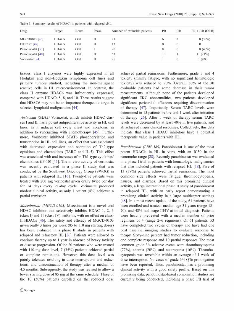
Table 1 Summary results of HDACi in patients with relapsed cHL
Drug Target Route Phase Number of evaluable patients PR CR PR + CR (ORR)
MGCD0103 [26] HDACs Oral II 21 6 2 8 (38%)
ITF2357 [49] HDACs Oral II 13 0 0 0
Panobinostat [51] HDACs Oral I 20 8 0 8 (40%)
Panobinostat [48] HDACs Oral II 53 10 1 11 (21%)
Vorinostat [24] HDACs Oral II 25 1 0 1 (4%)
S24 Invest New Drugs (2010) 28 (Suppl 1):S21–S27

panobinostat plus everolimus in patients with relapsed HLand non-Hodgkin lymphoma.
Entinostat (SNDX-275) Entinostat is an oral, class I iso-form selective HDAC inhibitor. Phase 1 studies in leukemiademonstrated the agent has a long half-life and that weeklyor every other week dosing is sufficient for antitumoractivity. In vitro experiments demonstrated a potent activityof entinostat in HL-derived cell lines, with an IC50 of0.4 μM [33]. At the molecular level, entinostat induced itsantiproliferative effect in HL cell lines by multiplemechanisms: it increased H3 acetylation, up-regulated p21protein expression, and activated the intrinsic apoptosispathway by down-regulating the anti-apoptotic X-linkedinhibitor or apoptosis (XIAP) protein, which was associatedwith activation of caspase 9 and 3 [33]. Furthermore,entinostat had synergistic effects when combined withgemcitabine and bortezomib. Moreover, entinostat increasedIL12 p40-70, IP10, and RANTES, and decreased the level ofIL13 and IL4, in HL cell lines supernatants, thus favoringTh1-type cytokines/chemokines. Finally, entinostat inducedthe expression of a variety of tumor-associated antigens,including SSX2 and MAGE-A [33]. Based on this encour-aging in vitro data, the safety and efficacy of entinostat iscurrently being evaluated in a phase 2 multi-center study inpatient with relapsed or refractory HL. In this studyentinostat is given 10 mg (two 5 mg tablets) orally, onceevery two weeks (days 1 and 15) in a 28 day cycleincreasing to 15 mg in absence of treatment related toxicity.Results of this study are expected to be available in 2010.
ITF 2357 ITF 2357 is a selective class I and class II HDACinhibitor. The efficacy, safety, and tolerability of daily 100-mg doses of ITF 2357 in relapsed/refractory HL wereinvestigated in a phase II clinical trial at the NationalTumors Institute of Milan [49]. The preliminary resultswere presented at the 2008 ASCO meeting: 15 patientswere enrolled and 13 were evaluable for response. Sevenpatients (54%) had a stable disease, with a reduction inFDG-PET uptake in six patients (46%) lasting at least3 months; six patients had disease progression. Interesting-ly, a correlation was found between a decrease in serumTARC levels and the response to treatment in this study. Onthe basis of the single-agent activity of ITF 2357 and thedocumented synergistic activity of ITF 2357 and mechlor-ethamine in HL cell lines, a phase II trial of ITF 2357combined with mechlorethamine was conducted by thesame group [50]. Nineteen patients who unsuccessfullyunderwent prior autologous/allogeneic stem cell transplan-tation were enrolled. Preliminary data from 17 evaluablepatients demonstrated that two patients (12%) achievedcomplete and three (18%) partial remissions. As expected,the major toxicity was hematologic, with seven patients
experiencing grade III/IV neutropenia and eight havingthrombocytopenia; four patients experienced infections duringtreatment. Taken together, these data suggest that ITF 2357 hasencouraging clinical activity in relapsed/refractory HL.
Conclusions
Class I and pan-DAC inhibitors are promising new agentsfor the treatment of patients with relapsed HL. Futurestudies should examine predictive biomarkers, such asTARC levels and the pattern of HDAC enzyme expressionin HL patients treated with HDACis to determine whether acertain pattern of expression may predict response totherapy. Furthermore, because HDAC inhibitors can mod-ulate a variety of survival proteins, combination regimenswith HDAC inhibitors should be investigated. Ultimately,these studies will hopefully improve our treatment strate-gies for patients with relapsed and refractory HL.
Conflict of interest Daniela Buglio: No conflict of interestsAnas Younes: No conflict of interestsLogistical support during submission of this article was provided
by Springer Healthcare LLC. This support was funded by Novartis.
Open Access This article is distributed under the terms of the CreativeCommons Attribution Noncommercial License which permits anynoncommercial use, distribution, and reproduction in any medium,provided the original author(s) and source are credited.
References
1. Jones PA, Baylin SB (2007) The epigenomics of cancer. Cell128:683–692
2. Issa JP (2007) DNA methylation as a therapeutic target in cancer.Clin Cancer Res 13:1634–1637
3. Glozak MA, Seto E (2007) Histone deacetylases and cancer.Oncogene 26:5420–5432
4. Baylin SB (2005) DNA methylation and gene silencing in cancer.Nat Clin Pract Oncol 2(Suppl 1):S4–S11
5. Marsoni S, Damia G, Camboni G (2008) A work in progress: theclinical development of histone deacetylase inhibitors. Epigenetics3:164–171
6. Heider U, Kaiser M, Sterz J et al (2006) Histone deacetylaseinhibitors reduce VEGF production and induce growth suppres-sion and apoptosis in human mantle cell lymphoma. Eur JHaematol 76:42–50
7. Wang S, Yan-Neale Y, Cai R, Alimov I, Cohen D (2006)Activation of mitochondrial pathway is crucial for tumor selectiveinduction of apoptosis by LAQ824. Cell Cycle 5:1662–1668
8. Brogdon JL, Xu Y, Szabo SJ et al (2007) Histone deacetylaseactivities are required for innate immune cell control of Th1 butnot Th2 effector cell function. Blood 109:1123–1130
9. Bolden JE, Peart MJ, Johnstone RW (2006) Anticancer activitiesof histone deacetylase inhibitors. Nat Rev Drug Discov 5:769–784
10. Minucci S, Pelicci PG (2006) Histone deacetylase inhibitors andthe promise of epigenetic (and more) treatments for cancer. NatRev Cancer 6:38–51
Invest New Drugs (2010) 28 (Suppl 1):S21–S27 S25
11. Glozak MA, Sengupta N, Zhang X, Seto E (2005) Acetylation anddeacetylation of non-histone proteins. Gene 363:15–23
12. Schrump DS (2009) Cytotoxicity mediated by histone deacetylaseinhibitors in cancer cells: mechanisms and potential clinicalimplications. Clin Cancer Res 15:3947–3957
13. Stimson L, Wood V, Khan O, Fotheringham S, La Thangue NB(2009) HDAC inhibitor-based therapies and haematologicalmalignancy. Ann Oncol 20:1293–1302
14. Duvic M, Talpur R, Ni X et al (2007) Phase 2 trial of oralvorinostat (suberoylanilide hydroxamic acid, SAHA) for refracto-ry cutaneous T-cell lymphoma (CTCL). Blood 109:31–39
15. Piekarz RL, Frye R, Turner M et al (2009) Phase II multi-institutional trial of the histone deacetylase inhibitor romidepsin asmonotherapy for patients with cutaneous T-cell lymphoma. J ClinOncol 27:5410–5417
16. Khan N, Jeffers M, Kumar S et al (2008) Determination of theclass and isoform selectivity of small-molecule histone deacety-lase inhibitors. Biochem J 409:581–589
17. Bi G, Jiang G (2006) The molecular mechanism of HDACinhibitors in anticancer effects. Cell Mol Immunol 3:285–290
18. Beumer JH, Tawbi H (2010) Role of histone deacetylases andtheir inhibitors in cancer biology and treatment. Curr ClinPharmacol 5(3):196-208
19. Lane AA, Chabner BA (2009) Histone deacetylase inhibitors incancer therapy. J Clin Oncol 27:5459–5468
20. Borbone E, Berlingieri MT, De Bellis F et al (2010) Histonedeacetylase inhibitors induce thyroid cancer-specific apoptosisthrough proteasome-dependent inhibition of TRAIL degradation.Oncogene 29:105–116
21. Prince HM, Bishton MJ, Harrison SJ (2009) Clinical studies ofhistone deacetylase inhibitors. Clin Cancer Res 15:3958–3969
22. Marks PA, Xu WS (2009) Histone deacetylase inhibitors:potential in cancer therapy. J Cell Biochem 107:600–608
23. O’Connor OA, Heaney ML, Schwartz L et al (2006) Clinicalexperience with intravenous and oral formulations of the novel histonedeacetylase inhibitor suberoylanilide hydroxamic acid in patients withadvanced hematologic malignancies. J Clin Oncol 24:166–173
24. Kirschbaum MH, Goldman BH, Zain JM et al (2007) Vorinostat(suberoylanilide hydroxamic acid) in relapsed or refractoryhodgkin lymphoma: SWOG 0517. Blood (ASH Annual MeetingAbstracts) 110:2574
25. Kirschbaum MH, Zain JM, Popplewell L et al (2007) A phase 2study of vorinostat (suberoylanilide hydroxamic acid, SAHA) inrelapsed or refractory indolent Non Hodgkin Lymphoma. ACalifornia Cancer Consortium Study. Blood (ASH AnnualMeeting Abstracts) 110:2568
26. Younes A, Pro B, Fanale M et al (2007) Isotype-selective HDACinhibitor MGCD0103 decreases serum TARC concentrations andproduces clinical responses in heavily pretreated patients withrelapsed classical Hodgkin Lymphoma (HL). Blood (ASH AnnualMeeting Abstracts) 110:2566
27. Younes A, Wedgwood A, McLaughlin P et al (2007) Treatmentof relapsed or refractory lymphoma with the oral isotype-selective histone deacetylase inhibitor MGCD0103: interimresults from a phase II study. Blood (ASH Annual MeetingAbstracts) 110:2571
28. Younes A (2009) Novel treatment strategies for patients withrelapsed classical Hodgkin lymphoma. Hematology 2009:507–519
29. Dickinson M, Ritchie D, DeAngelo DJ et al (2009) Preliminaryevidence of disease response to the pan deacetylase inhibitorpanobinostat (LBH589) in refractory Hodgkin Lymphoma. Br JHaematol 147:97–101
30. Schwering I, Brauninger A, Klein U et al (2003) Loss of the B-lineage-specific gene expression program in Hodgkin and Reed-Sternberg cells of Hodgkin lymphoma. Blood 101:1505–1512
31. Ushmorov A, Ritz O, Hummel M et al (2004) Epigeneticsilencing of the immunoglobulin heavy-chain gene in classicalHodgkin lymphoma-derived cell lines contributes to the loss ofimmunoglobulin expression. Blood 104:3326–3334
32. Ushmorov A, Leithauser F, Sakk O et al (2006) Epigeneticprocesses play a major role in B-cell-specific gene silencing inclassical Hodgkin lymphoma. Blood 107:2493–2500
33. Khaskhely NM, Buglio D, Shafer J, Bollard CM, Younes A(2009) The Histone Deacetylase (HDAC) Inhibitor Entinostat(SNDX-275) targets Hodgkin Lymphoma through a dual mecha-nism of immune modulation and apoptosis induction. Blood (ASHAnnual Meeting Abstracts) 114:1562
34. Buglio D, Mamidipudi V, Khaskhely NM et al (2009) The histonedeacetylase inhibitor MGCD0103 down regulates CD30, activatesNF-Kb, and synergizes with proteasome inhibitors by HDAC6independent mechanism in Hodgkin Lymphoma. Blood (ASHAnnual Meeting Abstracts) 114:3735
35. Buglio D, Georgiakis GV, Hanabuchi S et al (2008) Vorinostatinhibits STAT6-mediated TH2 cytokine and TARC production andinduces cell death in Hodgkin lymphoma cell lines. Blood112:1424–1433
36. Bollard CM, Aguilar L, Straathof KC et al (2004) Cytotoxic Tlymphocyte therapy for Epstein-Barr virus+ Hodgkin’s disease. JExp Med 200:1623–1633
37. Bollard CM, Gottschalk S, Leen AM et al (2007) Completeresponses of relapsed lymphoma following genetic modificationof tumor-antigen presenting cells and T-lymphocyte transfer.Blood 110:2838–2845
38. Israel BF, Kenney SC (2003) Virally targeted therapies for EBV-associated malignancies. Oncogene 22:5122–5130
39. Feng WH, Hong G, Delecluse HJ, Kenney SC (2004) Lyticinduction therapy for Epstein-Barr virus-positive B-cell lympho-mas. J Virol 78:1893–1902
40. Feng WH, Kenney SC (2006) Valproic acid enhances the efficacyof chemotherapy in EBV-positive tumors by increasing lytic viralgene expression. Cancer Res 66:8762–8769
41. Chambost H, Van Baren N, Brasseur F et al (2000) Expression ofgene MAGE-A4 in Reed-Sternberg cells. Blood 95:3530–3533
42. Colleoni GW, Capodieci P, Tickoo S, Cossman J, Filippa DA,Ladanyi M (2002) Expression of SSX genes in the neoplastic cellsof Hodgkin’s lymphoma. Hum Pathol 33:496–502
43. Shichijo S, Yamada A, Sagawa K et al (1996) Induction of MAGEgenes in lymphoid cells by the demethylating agent 5-aza-2′-deoxycytidine. Jpn J Cancer Res 87:751–756
44. Gloghini A, Buglio D, Khaskhely NM et al (2009) Expression ofhistone deacetylases in lymphoma: implication for the develop-ment of selective inhibitors. Br J Haematol 147:515–525
45. Buglio D, Georgakis GV, Hanabuchi S et al (2008) Vorinostatinhibits STAT6-mediated TH2 cytokine and TARC production andinduces cell death in Hodgkin lymphoma cell lines. Blood112:1424–1433
46. Fournel M, Bonfils C, Hou Y et al (2008) MGCD0103, a novelisotype-selective histone deacetylase inhibitor, has broad spectrumantitumor activity in vitro and in vivo. Mol Cancer Ther 7:759–768
47. Martell RE, Garcia-Manero G, Younes A et al (2009) Clinicaldevelopment of MGCD0103, an isotype-selective HDAC inhibi-tor: pericarditis/pericardial effusion in the context of overall safetyand efficacy. Blood (ASH Annual Meeting Abstracts) 114:4756
48. Younes A, Ong T-C, Ribrag V et al (2009) Efficacy ofpanobinostat in phase II study in patients with relapsed/refractoryHodgkin Lymphoma (HL) after high-dose chemotherapy withautologous stem cell transplant. Blood (ASH Annual MeetingAbstracts) 114:923
49. Viviani S, Bonfante V, Fasola C, Valagussa P, Gianni AM (2008)Phase II study of the histone-deacetylase inhibitor ITF2357 in
S26 Invest New Drugs (2010) 28 (Suppl 1):S21–S27

relapsed/refractory Hodgkin’s lymphoma patients. J Clin Oncol26:2008 (May suppl; abstract 8532)
50. Carlo-Stella C, Guidetti A, Viviani S et al (2008) Phase II trial ofcombination of the histone deacetylase inhibitor ITF2357 andmeclorethamine demonstrates clinical activity and safety inheavily pretreated patients with relapsed/refractory Hodgkin
Lymphoma (HL). Blood (ASH Annual Meeting Abstracts)112:2586
51. Spencer A, DeAngelo DJ, Prince HM et al (2008) Oralpanobinostat (LBH589), a novel deacetylase inhibitor (DACi)demonstrates clinical activity in relapsed/refractory Hodgkinlymphoma (HL). Ann Oncol 19:136, Abstract
Invest New Drugs (2010) 28 (Suppl 1):S21–S27 S27