histology of testis by dr mohammad manzoor mashwani
TRANSCRIPT
Histology of Testis
Dr Mohammad Manzoor Mashwani
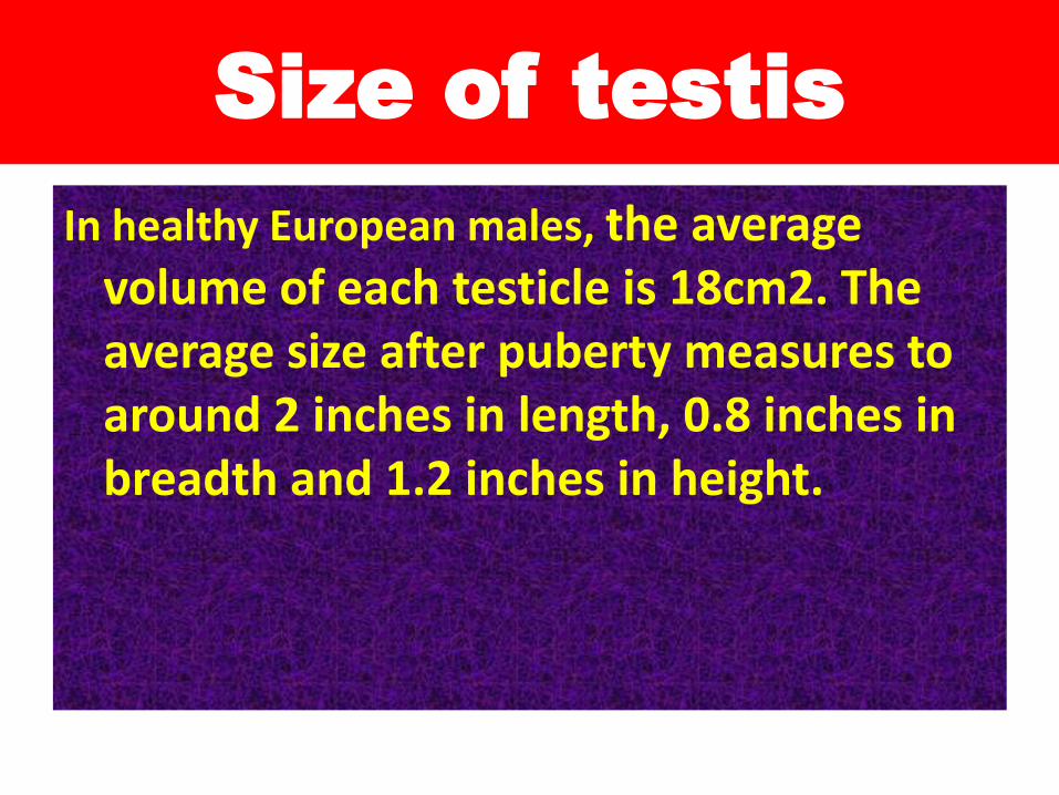
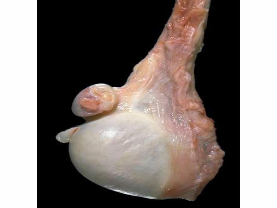
Size of testis
In healthy European males, the average volume of each testicle is 18cm2. The average size after puberty measures to around 2 inches in length, 0.8 inches in breadth and 1.2 inches in height.
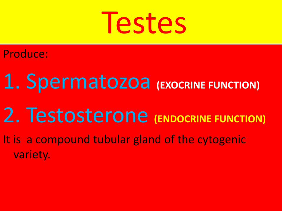
TestesProduce:
1. Spermatozoa (EXOCRINE FUNCTION)
2. Testosterone (ENDOCRINE FUNCTION)
It is a compound tubular gland of the cytogenic variety.
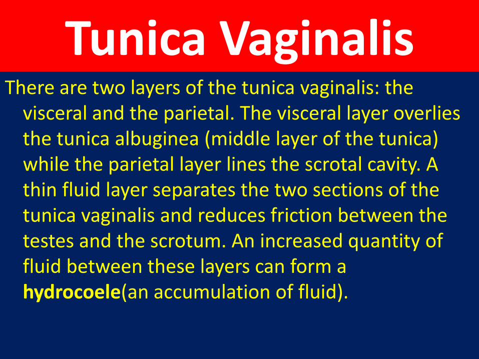
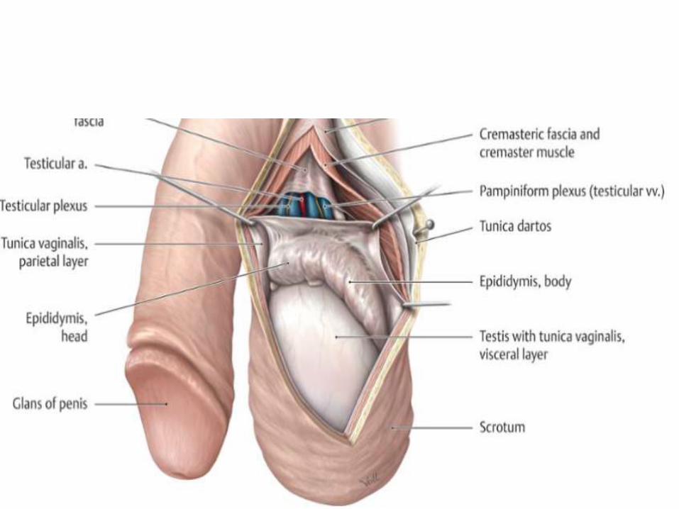
Tunica VaginalisThere are two layers of the tunica vaginalis: the
visceral and the parietal. The visceral layer overlies the tunica albuginea (middle layer of the tunica) while the parietal layer lines the scrotal cavity. A thin fluid layer separates the two sections of the tunica vaginalis and reduces friction between the testes and the scrotum. An increased quantity of fluid between these layers can form a hydrocoele(an accumulation of fluid).
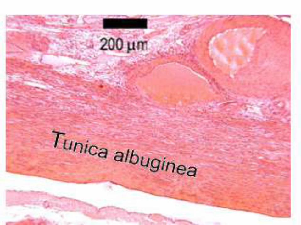
Tunica Albuginea/capsule
• Each testis is covered by a thick capsule
called Tunica Albuginea.
• It is composed mainly of collagenous fibers but also contains smooth muscle
cells.
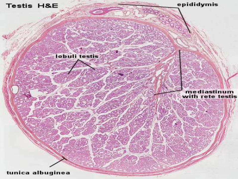
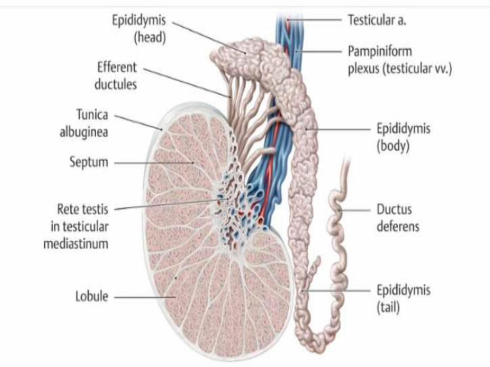
Mediastinum Testis Posteriorly the tunica albuginea
is thickened and projects into the gland as mediastinum testis.
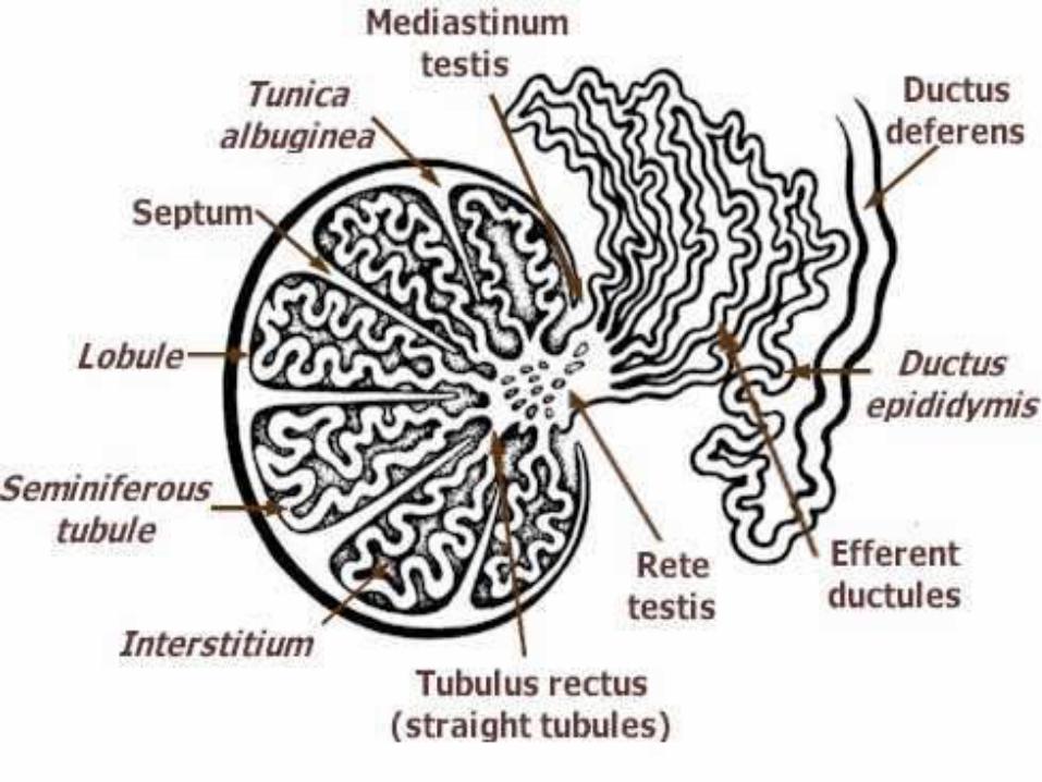
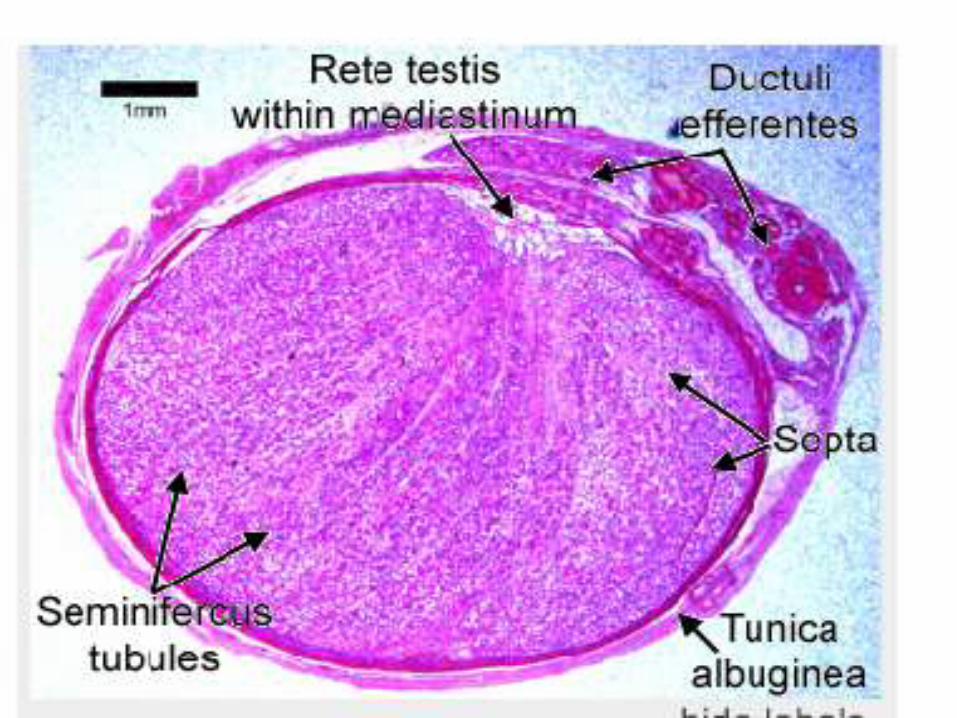
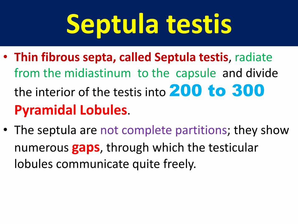
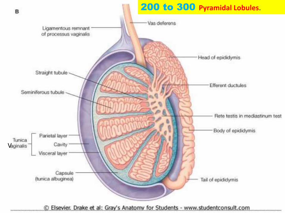
Septula testis• Thin fibrous septa, called Septula testis, radiate
from the midiastinum to the capsule and divide
the interior of the testis into 200 to 300
Pyramidal Lobules.
• The septula are not complete partitions; they show
numerous gaps, through which the testicular lobules communicate quite freely.
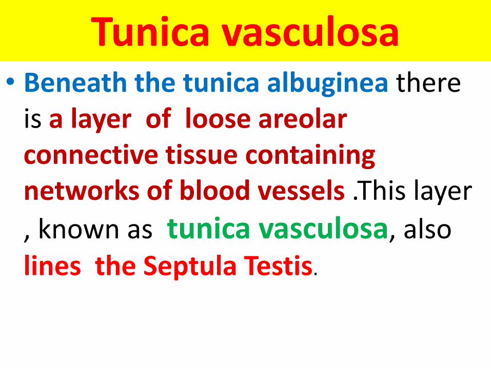
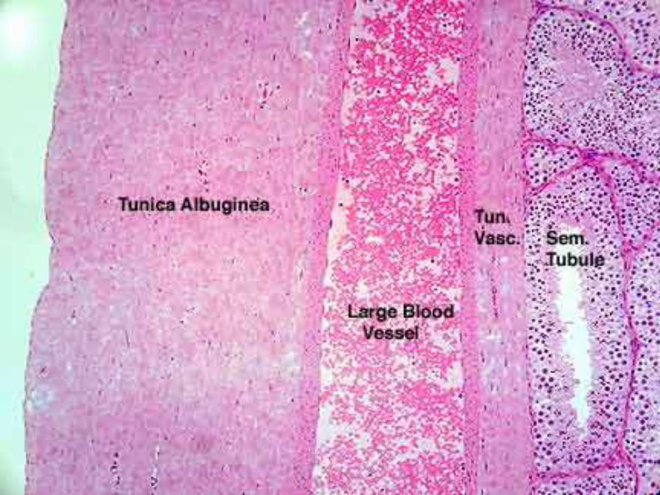
Tunica vasculosa• Beneath the tunica albuginea there
is a layer of loose areolar connective tissue containing networks of blood vessels .This layer
, known as tunica vasculosa, also lines the Septula Testis.
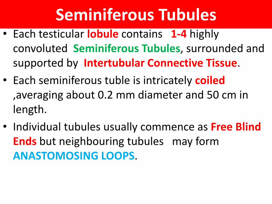
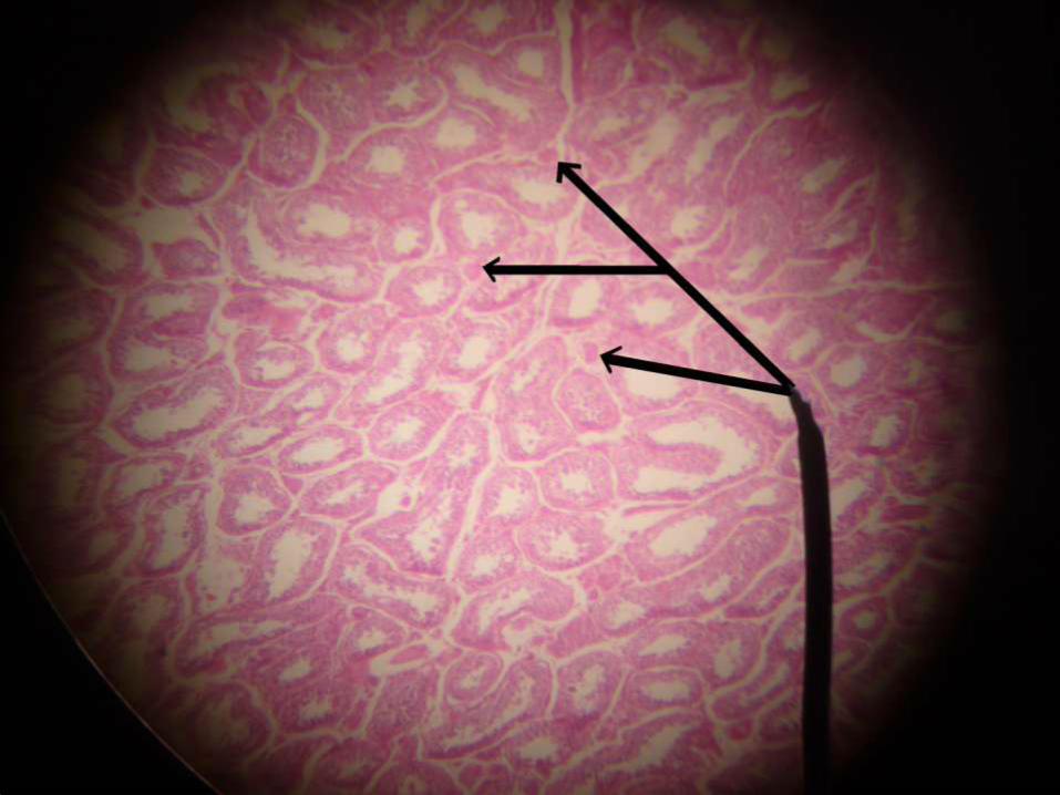
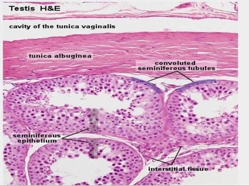
Seminiferous Tubules• Each testicular lobule contains 1-4 highly
convoluted Seminiferous Tubules, surrounded and supported by Intertubular Connective Tissue.
• Each seminiferous tuble is intricately coiled,averaging about 0.2 mm diameter and 50 cm in length.
• Individual tubules usually commence as Free Blind Ends but neighbouring tubules may form ANASTOMOSING LOOPS.
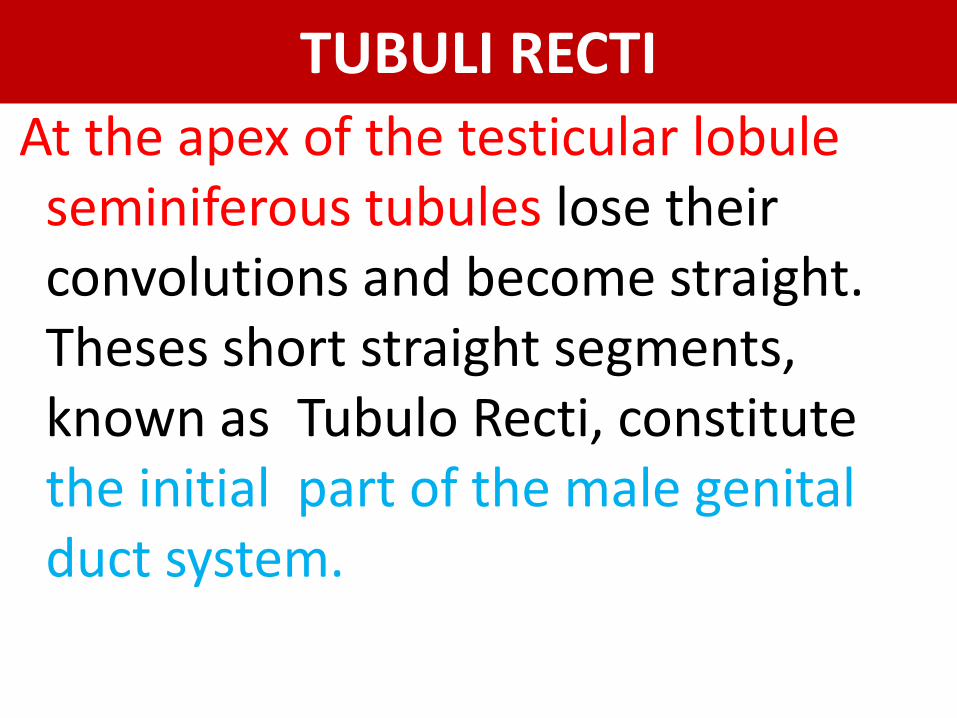
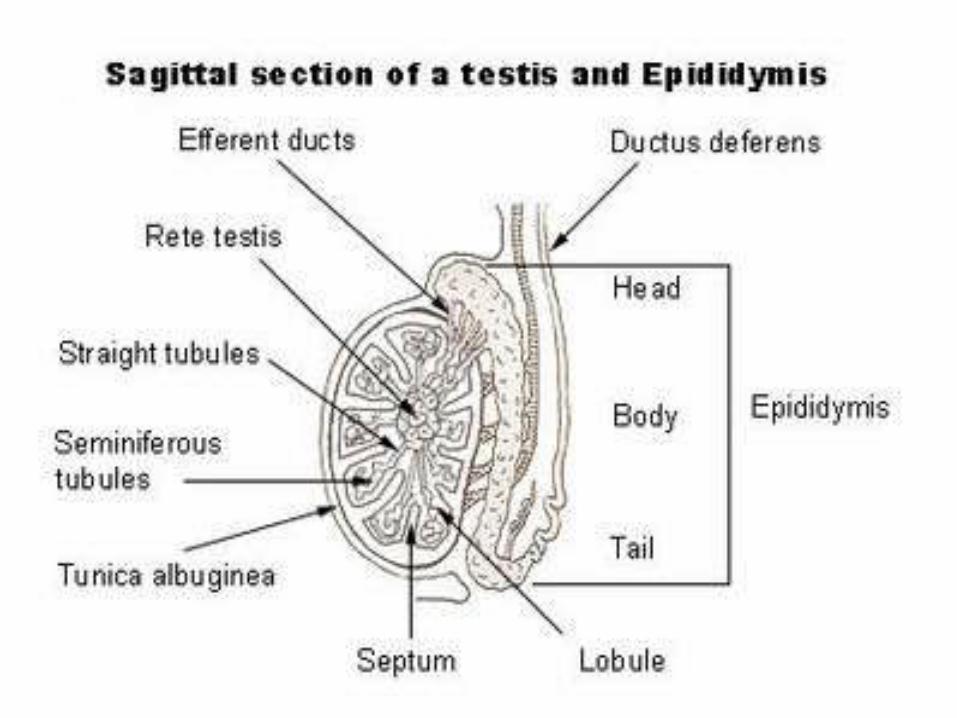
TUBULI RECTI
At the apex of the testicular lobule seminiferous tubules lose their convolutions and become straight. Theses short straight segments, known as Tubulo Recti, constitute the initial part of the male genital duct system.
V
200 to 300 Pyramidal Lobules.
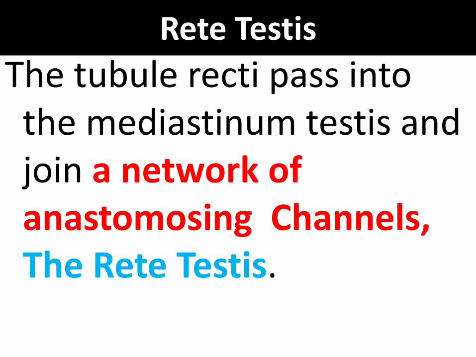
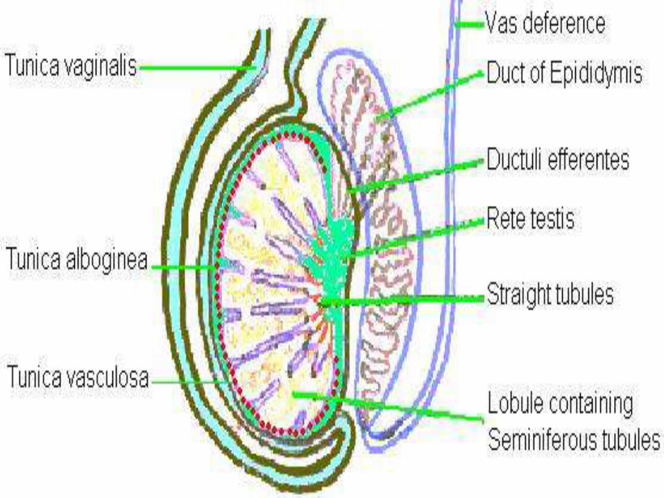
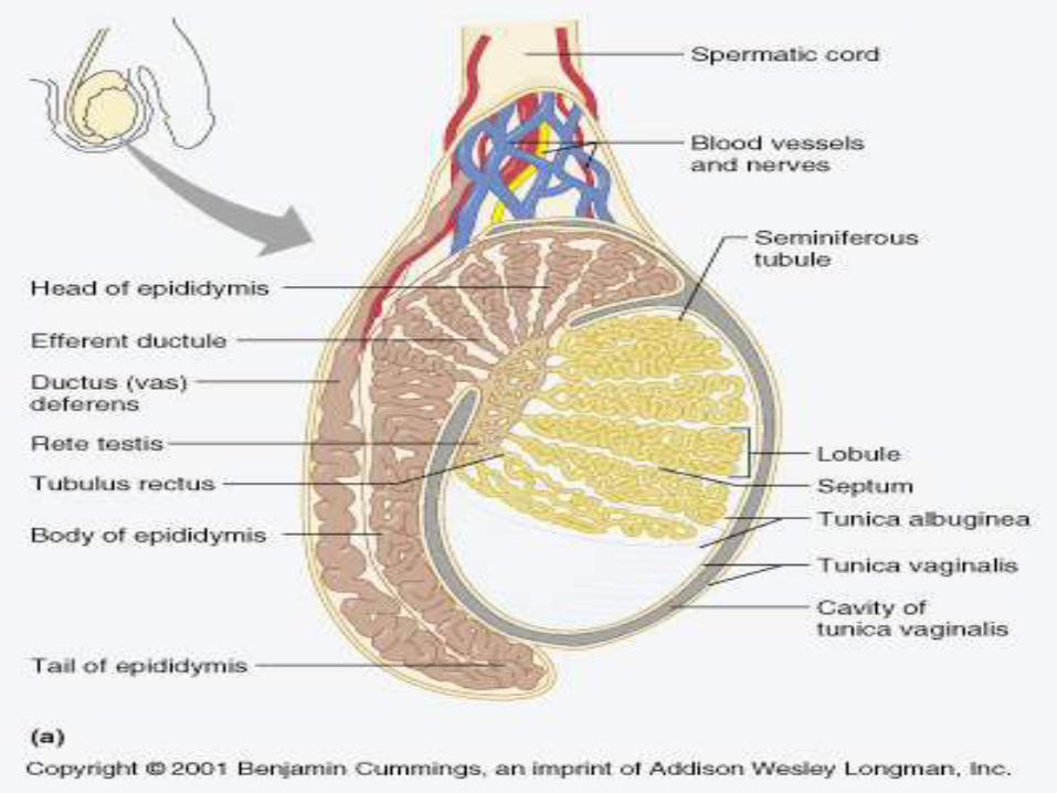
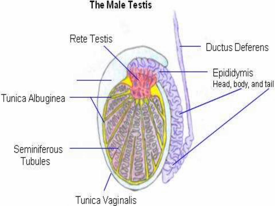
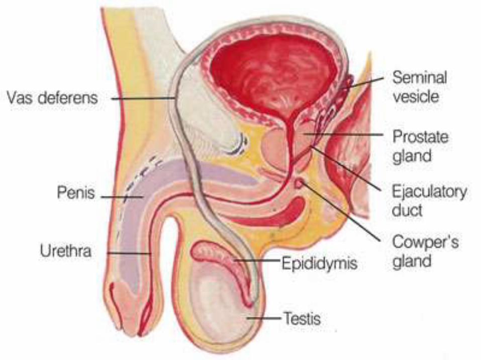
Rete Testis
The tubule recti pass into the mediastinum testis and join a network of anastomosing Channels,The Rete Testis.
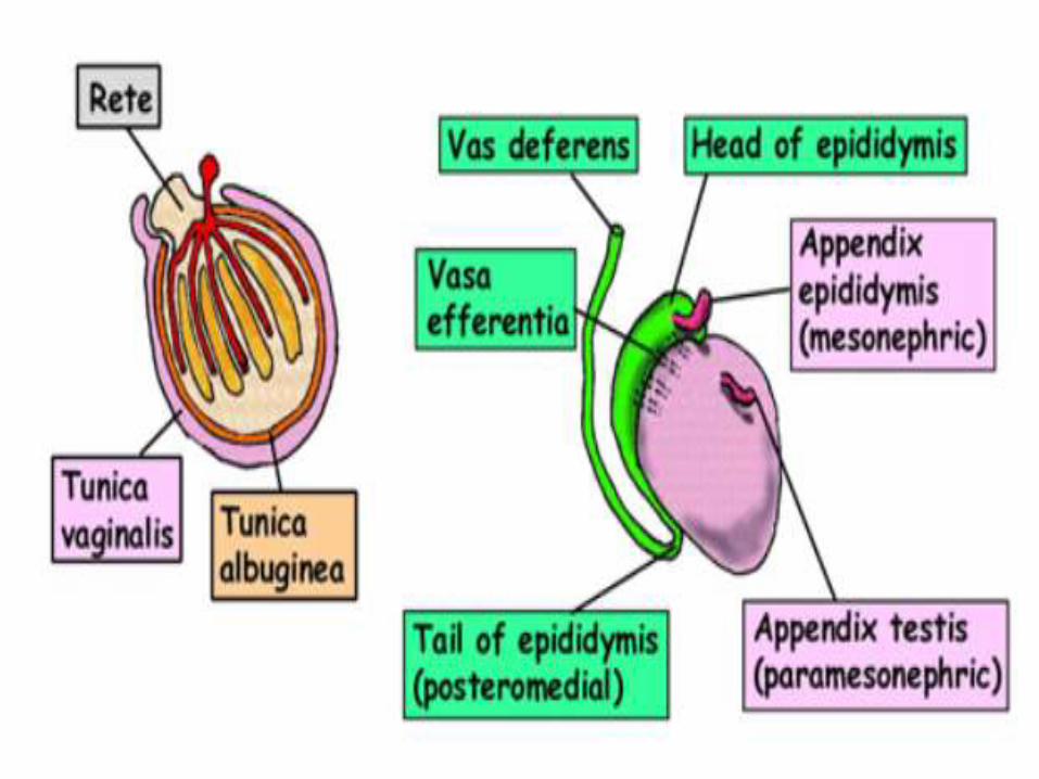
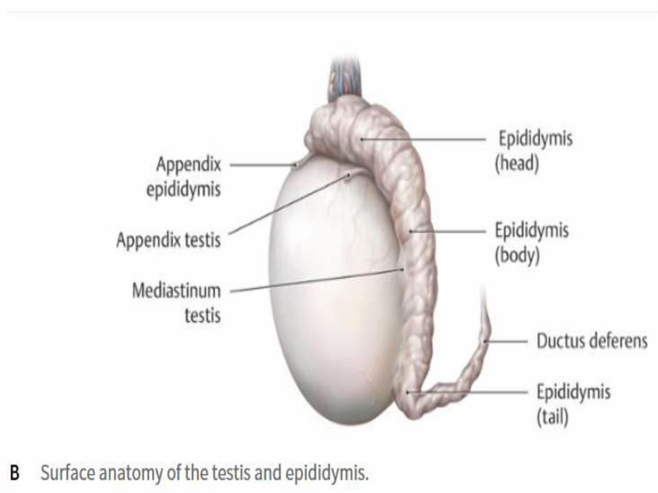
Ductus EfferentesFrom the Rete Testis arise 10-20 spirally wound efferent ductules ( Ductuli Efferentes) which leave the testis and open into the Duct of Epididymis.
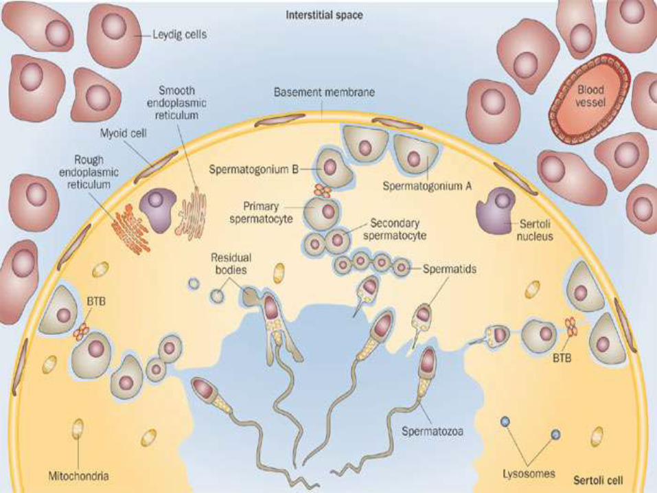
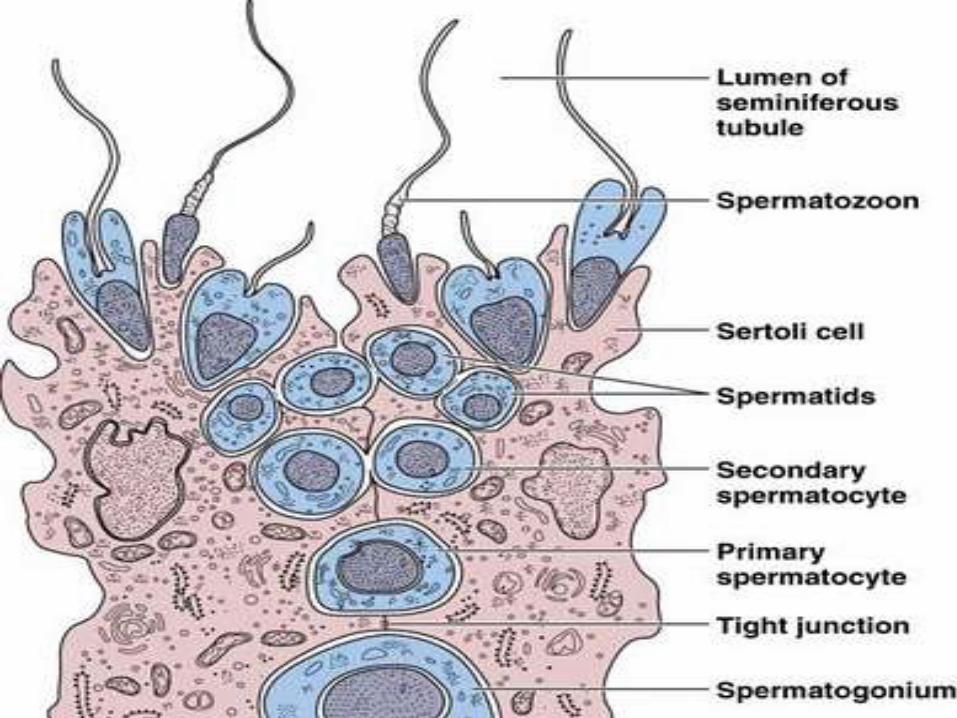
Seminiferous Epithelium
• Seminiferous tubule is lined by a complex Germinal or Seminiferous Epithelium.
• It is a modified stratified cuboidal
epithelium.
• The epithelium rests on a thin Basal Lamina.
Tunica PropriaSeminiferous Epithelium (Germinal
Epithelium) is surrounded externally by a thin layer of connective tissue called Tunica Propria.
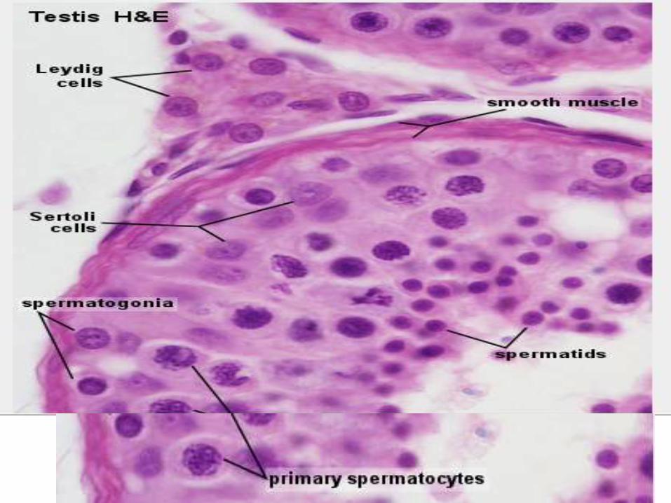
Spermatogenic or Germ Cells
• These cells lie Between The Sortoli Cells.
• They form a Stratified Epithelium.
• Consisting of Several Layers of cells which occupy the space between the Basal Lamina and Lumen of the Seminiferous Tubule.
Spermatogenic cells cont..
CHILD: In the testis of a child only the Primitive
Germ cells, called Spermatogonia are present.
• However, with the onset of Sexual maturity the process of spermatogenesis starts and the spermatogenic cells are seen in various stages of differentiation, arranged in an orderly manner.
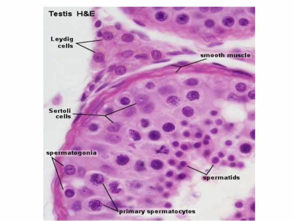
Spermatogonia (The primitive germ cells)
The most immature cells (Spermatogonia) are located near the basal lamina.
SpermatogoniaThe spermatogonia ( average diameter 12um)
are located directly inside the basal lamina of the seminiferous epithelium.
They are roughly spherical cells, each
containing a centrally located, round nucleus.
Types of SpermatogoniaDepending on the nuclear structure , the
spermatogonia can be classified into three types in an adult person.
These three types are:
i. Type A dark spermatogonia,
ii. Type A pale spermatogonia and
iii. Type B spermatogonia
The Type A Dark Spermatogonia
Contain an oval, darkly- staining nucleus , in which the nucleolus is located close to the nuclear membrane.
They are reserve cells and divide occasionally to maintain their own number and give rise to
Type A Pale Spermatogonia.
Type A Pale Spermatogonia
Contain a light –staining nucleus.
The nucleolus is located close to nuclear envelop.
These cells divide regularly to give rise to other Type A Pale Spermatogonia as well as
Type B Spermatogonia.
The Type B Spermatogonia
Have a spherical nucleus that shows
Darkely Staining Clumps of Chromotain located adjacent to the nuclear envelope.
The nucleolus is situated in the center of nucleus.
Each type B spermatogonium divide mitotically a few times to give rise to a number of daughter cells which do not divide further but transform into Primary Spermatocytes.
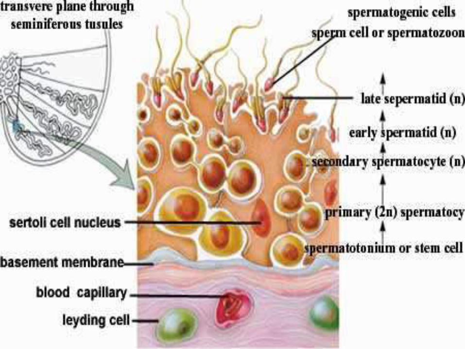
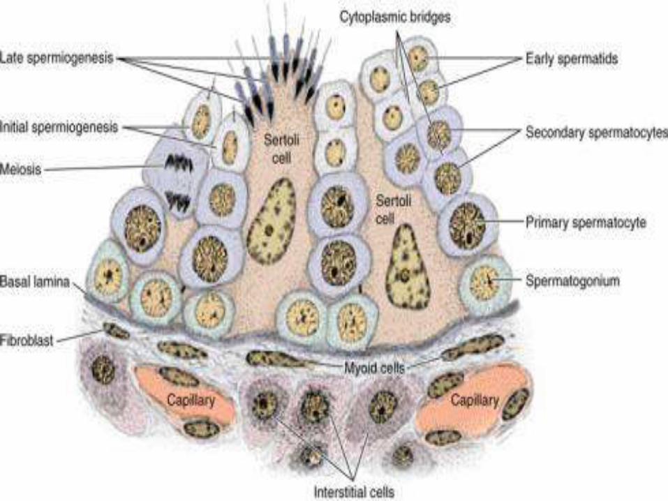
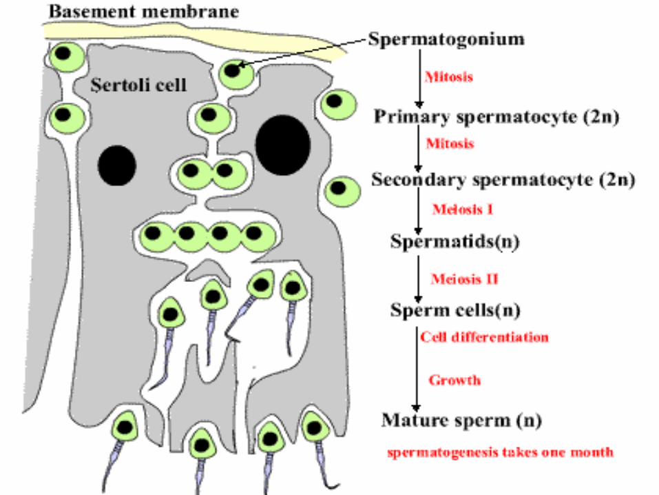
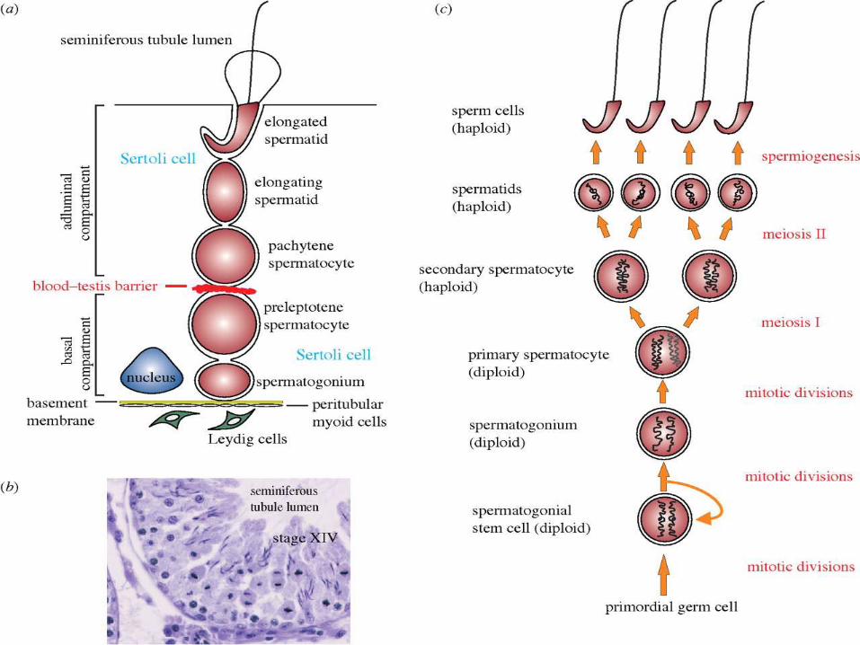
Stages of development of spermatogenic cells
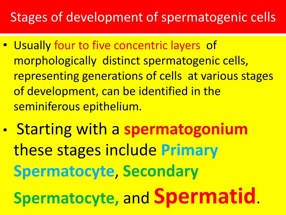
• Usually four to five concentric layers of morphologically distinct spermatogenic cells, representing generations of cells at various stages of development, can be identified in the seminiferous epithelium.
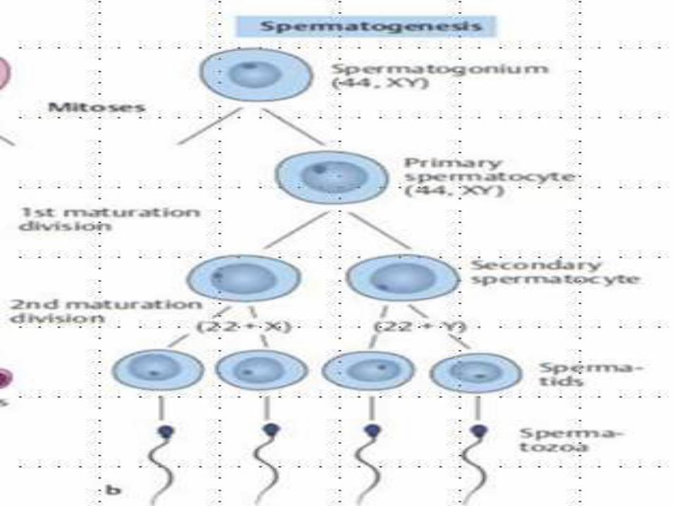
• Starting with a spermatogoniumthese stages include Primary Spermatocyte, Secondary
Spermatocyte, and Spermatid.
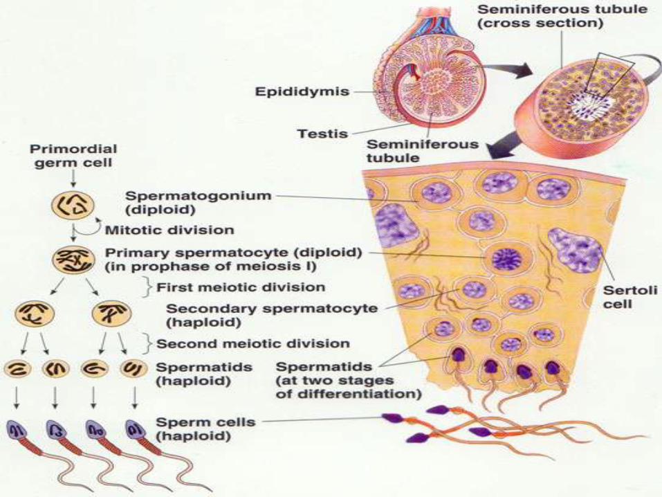
SpermatozoaAs the cells proliferate and undergo
differentiation, they gradually move
towards the lumen of the tubule.
Finally the cells come to lie at the luminal surface of the seminiferous tubule, where
they transform into spermatozoawhich become free to lie within the lumen of the tubule.

Primary Spermatocytes
The primary spermatocytes lie next to the spermatogonia.
They are large cells (diameter
18um) having vesicular nuclei.
Secondary Spermatocytes
These are smaller cells that arise from a primary spermatocyt as a result of First Meiotic Division.
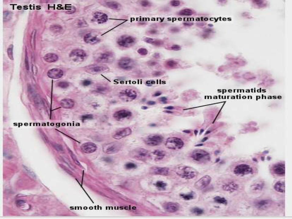
The SpermatidsProduced from the secondary spermatocytes by the
second meiotic division.
They lie adjacent to the lumen of the siminiferous tubule,
closely applied to the surface of Sertoli cells.
SpermatogenesisA spermatid does not divide further but is
transformed into a spermatozoonby a series of morphological changes
which are collectively known as
Spermatogenesis.
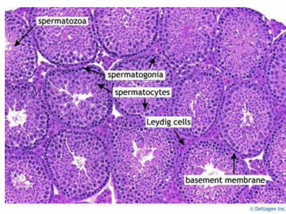
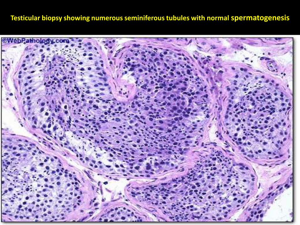
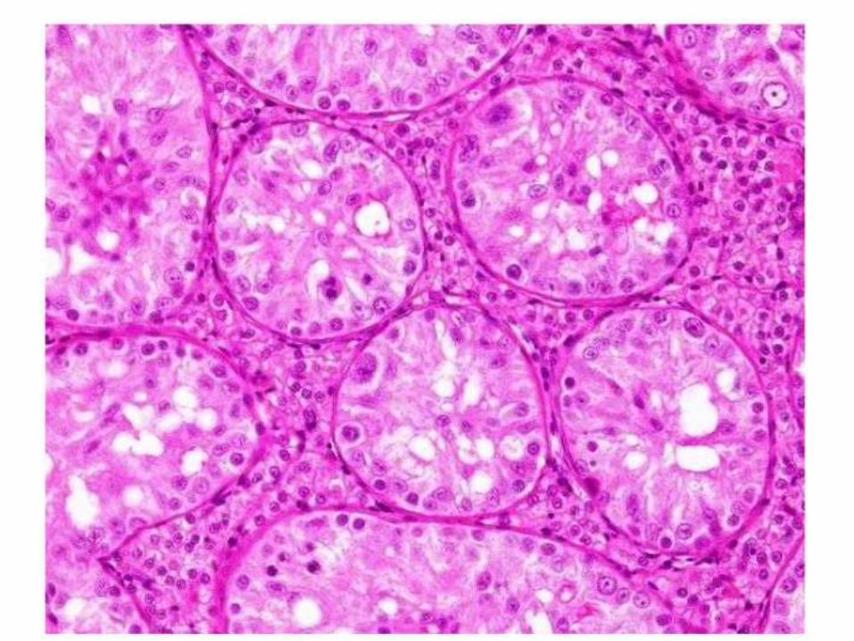
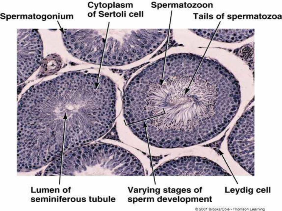
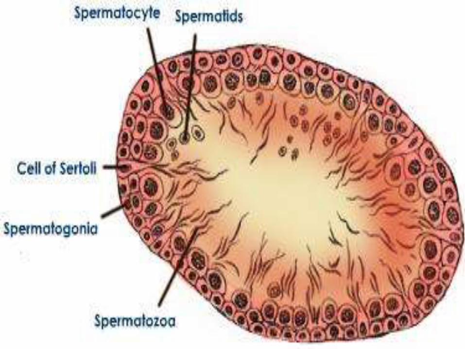
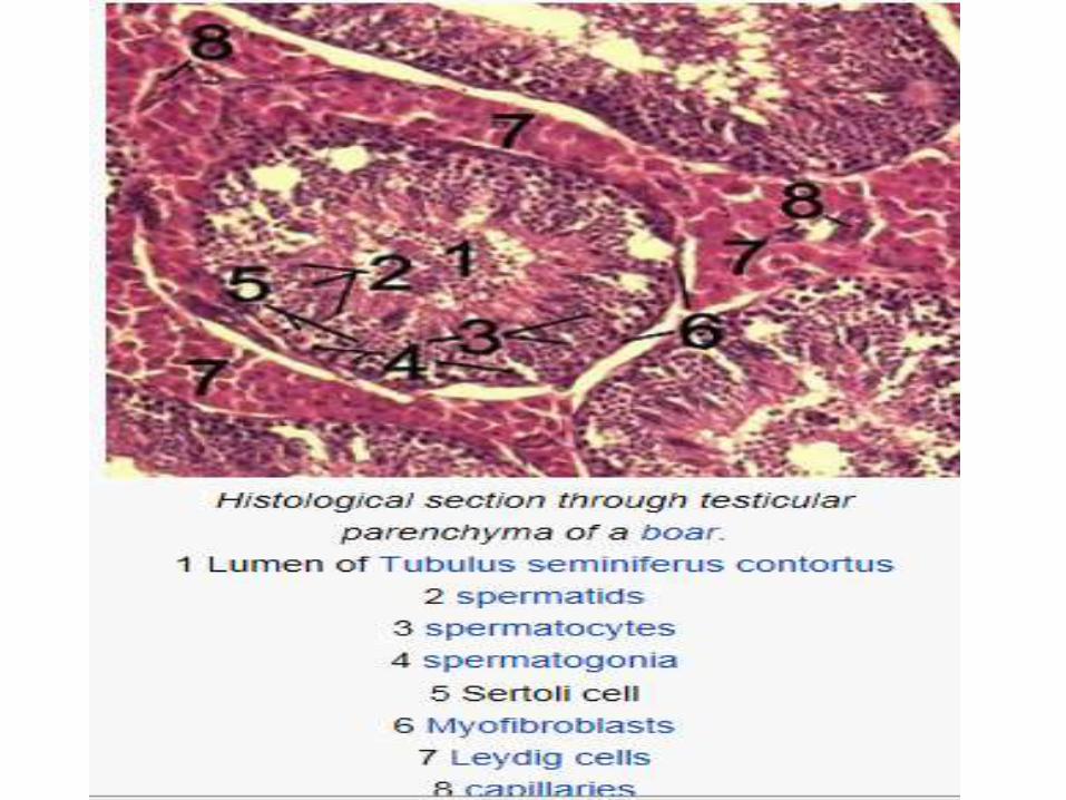
Testicular biopsy showing numerous seminiferous tubules with normal spermatogenesis
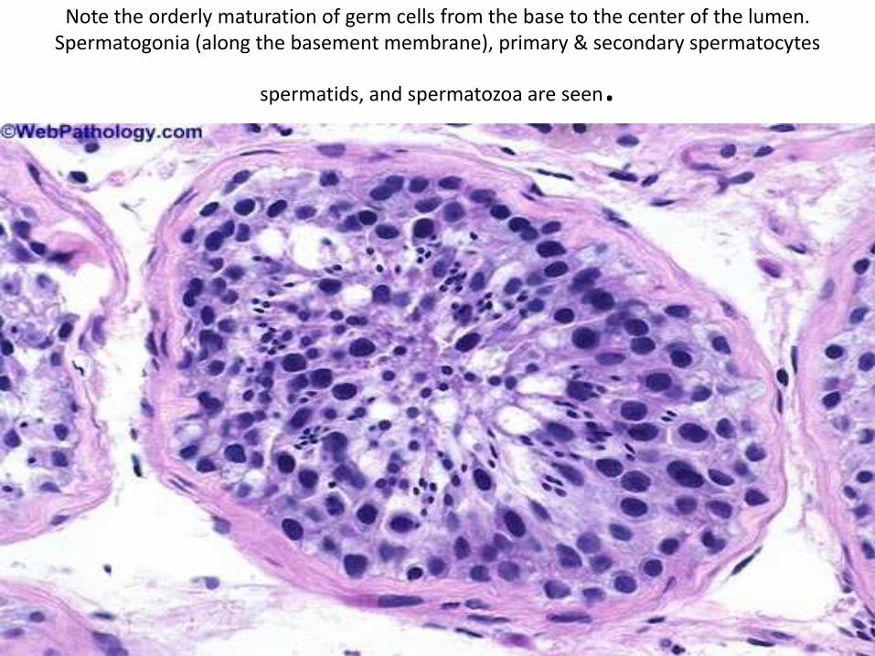
Note the orderly maturation of germ cells from the base to the center of the lumen. Spermatogonia (along the basement membrane), primary & secondary spermatocytes
spermatids, and spermatozoa are seen.
Sertoli cells/Sustentacular cells/Nurse cells/Mother cells/Tree cells/Stringy cells
• Sertoli cells are called so because of their eponym Enrico Sertoli, an Italian physiologist who discovered them while studying medicine in the University of Pavia, Italy.
• He published a description of this cell in 1865. In the 1865 publication, his first description used the terms "tree-like cell" or "stringy cell" and most importantly he referred to these "mother cells." It was other scientists who used Enrico's family name, Sertoli, to label these cell in publications, starting in 1888.
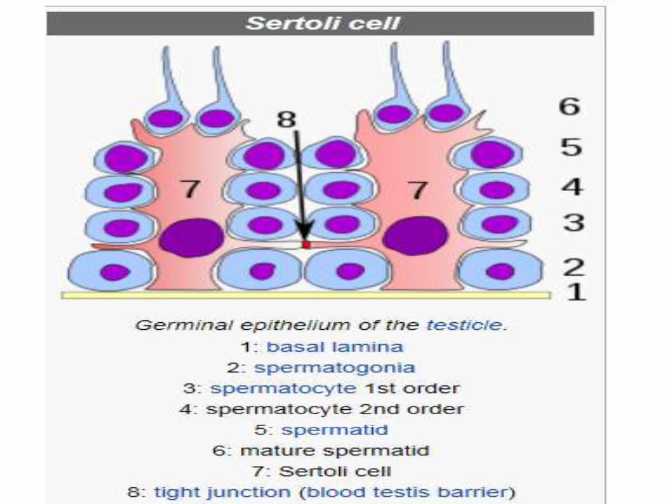
Sertoli Cells or Sustentacular Cells
• These are tall columnar cells that
• Extend from the basal lamina to the lumen of the seminiferous tubule,
• Interposed between the developing spermatogenic cells.
Sustentacular Cells or Sertoli Cells
• Each Sertoli cell is 70-90 um long and nearly 3o um wide.
• Due to their loose association with spermatogenic cells, the lateral margins of Sertoli cells are irregular and can not easily be distinguished under L/M.
Sustentacular cells
• Each sustentacular cell has a large pale- staining
(euchromatic) nucleus which is located in the basal portion of the cell.
• The nucleus exibits two distinctive features:
• 1) the nuclear envelope shows many infoldings, and,
• 2) the nucleolus is very prominent (Dark nucleolus).
Luminal surface of a Sertoli cell
shows many grooves in which heads of the
maturating spermatozoa are embeded.
Sustentacular or Sertoli cellsUnder L/M,
The cytoplasm of a Sertoli cell is seen to
contain:
1.Lipid droplets,
2. Glycogen granules and
3. Crystalloid.
Sertoli CellsE/M shows that these cells contain:
1. A well-developed Golgi apparatus,
2. An abundant smooth endoplasmic reticulum,
3. Some rough endoplasmic reticulum,
4. Numerous mitochondria and
5. Many lysosomes.
Functions of Sertoli cells
• A Sertoli cell is a 'nurse' cell or
‘mother’ cell of the testes.
• It is activated by follicle-stimulating hormone and has FSH-receptor on its membranes.
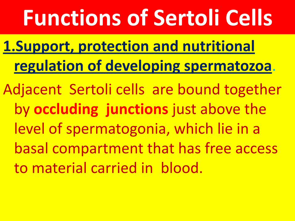
Functions of Sertoli Cells1.Support, protection and nutritional
regulation of developing spermatozoa.
Adjacent Sertoli cells are bound together by occluding junctions just above the level of spermatogonia, which lie in a basal compartment that has free access to material carried in blood.
Functions of Sertoli cells cont…
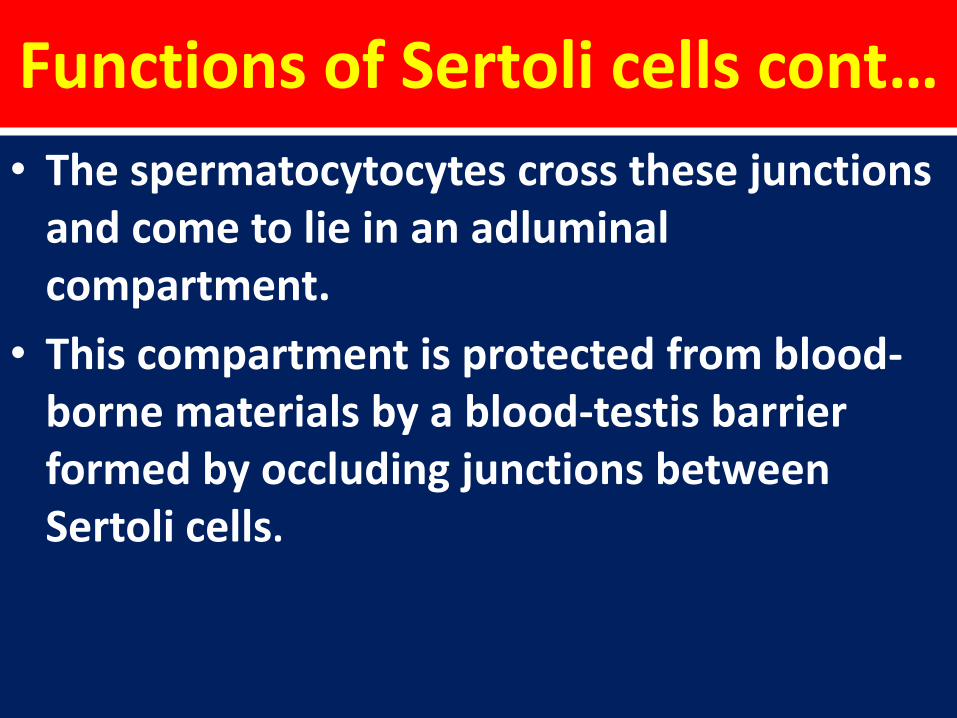
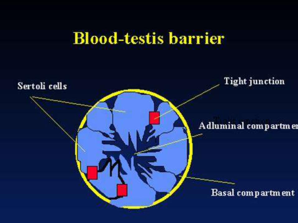
• The spermatocytocytes cross these junctions and come to lie in an adluminal compartment.
• This compartment is protected from blood-borne materials by a blood-testis barrier formed by occluding junctions between Sertoli cells.
Functions of Sertoli Cells cont..
• The spermatocytes, spermatids and developing spermatozoa are isolated from the blood circulation and depend on Sertoli cells to mediate the exchange of nutrients and metabolites.
• The blood-testis barrier protects the cells in the adluminal compartment from blood-born harmful substances and from autoimmune reaction against sperm-specific proteins.
Functions of Sertoli Cells cont…
2. Secretions: Sustentacular cells of Sertoli
secrete a fluid into the lumen of the seminiferou tubules which flows in the direction of genital ducts and is used for sperm transport.
• Sertoli cells secrete the following substances:
• anti-Müllerian hormone (AMH) - secreted during the early stages of fetal life.
• inhibin and activins - secreted after puberty, and work together to regulate FSH secretion
• androgen binding protein (also called testosteronebinding globulin) - increases testosterone concentration in the seminiferous tubules to stimulate spermatogenesis
• estradiol - aromatase from Sertoli cells convert testosterone to 17 beta estradiol to direct spermatogenesis
• glial cell line-derived neurotrophic factor (GDNF) -has been demonstrated to function in promoting undifferentiating spermatogonia, which ensures stem cell self-renewal during the perinatal period.
• the Ets related molecule (ERM transcription factor) -needed for maintenance of the spermatogonial stem cell in the adult testis.
• transferrin - a blood plasma protein for iron ion delivery.
Functions of Sertoli Cells cont….
3. Phagocytosis:
During sperminogenisis excess spermatid cytoplasm is shed as residual bodies which are phagocytized and broken down by Sertoli cells.
Functions of Sertoli Cells cont…
During the maturation phase of spermiogenesis, the Sertoli cells consume the unneeded portions of the spermatozoa.
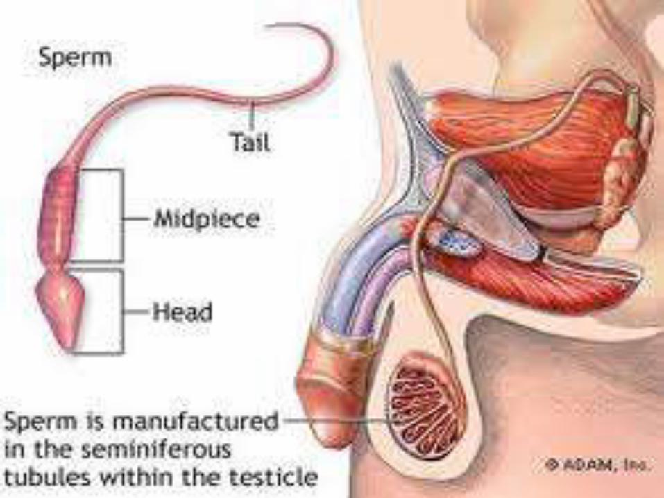
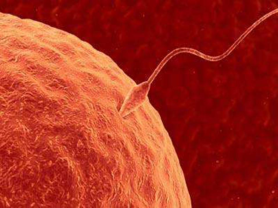
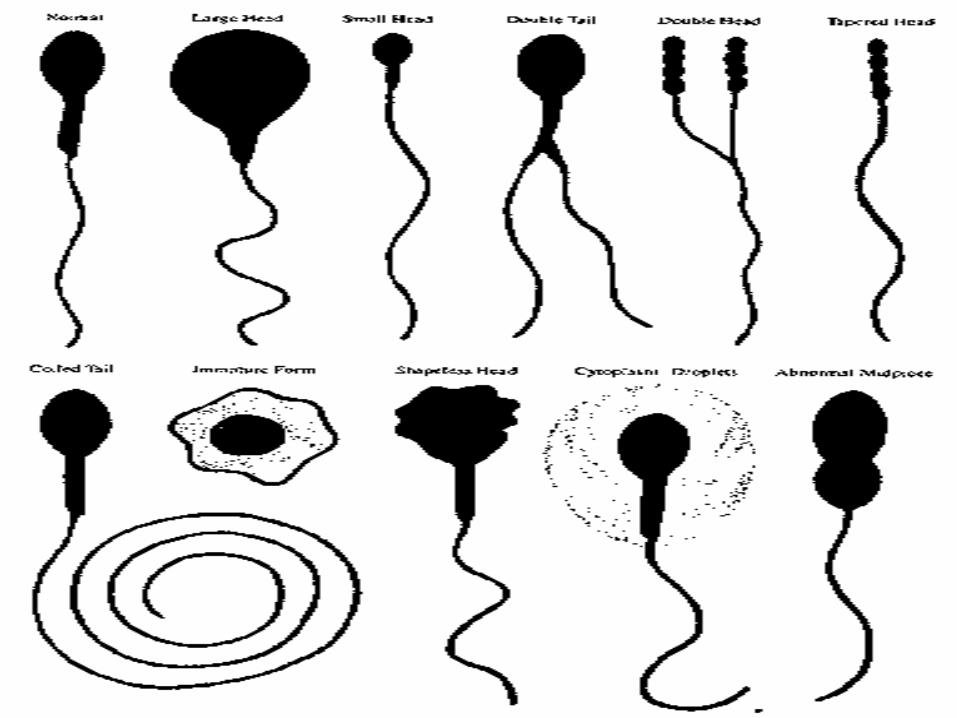
The Spermatozoa
• These are slender, motile, flagellate bodies, each having an average length of 60 um.
• A mature spermatozoon (Sperm) consist of two main parts, a head and a tail .
• The junctional region between these two parts is referred to as neck. All these parts are covered by a plasma membrane.
The Spermatozoa cont…
• Head: The head is a flattened, pear-shaped body
measuring about 4um in length, 3um in width and 1um in thickness.
• It consist chiefly of the condensed nucleus, present as a compact mass of chromatin enclosed in the nuclear envelope.
Acrosomal Cap
The anterior two third of the nucleus are covered by a flattened, membrane bound sac called acrosomal cap.
This cap has been found to contain hydrolytic enzymes like hyaluronidase, etc.
Plasmlemma. The entire sperm , including the head, is covered by a plasmalemma.
The Spermatozoa cont……
• The Neck: The neck is a short region between the head and tail of the sperm. It contains the proximal centriole running fibers that become continuous with those of the middle piece of the tail.
The Tail of spermatozoa
Length 55um
Has 3 segments:
1.Middle Piece
2.Principal Piece
3.End Piece


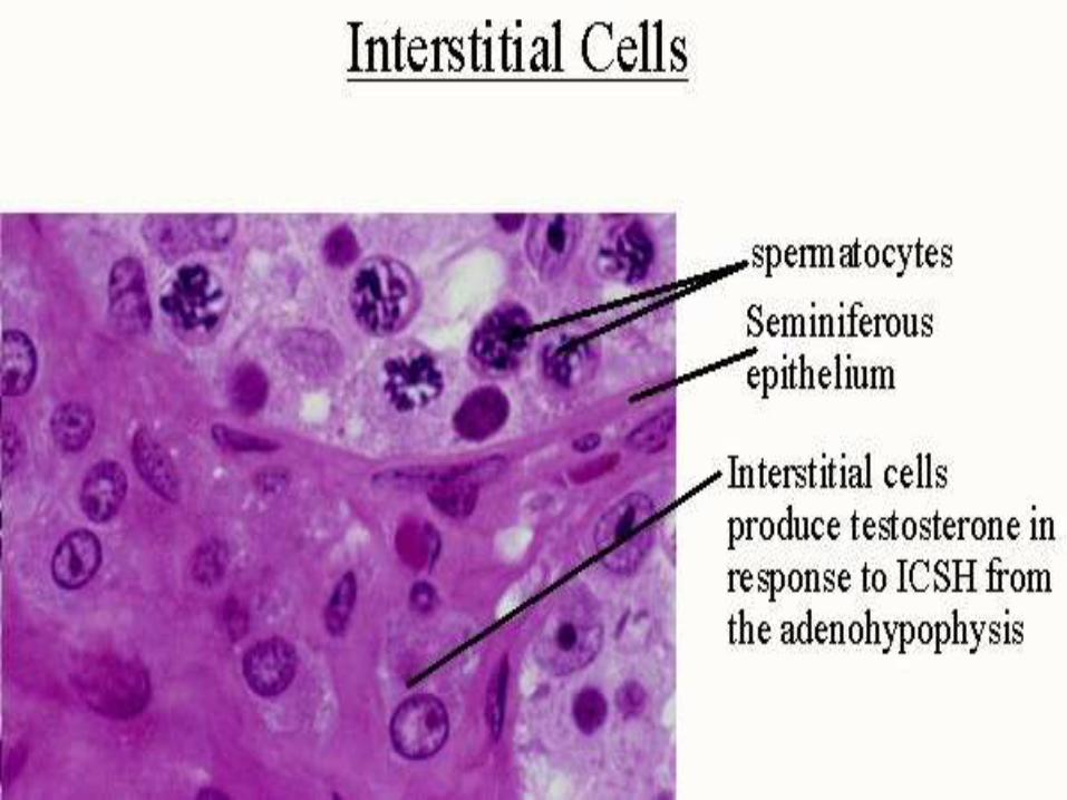
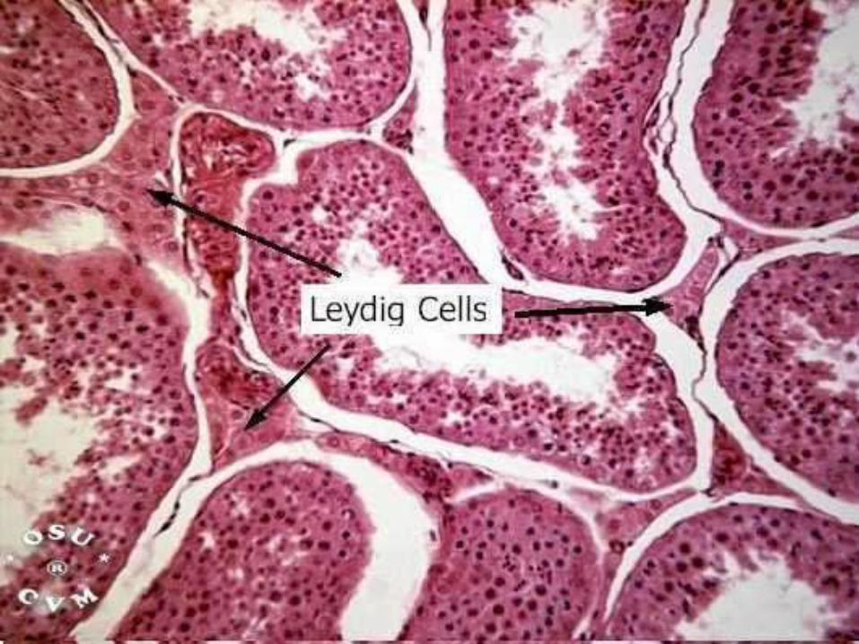
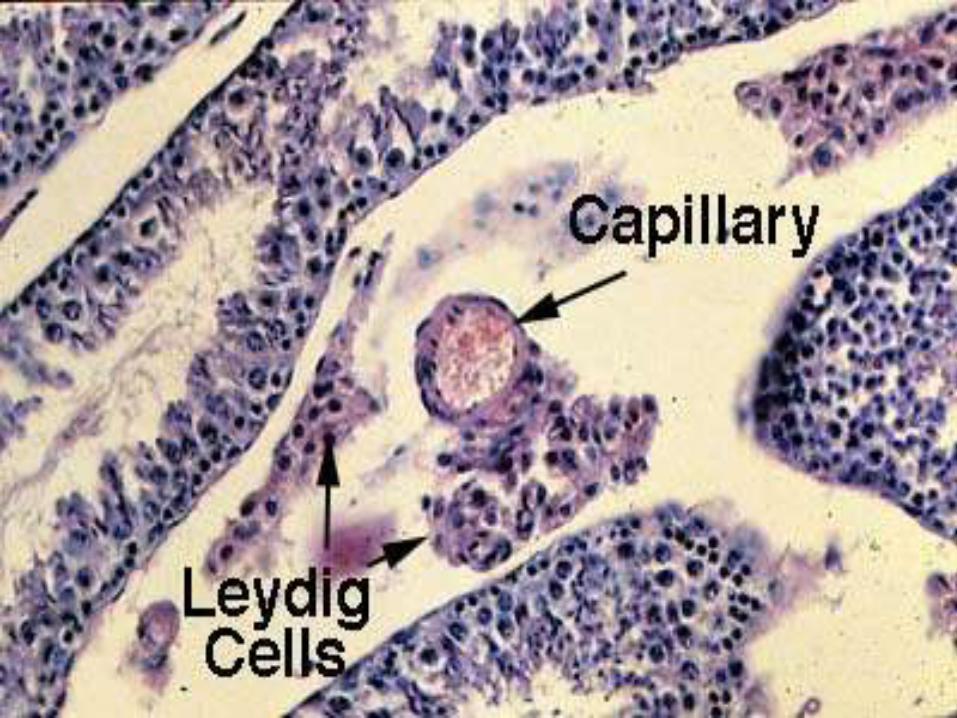
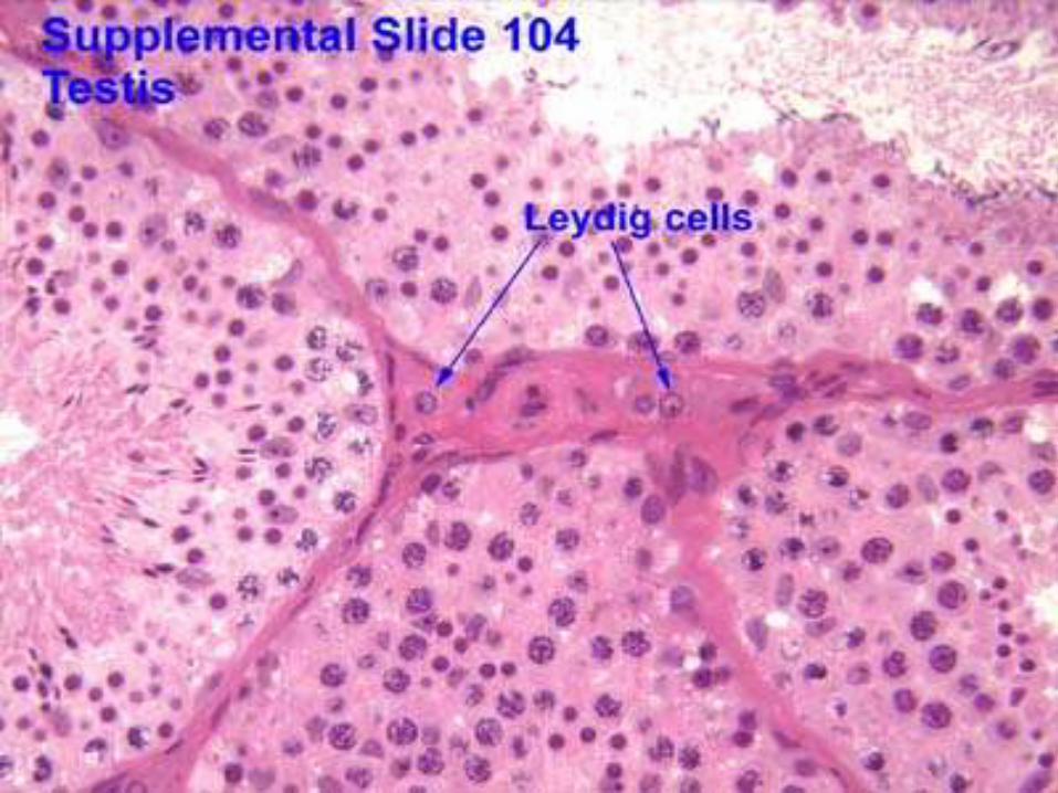
Interstitial Cells of Leydig
• Leydig cells, also known as interstitial cells of Leydig, are found adjacent to the seminiferous tubules in the testicle. They produce testosterone in the presence of luteinizing hormone (LH).
• Leydig cells are named after the German anatomist Franz Leydig, who discovered them in 1850
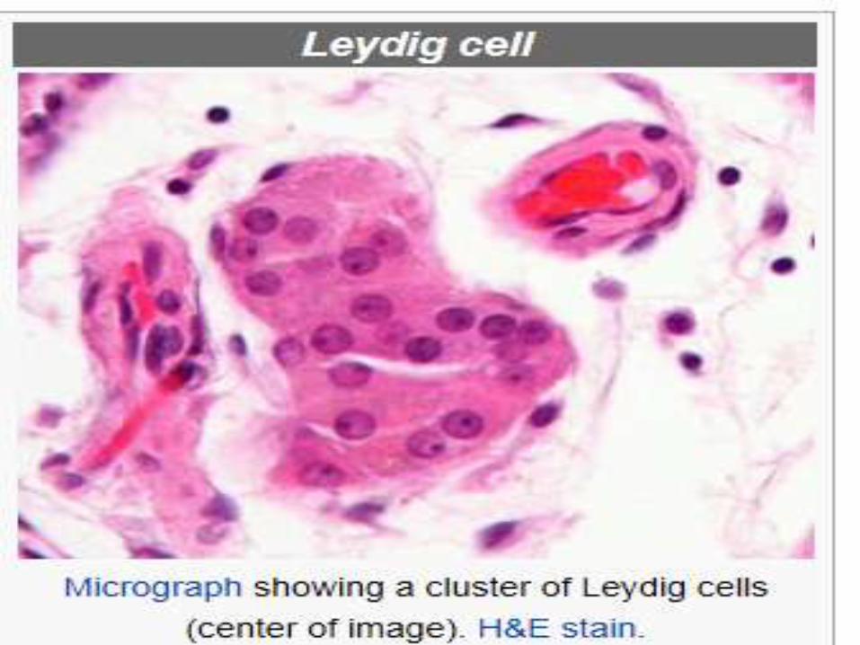
Structure
• Leydig cells are polyhedral in shape, display a large prominent nucleus, an eosinophilic cytoplasm and numerous lipid-filled vesicles.
Structure cont..
Leydig cell has a single eccentrically located ovoid nucleus. The nucleus
contains one to three prominent nucleoli and large amounts of dark-staining peripheral heterochromatin.
Structure cont…
• The acidophilic cytoplasm usually contains
numerous membrane-bound lipid droplets and large amounts of smooth endoplasmic
reticulum (SER).
Structure cont.
• Besides the obvious abundance of SER with scattered patches of rough endoplasmic reticulum, several mitochondria are also prominent within the cytoplasm.
Structure cont…..
• Frequently, lipofuscin pigment and rod-shaped crystal-like structures 3 to 20 micrometres in
diameter (Reinke's crystals) are found. These inclusions have no known function.