histiocytic ulcerative colitis in a french bulldog
TRANSCRIPT
J. small Anim. Pract. ( 1 978) 19,283-290
His tiocytic ulcerative colitis in a French Bulldog I N G R I D VAN D E R G A A G , * J I K VAN T O O R E N B U R G , ? G . V O O R H O U T , $ R . P. H A P P E t A N D R . H . G. AALFSg
* Institute ofveterinary Pathology, Biltstraat 166. t University Small Animal Clinic, Yalelaan 8. $ Institute ofveterinary Radiology, Yalelaan 10, Utrecht; Q Small Animal Practice Centre,
Randweg 34. Rotterdam, The Netherlands
ABSTRACT
A case of histiocytic ulcerative colitis (granulomatous colitis) in a one- year-old female French Bulldog is described. Until now this disease has been described only in the Boxer breed.
The dog had an increased frequency of defaecation and the faeces contained blood and mucus. Colonoscopy showed some irregularities and a few haemorrhages in the mucosa of the caudal part of the colon. At a distance of approximately 25 cm from the anus there was a stricture caused by rigidity of the'colon wall. Radiographic examination revealed marked shortening of the colon. There was also narrowing of the midpoint of the descending colon in some radiographs, but the colon appeared to be fully distensible when filled with barium. Histologically, periodic acid- Schiff (PAS)-positive staining histiocytes were found in biopsy and autopsy specimens from the colon.
I N T R O D U C T I O N
Histiocytic ulcerative colitis is a disease of Boxer dogs, clinically characterised by a chronic, intractable diarrhoea, generally without much loss of condition. I t occurs in young dogs, from about two months to four years of age, with equal frequency in male and females. The faeces contain variable amounts of fresh blood and mucus and the frequency of defaecation is increased.
These clinical signs are caused by specific granulomatous lesions in the mucosa, submucosa and sometimes in the muscular layers of the rectum, colon and caecum.
00204510/78/0500-0283 $2.00 0 1978 BSAVA
283
284 I N G R I D V A N D E R G A A G etal.
Microscopically there is a thickening of the lamina propria and the submucosa caused by histiocytes, plasma cells and lymphocytes and, in cases with ulceration, also neutrophils. The majority of these histiocytes are periodic acid-Schiff (PAS)- positive.
Thus far this disease has been reported exclusively in Boxer dogs in the United States (Van Kruiningen et al., 1965; Kennedy &Cello, 1966; Koch & Skelly, 1967; Sander & Langham, 1968; Starnes, 1969; Russell, Gomez & Trowbridge 197 1); in the Netherlands (Van der Gaag, Happe & Wolvekamp, 1975) and in West Germany (Van der Gaag, 1975). This report describes a one-year-old female French Bulldog with the typical features of canine histiocytic ulcerative colitis (CHUC) and typhlitis of the Boxer dog.
M E T H O D S
For colonoscopy (Olympus fiberscope, model CF-LB) the dog was sedated with 0.25 mg of acepromazine/kg.B.W (Vetranquil", Philips-Duphar B.V., Amster- dam). Food was withheld for 48 hours and an enema consisting of a mild soap solution was given one-half hour before the examination to cleanse the colon from persisting faeces. Biopsies were taken with the fibrescope.
Preparation for radiographic examination consisted of withholding food and water for 24 hours and sedation with 1-5 mg of triflupromazine HCL/kg.B.W. (SiquiP, E.R. Squib & Sons B.V., Rijswijk). Barium sulphate suspension at body temperature was introduced into the colon with a large syringe connected to a rectal tube with a distensible balloon..
After obtaining radiographs. in both right and left lateral recumbency and prone and supine positions, the positive contrast medium was removed and the colon was insufflated with air. Radiographs were again made in four directions, thus producing a double contrast image.
The biopsy specimens were fixed in Baker's fixative (Pearce, 1961) and autopsy specimens were fixed in 4% buffered formaldehyde. Paraffin sections 6 pm thick were stained with haematoxylin and eosin, van Gieson and PAS.
CASE R E P O R T
Clinical findings A one-year-old female French Bulldog was admitted to the Utrecht University
Small Animal Clinic with a six weeks' history of bloody, loose stools and increased frequency of defaecation. The dog showed some tenesmus when pro- ducing small quantities of faeces, these containing variable amounts of fresh blood and mucus. There was no history of vomiting or weight loss.
Two weeks after onset of the complaints the dog had been examined at a private clinic. The only notable physical findings at that time were that rectal examination elicited pain and the wall of the rectum was more folded than

H I S T I O C Y T I C U L C E R A T I V E C O L I T I S I N A F R E N C H B U L L D O G 285
normal. The dog was treated with chloramphenicol, several antidiarrhoeics and changes of diet. Because there was little or no response after four weeks of treatment, a barium enema examination was performed. This revealed a markedly shortened colon with some irregularities of the wall and an indication of a stricture approximately 25 cm cranial to the anus. The dog was then referred to the U trecht University Small Animal Clinic.
When presented at the Clinic the dog was alert and in good condition. Other than fresh blood in the rectum, no abnormalities were found at clinical examina- tion.
Results of routine blood examinations were normal except for a lowered packed cell volume (33%)). Microscopic examination of the faeces revealed no parasite ova and microchemical examination showed no abnormalities in diges- tion. Colonoscopy revealed that the mucosa of the colon and the rectum was smooth but there were some irregularities and a few small haemorrhages approxi- mately 5 cm cranial to the anus.
Passage of the fiberscope met with no difficulties until a stricture caused by rigidity of the colon wall was encountered 25 cm cranial to the anus. I t was possible to take a biopsy through this narrowed part, approximately 30 cm cranial to the anus, and additional biopsies were taken at 25, 20, 10 and 5 cm cranial to the anus.
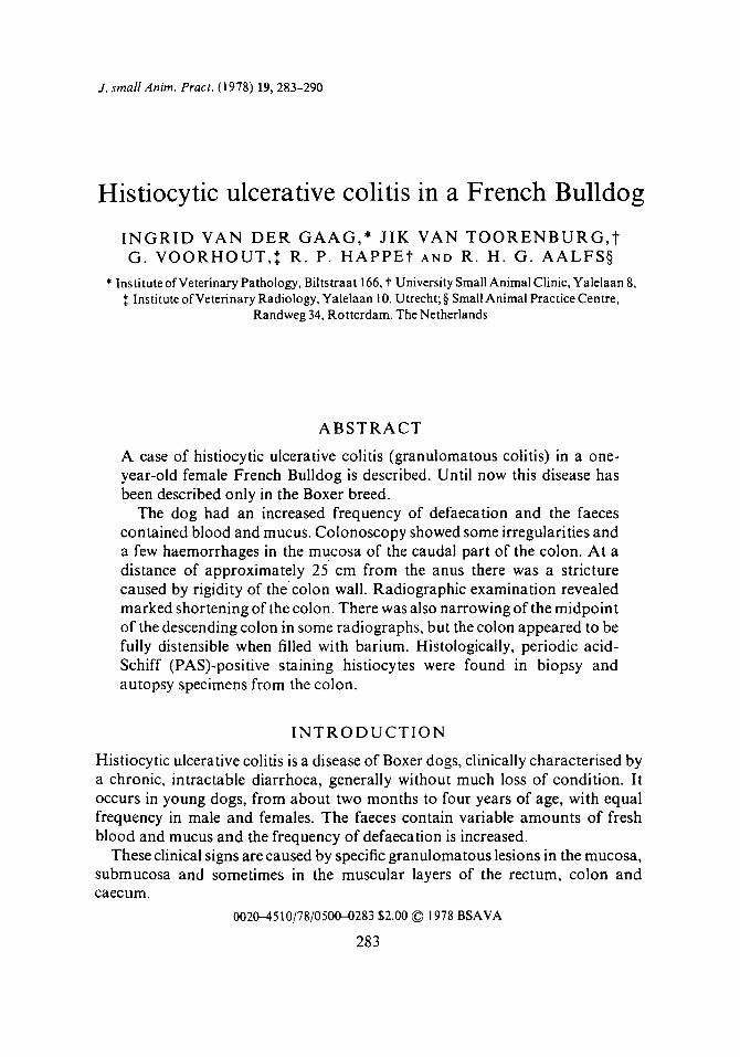
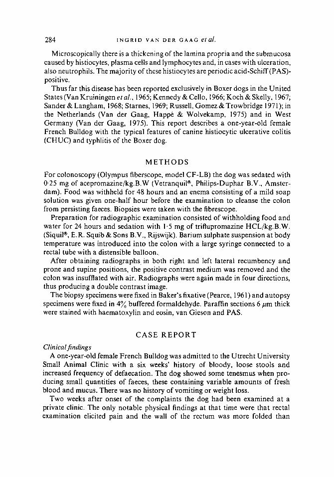
R A D I O L O G Y Plain films showed an irregularly distended, gas-filled descending colon. The thickness of its wall could not be evaluated. The narrowing of the proximal part of the descending colon, which had been observed in the previous barium enema study and by colonoscopy, was not a consistent finding in this examination. Although narrowing was found in some radiographs (Fig. l), in other the colon appeared to be fully distensible (Fig. 2), both after filling with barium sulphate suspension and during insufflation with air. There was marked shortening of the colon and the contrast medium not only filled the stretched caecum but passed easily into the ileum. The mucosal pattern of the colon was rather smooth and ulcers could not be detected.
B I O P S Y E X A M I N A T I O N
The diagnosis of histiocytic ulcerative colitis was based upon examination of the five biopsy specimens of the rectum and colon. The specimen obtained 5 cm from the anus revealed proctitis with erosions. The inflammatory infiltrate consisted of plasma cells, lymphocytes, neutrophils and large, pale histiocytes. The latter were packed tightly in the basal lamina propria.
At 10 cm from the anus there were only a few remnants of colon glands and the luminal surface was very irregular. Most df the cells in the lamina propria were
286 I N G R I D V A N D E R G A A G et al.
FIG. I . Double contrast radiograph, dorsoventral position. There is a narrowing a t the midpoint of the descending colon (large arrow). The narrowing in the distal colon is probably a temporary spasm due to the presence of the rectal tube. There is shortening of the colon and the caecum is stretched (small arrows).
histiocytes. The biopsies at 25 and 30 cm from the anus revealed essentially identical changes. In all of these specimens the histiocytes were PAS-positive. Only the biopsy at 20 cm contained no lesions.
C O U R S E
After the colonoscopy the dog was hospitalized for ten days, during which time treatment consisted of enemas containing 1 mg of betamethasone/kg B.W. (Betnesol lavement@, Glaxo B.V., Hoofddorp) twice daily and twice daily oral administration of 50 mg of salazosulfapyridine/kg B. W. (Salazopyrine@, Gist- Brocades N.V., Rijswijk). Although the dog did not respond well to this therapy, it
H I S T I O C Y T I C U L C E R A T I V E C O L I T I S I N A F R E N C H B U L L D O G 287
FIG. 2. Double contrast radiograph. ventrodorsal position. The entire colon is distended and the caecum is now filled with air.
was discharged from the clinic with a provisional maintenance therapy of 1 mg of prednisone/kg B.W. daily and 50 mg of salazosulfapyridine/kg B.W. twice daily. B vitamins and iron tablets (Fertanon@, B.V. Organon, Oss) were also given.
The dog was euthanized at the request of the owner six weeks after discharge from the clinic, because of insufficient improvement.
AUTOPSY F I N D I N G S The body was in good nutritional condition and gross lesions were limited to the lower part of the gastrointestinal tract. The caecum was short, straight and thickened (Fig. 3) and its contents were bloody. The colon was shortened to two-thirds normal length. There were no remarkable lesions in the cranial

288 I N G R I D V A N D E R G A A G e t a f .
one-half but there was a stricture at the midpoint (Fig. 3) and both the caudal half of the colon and the entire rectum were dilated. The submucosal lymphoid follicles in the caudal half of the colon and the rectum were enlarged, as were the colic and mesenteric lymph nodes.
FIG. 3. Gross appearance of the large intestine. Note the short colon and short. straight and thickened caecum (CE). There is a stricture of the middle of the colon (white arrow). The colic lymph node is enlarged (black arrow).
Other notable gross lesions included mild spondylosis and thinning of the adrenal cortices.
There were no histological lesions in the cranial 12 cm of the colon but in the remainder of the colon and in the caecum and rectum there were lesions essen- tially identical to those observed in the biopsy specimens. There were focal thickenings of the mucosa and submucosa due to infiltration of histiocytes in the lamina propria, especially the basal portion, and in the submucosa. In the latter, the infiltration was diffuse in some areas and in others it was concentrated near the muscularis mucosae, but there were few histiocytes in the muscularis mucosae itself. Histiocytes were also found in the circular muscular layer at the stricture as well as in the caecum and the terminal portion of the rectum. All of the histiocytes were PAS-positive (Fig. 4).
In the caecum and the caudal part of the colon there were many solitary lymphoid follicles. In the inflamed parts of the mucosa the colon glands were lifted from their basal attachment and the crypts of some glands were dilated. The epithelial cells were more cuboidal and stained less PAS-posi tive than normal, possibly indicating decreased secretory activity of the goblet cells. Many mitoses were present in these glands. There were aggregates of PAS-posi tive histiocytes in the paracortical zones and in the medullary cords of the colic lymph nodes. The mesenteric lymph nodes contained only a few histiocytes. Some of the splenic Malpighian bodies were necrotic and were surrounded by neutrophils but the periarteriolar lymphocyte sheaths were unaffected. The gross narrowing of the adrenal cortices was due to atrophy of the zona fasciculata, undoubtedly the result of the continuous prednisdne therapy during the six weeks prior to euth- anasia.
H I S T I O C Y T I C U L C E R A T I V E C O L I T I S I N A F R E N C H B U L L D O G 289
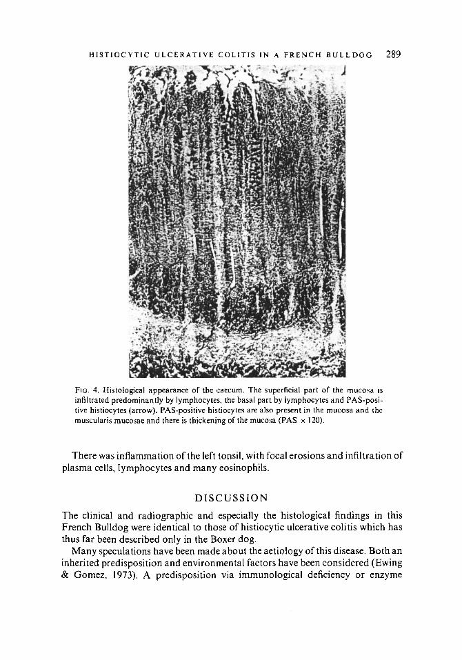
FIG. 4. Histological appearance of the caecum. The superficial part of the mucosa is infiltrated predominantly by lymphocytes. the basal part by lymphocytes and PAS-posi- tive histiocytes (arrow). PAS-positive histiocytes are also present in the mucosa and the muscularis mucosae and there is thickening of the mucosa (PAS x 120).
There was inflammation of the left tonsil, with focal erosions and infiltration of plasma cells, lymphocytes and many eosinophils.
D I S C U S S I O N
The clinical and radiographic and especially the histological findings in this French Bulldog were identical to those of histiocytic ulcerative colitis which has thus far been described only in the Boxer dog.
Many speculations have been made about the aetiology of this disease. Both an inherited predisposition and environmental factors have been considered (Ewing & Gomez, 1973). A predisposition via immunological deficiency or enzyme

290 I N G R I D V A N D E R G A A G e lul .
deficient histiocytes in the Boxer has also been suggested (as referred to by Kent & Moon, 1973).
Because of the lack of pedigree information the relationship of the Boxer dogs in the Netherlands with this disease is not known. In this regard it is of some interest that the disease has now been observed in a breed other than the Boxer and that this breed, the French Bulldog, has the same ancestral origin as the Boxer.
R E F E R E N C E S EWING, G.O. & GOMEZ, J.A. (1973) Canine ulcerative colitis. J. Am. Anim. Hosp. Ass. 9,395. KENNEDY, P.C. & CELLO, R.M. (1966) Colitis of Boxer dogs. Gastroenterology 51,926. KENT, T.H. & MOON, H.W. (1973) The comparative pathogenesis of some enteric diseases. Vet.
KOCH. S.A. & SKELLEY. J.F. ( I 967) Colitis in a dog resembling Whipple’s disease in man. J . Am. Vet.
F’EARCE, A.G.E. (1961) Hi.stiochemistry, 2nd edn. J. & A. Churchill, London. RUSSELL, S.W., GOMEZ, J.A. & TROWBRIDGE, J.O. ( 1 97 I ) Canine histiocytic ulcerative colitis. Lab.
SANDER. C.H. & LANGHAM. R.F. (1968) Canine histiocytic ulcerative colitis. Archs. Path. 85, 94. STARNES. D.D. (1969) Granulomatous colitis in a dog. S. West. Vet. , 22,234. VAN DER GAAG, I.. HAPPE, R.P. & WOLVEKAMP, WH. T.C. (1975) Histiocytic ulcerative colitis in the
Boxer. Proc. Voorjaarsdagen. Neth. Small Anim. Vet. Assn.. PA0 no. 6. 19. VAN DER GAAG. 1. ( I 975) Unpublished case referred from Prof. Dr M. Opitz (W. Berlin). VAN KRUININGEN. H.J.. MONTALI. R.J.. STRANDBERG, J.D. &KIRK, R.W. (1965) A granulomatous
colitis of dogs with histologic resemblance to Whipple’s disease. Pathologia vet. 2, 521.
Path. 10,414.
Med. Ass. 150, 22.
Invest. 25, 509.