hiroshi t. ito and erin m. schuman* - max planck society · hiroshi t. ito and erin m. schuman*...
TRANSCRIPT
Functional Division of Hippocampal Area CA1 ViaModulatory Gating of Entorhinal Cortical Inputs
Hiroshi T. Ito and Erin M. Schuman*
ABSTRACT: The hippocampus receives two streams of information,spatial and nonspatial, via major afferent inputs from the medial (MEC)and lateral entorhinal cortexes (LEC). The MEC and LEC projections inthe temporoammonic pathway are topographically organized along thetransverse-axis of area CA1. The potential for functional segregation ofarea CA1, however, remains relatively unexplored. Here, we demon-strated differential novelty-induced c-Fos expression along the trans-verse-axis of area CA1 corresponding to topographic projections of MECand LEC inputs. We found that, while novel place exposure induced auniform c-Fos expression along the transverse-axis of area CA1, novelobject exposure primarily activated the distal half of CA1 neurons. Inhippocampal slices, we observed distinct presynaptic propertiesbetween LEC and MEC terminals, and application of either DA or NEproduced a largely selective influence on one set of inputs (LEC).Finally, we demonstrated that differential c-Fos expression along thetransverse axis of area CA1 was largely abolished by an antagonist ofneuromodulatory receptors, clozapine. Our results suggest that neuro-modulators can control topographic TA projections allowing the hippo-campus to differentially encode new information along the transverseaxis of area CA1. VVC 2011 Wiley Periodicals, Inc.
KEY WORDS: temporoammonic pathway; medial/lateral entorhinalcortex; spatial/nonspatial information; dopamine; norepinephrine
INTRODUCTION
The brain has the capacity for parallel information processing, in whichsensory information received from the environment is segregated and inde-pendently processed based on particular features. For example, it is generallyaccepted that visual information is processed in two distinct informationstreams (Ungerleider and Haxby, 1994): a ventral stream that subserves objectrecognition, or ‘‘what’’ perception and a dorsal stream that primarily representsspatial information, or ‘‘where’’ perception. These distinct streams of informa-tion must be integrated somewhere in the brain for coherent perception(Engel and Singer, 2001). A number of studies indicate that the hippocampus,a brain structure important for episodic/declarative memory formation (Sco-ville and Milner, 1957; Squire et al., 2004), is one such integrative area that
combines the two streams of information (Witter andAmaral, 2004; Manns and Eichenbaum, 2006).
As initially described by Cajal (1911), the hippo-campus receives major afferent inputs from its adja-cent structure, the entorhinal cortex (EC). Anatomi-cally, the EC can be further divided into two subdivi-sions, the medial and lateral areas (MEC and LEC),based on cytoarchitecture and projection patterns(Blackstad, 1956; Dolorfo and Amaral, 1998; Witterand Amaral, 2004; Canto et al., 2008). Recent studiesfurther indicate that the MEC and LEC are function-ally distinct. For example, strong spatial modulationof neuronal activities is observed in the MEC (Fyhnet al., 2004; Hafting et al., 2005), but not in theLEC (Hargreaves et al., 2005). LEC neurons are, incontrast, likely to be involved in nonspatial informa-tion processing related to specific objects or cues inthe environment (Knierim et al., 2006). Targetedlesions of each subarea/projection led to distinct be-havioral deficits (Gauthier et al., 1983; Ferbinteanuet al., 1999; Hunsaker et al., 2007).
The projections from the MEC and LEC terminatein distinct parts of the hippocampus (Steward, 1976;Wyss, 1981; Witter and Amaral, 2004) (see Fig. 2).In the perforant pathway (originating from layer IIEC neurons), the fibers from the LEC terminate inthe outer third of the molecular layer in the dentategyrus (DG) and the superficial layer of stratum lacu-nosum moleculare (SLM) in area CA3, but the axonsfrom the MEC make synapses in the middle third ofthe DG molecular layer and the deep layer of theCA3 SLM. On the other hand, in the temporoam-monic (TA) projection (originating from layer III ECneurons) to area CA1, the fibers from the LEC makesynapses in the distal part (close to the subiculum) ofthe CA1 SLM, and the axons from the MEC termi-nate in the proximal part (close to area CA3). Thetopographic organization of LEC and MEC inputssuggests that neurons in proximal or distal CA1receive predominantly one set of entorhinal–corticalinputs via the TA pathway, implying the existence offunctional division along the transverse axis of areaCA1. This contrasts with the laminar organization ofthe perforant pathway, where each neuron in the DGor area CA3 receives both MEC and LEC inputs atdifferent dendritic locations.
Although the EC provides the hippocampus withboth spatial and nonspatial information, hippocampalneurons are not just passive responders to environ-
Division of Biology, Caltech/HHMI, Pasadena, California
Hiroshi T. Ito is currently at Kavli Institute for Systems Neuroscience andCentre for the Biology of Memory, Norwegian University of Science andTechnology, NO-7489 Trondheim, Norway.*Correspondence to: Erin M. Schuman, Max Planck Institute for BrainResearch, D-60528 Frankfurt am Main, Germany.E-mail: [email protected]
Additional Supporting Information may be found in the online version ofthis article.
Accepted for publication 29 September 2010DOI 10.1002/hipo.20909Published online 14 January 2011 in Wiley Online Library(wileyonlinelibrary.com).
HIPPOCAMPUS 22:372–387 (2012)
VVC 2011 WILEY PERIODICALS, INC.
mental stimuli. One prominent feature of hippocampal neuronactivity is a clear sensitivity to stimulus novelty (Knight, 1996;Stern et al., 1996; Dolan and Fletcher, 1997; Vinogradova,2001; Rutishauser et al., 2006), suggesting that the hippocam-pus may act as a novelty detector (Parkin, 1997; Kumaran andMaguire, 2007). Supporting this idea, novelty exposure influen-ces synaptic plasticity in the hippocampus (Manahan-Vaughanand Braunewell, 1999; Lemon and Manahan-Vaughan, 2006),and hippocampal-lesioned animals display deficits in noveltydetection (Buhusi et al., 1998; Allen et al., 2002; Hunsakeret al., 2008). The novelty-dependent activation of hippocampalneurons is likely to be a critical feature for learning, allowingcircuit modifications that optimize stimulus prediction. Howthe hippocampus acquires information about the novelty ofstimulus or context is still unclear, but a number of studieshave indicated a critical role of neuromodulators (Hasselmo,1995; Ranganath and Rainer, 2003; Lisman and Grace, 2005).
Neuromodulators play a key role in controlling informationflow among brain areas [see Ito and Schuman (2008) for areview]. Midbrain dopaminergic neurons in ventral tegmentalarea and substantial nigra compacta show increased activity afterexposure to novel stimuli (Schultz, 1998; Horvitz, 2000). Therelease of another major neuromodulator in the brain, norepi-nephrine (NE), is also controlled by novel stimuli (Vankovet al., 1995; Harley, 2004; Aston-Jones and Cohen, 2005; Sara,2009). The axons of these neuromodulator-releasing neuronsproject to the hippocampus (Swanson, 1987; Gasbarri et al.,1997) and release dopamine (DA) or NE after animals areexposed to a novel environment (Ihalainen et al., 1998). A num-ber of studies have indicated that DA or NE plays an importantrole in hippocampal-dependent learning (Gasbarri et al., 1996;El-Ghundi et al., 1999; Li et al., 2003; Murchison et al., 2004).
The interactions between neuromodulators and the hippo-campus may be crucial for constructing or updating representa-tions of environmental context, which requires the integrationof spatial and nonspatial information. Here, we investigatedhow neurons in the hippocampus are activated by differenttypes of stimulus novelty and examined if there is a functionaldivision in area CA1 afforded by the anatomical organizationof LEC and MEC inputs. We then examined the effects of NEand DA at entorhinal–cortical inputs as a potential mechanismfor the independent control of spatial and nonspatial informa-tion processing in the hippocampus.
MATERIALS AND METHODS
Hippocampal Slice Preparation
Slices were prepared from 21 to 30-day-old Sprague–Dawleyrats (Charles River). A vibrating microtome (Leica VT1000S)was used to make horizontal sections of the hippocampus(500-lm thickness for extracellular recordings and 300 lm forwhole-cell recordings) in ice-cold oxygenated artificial cerebro-spinal fluid (ACSF) containing (in mM) 119 NaCl, 2.5 KCl,1.3 MgSO4, 2.5 CaCl, 1.0 NaH2PO4, 26.2 NaHCO3, and
11.0 glucose. Slices made from 4 to 4.5 mm below the ventralsurface of the brain were used for all electrophysiology andimmunohistochemistry experiments (see Supporting Informa-tion Fig. 2). Slices were recovered at room temperature for atleast 2 h in an interface chamber and then transferred to a sub-merged recording chamber perfused with ACSF at 24.5–25.58C or 32–348C (for frequency-dependent analysis). For TApathway recordings, the DG and CA3 were removed to elimi-nate the possible activation of the trisynaptic pathway or perfo-rant path projection to area CA3. Concentric bipolar tungstenelectrodes (FHC: no. CBBRC75 or CABRC75; the inner polediameter was 25 lm, and the outer pole diameter was 200 lmfor the TA path or 125 lm for the perforant path stimulation.)and stimulus isolators (Axon Instruments) were used for thestimulation.
Electrophysiology
Extracellular field potential recordings were made with 1–3MX resistance microelectrodes filled with 3 M NaCl using abridge amplifier (Axoclamp 2B, Molecular Devices). We fol-lowed previous reports for the recordings from lateral TA-CA1synapses (Otmakhova and Lisman, 1999; Ito and Schuman,2007); the stimulating electrode was placed in the SLM of theCA1 border near the subiculum or the superficial molecular layerof the proximal subiculum (close to area CA1), and the record-ing electrode was inserted in the distal half of the CA1 SLM. Torecord from medial TA-CA1 synapses, the stimulating electrodewas placed in the SLM of the CA1 border near area CA2, andthe recording electrode was inserted in the proximal half of theCA1 SLM (see Fig. 3). The distance between each pair of stimu-lating and recording electrodes was 200–300 lm. We note herethat although not explicitly stated, our previous work (Ito andSchuman, 2007) described recordings exclusively from the lateralTA-CA1 synapses. The evoked synaptic potentials in the aboverecording configuration represent synaptic activity derived fromEC axons, but also from synapses with the nucleus reuniens(Herkenham, 1978; Wouterlood et al., 1990; Dolleman-VanDer Weel and Witter, 1996), postrhinal (Naber et al., 2001), orperirhinal cortex (Kosel et al., 1983; Naber et al., 1999). Thereuniens inputs, however, uniformly project to proximal and dis-tal parts of the CA1 SLM, thus will not contribute to the func-tional differences along the transverse-axis of CA1. The postrhi-nal and perirhinal projections to CA1 are topographically organ-ized as similar to those from MEC and LEC; however, theseprojections are relatively weak and make synapses at the extremeborders of CA1 (Naber et al., 1999, 2001), which we avoided inthe placement of the recording electrodes. Whole-cell voltage-clamp recordings from CA1 pyramidal neurons or DG granulecells were obtained without visualization with an Axopatch 200B(Molecular Devices). Internal solution of whole-cell patch pip-ettes was (in mM) 115 cesium gluconate, 20 KCl, 10 sodiumphosphocreatine, 10 HEPES, 0.2 EGTA, 2 MgATP, and0.3 NaGTP (pH 7.3). In addition, to minimize the possiblepostsynaptic current modulation by DA or NE, pipette solutionscontained (in mM) 5 QX314, 10 TEA, and 1 4AP. Membrane
Hippocampus
FUNCTIONAL DIVISION OF CA1 VIA MODULATORY GATING 373
voltage was clamped at 260 mV (without liquid junction poten-tial correction). Membrane capacitance was cancelled, and seriesresistance was compensated (60–70%). Recordings were dis-carded when the series resistance was over 20 MX or either seriesor membrane resistance changed more than 30% during data ac-quisition. For the analysis of frequency-dependent signal modu-lation, 100 pulses were applied at the indicated frequencies afterbaseline responses were stable for at least 10 min. The long-termpotentiation (LTP) induction protocol was 100 pulses at 100Hz, repeated twice with a 30-s interval. All stimulus pulses wereof the same length and amplitude as test pulses. Test pulses wereapplied once every 30 s for extracellular field recordings and ev-ery 10 s for whole cell recordings. Drugs were applied by dilu-tion of concentrated stock solutions into the perfusion medium.The final concentration of bath-applied DA or NE was 20 or 10lM, respectively. DA and NE were obtained from Sigma. Allother drugs were obtained from Tocris.
Behavioral Analysis
Animals used for behavioral analysis were male Sprague–Dawley rats, 24–30 days old. All the behavioral manipulationswere carried out at night (0–4:00 A.M.) to maximize active ex-ploration of the environment. The objects used for novel objectexposure were three small children’s toys, made of either plasticor wood. The new home cage for novel place exposure was inthe same color and shape as the original cage, but had newwoodchip flooring and did not have a food box on the ceiling.In DA -antagonist experiments, 250 ll of saline or clozapine(10 mg/kg, diluted in saline) was injected intraperitoneally inanimals. Clozapine blocks every subtype of DA receptors, butalso has a small antagonistic effect on serotonin receptors anda-2 adrenergic receptors (Baldessarini and Frankenburg, 1991).
Data Analysis
Electrophysiology
Data were collected using a custom-written program in Lab-View (National Instruments) for extracellular recordings, orDigiData 1200 and pClamp 9 (Molecular Devices) for whole-cell recordings. All numerical values listed represent mean 6s.e.m. For synaptic plasticity experiments, the baseline fEPSPwas normalized to the depressed state. For analysis of the wave-forms, during 100 pulse stimulation, stimulation artifacts andfiber volleys were excluded, and the gaps were linearly con-nected, and the last excitatory potential or current (100th stim-ulus response) was measured by a custom program in Matlab(MathWorks). A Wilcoxon rank sum test was performed to an-alyze the statistical significance of the data.
Immunohistochemistry
Slices (500 lm thickness) were prepared using the same pro-cedure as for electrophysiology recordings. After cutting, sliceswere quickly fixed in 4% paraformaldehyde in phosphate-buf-fered saline for at least 2 days. Thin (50 lm) sections were cutwith a vibrating microtome (Leica VT1000S). The sections
were incubated overnight with either of 1:250 concentration ofantic-Fos (sc-52) (Santa Cruz), 1:1,000 of anti-NeuN (Milli-pore), 1:1,000 of anti-Synapsin I (Millipore), and 1:1,000 ofanti-Bassoon (Stressgen) antibodies. The incubation was carriedout at room temperature in Tris-buffered saline containing0.2% Triton X-100, BSA 2%, and NGS 4%, followed by 4 hof secondary-antibody incubation with 1:1,000 of Alexa 488-conjugated antirabbit and 1:1,000 of Alexa 543-conjugatedanti-mouse antibodies (Invitrogen). For the analysis of immu-nohistochemistry experiments, images were obtained with ZeissLSM 510 laser scanning confocal microscopes using a Plan-Neofluor 103/0.3 air objective. Alexa 488 and 546 werevisualized by excitation with the 488 line of an argon ion laserand the 543 nm line of a He–Ne laser, respectively. The opticalsection was 20 lm, and fluorescent signals were acquiredthroughout the slice thickness (50 lm). Each 50-lm slice wasobtained from a different 500-lm section, and two slices wereanalyzed from each animal. Slices were obtained from the sameseptotemporal position in all experiments. To count the num-ber of c-Fos positive neurons, fluorescent signals less than themean 1 2SD were excluded (see Supporting Information Fig.1C). Then, automated particle analysis was carried out usingImageJ (NIH) based on the following criteria: particle sizemust be larger than 56 lm2 and the circularity larger than 0.5.For the analysis of dentate granule cells, particle sizes largerthan 39 lm2, instead 56 lm2, were used due to the smallersize of granule cells. Statistical differences between animalsgroups were assessed by ANOVA. Regional differences werestatistically analyzed by a Wilcoxon rank sum test.
RESULTS
Differential Activation of Distal and ProximalCA1 by Exposure to Novel Object or Place
Neurons in the hippocampus exhibit differential firing basedon the novelty or familiarity of stimuli encountered in the envi-ronment. We examined whether exposure to novel stimuli leadsto activation of neurons in area CA1, and if so, whether thereis differential activation along the transverse-axis of area CA1,due to the topographic organization of LEC and MEC projec-tions in the TA pathway. Following home-cage exposure forseveral days, pairs of individually housed rats were subjected toone of the following conditions: exposure to novel objects inthe home cage (experimental group: EXP) or sham exposure(cage opened but no objects introduced, control group: CON).After 2 h of novel object exposure, animals were sacrificed, andimmunohistochemistry was performed on brain slices. Toobserve activation of CA1 neurons, we stained with antibodiesfor an immediate early gene product, c-Fos (Morgan andCurran, 1991; Guzowski et al., 2005), together with a neuronalnuclear marker protein, NeuN (Fig. 1A). Animals exposed tothe novel objects exhibited a significantly higher number ofc-Fos-positive neurons in area CA1, when compared with con-
374 ITO AND SCHUMAN
Hippocampus
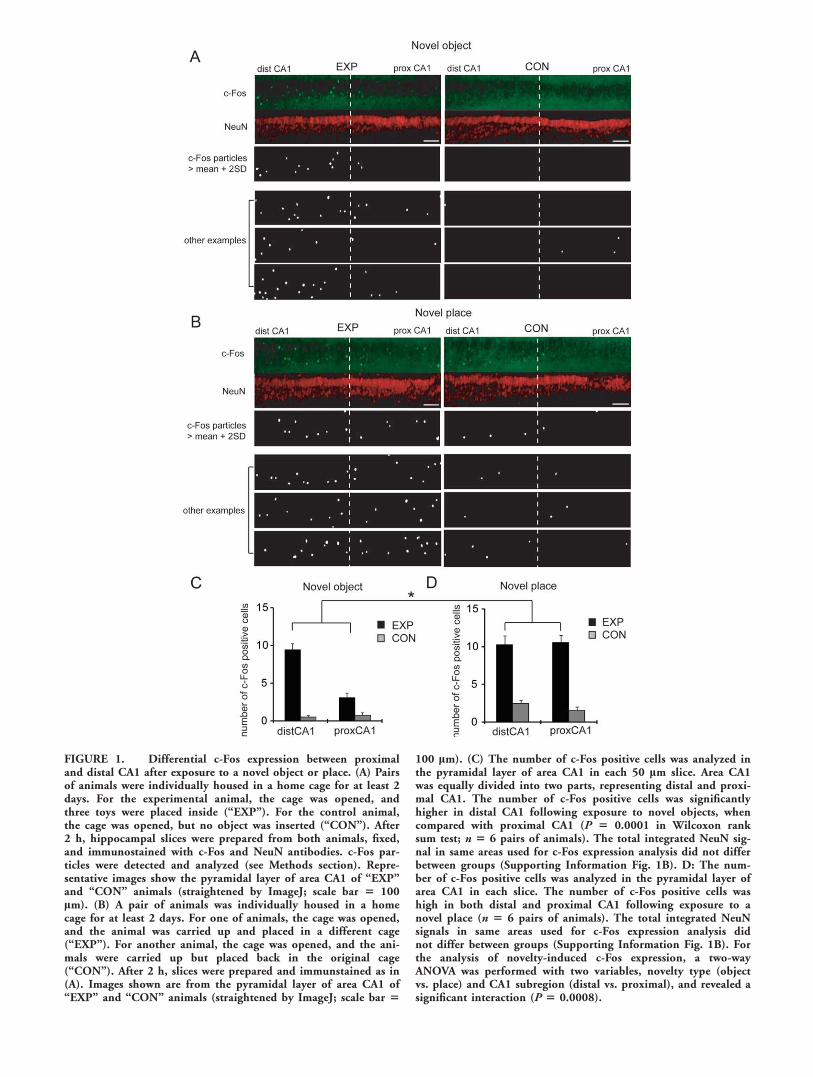
FIGURE 1. Differential c-Fos expression between proximaland distal CA1 after exposure to a novel object or place. (A) Pairsof animals were individually housed in a home cage for at least 2days. For the experimental animal, the cage was opened, andthree toys were placed inside (‘‘EXP’’). For the control animal,the cage was opened, but no object was inserted (‘‘CON’’). After2 h, hippocampal slices were prepared from both animals, fixed,and immunostained with c-Fos and NeuN antibodies. c-Fos par-ticles were detected and analyzed (see Methods section). Repre-sentative images show the pyramidal layer of area CA1 of ‘‘EXP’’and ‘‘CON’’ animals (straightened by ImageJ; scale bar 5 100lm). (B) A pair of animals was individually housed in a homecage for at least 2 days. For one of animals, the cage was opened,and the animal was carried up and placed in a different cage(‘‘EXP’’). For another animal, the cage was opened, and the ani-mals were carried up but placed back in the original cage(‘‘CON’’). After 2 h, slices were prepared and immunstained as in(A). Images shown are from the pyramidal layer of area CA1 of‘‘EXP’’ and ‘‘CON’’ animals (straightened by ImageJ; scale bar 5
100 lm). (C) The number of c-Fos positive cells was analyzed inthe pyramidal layer of area CA1 in each 50 lm slice. Area CA1was equally divided into two parts, representing distal and proxi-mal CA1. The number of c-Fos positive cells was significantlyhigher in distal CA1 following exposure to novel objects, whencompared with proximal CA1 (P 5 0.0001 in Wilcoxon ranksum test; n 5 6 pairs of animals). The total integrated NeuN sig-nal in same areas used for c-Fos expression analysis did not differbetween groups (Supporting Information Fig. 1B). D: The num-ber of c-Fos positive cells was analyzed in the pyramidal layer ofarea CA1 in each slice. The number of c-Fos positive cells washigh in both distal and proximal CA1 following exposure to anovel place (n 5 6 pairs of animals). The total integrated NeuNsignals in same areas used for c-Fos expression analysis didnot differ between groups (Supporting Information Fig. 1B). Forthe analysis of novelty-induced c-Fos expression, a two-wayANOVA was performed with two variables, novelty type (objectvs. place) and CA1 subregion (distal vs. proximal), and revealed asignificant interaction (P 5 0.0008).
trol animals. As the EC projection to area CA1 shows topo-graphic organization, we counted the number c-Fos-positiveneurons in distal (receiving LEC inputs) versus proximal(receiving MEC inputs) CA1. We observed a larger number ofc-Fos positive neurons in distal CA1 compared to proximalCA1 (c-Fos positive cells: distal 9.4 6 0.8, proximal 3.1 60.6; P 5 0.0001 in Wilcoxon rank sum test; Fig. 1C) in brainslices from EXP, but not control, animals.
To further examine the potential topographic representationof environmental signals to area CA1, we examined c-Fosexpression after animals were exposed to a novel environment.Using the same design as earlier, one animal was removed fromhis home cage and placed in a new cage (‘‘EXP’’ group), andthe paired control was removed and then reintroduced to hishome cage (‘‘CON’’ group). Exposure to a novel place signifi-cantly enhanced c-Fos expression in neurons in both proximaland distal CA1 (c-Fos positive cells: distal 10.3 6 1.2, proxi-mal 10.6 6 0.9; P 5 0.82 in Wilcoxon rank sum test; Figs.1B,D). These data indicate that proximal and distal CA1 canbe differentially activated depending on the different type ofstimulus novelty.
Differences in Presynaptic Properties BetweenLEC and MEC Inputs
The above difference in c-Fos immunoreactivity betweenproximal and distal CA1 indicates that topographic projectionsin the TA pathway are behaviorally relevant. We next investi-gated whether any differences can be detected between LECand MEC inputs at the synaptic scale. Because neurons in dif-ferent brain regions often express different level of presynapticproteins (Oyler et al., 1989; Melloni et al., 1993; Richteret al., 1999), we performed immunohistochemistry on hippo-campal slices to examine whether there are differences in pre-synaptic protein expression corresponding to LEC and MECsynapses. We found that synaptic terminal proteins, synapsin I,and bassoon showed differential distribution along the trans-verse axis of the CA1 SLM (Supporting Information Fig. 2).The transition of these differences occurs roughly at themidline of the CA1 transverse axis (Supporting InformationFig. 2D), corresponding to the slice section level, we chose (seeMethods section), which mapped onto the previously reportedanatomical distinction between areas receiving LEC and MECinputs (Steward, 1976; Wyss, 1981; Witter and Amaral, 2004).These immunostaining results suggest that proximal and distalparts of the CA1 SLM possess different axon terminals, provid-ing support for the topographic LEC and MEC projection.
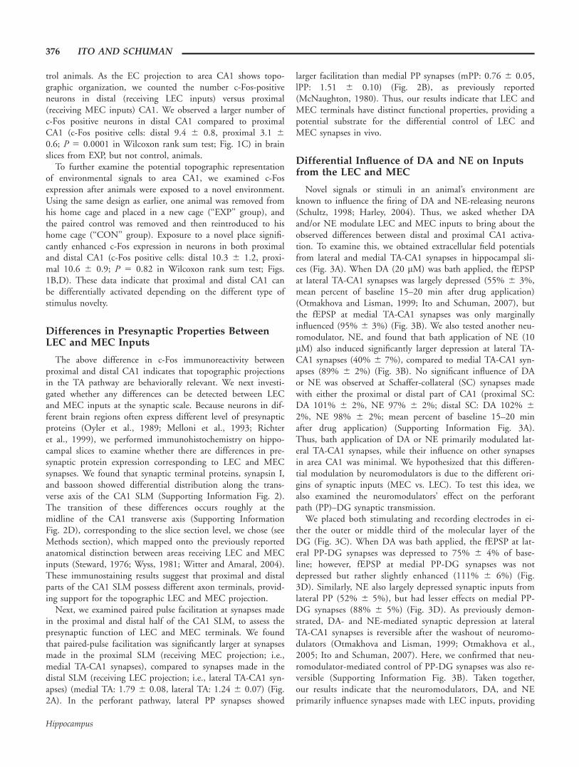
Next, we examined paired pulse facilitation at synapses madein the proximal and distal half of the CA1 SLM, to assess thepresynaptic function of LEC and MEC terminals. We foundthat paired-pulse facilitation was significantly larger at synapsesmade in the proximal SLM (receiving MEC projection; i.e.,medial TA-CA1 synapses), compared to synapses made in thedistal SLM (receiving LEC projection; i.e., lateral TA-CA1 syn-apses) (medial TA: 1.79 6 0.08, lateral TA: 1.24 6 0.07) (Fig.2A). In the perforant pathway, lateral PP synapses showed
larger facilitation than medial PP synapses (mPP: 0.76 6 0.05,lPP: 1.51 6 0.10) (Fig. 2B), as previously reported(McNaughton, 1980). Thus, our results indicate that LEC andMEC terminals have distinct functional properties, providing apotential substrate for the differential control of LEC andMEC synapses in vivo.
Differential Influence of DA and NE on Inputsfrom the LEC and MEC
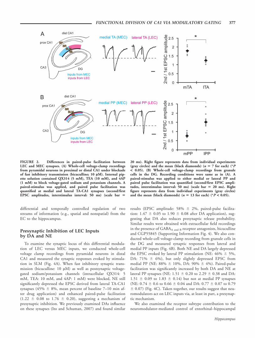
Novel signals or stimuli in an animal’s environment areknown to influence the firing of DA and NE-releasing neurons(Schultz, 1998; Harley, 2004). Thus, we asked whether DAand/or NE modulate LEC and MEC inputs to bring about theobserved differences between distal and proximal CA1 activa-tion. To examine this, we obtained extracellular field potentialsfrom lateral and medial TA-CA1 synapses in hippocampal sli-ces (Fig. 3A). When DA (20 lM) was bath applied, the fEPSPat lateral TA-CA1 synapses was largely depressed (55% 6 3%,mean percent of baseline 15–20 min after drug application)(Otmakhova and Lisman, 1999; Ito and Schuman, 2007), butthe fEPSP at medial TA-CA1 synapses was only marginallyinfluenced (95% 6 3%) (Fig. 3B). We also tested another neu-romodulator, NE, and found that bath application of NE (10lM) also induced significantly larger depression at lateral TA-CA1 synapses (40% 6 7%), compared to medial TA-CA1 syn-apses (89% 6 2%) (Fig. 3B). No significant influence of DAor NE was observed at Schaffer-collateral (SC) synapses madewith either the proximal or distal part of CA1 (proximal SC:DA 101% 6 2%, NE 97% 6 2%; distal SC: DA 102% 62%, NE 98% 6 2%; mean percent of baseline 15–20 minafter drug application) (Supporting Information Fig. 3A).Thus, bath application of DA or NE primarily modulated lat-eral TA-CA1 synapses, while their influence on other synapsesin area CA1 was minimal. We hypothesized that this differen-tial modulation by neuromodulators is due to the different ori-gins of synaptic inputs (MEC vs. LEC). To test this idea, wealso examined the neuromodulators’ effect on the perforantpath (PP)–DG synaptic transmission.
We placed both stimulating and recording electrodes in ei-ther the outer or middle third of the molecular layer of theDG (Fig. 3C). When DA was bath applied, the fEPSP at lat-eral PP-DG synapses was depressed to 75% 6 4% of base-line; however, fEPSP at medial PP-DG synapses was notdepressed but rather slightly enhanced (111% 6 6%) (Fig.3D). Similarly, NE also largely depressed synaptic inputs fromlateral PP (52% 6 5%), but had lesser effects on medial PP-DG synapses (88% 6 5%) (Fig. 3D). As previously demon-strated, DA- and NE-mediated synaptic depression at lateralTA-CA1 synapses is reversible after the washout of neuromo-dulators (Otmakhova and Lisman, 1999; Otmakhova et al.,2005; Ito and Schuman, 2007). Here, we confirmed that neu-romodulator-mediated control of PP-DG synapses was also re-versible (Supporting Information Fig. 3B). Taken together,our results indicate that the neuromodulators, DA, and NEprimarily influence synapses made with LEC inputs, providing
376 ITO AND SCHUMAN
Hippocampus
differential and temporally controlled regulation of twostreams of information (e.g., spatial and nonspatial) from theEC to the hippocampus.
Presynaptic Inhibition of LEC Inputsby DA and NE
To examine the synaptic locus of this differential modula-tion of LEC versus MEC inputs, we conducted whole-cellvoltage clamp recordings from pyramidal neurons in distalCA1 and measured the synaptic responses evoked by stimula-tion in SLM (Fig. 4A). When fast inhibitory synaptic trans-mission (bicuculline: 10 lM) as well as postsynaptic voltage-gated sodium/potassium channels (intracellular QX314: 5mM, TEA: 10 mM, and 4AP: 1 mM) were blocked, NE stillsignificantly depressed the EPSC derived from lateral TA-CA1synapses (45% 6 8%, mean percent of baseline 7–10 min af-ter drug application) and enhanced paired-pulse facilitation(1.22 6 0.08 to 1.76 6 0.20), suggesting a mechanism ofpresynaptic inhibition. We previously examined DAs influenceon these synapses (Ito and Schuman, 2007) and found similar
results (EPSC amplitude: 58% 6 2%, paired-pulse facilita-tion: 1.47 6 0.05 to 1.90 6 0.08 after DA application), sug-gesting that DA also reduces presynaptic release probability.Similar results were obtained with extracellular field recordingsin the presence of GABAA and B receptor antagonists, bicucullineand CGP55845 (Supporting Information Fig. 4). We also con-ducted whole-cell voltage-clamp recording from granule cells inthe DG and measured synaptic responses from lateral andmedial PP inputs (Fig. 4B). Both NE and DA largely depressedthe EPSC evoked by lateral PP stimulation (NE: 46% 6 5%,DA: 71% 6 6%), but only slightly depressed EPSC frommedial PP (NE: 88% 6 10%, DA: 90% 6 4%). Paired-pulsefacilitation was significantly increased by both DA and NE atlateral PP synapses (NE: 1.51 6 0.20 to 2.29 6 0.38 and DA:1.51 6 0.09 to 1.83 6 0.14) but not at medial PP synapses(NE: 0.74 6 0.6 to 0.66 6 0.04 and DA: 0.77 6 0.07 to 0.796 0.07) (Fig. 4C). Taken together, our results suggest that neu-romodulators act on LEC inputs via, at least in part, a presynap-tic mechanism.
We also examined the receptor subtype contribution to theneuromodulator-mediated control of entorhinal–hippocampal
FIGURE 2. Differences in paired-pulse facilitation betweenLEC and MEC synapses. (A) Whole-cell voltage-clamp recordingsfrom pyramidal neurons in proximal or distal CA1 under blockadeof fast inhibitory transmission (bicuculline; 10 lM). Internal pip-ette solution contained QX314 (5 mM), TEA (10 mM), and 4AP(1 mM) to block voltage-gated sodium and potassium channels. Apaired-stimulus was applied, and paired pulse facilitation wasquantified at medial and lateral TA-CA1 synapses (second/firstEPSC amplitudes, interstimulus interval: 50 ms) (scale bar 5
20 ms). Right figure represents data from individual experiments(gray circles) and the mean (black diamonds) (n 5 7 for each) (*P< 0.05). (B) Whole-cell voltage-clamp recordings from granulecells in the DG. Recording conditions were same as in (A). Apaired-stimulus was applied to either medial or lateral PP andpaired pulse facilitation was quantified (second/first EPSC ampli-tudes, interstimulus interval: 50 ms) (scale bar 5 20 ms). Rightfigure represents data from individual experiments (gray circles)and the mean (black diamonds) (n 5 13 for each) (*P < 0.05).
FUNCTIONAL DIVISION OF CA1 VIA MODULATORY GATING 377
Hippocampus
FIGURE 3. Differential modulation of LEC inputs by DA andNE in two different hippocampal pathways. (A) Scheme of entorhi-nal cortical inputs to SLM of area CA1. MEC inputs primarily makesynapses at the proximal (relative to CA3) CA1 SLM (medial TApathway), but LEC inputs are enriched in the distal CA1 SLM (lat-eral TA pathway). Appropriate positioning of recording electrode ineither proximal or distal parts of CA1 SLM allows the measurementof synaptic responses derived from each input (see Methods section).Two stimulating electrodes were positioned, so that the distancebetween stimulating and recording electrodes was equal for eachinput. Representative waveforms show field potentials in proximaland distal SLM after the stimulation of TA axons by each stimulatingelectrode (scale bar 5 0.1 mV and 5 ms). (B) Either DA (20 lM) orNE (10 lM) application caused a large depression of the field EPSPresulting from lateral, but not medial TA-CA1 synapses (DA: n 5 7;NE: n 5 5). Field EPSP waveforms before (black) and after (gray)DA or NE application are shown (scale bar 5 0.1 mV and 5 ms). (C)
Scheme of entorhinal cortical inputs to the molecular layer of DG.LEC inputs make synapses in the outer molecular layer (OML; lat-eral PP), but MEC inputs project the middle molecular layer (MML;medial PP). Appropriate positioning of stimulating and recordingelectrodes allows the measurement of synaptic responses from eachpathway. Pathway selectivity was further confirmed by sink-sourcewaveform analysis of field potentials. Representative field potentialsdepict negative-going potentials in MML and positive-going poten-tials in the OML in response to medial PP stimulation. On the otherhand, lateral PP stimulation induces a negative-going potential inthe OML and a positive-going potential in the MML (scale bar 50.1 mV and 5 ms). (D) Either DA or NE application induced largesynaptic depression at the lateral PP–DG, but not in medial PP-DGsynapse (DA: n 5 6; NE: n 5 5). Field EPSP waveforms before(black) and after (gray) DA or NE application are shown (scale bar5 0.1 mVand 5 ms).
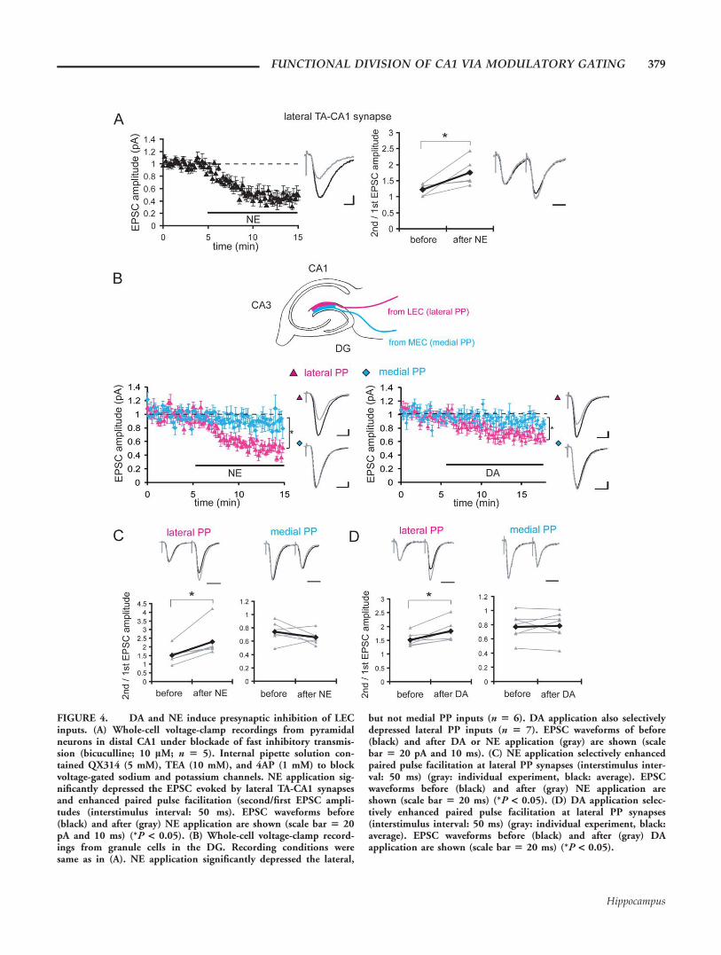
FIGURE 4. DA and NE induce presynaptic inhibition of LECinputs. (A) Whole-cell voltage-clamp recordings from pyramidalneurons in distal CA1 under blockade of fast inhibitory transmis-sion (bicuculline; 10 lM; n 5 5). Internal pipette solution con-tained QX314 (5 mM), TEA (10 mM), and 4AP (1 mM) to blockvoltage-gated sodium and potassium channels. NE application sig-nificantly depressed the EPSC evoked by lateral TA-CA1 synapsesand enhanced paired pulse facilitation (second/first EPSC ampli-tudes (interstimulus interval: 50 ms). EPSC waveforms before(black) and after (gray) NE application are shown (scale bar 5 20pA and 10 ms) (*P < 0.05). (B) Whole-cell voltage-clamp record-ings from granule cells in the DG. Recording conditions weresame as in (A). NE application significantly depressed the lateral,
but not medial PP inputs (n 5 6). DA application also selectivelydepressed lateral PP inputs (n 5 7). EPSC waveforms of before(black) and after DA or NE application (gray) are shown (scalebar 5 20 pA and 10 ms). (C) NE application selectively enhancedpaired pulse facilitation at lateral PP synapses (interstimulus inter-val: 50 ms) (gray: individual experiment, black: average). EPSCwaveforms before (black) and after (gray) NE application areshown (scale bar 5 20 ms) (*P < 0.05). (D) DA application selec-tively enhanced paired pulse facilitation at lateral PP synapses(interstimulus interval: 50 ms) (gray: individual experiment, black:average). EPSC waveforms before (black) and after (gray) DAapplication are shown (scale bar 5 20 ms) (*P < 0.05).
FUNCTIONAL DIVISION OF CA1 VIA MODULATORY GATING 379
Hippocampus

connections, using a variety of receptor antagonists. Thesestudies suggest the contribution of both DA D1-like and D2-like receptors to DA-induced synaptic depression and the a-2adrenergic receptor to NE-induced depression at both the TAand perforant pathway synapses (Supporting Information Fig.4). Thus, the TA and perforant pathway appear to utilize asimilar signal transduction pathway for the neuromodulator-mediated control of LEC inputs, irrespective of the origin dif-ferences between the TA pathway (layer III of the EC) andthe perforant pathway (layer II of the EC). The differentialcontrol of lateral and medial perforant path by NE, weobserved here, is mediated by a distinct mechanism from pre-vious reports on b adrenergic receptors (Dahl and Sarvey,1989; Pelletier et al., 1994). Because a-2 adrenergic receptorsare primarily located at presynaptic terminals in the hippo-campus (Milner et al., 1998; Hillman et al., 2005), ourresults are consistent with the idea that differential control ofMEC and LEC synapses is mediated by presynaptic terminaldifferences.
Differential Modulation of High-FrequencySignal Transmission by DA or NE atLateral TA-CA1 Synapses
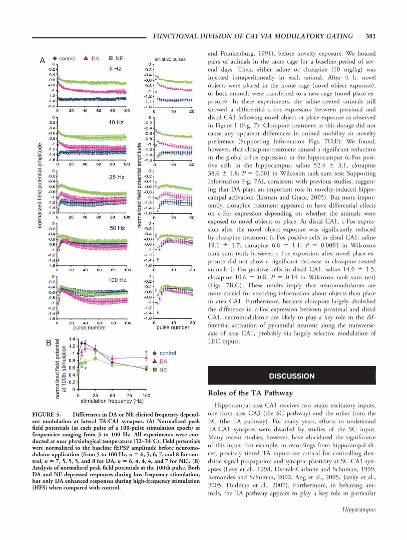
Signal transmission in neuronal networks is intrinsically non-linear and strongly influenced by the frequency of the input sig-nals (Markram et al., 1998). Dynamic changes in the amplitude,frequency, and phase-coordination of oscillations in vivo appearto be functionally linked to animal behavior (Buzsaki and Dra-guhn, 2004), suggesting that oscillatory activities may participatein signal gating (Laurent, 2002; Ito and Schuman, 2008). AsDA modulates frequency-dependent signal transmission at distalTA-CA1 synapses by enhancing high-frequency input signalsbut depressing low-frequency inputs (Ito and Schuman, 2007;Fig. 5), we next examined how NE influences frequency-depend-ent signal transmission at these same synapses.
We applied 100 pulses of stimulation to the TA pathway atdifferent ranges of stimulation frequency from 5 to 100 Hz(Fig. 5A). As in our previous report (Ito and Schuman, 2007),we focused on the analysis of steady-state potentials, becausetransient potentials during the first few stimuli can be influ-enced by the prestimulus state of the neuronal network. Whenlow-frequency (<50 Hz) stimulation was applied, NE stronglydepressed the steady-state potentials when compared with con-trol. However, as the stimulation frequency increased, the dif-ference between control and NE became smaller (Fig. 5B),with no modulation observed at high frequencies (>50 Hz).Although the depression of low-frequency signals by NE is sim-ilar to DA-induced modulation, a major difference appearedduring high-frequency stimulation: only DA enhanced high-fre-quency signals.
Thus, although no difference between DA- and NE-medi-ated synaptic modulation was evident in our analysis of basalsynaptic transmission (0.033 Hz) (Fig. 3B), the frequency-response analysis revealed a clear difference in high-frequencysignal modulation by DA and NE at lateral TA-CA1 synapses.
We also examined frequency-dependent signal transmission atmedial TA-CA1 synapses and found that neither DA nor NEinfluences the transmission of 5–100 Hz signals at these synap-ses (Supporting Information Fig. 5).
Selective Influence of DA or NEon LTP at Medial and LateralTA-CA1 Synapses
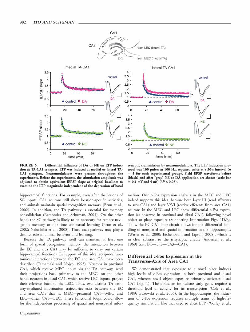
The above differences in frequency-response modulationbetween NE and DA may influence synaptic plasticity induc-tion, due to the differential handling of high frequency signalslike those commonly used to elicit long-term potentiation(LTP) (e.g., 100 Hz). We tested this idea by recording fromlateral TA-CA1 synapses (Fig. 6). The application of high-fre-quency (100 Hz) stimulation induced LTP of a modest magni-tude at these synapses (control: 112% 6 3%, mean percent ofbaseline 55–60 min after LTP induction). On the other hand,slices exposed to DA exhibited LTP of a greater magnitude, aswe previously reported (Ito and Schuman, 2007) (DA: 149%6 4%; Fig. 6, upper right). On the contrary, NE applicationdid not enhance LTP magnitude, when compared with control(NE: 114% 6 3%; Fig. 6, lower right). This is predicted bythe differences in high-frequency signal modulation betweenDA and NE that we observed (Fig. 5). We also tested whetherneuromodulators influence LTP induction at medial TA-CA1synapses, but neither DA nor NE altered LTP magnitude atthese synapses (control: 119% 6 3%, DA: 122% 6 3%, NE:122% 6 3%; Fig. 6, left). Thus, neither DA nor NE influen-ces the transmission of 5–100 Hz signals (Supporting Informa-tion Fig. 5) or plasticity at medial TA-CA1 synapses. In con-trast, DA, but not NE, modulated synaptic plasticity at lateralTA-CA1 synapses.
Although the above LTP experiments were done in the con-tinuous presence of neuromodulators, we also determinedwhether an acute application of neuromodulators is sufficientto influence synaptic plasticity. An application of DA or NE at10 s before the LTP induction still differentially controlled theLTP magnitude at lateral TA-CA1 synapses (Ito and Schuman,2007; Supporting Information Fig. 6A), suggesting that a pha-sic release of neuromodulators in vivo could control synapticplasticity at these synapses.
Clozapine Reduces Novelty-InducedDifferential Activation of Distaland Proximal CA1
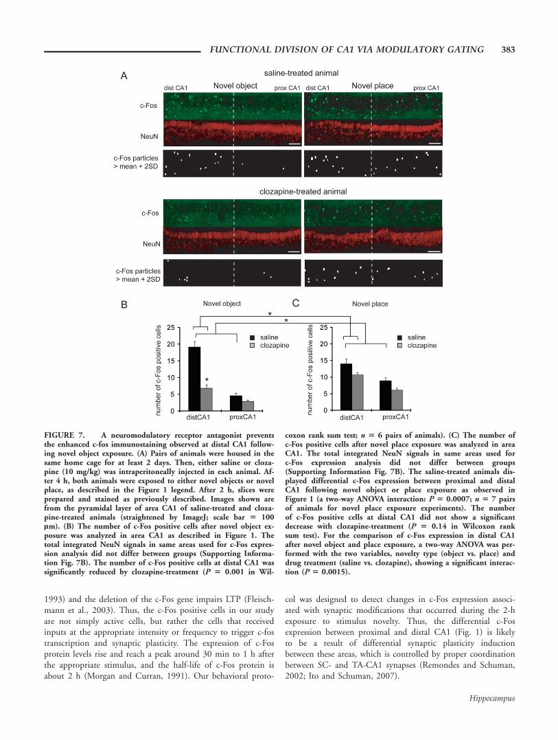
Our slice electrophysiology results suggest that neuromodula-tors primarily modulate synaptic transmission at lateral TA-CA1 synapses, raising the possibility that the novelty-inducedenhancement of c-fos transcription is mediated by this influ-ence. Thus, we asked whether an antagonist of neuromodula-tors influences the differential c-Fos expression along the trans-verse-axis of area CA1 observed in our behavioral experiments(Fig. 1). To test this, we examined c-Fos expression in animalstreated with a broad spectrum receptor antagonist that blocksboth DA and NE-mediated signaling, clozapine (Baldessarini
380 ITO AND SCHUMAN
Hippocampus
and Frankenburg, 1991), before novelty exposure. We housedpairs of animals in the same cage for a baseline period of sev-eral days. Then, either saline or clozapine (10 mg/kg) wasinjected intraperitoneally in each animal. After 4 h, novelobjects were placed in the home cage (novel object exposure),or both animals were transferred to a new cage (novel place ex-posure). In these experiments, the saline-treated animals stillshowed a differential c-Fos expression between proximal anddistal CA1 following novel object or place exposure as observedin Figure 1 (Fig. 7). Clozapine-treatment at this dosage did notcause any apparent differences in animal mobility or noveltypreference (Supporting Information Figs. 7D,E). We found,however, that clozapine-treatment caused a significant reductionin the global c-Fos expression in the hippocampus (c-Fos posi-tive cells in the hippocampus: saline 52.4 6 3.1, clozapine38.6 6 1.8; P 5 0.001 in Wilcoxon rank sum test; SupportingInformation Fig. 7A), consistent with previous studies, suggest-ing that DA plays an important role in novelty-induced hippo-campal activation (Lisman and Grace, 2005). But more impor-tantly, clozapine treatment appeared to have differential effectson c-Fos expression depending on whether the animals wereexposed to novel objects or place. At distal CA1, c-Fos expres-sion after the novel object exposure was significantly reducedby clozapine-treatment (c-Fos positive cells in distal CA1: saline19.1 6 1.7, clozapine 6.8 6 1.1; P 5 0.0001 in Wilcoxonrank sum test); however, c-Fos expression after novel place ex-posure did not show a significant decrease in clozapine-treatedanimals (c-Fos positive cells in distal CA1: saline 14.0 6 1.5,clozapine 10.6 6 0.8; P 5 0.14 in Wilcoxon rank sum test)(Figs. 7B,C). These results imply that neuromodulators aremore crucial for encoding information about objects than placein area CA1. Furthermore, because clozapine largely abolishedthe difference in c-Fos expression between proximal and distalCA1, neuromodulators are likely to play a key role in the dif-ferential activation of pyramidal neurons along the transverse-axis of area CA1, probably via largely selective modulation ofLEC inputs.
DISCUSSION
Roles of the TA Pathway
Hippocampal area CA1 receives two major excitatory inputs,one from area CA3 (the SC pathway) and the other from theEC (the TA pathway). For many years, efforts to understandTA-CA1 synapses were dwarfed by studies of the SC input.Many recent studies, however, have elucidated the significanceof this input. For example, in recordings from hippocampal sli-ces, precisely timed TA inputs are critical for controlling den-dritic signal propagation and synaptic plasticity at SC-CA1 syn-apses (Levy et al., 1998; Dvorak-Carbone and Schuman, 1999;Remondes and Schuman, 2002; Ang et al., 2005; Jarsky et al.,2005; Dudman et al., 2007). Furthermore, in behaving ani-mals, the TA pathway appears to play a key role in particular
FIGURE 5. Differences in DA or NE elicited frequency depend-ent modulation at lateral TA-CA1 synapses. (A) Normalized peakfield potentials (at each pulse of a 100-pulse stimulation epoch) atfrequencies ranging from 5 to 100 Hz. All experiments were con-ducted at near physiological temperature (32–348C). Field potentialswere normalized to the baseline fEPSP amplitude before neuromo-dulator application (from 5 to 100 Hz, n 5 6, 5, 6, 7, and 8 for con-trol; n 5 7, 5, 5, 5, and 8 for DA; n 5 6, 4, 4, 4, and 7 for NE). (B)Analysis of normalized peak field potentials at the 100th pulse. BothDA and NE depressed responses during low-frequency stimulation,but only DA enhanced responses during high-frequency stimulation(HFS) when compared with control.
FUNCTIONAL DIVISION OF CA1 VIA MODULATORY GATING 381
Hippocampus
hippocampal functions. For example, even after the lesions ofSC inputs, CA1 neurons still show location-specific activities,and animals maintain spatial recognition memory (Brun et al.,2002). In addition, the TA pathway is essential for memoryconsolidation (Remondes and Schuman, 2004). On the otherhand, the SC pathway is likely to be necessary for remote navi-gation memory or one-time contextual learning (Brun et al.,2002; Nakashiba et al., 2008). Thus, each pathway may play adistinct role in animal behavior and learning.
Because the TA pathway itself can maintain at least oneform of spatial recognition memory, the interaction betweenthe EC and area CA1 may be sufficient to carry out certainhippocampal functions. In support of this idea, reciprocal ana-tomical interactions between the EC and area CA1 have beendescribed (Tamamaki and Nojyo, 1995). Neurons in proximalCA1, which receive MEC inputs via the TA pathway, sendtheir projections back primarily to the MEC; on the otherhand, neurons in distal CA1, which receive LEC inputs, projecttheir efferents back to the LEC. Thus, two distinct TA-path-way-mediated information trajectories exist between the ECand area CA1; that is, MEC—proximal CA1—MEC andLEC—distal CA1—LEC. These functional loops could allowfor the independent processing of spatial and nonspatial infor-
mation. Our c-Fos expression analysis in the MEC and LECindeed supports this idea, because both layer III (send afferentsto area CA1) and layer V/VI (receive efferents from area CA1)neurons in the MEC and LEC show differential c-Fos expres-sion (as observed in proximal and distal CA1), following novelobject or place exposure (Supporting Information Figs. 1D,E).Thus, the EC-CA1 loop circuit allows for the differential han-dling of nonspatial and spatial information in the hippocampus(Witter et al., 2000; Eichenbaum and Lipton, 2008), which isin clear contrast to the trisynaptic circuit (Andersen et al.,1969) (i.e., EC—DG—CA3—CA1).
Differential c-Fos Expression in theTransverse-Axis of Area CA1
We demonstrated that exposure to a novel place induceshigh levels of c-Fos expression in both proximal and distalCA1, whereas novel object exposure primarily activates distalCA1 (Fig. 1). The c-Fos, an immediate early gene, requires athreshold level of activity for its transcription (Cole et al.,1989; Guzowski et al., 2005). In the hippocampus, the induc-tion of c-Fos expression requires multiple trains of high-fre-quency stimulation, like that used to elicit LTP (Worley et al.,
FIGURE 6. Differential influence of DA or NE on LTP induc-tion at TA-CA1 synapses. LTP was induced at medial or lateral TA-CA1 synapses. Neuromodulators were present throughout theexperiments. Before the experiments, the stimulation amplitude wasadjusted to obtain equivalent fEPSP slope as original baselines toexamine the LTP magnitude independent of the depression of basal
synaptic transmission by neuromodulators. The LTP induction pro-tocol was 100 pulses at 100 Hz, repeated twice at a 30-s interval (n5 5 for each experimental group). Field EPSP waveforms before(black) and after (gray) NE or DA application are shown (scale bar5 0.1 mV and 5 ms) (*P < 0.05).
382 ITO AND SCHUMAN
Hippocampus
1993) and the deletion of the c-Fos gene impairs LTP (Fleisch-mann et al., 2003). Thus, the c-Fos positive cells in our studyare not simply active cells, but rather the cells that receivedinputs at the appropriate intensity or frequency to trigger c-fostranscription and synaptic plasticity. The expression of c-Fosprotein levels rise and reach a peak around 30 min to 1 h afterthe appropriate stimulus, and the half-life of c-Fos protein isabout 2 h (Morgan and Curran, 1991). Our behavioral proto-
col was designed to detect changes in c-Fos expression associ-ated with synaptic modifications that occurred during the 2-hexposure to stimulus novelty. Thus, the differential c-Fosexpression between proximal and distal CA1 (Fig. 1) is likelyto be a result of differential synaptic plasticity inductionbetween these areas, which is controlled by proper coordinationbetween SC- and TA-CA1 synapses (Remondes and Schuman,2002; Ito and Schuman, 2007).
FIGURE 7. A neuromodulatory receptor antagonist preventsthe enhanced c-fos immunostaining observed at distal CA1 follow-ing novel object exposure. (A) Pairs of animals were housed in thesame home cage for at least 2 days. Then, either saline or cloza-pine (10 mg/kg) was intraperitoneally injected in each animal. Af-ter 4 h, both animals were exposed to either novel objects or novelplace, as described in the Figure 1 legend. After 2 h, slices wereprepared and stained as previously described. Images shown arefrom the pyramidal layer of area CA1 of saline-treated and cloza-pine-treated animals (straightened by ImageJ; scale bar 5 100lm). (B) The number of c-Fos positive cells after novel object ex-posure was analyzed in area CA1 as described in Figure 1. Thetotal integrated NeuN signals in same areas used for c-Fos expres-sion analysis did not differ between groups (Supporting Informa-tion Fig. 7B). The number of c-Fos positive cells at distal CA1 wassignificantly reduced by clozapine-treatment (P 5 0.001 in Wil-
coxon rank sum test; n 5 6 pairs of animals). (C) The number ofc-Fos positive cells after novel place exposure was analyzed in areaCA1. The total integrated NeuN signals in same areas used forc-Fos expression analysis did not differ between groups(Supporting Information Fig. 7B). The saline-treated animals dis-played differential c-Fos expression between proximal and distalCA1 following novel object or place exposure as observed inFigure 1 (a two-way ANOVA interaction: P 5 0.0007; n 5 7 pairsof animals for novel place exposure experiments). The numberof c-Fos positive cells at distal CA1 did not show a significantdecrease with clozapine-treatment (P 5 0.14 in Wilcoxon ranksum test). For the comparison of c-Fos expression in distal CA1after novel object and place exposure, a two-way ANOVA was per-formed with the two variables, novelty type (object vs. place) anddrug treatment (saline vs. clozapine), showing a significant interac-tion (P 5 0.0015).
FUNCTIONAL DIVISION OF CA1 VIA MODULATORY GATING 383
Hippocampus

Our observation of the differential c-Fos expression betweenproximal and distal CA1 corresponds to anatomical projectionsfrom the MEC (spatial information) and the LEC (nonspatialinformation). These results, together with previous studies onc-Fos expression analysis in perirhinal and postrhinal cortexes(Wan et al., 1999; Vann et al., 2000), support the idea of twostreams of information to the hippocampus (Manns andEichenbaum, 2006). It is important to note that the exposureto novel objects or place does not simply correspond to amanipulation of either nonspatial or spatial information alone,because object recognition requires information about both theidentity and location of the object, and the encoding of a novelplace also requires both spatial and nonspatial information(e.g., novel wood-chip flooring or odor of the environment).We indeed observed strong c-Fos expression in both proximaland distal CA1 following novel place exposure, which probablyresults from the integration of nonspatial and spatial informa-tion to encode a new environment. The inputs from CA3 neu-rons (SC pathway) will also influence c-Fos expression in CA1neurons. Recent in vivo recording studies indicate that theplace-fields of CA3 neurons show differential place-field remap-ping, depending on the type of environmental manipulation(i.e., rate vs. global remapping) [see Colgin et al. (2008) forreview]. According to this idea, exposure to a novel place willlikely change the peak-firing location of CA3 neurons (i.e.,global remapping), but exposure to a novel object will not.The changes in the peak-firing location of CA3 neurons willgenerate different temporal patterns of SC inputs, which mayinduce synaptic plasticity in both proximal and distal CA1 neu-rons via spike-timing-dependent mechanism [see Dan and Poo(2004) for review]. Here, in contrast to the TA pathway, theSC pathway is likely to provide a similar impact on both proxi-mal and distal CA1 without differential modulation by DA orNE (Supporting Information Fig. 3A). Irrespective of the inter-pretation, our findings support the idea that new informationis differentially encoded along the transverse-axis of area CA1depending on the type of stimulus novelty.
Neuromodulators in the Hippocampus
A number of studies have indicated key roles for neuromo-dulators in hippocampal function (Hasselmo, 1995; Harley,2004; Lisman and Grace, 2005). How neuromodulatorsexactly influence information processing is, however, unclear.Here, we demonstrated the dynamic influence of neuromodu-lators, DA and NE, on synaptic transmission at entorhinal–hippocampal connections. The neuromodulator-mediatedeffect on synaptic transmission and plasticity at these synapsesspecifically requires an exogenous application of neuromodula-tors (in lM range) (Supporting Information Fig. 6), althoughseveral studies have demonstrated the action of endogenousDA in acute hippocampal slices (Frey et al., 1990; Otma-khova and Lisman, 1996). It is important to note that DA-mediated signaling in the brain is not a simple on/off binaryswitch, but rather, DA concentration is dynamically regulatedto various levels by the activity of dopaminergic neurons (e.g.,
tonic or phasic activation) (Grace, 1991). Indeed, microdialy-sis and voltametry studies show that there is residual DA inresting animals (in nM range) and that the concentrationincreases dramatically (to lM range) after animals are exposedto novel stimuli or following electrical stimulation of dopami-nergic neurons (e.g., Garris et al., 1997; Ihalainen et al.,1999; Chen and Budygin, 2007). In addition, each subtype ofDA receptor has a different affinity for DA (Gingrich andCaron, 1993). Thus, a phasic surge in DA concentration invivo will influence a different subset of DA receptors, produc-ing distinct modulatory actions.
The neuromodulators, DA and NE, primarily influenceinputs from the LEC at both TA-CA1 and PP-DG synapses, inspite of the differences in neuronal morphology (Canto et al.,2008), presynaptic protein expression (Supporting InformationFig. 2), and PPF ratios (Fig. 2) between layer II (send axons tothe DG) and III (send axons to area CA1) neurons in the EC.This implies that neuromodulator-mediated influence on theentorhinal–hippocampal connections might be based on the in-formation-modality. We note here that we observed a differentpattern of neuromodulator-mediated control in the mouse(C57BL/6J) hippocampus, where both DA and NE elicit sig-nificantly larger depression at medial than lateral TA-CA1 syn-apses (Ito et al., 2010). This modulatory difference in medial,and lateral TA-CA1 synapses implies a functional difference ofthe TA pathway between rat and mouse brain, which mayexplain the distinct behavioral deficits observed after the lesion/inactivation of SC-CA1 inputs in rat or mouse studies (Brunet al., 2002; Nakashiba et al., 2008).
In distal CA1, we previously demonstrated DA-induced dis-inhibition that reduces TA-pathway-mediated excitation ofinterneurons, resulting in frequency-dependent signal modula-tion as well as the enhancement of LTP at both TA- andSC-CA1 synapses (Ito and Schuman, 2007). The TA-pathwaystimulation is known to evoke strong inhibitory responses inCA1 pyramidal neurons in slices (Empson and Heinemann,1995; Dvorak-Carbone and Schuman, 1999); thus, disinhibi-tion in this pathway will have a great impact on area CA1output.
The EC, the origin of the TA pathway, is a major source oftheta (4–12 Hz) and gamma (40–100 Hz) oscillatory activities(Chrobak et al., 2000); thus, the frequency-dependent effects,we observed, are predicted to modulate information flow in thecircuit. We demonstrated the differential modulation of fre-quency-dependent signal transmission between DA and NE atlateral TA-CA1 synapses. Although both DA and NE similarlydepressed low-frequency signals, only DA enhanced high-fre-quency signal transmission when compared with control. Thus,DA and NE may differentially gate a high-frequency range ofoscillatory activities (e.g., gamma oscillations) in vivo.
We showed that the receptor antagonist clozapine abolishedthe differential c-Fos expression between proximal and distalCA1 observed following novel object or place exposure (Fig. 7),indicating the importance of neuromodulatory control in this cir-cuit. We further observed a differential clozapine-sensitivity of c-Fos activation in distal CA1, depending on whether animals are
384 ITO AND SCHUMAN
Hippocampus
exposed to novel objects or place (Fig. 7), which suggests that thehippocampus may use distinct encoding modes, with one modebeing more neuromodulator-sensitive than the other. The mainsites of clozapine’s action in our study are not clear, because it wasadministered systemically. We did not observe, however, a differ-ential clozapine-sensitivity in proximal CA1 or CA3 betweennovel object and place exposure (Supporting Information Fig.7A). Because neuromodulators primarily influence lateral TA-CA1 synapses in area CA1, these data are consistent with the ideathat neuromodulators may allow differential encoding of nonspa-tial information in the hippocampus by controlling LEC inputs.
The hippocampus and its associated medial temporal lobestructures are thought to represent information about the envi-ronmental context (Myers and Gluck, 1994; Clark and Martin,2005; Smith and Mizumori, 2006). To acquire such a represen-tation, individual sensory inputs must be associated with thespatial geometry of the environment. Our data demonstratethat neuromodulators differentially control spatial and nonspa-tial information flow in entorhinal–hippocampal connections,which reveals a clear functional division along the transverse-axis of area CA1, emphasizing the importance of the EC-CA1circuit. The differential gating of spatial and nonspatial infor-mation will extend the capacity of the hippocampus with opti-mal registering of new information in the circuit, allowing ani-mals a more flexible adaptation to various changes in the envi-ronmental context.
Acknowledgments
We thank the members of the Schuman laboratory for dis-cussions. E.M.S. was an Investigator of the Howard HughesMedical Institute.
REFERENCES
Allen MT, Padilla Y, Myers CE, Gluck MA. 2002. Selective hippocampallesions disrupt a novel cue effect but fail to eliminate blocking in rab-bit eyeblink conditioning. Cogn Affect Behav Neurosci 2:318–328.
Andersen P, Bliss TV, Lomo T, Olsen LI, Skrede KK. 1969. Lamellarorganization of hippocampal excitatory pathways. Acta PhysiolScand 76:4A–5A.
Ang CW, Carlson GC, Coulter DA. 2005. Hippocampal CA1 cir-cuitry dynamically gates direct cortical inputs preferentially at thetafrequencies. J Neurosci 25:9567–9580.
Aston-Jones G, Cohen JD. 2005. An integrative theory of locus coeru-leus-norepinephrine function: Adaptive gain and optimal perform-ance. Annu Rev Neurosci 28:403–450.
Baldessarini RJ, Frankenburg FR. 1991. Clozapine. A novel antipsy-chotic agent. N Engl J Med 324:746–754.
Blackstad TW. 1956. Commissural connections of the hippocampalregion in the rat, with special reference to their mode of termina-tion. J Comp Neurol 105:417–537.
Brun VH, Otnass MK, Molden S, Steffenach HA, Witter MP, MoserMB, Moser EI. 2002. Place cells and place recognition maintained bydirect entorhinal-hippocampal circuitry. Science 296:2243–2246.
Buhusi CV, Gray JA, Schmajuk NA. 1998. Perplexing effects of hip-pocampal lesions on latent inhibition: A neural network solution.Behav Neurosci 112:316–351.
Buzsaki G, Draguhn A. 2004. Neuronal oscillations in cortical net-works. Science 304:1926–1929.
Cajal SRY. 1911. Histologie du Systeme Nerveux de l’Hommes et desVertebres. Paris: Maloine.
Canto CB, Wouterlood FG, Witter MP. 2008. What does the anatom-ical organization of the entorhinal cortex tell us? Neural Plast2008:381243.
Chen KC, Budygin EA. 2007. Extracting the basal extracellular dopa-mine concentrations from the evoked responses: Re-analysis of thedopamine kinetics. J Neurosci Methods 164:27–42.
Chrobak JJ, Lorincz A, Buzsaki G. 2000. Physiological patterns in thehippocampo-entorhinal cortex system. Hippocampus 10:457–465.
Clark RE, Martin SJ. 2005. Interrogating rodents regarding theirobject and spatial memory. Curr Opin Neurobiol 15:593–598.
Cole AJ, Saffen DW, Baraban JM, Worley PF. 1989. Rapid increase ofan immediate early gene messenger RNA in hippocampal neuronsby synaptic NMDA receptor activation. Nature 340:474–476.
Colgin LL, Moser EI, Moser MB. 2008. Understanding memorythrough hippocampal remapping. Trends Neurosci 31:469–477.
Dahl D, Sarvey JM. 1989. Norepinephrine induces pathway-specificlong-lasting potentiation and depression in the hippocampal den-tate gyrus. Proc Natl Acad Sci USA 86:4776–4780.
Dan Y, Poo MM. 2004. Spike timing-dependent plasticity of neuralcircuits. Neuron 44:23–30.
Dolan RJ, Fletcher PC. 1997. Dissociating prefrontal and hippocampalfunction in episodic memory encoding. Nature 388:582–585.
Dolleman-Van Der Weel MJ, Witter MP. 1996. Projections from thenucleus reuniens thalami to the entorhinal cortex, hippocampalfield CA1, and the subiculum in the rat arise from different popu-lations of neurons. J Comp Neurol 364:637–650.
Dolorfo CL, Amaral DG. 1998. Entorhinal cortex of the rat: Organi-zation of intrinsic connections. J Comp Neurol 398:49–82.
Dudman JT, Tsay D, Siegelbaum SA. 2007. A role for synaptic inputsat distal dendrites: Instructive signals for hippocampal long-termplasticity. Neuron 56:866–879.
Dvorak-Carbone H, Schuman EM. 1999. Patterned activity in stratumlacunosum moleculare inhibits CA1 pyramidal neuron firing.J Neurophysiol 82:3213–3222.
Eichenbaum H, Lipton PA. 2008. Towards a functional organizationof the medial temporal lobe memory system: Role of the parahip-pocampal and medial entorhinal cortical areas. Hippocampus18:1314–1324.
El-Ghundi M, Fletcher PJ, Drago J, Sibley DR, O’Dowd BF, GeorgeSR. 1999. Spatial learning deficit in dopamine D1 receptor knock-out mice. Eur J Pharmacol 383:95–106.
Empson RM, Heinemann U. 1995. The perforant path projection tohippocampal area CA1 in the rat hippocampal-entorhinal cortexcombined slice. J Physiol 484 (Pt 3):707–720.
Engel AK, Singer W. 2001. Temporal binding and the neural corre-lates of sensory awareness. Trends Cogn Sci 5:16–25.
Ferbinteanu J, Holsinger RM, McDonald RJ. 1999. Lesions of themedial or lateral perforant path have different effects on hippocam-pal contributions to place learning and on fear conditioning tocontext. Behav Brain Res 101:65–84.
Fleischmann A, Hvalby O, Jensen V, Strekalova T, Zacher C, LayerLE, Kvello A, Reschke M, Spanagel R, Sprengel R, Wagner EF,Gass P. 2003. Impaired long-term memory and NR2A-typeNMDA receptor-dependent synaptic plasticity in mice lacking c-Fos in the CNS. J Neurosci 23:9116–9122.
Frey U, Schroeder H, Matthies H. 1990. Dopaminergic antagonistsprevent long-term maintenance of posttetanic LTP in the CA1region of rat hippocampal slices. Brain Res 522:69–75.
Fyhn M, Molden S, Witter MP, Moser EI, Moser MB. 2004. Spatialrepresentation in the entorhinal cortex. Science 305:1258–1264.
Garris PA, Christensen JR, Rebec GV, Wightman RM. 1997. Real-time measurement of electrically evoked extracellular dopamine inthe striatum of freely moving rats. J Neurochem 68:152–161.
FUNCTIONAL DIVISION OF CA1 VIA MODULATORY GATING 385
Hippocampus
Gasbarri A, Sulli A, Innocenzi R, Pacitti C, Brioni JD. 1996. Spatialmemory impairment induced by lesion of the mesohippocampaldopaminergic system in the rat. Neuroscience 74:1037–1044.
Gasbarri A, Sulli A, Packard MG. 1997. The dopaminergic mesence-phalic projections to the hippocampal formation in the rat. ProgNeuropsychopharmacol Biol Psychiatry 21:1–22.
Gauthier M, Destrade C, Soumireu-Mourat B. 1983. Functional dis-sociation between lateral and medial entorhinal cortex in memoryprocesses in mice. Behav Brain Res 9:111–117.
Gingrich JA, Caron MG. 1993. Recent advances in the molecularbiology of dopamine receptors. Annu Rev Neurosci 16:299–321.
Grace AA. 1991. Phasic versus tonic dopamine release and the modu-lation of dopamine system responsivity: A hypothesis for the etiol-ogy of schizophrenia. Neuroscience 41:1–24.
Guzowski JF, Timlin JA, Roysam B, McNaughton BL, Worley PF,Barnes CA. 2005. Mapping behaviorally relevant neural circuitswith immediate-early gene expression. Curr Opin Neurobiol15:599–606.
Hargreaves EL, Rao G, Lee I, Knierim JJ. 2005. Major dissociationbetween medial and lateral entorhinal input to dorsal hippocam-pus. Science 308:1792–1794.
Harley CW. 2004. Norepinephrine and dopamine as learning signals.Neural Plast 11:191–204.
Hasselmo ME. 1995. Neuromodulation and cortical function: Model-ing the physiological basis of behavior. Behav Brain Res 67:1–27.
Herkenham M. 1978. The connections of the nucleus reuniens thal-ami: Evidence for a direct thalamo-hippocampal pathway in therat. J Comp Neurol 177:589–610.
Hillman KL, Knudson CA, Carr PA, Doze VA, Porter JE. 2005. Adre-nergic receptor characterization of CA1 hippocampal neurons usingreal time single cell RT-PCR. Brain Res Mol Brain Res 139:267–276.
Horvitz JC. 2000. Mesolimbocortical and nigrostriatal dopamineresponses to salient non-reward events. Neuroscience 96:651–656.
Hunsaker MR, Mooy GG, Swift JS, Kesner RP. 2007. Dissociations ofthe medial and lateral perforant path projections into dorsal DG,CA3, and CA1 for spatial and nonspatial (visual object) informa-tion processing. Behav Neurosci 121:742–750.
Hunsaker MR, Rosenberg JS, Kesner RP. 2008. The role of the den-tate gyrus, CA3a, b, and CA3c for detecting spatial and environ-mental novelty. Hippocampus 18:1064–1073.
Ihalainen JA, Riekkinen P Jr., Feenstra MG. 1999. Comparison ofdopamine and noradrenaline release in mouse prefrontal cortex, stria-tum and hippocampus using microdialysis. Neurosci Lett 277:71–74.
Ito HT, Schuman EM. 2007. Frequency-dependent gating ofsynaptic transmission and plasticity by dopamine. Front NeuralCircuits 1:1.
Ito HT, Schuman EM. 2008. Frequency-dependent signal transmissionand modulation by neuromodulators. Front Neurosci 2:138–144.
Ito HT, Smith SE, Hsiao E, Patterson PH. 2010. Maternal immuneactivation alters nonspatial information processing in the hippo-campus of the adult offspring. Brain Behav Immun 24:930–941.
Jarsky T, Roxin A, Kath WL, Spruston N. 2005. Conditionaldendritic spike propagation following distal synaptic activationof hippocampal CA1 pyramidal neurons. Nat Neurosci 8:1667–1676.
Knierim JJ, Lee I, Hargreaves EL. 2006. Hippocampal place cells:Parallel input streams, subregional processing, and implications forepisodic memory. Hippocampus 16:755–764.
Knight R. 1996. Contribution of human hippocampal region to nov-elty detection. Nature 383:256–259.
Kosel KC, Van Hoesen GW, Rosene DL. 1983. A direct projectionfrom the perirhinal cortex (area 35) to the subiculum in the rat.Brain Res 269:347–351.
Kumaran D, Maguire EA. 2007. Which computational mechanismsoperate in the hippocampus during novelty detection? Hippocam-pus 17:735–748.
Laurent G. 2002. Olfactory network dynamics and the coding of mul-tidimensional signals. Nat Rev Neurosci 3:884–895.
Lemon N, Manahan-Vaughan D. 2006. Dopamine D1/D5 receptorsgate the acquisition of novel information through hippocampallong-term potentiation and long-term depression. J Neurosci26:7723–7729.
Levy WB, Desmond NL, Zhang DX. 1998. Perforant path activationmodulates the induction of long-term potentiation of the schaffercollateral–hippocampal CA1 response: Theoretical and experimen-tal analyses. Learn Mem 4:510–518.
Li S, Cullen WK, Anwyl R, Rowan MJ. 2003. Dopamine-dependentfacilitation of LTP induction in hippocampal CA1 by exposure tospatial novelty. Nat Neurosci 6:526–531.
Lisman JE, Grace AA. 2005. The hippocampal-VTA loop: Controllingthe entry of information into long-term memory. Neuron 46:703–713.
Manahan-Vaughan D, Braunewell KH. 1999. Novelty acquisition isassociated with induction of hippocampal long-term depression.Proc Natl Acad Sci USA 96:8739–8744.
Manns JR, Eichenbaum H. 2006. Evolution of declarative memory.Hippocampus 16:795–808.
Markram H, Gupta A, Uziel A, Wang Y, Tsodyks M. 1998. Informa-tion processing with frequency-dependent synaptic connections.Neurobiol Learn Mem 70:101–112.
McNaughton BL. 1980. Evidence for two physiologically distinct per-forant pathways to the fascia dentata. Brain Res 199:1–19.
Melloni RH Jr., Hemmendinger LM, Hamos JE, DeGennaro LJ.1993. Synapsin I gene expression in the adult rat brain with com-parative analysis of mRNA and protein in the hippocampus. JComp Neurol 327:507–520.
Milner TA, Lee A, Aicher SA, Rosin DL. 1998. Hippocampal a2a-ad-renergic receptors are located predominantly presynaptically but arealso found postsynaptically and in selective astrocytes. J CompNeurol 395:310–327.
Morgan JI, Curran T. 1991. Stimulus-transcription coupling in thenervous system: Involvement of the inducible proto-oncogenes fosand jun. Annu Rev Neurosci 14:421–451.
Murchison CF, Zhang XY, Zhang WP, Ouyang M, Lee A, ThomasSA. 2004. A distinct role for norepinephrine in memory retrieval.Cell 117:131–143.
Myers CE, Gluck MA. 1994. Context, conditioning, and hippocampalrerepresentation in animal learning. Behav Neurosci 108:835–847.
Naber PA, Witter MP, Lopes da Silva FH. 2001. Evidence for a directprojection from the postrhinal cortex to the subiculum in the rat.Hippocampus 11:105–117.
Naber PA, Witter MP, Lopez da Silva FH. 1999. Perirhinal cortexinput to the hippocampus in the rat: evidence for parallel path-ways, both direct and indirect. A combined physiological and ana-tomical study. Eur J Neurosci 11:4119–4133.
Nakashiba T, Young JZ, McHugh TJ, Buhl DL, Tonegawa S. 2008.Transgenic inhibition of synaptic transmission reveals role of CA3output in hippocampal learning. Science 319:1260–1264.
Otmakhova NA, Lisman JE. 1996. D1/D5 dopamine receptor activa-tion increases the magnitude of early long-term potentiation atCA1 hippocampal synapses. J Neurosci 16:7478–7486.
Otmakhova NA, Lisman JE. 1999. Dopamine selectively inhibits thedirect cortical pathway to the CA1 hippocampal region. J Neurosci19:1437–1445.
Otmakhova NA, Lewey J, Asrican B, Lisman JE. 2005. Inhibition ofperforant path input to the CA1 region by serotonin and noradren-aline. J Neurophysiol 94:1413–1422.
Oyler GA, Higgins GA, Hart RA, Battenberg E, Billingsley M, BloomFE, Wilson MC. 1989. The identification of a novel synaptoso-mal-associated protein, SNAP-25, differentially expressed by neuro-nal subpopulations. J Cell Biol 109(6, Pt 1):3039–3052.
Parkin AJ. 1997. Human memory: Novelty, association and the brain.Curr Biol 7:R768–R769.
386 ITO AND SCHUMAN
Hippocampus
Pelletier MR, Kirkby RD, Jones SJ, Corcoran ME. 1994. Pathwayspecificity of noradrenergic plasticity in the dentate gyrus. Hippo-campus 4:181–188.
Ranganath C, Rainer G. 2003. Neural mechanisms for detect-ing and remembering novel events. Nat Rev Neurosci 4:193–202.
Remondes M, Schuman EM. 2002. Direct cortical input modulatesplasticity and spiking in CA1 pyramidal neurons. Nature 416:736–740.
Remondes M, Schuman EM. 2004. Role for a cortical input to hippo-campal area CA1 in the consolidation of a long-term memory. Na-ture 431:699–703.
Richter K, Langnaese K, Kreutz MR, Olias G, Zhai R, Scheich H,Garner CC, Gundelfinger ED. 1999. Presynaptic cytomatrix pro-tein bassoon is localized at both excitatory and inhibitory synapsesof rat brain. J Comp Neurol 408:437–448.
Rutishauser U, Mamelak AN, Schuman EM. 2006. Single-trial learn-ing of novel stimuli by individual neurons of the human hippo-campus-amygdala complex. Neuron 49:805–813.
Sara SJ. 2009. The locus coeruleus and noradrenergic modulation ofcognition. Nat Rev Neurosci 10:211–223.
Schultz W. 1998. Predictive reward signal of dopamine neurons. JNeurophysiol 80:1–27.
Scoville WB, Milner B. 1957. Loss of recent memory after bilateralhippocampal lesions. J Neurochem 20:11–21.
Smith DM, Mizumori SJ. 2006. Hippocampal place cells, context,and episodic memory. Hippocampus 16:716–729.
Squire LR, Stark CE, Clark RE. 2004. The medial temporal lobe.Annu Rev Neurosci 27:279–306.
Stern CE, Corkin S, Gonzalez RG, Guimaraes AR, Baker JR, JenningsPJ, Carr CA, Sugiura RM, Vedantham V, Rosen BR. 1996. Thehippocampal formation participates in novel picture encoding: Evi-dence from functional magnetic resonance imaging. Proc NatlAcad Sci USA 93:8660–8665.
Steward O. 1976. Topographic organization of the projections fromthe entorhinal area to the hippocampal formation of the rat. JComp Neurol 167:285–314.
Swanson L. 1987. The Limbic Region I. The Septohippocampal Sys-tem. Handbook of Chemical neuroanatomy. Amsterdam: Elsevier.pp 125–227.
Tamamaki N, Nojyo Y. 1995. Preservation of topography in the con-nections between the subiculum, field CA1, and the entorhinal cor-tex in rats. J Comp Neurol 353:379–390.
Ungerleider LG, Haxby JV. 1994. ‘What’ and ‘where’ in the humanbrain. Curr Opin Neurobiol 4:157–165.
Vankov A, Herve-Minvielle A, Sara SJ. 1995. Response to novelty andits rapid habituation in locus coeruleus neurons of the freelyexploring rat. Eur J Neurosci 7:1180–1187.
Vann SD, Brown MW, Erichsen JT, Aggleton JP. 2000. Fos imagingreveals differential patterns of hippocampal and parahippocampalsubfield activation in rats in response to different spatial memorytests. J Neurosci 20:2711–2718.
Vinogradova OS. 2001. Hippocampus as comparator: Role of the twoinput and two output systems of the hippocampus in selection andregistration of information. Hippocampus 11:578–598.
Wan H, Aggleton JP, Brown MW. 1999. Different contributions ofthe hippocampus and perirhinal cortex to recognition memory. JNeurosci 19:1142–1148.
Witter MP, Amaral D.G. 2004. Hippocampal Formation. In: PaxinosG, editor. The Rat Nervous System. Amsterdam: Elsevier.
Witter MP, Naber PA, van Haeften T, Machielsen WC, RomboutsSA, Barkhof F, Scheltens P, Lopes da Silva FH. 2000. Cortico-hip-pocampal communication by way of parallel parahippocampal-sub-icular pathways. Hippocampus 10:398–410.
Worley PF, Bhat RV, Baraban JM, Erickson CA, McNaughton BL,Barnes CA. 1993. Thresholds for synaptic activation of transcrip-tion factors in hippocampus: Correlation with long-term enhance-ment. J Neurosci 13:4776–4786.
Wouterlood FG, Saldana E, Witter MP. 1990. Projection from thenucleus reuniens thalami to the hippocampal region: Light and elec-tron microscopic tracing study in the rat with the anterograde tracerPhaseolus vulgaris-leucoagglutinin. J Comp Neurol 296:179–203.
Wyss JM. 1981. An autoradiographic study of the efferent connectionsof the entorhinal cortex in the rat. J Comp Neurol 199:495–512.
FUNCTIONAL DIVISION OF CA1 VIA MODULATORY GATING 387
Hippocampus