heterobasidion – conifer pathosystempub.epsilon.slu.se/2222/1/lunden_k_100205.pdf ·...
TRANSCRIPT
– Conifer Pathosystem
Heterologous array analysis and Transcriptional shift from Saprotrophic to Necrotrophic growth
Karl Lundén Faculty of Natural Resources and Agricultural Sciences
Department of Forest Mycology and Pathology Uppsala
Doctoral Thesis Swedish University of Agricultural Sciences
Uppsala 2010
Heterobasidion
Acta Universitatis agriculturae Sueciae
2010:19
ISSN 1652-6880 ISBN 978-91-576-7496-8 © 2010 Karl Lundén, Uppsala Print: SLU Service/Repro, Uppsala 2010
Heterobasidion – Conifer Pathosystem: Heterologous array analysis and transcriptional shift from saprotrophic to necrotrophic growth
Abstract In this thesis the Heterobasidion – conifer pathosystem is discussed in a symbiosis context. Heterobasidion annosum (Fr.)Bref. s.l. is a species complex with closely related species with partly overlapping host range. There are three European Heterobasidion species, H. annosum, H. abietinum and H. parviporum. In the first study it was shown that cDNA arrays printed for one species can be used to study gene expression in the other species.
H. annosum can grow both as a saprotroph on dead wood or kill its conifer host as a necrotroph. This possibility to switch nutritional mode has impact on forest management as H.annosum can prevail in old wood for decades until infecting the next generation of trees. Gene expression patterns during the transition from saprotrophic to necrotrophic growth were studied in a nutrient limited microcosm system with dead and living Pinus sylvestris seedlings connected by a common mycelium. These results were compared with gene expression patterns of H. annosum, Phanerochaete chrysosporium (saprotroph) and Paxillus involutus (mutualist) growing in nutrient rich systems. In the nutrient rich comparison a higher correlation was found, than between the saprotrophic and necrotrophic growth of H. annosum where no differentially expressed genes were identified. However differences were found when the genes were annotated into functional categories by KOG groups. This suggests that differences between the two growth modes might depend on the magnitude of gene expression rather than distinct qualitative differences.
The specificity of two mycorrhiza-associated Pinus genes (similar to Clavata 1 and MtN21) in comparison to known auxin-induced and defence genes through early signalling and ECM development with and without the auxin transport inhibitor TIBA was further investigated. The Clv-1-like gene seems to be associated with lateral root formation since expression was detected in root primordia during lateral root formation and in mycorrhizal roots. Keywords: Heterobasidion annosum, symbiosis, nutritional mode, saprotroph, Clv-l-like, gene expression, Pinus sylvestris, array, auxin, mycorrhiza Author’s address: Karl Lundén, Department of Forest Mycology and Pathology, P.O. Box 7026, 75007 Uppsala, Sweden E-mail: [email protected]

Dedication
Till Manne och Klara
Contents List of Publications 7Abbreviations 9
1 Introduction 111.1 Nutritional modes of fungi in the boreal forest 12
1.1.1 Saprotrophic fungi 131.1.2 Pathogenic fungal symbionts 131.1.3 Commensalistic fungal symbionts 151.1.4 Mutualistic symbionts 15
1.2 Evolution of symbiosis 161.3 Microbe Associated Molecular Patterns (MAMP) 181.4 The role of auxin in symbiosis 20
2 Objectives 23
3 Materials and Methods 253.1 Fungal material 253.2 Plant material 263.3 Infection systems and culture conditions 263.4 Macroarray hybridization and analysis 273.5 Amplification of RNA 293.6 Q-PCR 30
4 Results and discussion 334.1 Paper I. Heterologous array analysis in Heterobasidion: Hybridisation
of cDNA arrays with probe from mycelium of S, P or F-types 334.2 Paper II Analysis of gene expression during transition from saprotrophic
to necrotrophic growth stage in Heterobasidion annosum 344.3 Paper III Response of Pinus sylvestris to pathogenic, saprotrophic or
symbiotic fungi: analysis of novel Clavata1-like and Nodulin 21-like genes. 37
5 Conclusions 41
6 Future perspectives 43

References 45
Acknowledgements 57
7
List of Publications
This thesis is based on the work contained in the following papers, referred to by Roman numerals in the text:
I K. Lundén, M. Eklund, R. Finlay, J. Stenlid, F.O. Asiegbu (2008).
Heterologous array analysis in Heterobasidion: Hybridisation of cDNA arrays with probe from mycelium of S, P or F-types Journal of Microbiological Methods 75, 219–224
II K. Lundén, G. Li, M. Elfstrand, R. Finlay, J. Stenlid, F. O. Asiegbu. Analysis of gene expression during transition from saprotrophic to necrotrophic growth stage in Heterobasidion annosum. (Manuscript).
III G. Heller, K. Lundén, R. D. Finlay, F. O. Asiegbu, M. Elfstrand. Response of Pinus sylvestris to pathogenic, saprotrophic or symbiotic fungi: analysis of novel Clavata1-like and Nodulin 21-like genes. (Manuscript)
Papers I-III are reproduced with the permission of the publishers.
8
The contribution of Karl Lundén to the papers included in this thesis was as follows:
I Made the experiments and wrote the article
II Conceived the study, made the experiments and wrote the manuscript
III Did parts of the experiment and drafted the manuscript
9
Abbreviations
AM Arbuscular Mycorrhiza cDNA Complementary Deoxyribonucleic acid Clv1-like Clavata 1 like DNA Deoxyribonucleic acid Dpi Days post inoculation ECM EctoMycorrhizal symbiosis EST Expressed Sequence Tags HR Hypersensitive Response IBA indole-3-butyric acid IAA Indole acetic acid IGs Indole Glucosinolates MtN21 Medicago truncatula Nodulin 21 MAMP Microbe Associated Molecular Patterns MAPK Mitogen-activated protein kinase NPA 1-naphthylthalamic acid PAMP Pathogen Associated Molecular Patterns PIN1 Peptidylprolyl cis/trans isomerase, NIMA-interacting 1 PR-proteins
Pathogenesis Related Proteins
PTI Pathogen Triggered Immunity QTL Quantitative Real time Polymerase Chain Reaction R-genes Resistance related genes RNA Ribonucleic acid ROS Reactive Oxygen Species s.l. senso lato s.s senso stricto TIBA 2,3,5-triiodobenzoic acid RLK Receptor-like protein kinase

10
11
1 Introduction
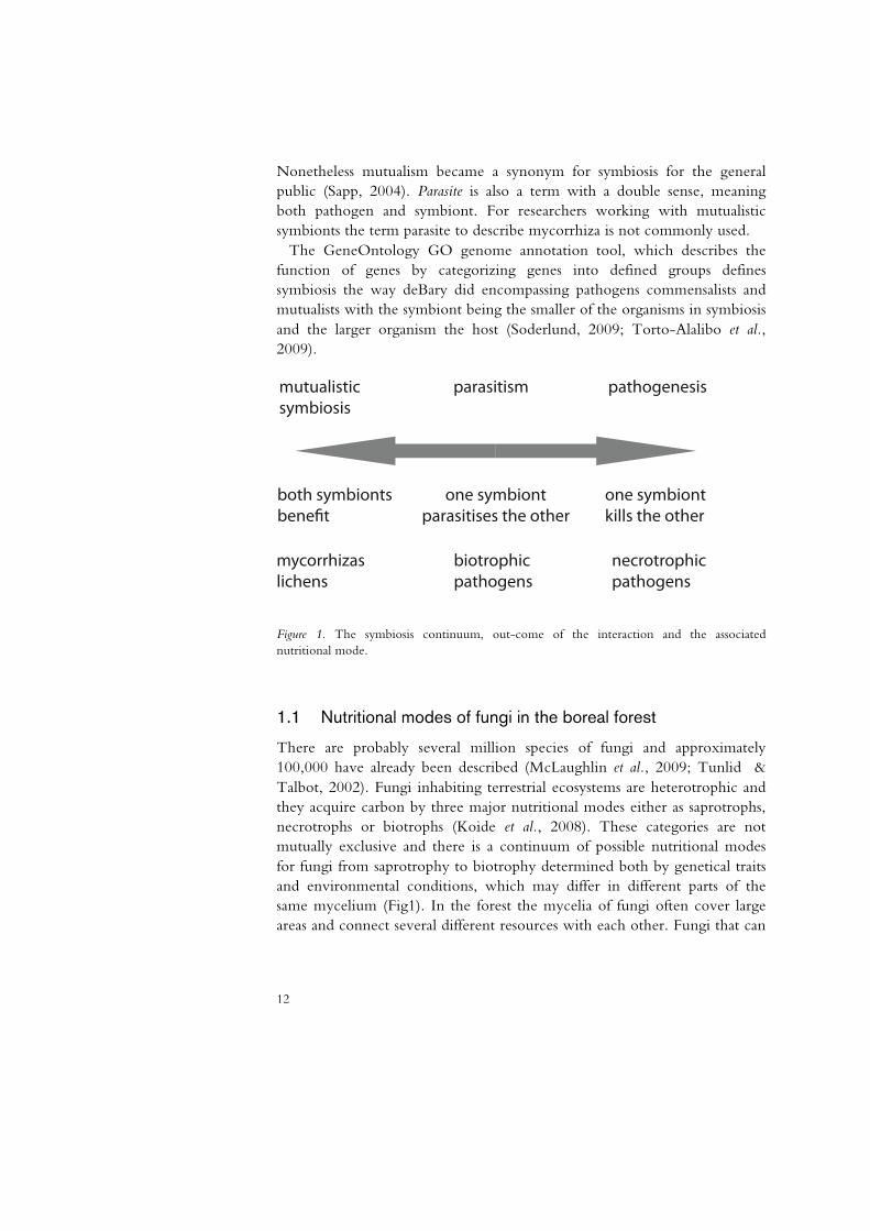
Some 150 years ago Darwin published his ideas about evolution that have ever since provided a framework for researchers to explain their findings. Associations between plants and microorganisms were noted early in human history with observations already in ancient Greece that fungi were present on destroyed crops or on dying trees, but not until the late 19th century did the study of fungi become more formalized (Manion, 1981). In 1879 the father of plant pathology, German Antoine deBary, used the term symbiosis to describe “the living together of unlike organisms”. He used the term to describe a proof of evolution that lichens were both algae and fungi (Sapp, 2004). The partners in symbiosis are called symbionts, or host and symbiont, in which case the symbiont is the microorganism and the host the macro organism. The nature of the symbiosis can be parasitism, mutualism or commensalism. In parasitism one symbiont benefits whilst the other is harmed. In mutualism both symbionts benefit and in commensalism one organism benefits and the other is unaffected. The connection between symbiosis and parasitism is strong not only in relation to scientific history but also in relation to their definition and evolutionary history. The question as to what factors determine the development into mutualist, pathogen or saprotrophs, which were formulated at the end of the 19th century is still puzzling researchers today even though the tools available to answer the questions have changed.
The term symbiosis has been interpreted not only as the broad including definition of “living together of unlike organisms “ but also as a synonym for mutualism. When Frank in 1885 coined the term mycorrhiza for the fungus root structure he observed he claimed the symbiosis to be mutually beneficial, however his critics claimed that microbes were parasites only. Frank himself had, two years before deBary invented the term symbiotismus for coexisting organisms, a term neutral to the role the organisms had.
12
Nonetheless mutualism became a synonym for symbiosis for the general public (Sapp, 2004). Parasite is also a term with a double sense, meaning both pathogen and symbiont. For researchers working with mutualistic symbionts the term parasite to describe mycorrhiza is not commonly used.
The GeneOntology GO genome annotation tool, which describes the function of genes by categorizing genes into defined groups defines symbiosis the way deBary did encompassing pathogens commensalists and mutualists with the symbiont being the smaller of the organisms in symbiosis and the larger organism the host (Soderlund, 2009; Torto-Alalibo et al., 2009).
mutualisticsymbiosis
parasitism pathogenesis
both symbiontsbenefit
one symbiontparasitises the other
one symbiontkills the other
mycorrhizaslichens
biotrophicpathogens
necrotrophicpathogens
Figure 1. The symbiosis continuum, out-come of the interaction and the associated nutritional mode.
1.1 Nutritional modes of fungi in the boreal forest
There are probably several million species of fungi and approximately 100,000 have already been described (McLaughlin et al., 2009; Tunlid & Talbot, 2002). Fungi inhabiting terrestrial ecosystems are heterotrophic and they acquire carbon by three major nutritional modes either as saprotrophs, necrotrophs or biotrophs (Koide et al., 2008). These categories are not mutually exclusive and there is a continuum of possible nutritional modes for fungi from saprotrophy to biotrophy determined both by genetical traits and environmental conditions, which may differ in different parts of the same mycelium (Fig1). In the forest the mycelia of fungi often cover large areas and connect several different resources with each other. Fungi that can

13
act as parasites may therefore do so in one part of the mycelium and grow as saprotrophs in another part of the mycelium.
1.1.1 Saprotrophic fungi
Most fungal species are saprotrophs, which implies that they can survive on dead organic matter. Many saprotrophs live on easily accessible compounds and soluble sugars but others have a more complex set of machinery to acquire nutrients. Several saprotrophic fungi have genes encoding enzymes for cellulose and lignin degradation and for phenol oxidation (Zabel & Morell, 1992; Eriksson et al., 1990), as well as those coding for pectinases. The decay capabilities of fungi cause devastating economical losses during storage of diverse products of crops and wood, due to fungi such as the dry rot fungus Serpula lacrymans that grows in timber in houses and Aspergillus flavus that produce toxin that renders peanuts hazardous to consume (Carlile et al., 1994). However, the unique ability of fungi to degrade various compounds is crucial for nutrient cycling in forest ecosystems (Rayner & Boddy, 1988). In boreal forests that are dominated by conifer trees, the soils are generally acidic and stratified into layers of litter at different stages of decomposition. These soils are poor in mineral nutrients and particularly in nitrogen (Berg & Tamm, 1991). The litter from the conifers is highly lignified and many fungi are capable of breaking down lignin (Osono & Takeda, 2002). The stratification creates different niches with species adapted to the nutrient content in each stratum. Saprotrophic litter decomposing fungi are more abundant in the upper layers than in the deeper layers (Lindahl et al., 2007).
Mycelia of saprotrophic fungi can occupy large areas and persist for years (Smith et al., 1992) The saprotrophic fungi are often combative and have means of defending their resources by producing antimicrobial toxins, or by translocating them to another part of the mycelium (Boddy et al., 2009).
1.1.2 Pathogenic fungal symbionts
In nature, only around ten percent of all fungal species are phytopathogenic (Tunlid & Talbot, 2002). The pathogenic fungi are parasites that obtain their carbon as pathogens by damaging or killing their host. Some are obligate parasites and need a host to complete their lifecycle whereas others are facultative parasites and have the option to switch nutritional modes. The parasites can also be classified as biotrophs that need a living host or necrotrophs that kill the host (Lewis, 1973). Hemibiotrophs are fungi that require a living host, but then switch to a necrotrophic mode (Oliver & Ipcho, 2004; Perfect & Green, 2001).
14
Biotrophs and hemibiotrophs have specialized feeding structures, haustoria to acquire nutrients whereas necrotrophs kill their host with toxins and lytic enzymes (Oliver & Ipcho, 2004). Both biotrophic fungal pathogens and necrotrophic fungal pathogens can infect their host with appressoria that penetrate the host surface by mechanical force. Entering via stomata or direct penetration by the hypha are also other options (Agrios, 1997).
One of the most harmful and destructive forest pathogen in the boreal forest, Heterobasidion annosum (Fries 1821) Bref. was first described by Elias Fries as Polyporus annosus in 1821 and was later given the name Heterobasidion by Oscar Brefeld in 1888 because of its conidiophores that somewhat resemble basidia. However it was the forest pathologist Robert Hartig that adopted Moritz Willkomms ideas that H. annosum was a decay causing fungus and described it in “Important diseases of forest trees” from 1874, considered the birth of forest pathology (Huettermann & Woodward, 1998).
Several conifer species (Norway spruce, Scots pine, Douglas fir) serve as host to the three forms of H. annosum s.l., the S, P and F-types, respectively. In northern Europe there are two intersterility types of Heterobasidion annosum s.l.: The P-type and the S-type now called H. annosum s.s. and H. parviporum. The P-type, H. annosum s.s., attacks mostly pines such as Pinus sylvestris but it may also attack many other trees especially when they grow mixed with pine trees (Asiegbu et al., 2005). Picea abies, Juniperus communis, and even Betula pendula are particularly susceptible. The P-type H. annosum s.s. can be found all over Europe where there are pine trees. The S-type H. parviporum attacks spruce (Picea abies) even though it occasionally attacks pine, birch and exotics. It is common in northern and eastern Europe (Asiegbu et al., 2005). In southern Europe and especially in Italy a third intersterility group (F-type) H. abietinum attacks fir, mainly Abies alba. In North America there are P-types and S-types of H. annosum s.l. named H. irregularis and H. occidentalis which differ significantly from the European species (Otrosina & Garbelotto), H. occidentalis for example do not have the same narrow host range as the European variant. H. occidentalis is in the west and H. irregularis in the south-east (Korhonen et al., 1998).
Heterobasidion has the capability of switching between saprotophic and pathogenic nutritional modes. The fact that these organisms are white-rot fungi (i.e. they degrade both lignin and cellulose components of wood), contributes to their action as strong parasites and saprotrophs, able to infect and destroy living conifer roots and stems of all ages, as well as dead trees (Daniel et al., 1998).
15
1.1.3 Commensalistic fungal symbionts
Endophytic fungi live entirely within a host in roots, stems or leaves, not entering the rhizosphere seemingly without directly affecting it and can be viewed as commensalists. There are clavicipitaceous endophytes that colonize grasses and three classes of non-clavicipitaceous endophytes with a broad host range from grasses to conifers.
Indirect beneficial effects for the host can be due to endophytes like with the grass endophyte which produces a toxin that renders the grass inedible for grazers, and is hence benign to the plant, or those that increase stress tolerance to abiotic factors such as drought and heat. It has been demonstrated that without grazers there is a cost associated with the endophytes (Rodriguez et al., 2009). Healthy spruces can have around 200 fungal endophytes in the stem and branches (Muller & Hallaksela, 2000). Dark septate fungi, the fourth class of endophytes are very common in the boreal forest and Phialocephala sp. form mycorrhizal like structures on a wide range of trees hosts, but are also present inside decaying wood (Rodriguez et al., 2009; Menkis et al., 2004).
1.1.4 Mutualistic symbionts
At the other extreme of the pathogen–mutualist continuum are symbiotic mycorrhizal fungi that contribute to the vitality and vigor of their host plants. About 95% of all plant species are colonized by mycorrhizal fungi. These symbiotic fungi facilitate nutrient acquisition for their host plant and receive carbon in return. The fungal mycelium increases the surface area available for nutrient absorption compared to uncolonized roots and provides some physical protection against pathogens. The association needs to be controlled by the host, since the outcome can be close to reciprocal parasitism (Finlay, 2008; Francis & Read, 1995).
The association can be both intracellular- as with arbuscular and ericoid mycorrhizal fungi and extracellular as with ectomycorrhizal fungi. Arbuscular mycorrhiza form tree like structures inside the host cell for nutrient exchange, that like haustoria do not breach the cell membrane (Perfect & Green, 2001; Smith & Read, 1997). Ectomycorrhiza enclose the lateral roots of their hosts with a fungal sheet and host cortical cells with a reticulated hyphal structure, the Hartig net, which forms the interface for exchange of carbon and nutrients. Symbiotic ectomycorrhizal fungi colonize the vast majority of tree roots in boreal forests, forming the mutualistic associations which are necessary for successful nutrient acquisition and growth (Smith & Read, 1997). Formation of ectomycorrhizal roots involves both structural and metabolic integration of the symbionts and modification

16
of both plant and fungal gene expression. The development appears to be the result of highly coordinated molecular processes and governed by morphogenetic patterns, which may respond to pre-established programs in both partners (Martin, 2008; Le Quere et al., 2005; Podila et al., 2002). Usually, the plant shows a transient uncoordinated weak defence response early in the colonization process in both arbuscular mycorrhiza (AM) and ectomycorrhiza (ECM), during colonization (Liu et al., 2003; Harrison & Dixon, 1994). The responses have been shown to be weaker than towards a pathogen (Adomas et al., 2007; García-Garrido & Ocampo, 2002) (Jane Barker et al., 1998).
1.2 Evolution of symbiosis
Symbiotic relations of some sort exist in diverse organisms among the eukaryotes from lichens to humans. The microbial symbionts fix nitrogen, photosynthesize, metabolize sulphur and perform other physiological processes their plant hosts cannot do on their own (Sapp, 2004). The genetic program for symbiosis is thus fundamental for all land living organisms. The energy producing mitochondria in eukaryotic cells are a product of evolution of endosymbiotic bacteria (Gray et al., 1999), the chloroplasts of plants are ancient cyanobacteria (Bhattacharya et al., 2004; Yoon et al., 2004). Even the nucleus of the eukaryotic cell has been suggested to be a result of fusion between bacteria (Sapp, 2004). The mitochondria have transferred large parts of their genome to the host genome but the product of the genes transferred are often retargeted to the mitochondria (Pesaresi et al., 2007) (Kleine et al., 2009) .
The entire concept of organisms has been discussed in relation to symbiosis. Symbiome is a term used to describe the totality of an organism’s symbionts, from organelles to microbes and bacteria inside and outside of the organism (Sapp, 2004). The hologenome evolution theory states that the symbiome or holobiome is the important unit of evolution, not the organism alone. The importance of environmental factors in evolution is considered since they affect what microbes form symbiotic associations with the host (Zilber-Rosenberg & Rosenberg, 2008). Lamarckian theories of evolution by acquired traits can be relevant if symbionts that are transferred to the next generations are considered acquired traits. Such symbionts are for example organelles like mitochondria, but also plant seed transmitted fungi (Rosenberg et al., 2009).
There are fossil evidence that supports the idea that land itself was colonized by plants in symbiotic association with ancestral arbuscular
17
mycorrhiza some 460 million years ago (Redecker et al., 2000; Remy et al., 1994; Pirozynski & Malloch, 1975).
Genetic programs governing symbiosis are therefore likely to be present to a varying degree within all fungi and plants as well. Even non-mycorrhizal plants like Arabidopsis thaliana have been shown to have endophytic fungi and react to the presence of mycorrhizal fungi (Felten et al., 2009; Le Quere et al., 2004; Peskan-Berghofer et al., 2004). On the host side, there are common genetic programs for symbiosis, Güimil et al showed common patterns of expression in rice when colonized by an arbuscular mycorrhiza, a hemibiotroph and a necrotroph (Guimil et al., 2005).
The ability to form ectomycorrhiza has evolved and disappeared several times through evolution but ectomycorrhizal fungi are thought to have evolved 180 million years ago. Fossils of ECM roots 50 million years old have been found (Hibbett & Matheny, 2009; LePage et al., 1997). There are three main hypothesises about the origin of ectomycorrhiza, 1) that they evolved from saprotrophs, 2) that they evolved from saprotrophs and some lineages reversed to saprotrophism and 3) that they had a common ectomycorrhizal ancestor but lost the ability to form mycorrhiza on numerous occasions (Matheny et al., 2006). The ECM are unlikely to a have evolved in the way the third theory states as the hosts are evolutionary younger than the fungi (Hibbett & Matheny, 2009). Whatever the origin they are not a monophyletic group, there are some examples among the ascomycetes and they are present in several different orders among the basidiomycetes. They are often close relatives to saprotrophic fungi as well as pathogenic fungi (Matheny et al., 2006; Miller et al., 2006)(Fig1).
Phytopathogenic fungi are not common but widespread in the fungal kingdom (Soanes et al., 2007; James et al., 2006) and the phytopathogenic trait has evolved on several occasions (Cornell et al., 2007; Fitzpatrick et al., 2006). Among the phytopatogenic ascomycetes, the sequenced genomes are different to such a degree that the evolutionary background and trait of phytopathogenicity has been clouded (Soanes et al., 2007). Some classes of genes have been suggested to be important for phytopathogenicity based on the frequency of their occurrence in genomes. There are only three cell surface receptors like G-protein coupled receptors linked to signal transduction pathways as MAPK in the saprotrophic yeast Saccharomyces cerevisiae but 61 in the rice pathogen Magnaporthe grisae and 84 in Fusarium graminearum. This enables the pathogens to respond to a variety of environmental conditions(Cuomo et al., 2007; Soanes et al., 2007; Dean et al., 2005). Biotrophic fungi seem to have few cellwall degrading enzymes. In ascomycete phytopathogenic fungi there are unknown secreted proteins
18
not present in saprotrophic fungi (Soanes et al., 2008) a similar basidiomycete analysis have to the authors knowledge not yet been done
The ability to form ectomycorrhiza is likely to rely on a few unique genes that determines that functional category (Hibbet et al., 2000). This is illustrated by the mutualist Paxillus involutus as several other closely related species in the genus Paxillus are saprophytes (Le Quere et al., 2004). The remaining genes are probably the same genes that are present in a saprotroph. The sequencing of the ectomycorrhiza Laccaria bicolor has not yet solved the debated question whether ectomycorrhizas can be facultative free-living saprotrophs in nature (Baldrian, 2009; Cullings & Courty, 2009; Martin & Selosse, 2008; Taylor & Alexander, 2005). One example of genes possibly defining an ectomycorrhizal fungus is the glycosidases GH32 gene family which seems to be missing in mycorrhizal fungi (Parrent et al., 2009).
It is possible that the genetic control of pathogenesis in the necrotrophic pathogen H. annosum that kills its host and then prevail as a saprotroph also relies on a limited number of genes. Although obligate saprotrophs and mutualists are known to contain similar kinds of genes, the activity of certain key genes relevant in cell wall degradation such as lignin degrading enzymes is known to differ between mycorrhizal and pathogenic fungi (Martin et al., 2008). The basis for this difference may stem from the way in which such genes are regulated. Distinctions between saprotrophic fungi and pathogenic fungi have also been found in expansions in families of certain key genes that are potentially relevant in pathogenesis (Dean et al., 2005). When EST from saprotrophic and pathogenic fungi were compared no significant difference was found between them (Soanes & Talbot, 2006). When protein clusters of 36 genomes of fungi and oomycetes were compared, no unique phytopathogenic protein cluster was found (Soanes 2008). In the H. annosum pathosystem, little is however known about what kind of genes are under expansion nor have regulatory patterns of key potential pathogenicity genes been compared to those of non-pathogenic models such as obligate saprotrophs and mutualists.
1.3 Microbe Associated Molecular Patterns (MAMP)
Parasites need to have a compatible host in order to complete colonization successfully. Plants lack the option of fleeing a potential threat and they do not have an adaptive immune system like animals. Instead they rely on, an innate immune system as means of recognizing the microbes in their surroundings in order to react appropriately. That immune system use transmembrane receptors that respond to slowly evolving microbe-
19
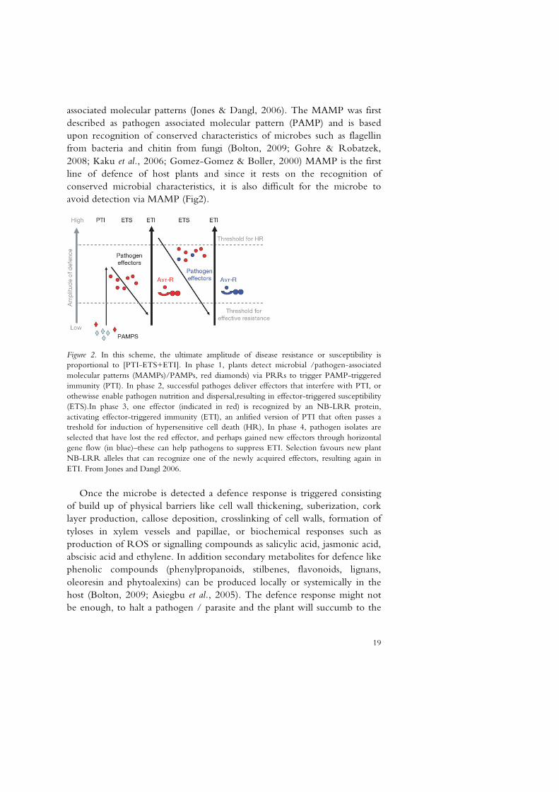
associated molecular patterns (Jones & Dangl, 2006). The MAMP was first described as pathogen associated molecular pattern (PAMP) and is based upon recognition of conserved characteristics of microbes such as flagellin from bacteria and chitin from fungi (Bolton, 2009; Gohre & Robatzek, 2008; Kaku et al., 2006; Gomez-Gomez & Boller, 2000) MAMP is the first line of defence of host plants and since it rests on the recognition of conserved microbial characteristics, it is also difficult for the microbe to avoid detection via MAMP (Fig2).
Figure 2. In this scheme, the ultimate amplitude of disease resistance or susceptibility is proportional to [PTI-ETS+ETI]. In phase 1, plants detect microbial /pathogen-associated molecular patterns (MAMPs)/PAMPs, red diamonds) via PRRs to trigger PAMP-triggered immunity (PTI). In phase 2, successful pathoges deliver effectors that interfere with PTI, or othewisse enable pathogen nutrition and dispersal,resulting in effector-triggered susceptibility (ETS).In phase 3, one effector (indicated in red) is recognized by an NB-LRR protein, activating effector-triggered immunity (ETI), an anlified version of PTI that often passes a treshold for induction of hypersensitive cell death (HR), In phase 4, pathogen isolates are selected that have lost the red effector, and perhaps gained new effectors through horizontal gene flow (in blue)–these can help pathogens to suppress ETI. Selection favours new plant NB-LRR alleles that can recognize one of the newly acquired effectors, resulting again in ETI. From Jones and Dangl 2006.
Once the microbe is detected a defence response is triggered consisting of build up of physical barriers like cell wall thickening, suberization, cork layer production, callose deposition, crosslinking of cell walls, formation of tyloses in xylem vessels and papillae, or biochemical responses such as production of ROS or signalling compounds as salicylic acid, jasmonic acid, abscisic acid and ethylene. In addition secondary metabolites for defence like phenolic compounds (phenylpropanoids, stilbenes, flavonoids, lignans, oleoresin and phytoalexins) can be produced locally or systemically in the host (Bolton, 2009; Asiegbu et al., 2005). The defence response might not be enough, to halt a pathogen / parasite and the plant will succumb to the
20
infection. In that case the intracellular defence system based on R-genes may be activated. R-genes recognize pathogen effectors (avr-genes) and activate defence responses accordingly. R-gene mediated resistance is primarily effective against biotrophic or hemibiotrophic pathogens, but not against necrotrophs (Jones & Dangl, 2006).
Induced defence is costly, energy consuming both in microbe and host, particularly since the host often tries all possible defences at once. During defence and attack, normal growth is often suspended, plant mutants constitutively expressing defence responses are small and have low ability to reproduce (Heil & Baldwin, 2002) Fungal effectors includes toxins with a broad definition of the effector concept. Heterobasidion annosum produces a dozen toxins, mostly benzofuran derivates such as fommannosin, fommanoxin, fommanoxin acid, oosponol and oospoglycol (Asiegbu et al., 1998; Sonnenbichler et al., 1994; Sonnenbichler et al., 1989; Sonnenbichler et al., 1983; Bassett et al., 1967). Host specific toxins often display great degree of polymorphism and seem to recognize the same R-gene receptors that bind avr-genes, indicating that the relative abundance of biotrophic and necrotrophic pathogens determine the evolution of R-genes (Stukenbrock & McDonald, 2009) Necrotrophic fungi seem to have few avr-genes compared to biotrophic fungi, most likely because it does not require living host cells for infection (de Wit et al., 2009).
Chitin is one of the best-known MAMPs with receptors identified in Arabidopsis and rice (de Wit et al., 2009). Fungal chitin can provoke a defence response in conifers versus for example H.annosum or Hebeloma crustiliniforme (Hietala et al., 2004). In the late 90’s it was hypothesized that these chitinous elicitors are degraded by plant chitinases during active mycorrhizal formation thereby limiting active host defence reactions (Salzer et al., 1997). Chitinases are induced during colonization of ECM in lateral roots (Frettinger et al., 2006).
1.4 The role of auxin in symbiosis
Plants have a number of different hormones, carrying out different functions in the organism. Plant growth regulators, regulating cell cycles and morphogenesis (auxin and cytokinins), flowering, senescence and stress (GA, ABA, ethylene) (Raven et al., 1998) Defence responses are mediated by brassinosteroids, salicylic acid (SA), jasmonic acid(JA). SA is mediating resistance against biotrophs while JA mediates resistances against necrotrophs (Kazan and Manners 2009). Secondary metabolites such as strigolactones play a crucial role in interactions with mutualists and pathogens (Akiyama &
21
Hayashi, 2006). Some plant growth regulators like auxin and ethylene have clear functions as regulators of defence responses in addition to their other functions.
Auxin plays an important role in regulation of mutualistic interactions, for instance in the rhizobial symbiosis between Rhizobium spp. and legumes, the root auxin balance as a prerequisite for nodule formation (Mathesius, 2008) and pathogenic interactions such as Crown gall disease (Spaepen et al., 2007). Auxin transport and signalling has recently been connected to the plant defence, in common with the signalling pathways of the defence-associated plant hormones SA and JA, auxin signalling affects resistance to different parasitic life styles differently. Furthermore it seems that the auxin and SA pathways acts in a mutually antagonistic manner during plant defence (Cuzick et al., 2009; Llorente et al., 2008), while auxin and JA signalling share regulatory steps (Kazan & Manners, 2009; Gray et al., 2003; Tiryaki & Staswick, 2002; Tor et al., 2002). SA is mediating resistance against biotrophs while JA mediates resistances against necrotrophs
Auxin moves polar from the shoot towards the root via the vascular cambium and the phloem (Raven et al., 1998; Raven, 1975). Functioning auxin transportation and signalling is a prerequisite for lateral root formation (Fukaki et al., 2007; Bhalerao et al., 2002) which is a central step in ECM formation (Felten et al., 2009). Addition of the auxin IAA to pine roots mimics swellings and branching of roots obtained by application of culture media from the mycorrhizal fungus Boletus luteus (Barker & Tagu, 2000; Slankis, 1973a; Gruen, 1959). The nature of the signal that induces the Lateral root formation in ECM is not known, but Ectomycorrhizal fungi have been demonstrated to produce auxin (Gay, 1988; Ek et al., 1983). It has thus been suggested that it may be fungal auxin (Niemi et al., 2002; Karabaghli-Degron et al., 1998; Slankis, 1973b). The use of auxin transport inhibitors supports this hypothesis to some degree (Felten et al., 2009; Laajanen et al., 2007; Rincon et al., 2003; Rincon et al., 2001; Karabaghli-Degron et al., 1998). However the exact role of auxin in mycorrhiza formation is still unclear.

23
2 Objectives
Symbiotic, mycorrhizal associations and saprotrophic fungi play important and economically significant roles in the nutrition, growth and health of forest trees as well as in decomposition and nutrient cycling. Mycorrhizal and pathogenic interactions share some common features but they differ in distinct ways with respect to which the different types of interactions are induced and regulated. The primary objective of the studies described in this thesis was to elucidate the patterns of gene expression in Heterobasidion species during growth on their conifer tree host and in artificial media. The results were compared with similar studies using non-pathogen fungi. More specifically, we investigated:
Paper I. If the gene expression of three Heterobasidion species correlated in a subset of genes such that cDNA arrays of one can be used to study the other.
Paper II. The gene expression during transition from saprotrophic to necrotrophic growth stage in Heterobasidion annosum Paper III. The expression of of a novel Clavata1-like and a Nodulin 21-like gene during ectomycorrhizal development in Pinus sylvestris and in response to pathogenic, saprotrophic or symbiotic fungi

25
3 Materials and Methods
3.1 Fungal material
The Heterobasidion isolates used in this study were representatives from Europe and North America that have all been previously studied in earlier publications and the choice of the isolates was based on this fact. In the first study three European Heterobasidion species representing the different host preferences in the Heterobasidion species complex were choosen with Heterobasidion annosum s.s. isolate FP5 (Korhonen) H.parviporum FS6 and H.abietinum Faf4-6 (Karlsson & Stenlid, 1991; Stenlid & Karlsson, 1991). For H.parviporum a library of ESTs (Expressed Sequence Tags) were available (Abu et al., 2004). In the second study the north American P-type of H. annosum s.l. presently called H. irregularis TC-32-1 (Chase, 1985) a homokaryotic isolate which has been the model “lab rat” with EST resources and QTL map and now the whole genome available (Lind et al., 2007; Lind et al., 2005; Karlsson et al., 2003; Chase, 1985), (http://genome.jgi-psf.org/Hetan1/Hetan1.home.html). The saprotroph Phanerochaete chrysosporium RP78 (Jill Gaskell) (Stewart et al., 2000) and the mutualist Paxillus involutus were included. In the third study H.parviporum, FS6 was used together with the saprotroph P. chrysosporium RP78 (http://genome.jgi-psf.org/Phchr1/Phchr1.home.html). The mutualists Paxillus involutus MAJ and the sequenced Laccaria bicolor 238A(http://genome.jgi-psf.org/Lacbi1/Lacbi1.home.html) were included in paper III
26
3.2 Plant material
Pinus sylvestris (L.) Saleby FP45 seeds were surface sterilized in 33% hydrogen peroxide for 15 minutes under stirring and rinsed in excess sterilized water and grown on 1% water agar.
3.3 Infection systems and culture conditions
To study fungi, pure cultures of fungi isolated from the environment are essential. With fungi isolated in pure cultures from plants with disease symptoms Koch’s postulate can be tested, by determining whether diseases can be induced by inoculation of healthy plants with the isolated fungi. There is a multitude of different media that have been developed to suit the requirements of various symbionts, however many fungi such as obligate parasites are still impossible to grow in pure culture. The balance of nutrients in the medium is crucial and deviation from what the fungus encounter in its natural habitat may induce physiological abnormalities and misinterpretations of the biology of the fungus. Typically the natural conditions may be altered with having too much exogenous carbon available, even the host range may get changed (Langer et al., 2008; Duddridge, 1986). Nitrogen starvation has been used as a mean of mimicking the growth of pathogens in planta and study virulence genes in vitro (Bolton & Thomma, 2008; Coleman et al., 1997). The balance of C:N ratio in a substrate is important for the efficiency of decomposer fungi to break down the substrate (Boberg, 2009).
We used an axenic model system with juvenile seedlings to represent the hosts of our fungi. The use of juvenile seedlings instead of full grown trees has its pro’s and con’s as it facilitates easy handling and allows a sterile controlled environment, however since the seedlings lack several of the features of adult trees they do not in all aspects reflects the full grown trees, i.e. suberin and bark. However, even mature trees have root tips of a similar age to the roots of seedling plants used in the present study. Studies with seedlings have been used in previous studies (Olson & Stenlid, 2001; Asiegbu et al., 1999; Asiegbu et al., 1994).
In paper II the infection systems was constructed to mimic the nutrient limited environment that a boreal forest consists of, with intact mycelia to connect the inhabited resource with the uncolonized substrate. The colonized dead seedling was the only nutrient sources until inoculation with a new living seedling. The experimental system was constructed to minimize the difference between the nutrient sources such that there was no other major difference other than the host being alive or dead. Pinus
27
sylvestris seedlings of two weeks age were grown on 1% water agar and dried for a week at 60 °C. The dried Scots pine seedlings were then transferred to Hagem agar medium (Stenlid, 1985) Petri dishes inoculated with the H. irregularis TC32 and left for colonization. The dead colonized pine was transferred to water agar plate containing cellophane membrane and left until the mycelium had almost covered the plate. A new living seedling was then applied to the mycelial front 5cm from the dead seedling. The living seedling was removed after 1, 6 and 15 days post infection and the mycelium from a 1cm border of the living seedling as well as a 1cm strip of mycelium from the dead colonized seedling was harvested. The mycelium was shock frozen in liquid nitrogen and stored at -80°C until used for RNA extraction.
The hydroponic box system in paper 3 was constructed as a way to produce samples with many seedlings all subjected to the same treatment. Each box contained almost hundred seedlings in a 300 ml liquid solution of modified Melin Norkrans medium (Marx, 1969). There is one box per a sample. The seedlings had been grown for 14 days on water agar when transferred under sterile condition to the box system. Inoculation of the P. sylvestris seedlings was done directly into the medium by adding the treatment solutions. The treatments were; Auxin indole-3-butyric acid (IBA) 100uM, Laccaria bicolor mixture C alone or in combination with the auxin transport inhibitor 2,3,5-triiodobenzoic acid (TIBA) 10uM. The seedlings were cultured in a 16h photo period at 21 °C. After 1, 5, 15 and 30 days the seedlings were harvested in liquid nitrogen or in the RNA stabilizing solution RNA later™ for the spatial distribution samples.
3.4 Macroarray hybridization and analysis
In study I (Heterologous array analysis) and II (Saprotrophic-Pathogenic Switch) we use an array hybridization technique, in which the affinity of nucleotides to bind to complementary sequences is used. There are several automated ways of making the hybridizations usually called microarrays. In micro-arrays, nucleotides either amplified DNA or cDNA or synthetically designed probes are bound to a glass slide in spots. The microarrays can contain a small custom-made set of nucleotides representing genes of interest or entire genomes, depending on the printing capacity of the automated system and the request of the researcher. The glass slides are either single or two-channelled, in single channelled microarrays there is only one representative of each DNA fragment per printgroup since the RNA to be hybridized on the array only contains one fluorescent colour. In
28
two channelled microarrays two different RNA sources are compared by marking them with a red or a green fluorescent colour and then measuring the difference between the colour ratio, one spot of printed DNA fragment is used per colour. However, macroarrays used in this study are single channel arrays printed on a membrane.
In macroarrays clones with known or unknown sequences are bound to a membrane and in a buffer they are heated together with the RNA or cDNA that was harvested during the experiment, this causes the experimental RNA to bind to the clones on the paper. Then the RNA from the experiment is washed away unless it binds strongly, which it does when the same sequence is in the clone as in the experimental RNA. The temperature can be altered to increase or lower the stringency of both the hybridization and the washing in such a way that an increased temperature requires a better match between the nucleotides on the array and the experimental RNA. As the experimental RNA is marked with either a radioactive label or a chemifluorescent dye, pictures can be taken of the macroarray and an estimate of the gene expression of the clones present on the array in the experimental RNA can be made.
In order to estimate gene expression there are several necessary steps to analyze the pictures taken. Firstly, the digital image processing of each spot on the array is examined for characters such as shape, signal intensity and background signal intensity. There is a multitude of software (Agilent, Genepix, Imagene, Spot, etc.) that analyze the fluorescence of arrays, usually in concurence with the hybridization and in standardized output formats, but it is also possible to examine the fluorescence using separate image analysis software not tailor made for array analysis such as Quantity One™.
For estimation of signal intensity there are commonly control samples on the array with known amounts per spot either positive samples in dilutions or negative controls with samples unrelated to the experimental RNA. The background intensity is either determined locally around every spot or globally as an estimate of the representative background intensities based on either a model like Lowess smoothing or calculations of the mean background
The signal intensity is bound to the false background intensity but there are many methods to determine how. The simplest methods are either to ignore the existence of background or to use direct subtraction, but there are many drawbacks of both methods (as background may be due to leakage of correctly hybridized but non stringently removed or just non biological relevant noise). Several versions of the subtraction methods exist dealing with the normalization step in appropriate possible negative values (ex Minimum, Half, Camberra, Edwards etc)(Smyth, 2005). There are also
29
methods that treat the background as a part to be added to the signal intensity, the RMA algorithm and the more recent Normexp method are variants where the signal intensity is a model where the normal distribution of the local background is added to the signal intensity (Silver et al., 2009; Ritchie et al., 2007).
Normalization of the arrays is the next step and once again there are several alternatives available but they all aim to reduce differences in order to make it possible to compare genes within the arrays with each other as well as to compare different arrays.
For single channel arrays, the between array methods are appropriate. The Quantile normalization method, in which the expression of each array is centred to a common quantile, is common.
The open source statistical analysis software R contains several packages aimed at analyzing microarrays, both single channel and two-channel microarrays. (The single channel software is almost exclusively aimed at the well defined Affymetrix platform which hinders the use of non-conventional arrays as macroarrays. Instead adaptions of the two-colour software are necessary too squeeze in the single channel format of macroarrays into the two channel analysis. There is one dominant software; Limma in R, that deals with processing of microarrays, including background subtraction, normalization and gene expression (Smyth, 2005). The data are read into the software as matrices, which can be created from sources other than the standard output files from the automatic hybridization, all data are log transformed to obtain normal distribution. The gene expression in Limma is mostly based on Bayesian statistics via the function eBayes (Smyth, 2004) which calculates likeliness of differential expression for gene matrices with expression data fitted to linear models and compared in contrast models.
3.5 Amplification of RNA
When the polymerase chain reaction PCR was automated it revolutionized molecular biology since it became possible to obtain high quantities of DNA via amplification of the initial material. The PCR has made it possible to circumvent the need to culture hard cultured organisms, by sequencing of DNA, taxonomy has become based on DNA instead of morphological characters, etc, basically all aspects of biology can now be studied with the aid of PCR (Peay et al., 2008). For array studies, amplification of the initial material is often necessary, but since the researcher is interested in the initial quantity of different RNA transcripts in the cell or at least their relative abundance direct PCR amplification of the cDNA is not sufficient. The
30
problem is that the relative abundance of transcript can be altered after PCR amplification. There are however alternative methods such as the T7 eberwine method where amplification is linear (Vangelder et al., 1990).
3.6 Q-PCR
Quantitative PCR is the other technique widely used in the work described in this thesis. QPCR or Quantitative Real Time PCR is a way to follow the build up of a PCR product in a PCR reaction cycle by cycle and thus quantify the amount of the targeted transcript. This is realized by signal intensity measurement of fluorescence markers incorporated into the PCR product. The number of cycles it takes to amplify so much of the desired PCR product (amplicon) that the reaction is unlimited by deficient quantities of DNA or unused enzyme and nucleotides is determined by setting a cycle threshold where the signal raises above the background noise (Nolan et al., 2006; Bustin, 2000).
The quantification can essentially be done in two ways: absolute quantification or relative quantification. In absolute quantification, a known amount of transcripts is analyzed with QPCR in a dilution series so that the cycle threshold (Ct) for each dilution is known and used to derive the amount of transcripts in the unknown samples. The result is expressed as an exact number of molecules or moles of nucleic acid. The more commonly used technique is relative quantification, it is concerned with relative changes between samples or genes and not amounts of transcripts. Ct value for a test sample is compared to the Ct of a calibrator sample, the difference is the deltaCt. The fold change is then two to the power of deltaCt. The gene expression in cycle threshold values must be normalized with a reference gene to correct for sample to sample variation therefore more refined methods of relative quantification have been developed. The deltadelta Ct is such a method where the test sample and the calibrator are both compared to a reference gene for normalization, thus providing deltaCtsample-reference gene and delta Ctcalibrator-reference gene. The two normalized deltaCt values, sample and calibrator, are then compared to obtain the deltadeltaCt value. Lately the dominating normalization methods are Vandesompele’s (Vandesompele et al., 2002) which requires two reference genes for normalization or the method by Pfaffl, which uses one reference gene (Pfaffl, 2001).
Ideally the expression of the reference gene should be evaluated so that they are not differentially expressed in the samples examined, but there are several standard genes so called house-keeping genes commonly used for
31
normalization in QPCR experiments (Brunner et al., 2004). QPCR is commonly used in conjunction with array experiments to validate the expression of the array. There are however often dissimilarities between the gene expression determined by the array and by QPCR and the gene expression from QPCR is often higher than that measured by the array. The correlation between the two methods differs, mostly depending on the method used for the array analyses. When compared with other methods the RMA algorithm has proven to be the best choice (Millenaar et al., 2006).

33
4 Results and discussion
4.1 Paper I. Heterologous array analysis in Heterobasidion: Hybridisation of cDNA arrays with probe from mycelium of S, P or F-types
The creation of some genetic resources like clone libraries with EST from Heterobasidion (Abu et al., 2004; Karlsson et al., 2003) has allowed us to draw conclusions about gene expression under different biological conditions such as interaction with seedlings (Karlsson et al., 2003) and spore germination (Abu et al., 2004). The genomes of related species have evolved by mutations involving insertions, substitutions and deletions of single nucleotides and rearrangements of the genomes, but also share a common ancestry and are similar in many features. There has been interest in using established resources from well-known model organisms to study related organisms. One such example is the Pinus taeda arrays, which have been used to perform gene expression studies in other pine species (Adomas et al., 2008; Brinker et al., 2004; van Zyl et al., 2002).
In the first study, we investigated the gene expression in the three different European Heterobasidion species with different host preferences on a non-species specific medium to see whether or not the gene expression differed. We also evaluated whether it was possible to study gene expression in one species with the cDNA arrays of another related species. We concluded that although some differences were observed they were not due to hybridization differences or to differences in sequences, but rather to actual differences in gene expression patterns. The most striking observation was that the pattern of gene expression in all three species was similar on a non-selective medium.
34
The results further showed that labelled cDNA from the two Heterobasidion species (P and F-group) hybridized to the H. parviporum (S-group) cDNA arrays with comparable efficiency. This is to be expected in view of the high sequence similarity of the very few genes that have been sequenced in both H. annosum and H. parviporum. Pairwise comparison was then used to assess variations in gene expression patterns of the three intersterility groups. The result revealed a Pearson correlation of 0.81 for gene expression in P versus S, which was much higher than a value of 0.49 obtained for S versus F. The result further confirms that S and F are both genetically and physiologically distinct. The result is along the lines of earlier studies that demonstrate that the F and S intersterility group separated early from each other. Technically, some of the observed variations in the gene expression pattern among the isolates could be due to differences in the degree of cross hybridisation among genes belonging to a gene family. Other authors have also explained that the hybridisation degree might be influenced by cross hybridisation of genes belonging to the same gene family. An additional conclusion from this study was that a cDNA array made for one intersterility group (S, P, F) of H. annosum could be used for gene expression studies in the other intersterility groups.
4.2 Paper II Analysis of gene expression during transition from saprotrophic to necrotrophic growth stage in Heterobasidion annosum
Heterobasidion as a necrotroph lives by killing its host and as a saprotroph it survives on dead wood by breaking down lignin and cellulose. The fungus spreads from the dead or dying host by means of spores or by direct contact with a new host tree via root contact. The spread via spores has been recognized as the major means of starting new infection foci(Redfern & Stenlid, 1998)(Fig.4).
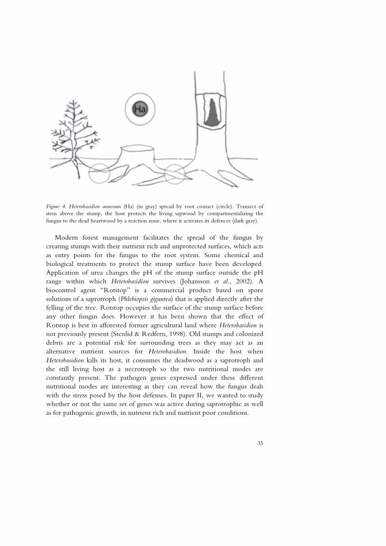
35
Figure 4. Heterobasidion annosum (Ha) (in gray) spread by root contact (circle). Transect of stem above the stump, the host protects the living sapwood by compartmentalizing the fungus to the dead heartwood by a reaction zone, where it activates its defences (dark gray).
Modern forest management facilitates the spread of the fungus by creating stumps with their nutrient rich and unprotected surfaces, which acts as entry points for the fungus to the root system. Some chemical and biological treatments to protect the stump surface have been developed. Application of urea changes the pH of the stump surface outside the pH range within which Heterobasidion survives (Johansson et al., 2002). A biocontrol agent “Rotstop” is a commercial product based on spore solutions of a saprotroph (Phlebiopsis gigantea) that is applied directly after the felling of the tree. Rotstop occupies the surface of the stump surface before any other fungus does. However it has been shown that the effect of Rotstop is best in afforested former agricultural land where Heterobasidion is not previously present (Stenlid & Redfern, 1998). Old stumps and colonized debris are a potential risk for surrounding trees as they may act as an alternative nutrient sources for Heterobasidion. Inside the host when Heterobasidion kills its host, it consumes the deadwood as a saprotroph and the still living host as a necrotroph so the two nutritional modes are constantly present. The pathogen genes expressed under these different nutritional modes are interesting as they can reveal how the fungus deals with the stress posed by the host defenses. In paper II, we wanted to study whether or not the same set of genes was active during saprotrophic as well as for pathogenic growth, in nutrient rich and nutrient poor conditions.
36
Consequently, the switch from saprotrophic to pathogenic growth was studied by gene expression studies of the growth of H. annosum on dead and living P. sylvestris seedlings. The experimental system was constructed to minimize the difference between the nutrient sources such that there was no other major difference than the host being alive or dead. The study consisted of one 384 clone macroarray which was analyzed with RNA harvested at one and six days post inoculation (dpi). An extra timepoint of 15dpi was studied with QPCR. The result showed that in the set of genes that we included in the study only a handful differed between the saprotrophic and pathogenic growth. The genes that do differ have functions related to detoxification and energy production (e,g. transcript antisense to ribosomal RNA Tar1p homologue).
The experimental set up allowed us to investigate the transitional shift in gene expression from saprotrophic to pathogenic growth in a nutrient limited environment. A dead pine seedling was the nutrient source in a mycelium connected to a living pine seedling. Our hypothesis was that the genes required for basic metabolic processes differ from those responsible for the pathogenicity of the fungus. There was a statistically significant switch in gene expression in the pathogenic and saprotrophic growth stage compared to the nutrient rich control. Between the pathogenic and saprotrophic mode of growth no clear significant shift in gene expression was found between the saprotrophic samples and the pathogenic samples. However, several trends were detected that suggests interesting differences in gene expression between the pathogenic and the saprotrophic stage.
The data can be interpreted as a reflection of the struggle for and the exploitation of a new resource. The expression pattern of GST homolog and CoQ5 is likely to be high during the interaction and to decrease when the host’s defences are overcome. The high expression pattern of GST homolog and CoQ5 during the saprotrophic stage compared to pathogenic stage at 6dpi was however contrary to our expectation. The decreases in the transcript levels recorded at 15 dpi probably reflect a depletion of the inductive nutrient source. GST is a gene which was expected to be up-regulated during active colonization and host defence reactions (Adomas et al., 2007). Although it was up-regulated during the colonization of the living host it never reached the levels seen under growth on the dead pine seedling. GSTs have multiple functions in fungi (McGoldrick et al., 2005) including detoxification of xenobiotics, transport and protection against oxidative stress. CoQ5 on the other hand is part of the electron transfer system in the mitochondria important for the energy system. There is a high demand for energy during pathogenicity, infection and the genetical
37
composition of the mitochondria has been implicated as virulence factor in H. annosum (Olson & Stenlid, 2001). However, in the present study, significant increases were recorded only during saprotrophic growth. It is possible that the primary function of CoQ5 is to protect the pathogen from its own radicals or ROS produced by the host during the infection. Finally, although no genes could be identified as belonging uniquely to either saprotrophic or pathogenic stages, our results suggests that differences between the two stages might hinge on the magnitude of gene expression during the various growth conditions.
4.3 Paper III Response of Pinus sylvestris to pathogenic, saprotrophic or symbiotic fungi: analysis of novel Clavata1-like and Nodulin 21-like genes.
The ecology and physiology of ECM symbiosis with conifer trees are well documented, however very little is however known about the molecular regulation of these associations. Similarly, the study of the genomics of the response of conifer tree tissues to pathogenic challenge is still in its infancy. The initiation, development and maintenance of ectomycorrhizas involve signal exchange and coordinated changes in gene expression from both partners (Le Quere et al., 2005; Podila et al., 2002; Voiblet et al., 2001; Slankis, 1973b). Studies describing transcriptome changes during ectomycorrhiza formation have indicated that the number of mycorrhiza-specific genes in both partners may be relatively small (Duplessis et al., 2005; Le Quere et al., 2005; Wright et al., 2005; Podila et al., 2002; Voiblet et al., 2001; Slankis, 1973b) and to date little is known about the mechanisms by which the fungus and the plant communicate during ectomycorrhizal formation. (Reddy et al., 2006; Reddy et al., 2003; Charvet-Candela et al., 2002a; Charvet-Candela et al., 2002b). Recent reports have revealed three ESTs which are involved in early stages of ectomycorrhizal development in Pinus sylvestris with Laccaria bicolor (Heller et al., 2008) that also show similarity to Clavata 1 RLK gene of Arabidopsis (Clv1-like) and to a MtN21 nodulin-like gene of Medicago truncatula (two clones MtN21-like-a and MtN21-like-b).
In paper III, the primary objectives were to investigate the specificity of response of these tree mycorrhiza-associated ESTs to either saprotrophic (Phanerochaete chrysosporium), mutualistic (Paxillus involutus and L. bicolor) or pathogenic (Heterobasidion annosum) fungi and to follow the expression of these genes during rhizogenesis and ECM development. We also characterized the expression patterns of the auxin homeostasis gene GH3
38
and a auxin regulated iaa88-like gene together with the mycorrhiza-associated EST after treatment with the auxin indole-3-butyric acid (IBA). The transcription of these genes was also followed in a time course experiment during ectomycorrhizal interaction with L. bicolor with and without the co-inoculation of the auxin transport inhibitor 2,3,5-triiodobenzoic acid (TIBA).
Generally, all four fungal species with diverse nutritional life styles provoked induction of host defence genes. This was in line with the result of our earlier study, although such increases in transcript levels were attenuated after prolonged infection with the non-pathogenic fungi (Adomas et al., 2008). The results also confirm our earlier observation that conifer tree tissues, even at immature stages, possess the right set of gene machinery for rapid response to extraneous factors (Asiegbu et al., 1999). The up-regulation of the auxin homeostasis gene GH3 in P. sylvestris in the presence of IBA and the early up-regulation and consequent down regulation during colonization by L. bicolor is in line with the findings in Pinus pinaster where the transcript levels of Pp-GH3.16 decreased gradually in the presence of the ECM fungus Hebeloma cylindrosporum but increased in auxin treatments (Reddy et al., 2006). These results indicate that the expression of plant genes associated with ectomycorrhizal formation is similar between closely related plant species and not necessarily specifically dependent on the colonizing fungal partner, also supporting the concept of a general “mycorrhiza” associated transcription program in ectomycorrhiza. This is further underlined by the observation of the expression of iaa88-like genes during ECM colonization. The Pp-iaa88 gene is reported to be highly expressed during fungal sheath formation (Charvet-Candela et al., 2002a). The P. sylvestris iaa88-like sequence is most highly expressed at 15 dpi which also coincides with fungal sheath formation (Heller et al., 2008). The expression patterns of the GH3- and iaa88-like genes in P. sylvestris underline the similarities of the regulation in the P. sylvestris/L. bicolor system to the transcriptional regulation in other symbiotic interactions with the genus Pinus.
The MtN21-like a/b gene represented by two ESTs is up-regulated during ECM formation. They are likely to originate from the same gene and, just like the P. taeda gene 5NG4 (Busov et al., 2004), they share homology with the nodulin 21 from Medicago truncatula (Gamas et al., 1996), a gene with unknown function. In our system, the P. sylvestris homologs 5NG4 and MtN21-like-a /b did not respond similarly either to auxin or to treatment with fungi. Expression of MtN21-like-a/b was not influenced by IBA treatment, whereas co-cultivation with L. bicolor led to a transient increase in
39
gene activity at 5 dpi. Treatment with an auxin inhibitor (TIBA) did not influence the response of MtN21-like-a/b to L. bicolor colonization. The application of auxin transport inhibitors results in a local increase in auxin concentration in plant tissues through a blockage of the PIN1 cycling(Geldner et al., 2001). It has been shown that polar auxin transport is necessary for lateral root formation in the early L.bicolor-plant interaction; Inoculation with L. bicolor induces lateral root formation in P. tremula x P. tremuloides and A. thaliana even without physical contact, this induction can be completely inhibited by the addition an auxin transport inhibitor (Felten et al., 2009). We also observed an almost complete inhibition of L. bicolor-induced lateral root formation in our material after treatment with TIBA. It is tempting to speculate on the basis of our results that the response of the MtN21-like-a/b gene in ectomycorrhizal formation is, at least partially, independent of auxin, and that the gene could be specifically associated with ectomycorrhiza formation between Pinus and L. bicolor. Treatment with L.bicolor also generated the strongest response in the study on co-cultivation with established fungal colonies, while H.annosum did not induce any response. Irrespective of the regulatory mechanisms of the MtN21-like-a/b gene, it is an interesting candidate for more detailed studies.
The Clv1-like EST falls into the LRR-XI superfamily (Shiu & Bleecker, 2001) of RLKs. There are at least 38 contigs with similarity to the LRR-XI superfamily in the Pine EST databases out of which the expression pattern has been described for only one gene up to now (Avila et al., 2006). The Clv1-like gene seem to be associated with the formation of lateral roots as expression can be detected in root primordias and during the formation of mycorrhizal roots. The Clv1-like transcript accumulates in response to to L. bicolor and to auxin treatment. The association between Clv1-like gene expression and lateral root initiation is supported by the absence of Clv1-like expression in the co-cultivation study as the roots had no lateral roots and root primordias could not be formed within the harvesting period, otherwise one could have expected P. involutus to induce Clv1-like gene expression in P. sylvestris.

41
5 Conclusions
The thesis relies on the central dogma (Crick, 1970) i.e. that the DNA encodes RNA and that the RNA encodes the proteins produced and therefore reflect the activity of the organism studied. This based on the fact that we have used RNA to construct cDNA and interpret the activity of the organisms studied instead of studying enzyme activity.
In global gene expression analysis, a first step in studying the basis for fungal diversity was to investigate whether cDNA arrays printed for one species give useful information from hybridization with labelled cDNA from other related species with the aid of macroarray hybridization techniques. Due to the high level of correlation, in the gene expression observed among the European Heterobasidion species, we concluded that the cDNA array of one specie can be used to study gene expression in the others. Furthermore, the gene expression data also supported the recent separation of the intersterility groups into the three separate species, Heterobasidion parviporum, Heterobasidion annosum, Heterobasidion abietinum representing the European S, P and F groups respectively.
Additionally, the present thesis has further confirmed sequence conservation amongst the investigated basidiomycetes. It also showed that no genes could be identified as uniquely belonging to either saprotrophic or pathogenic stages of H.annosum, which suggests that differences between the two stages might hinge on the magnitude of gene expression rather than distinct qualitative differences. In the near future the entire genome of H. annosum will be available which will allow for more complete expression studies using whole genome arrays as well as detailed studies on the promoter regions of the genes.
Furthermore, we investigated two symbiosis regulated genes and one gene homologous to the Medicago truncatula nodulin MtN21, and a homolog to the receptor like kinase Clavata1 like gene in interactions between Laccaria

42
bicolor and Pinus sylvestris (Scots pine), all belonging to gene families with members that can be regulated by auxin. Our results have revealed a different role played by the different nodulin homologs found in P. sylvestris and a possible role of Clv1-like in lateral root initiation during ectomycorrhizal development.
43
6 Future perspectives
This is an interesting time for everyone with a fascination for molecular biology as large genome sequencing projects are underway. No doubt, to intervene in disease and understand the basis of biological control or symbiotic relationships, a concerted and co-ordinated genomic analysis of fungi is essential. With the advent of such large-scale sequencing and novel functional genomics tools, there is an ample opportunity to understand the nature of beneficial and pathogenic fungus – tree interactions at levels of resolution never before possible. Since the first fungal genome was sequenced in 1996 the combined database of sequencing information has grown and the hope is that the resulting databases will allow for a comprehensive analysis of developmental processes that are characteristic of fungi. Such information will contribute to basic understanding of not only the mechanics of infection, symbiosis or saprotrophism but also of the evolution of pathogenicity and mutualism. To date there are around 100 fungal genomes sequenced (Stajich, 2009) and with the forthcoming sequencing of H. annosum and closely related species among the Russulales such as the ectomycorrhizal fungus Lactarius quietus and the mainly saprotrophic Phlebiopsis gigantea, new comparisons can be made which will further our knowledge of symbiosis in general and Heterobasidion biology in particular.
With development of novel molecular biological tools as well as efficient DNA transformation techniques for Heterobasidion functional characterization of genes important for key developmental or biological processes can be made via knockouts or RNA silencing. The regulation can then be studied in further detail by fusing promoters to GFP.
The availability of the genome sequence for Populus has no doubt facilitated basic studies in host–microbe interactions relevant for angiosperm tree ecology. The sequencing of parts of the symbiome of Populus with the
44
mutualistic Laccaria bicolor, Glomus intraradices and the pathogen Melampsora larici as well as the sequencing of the human gut is a likely indicator of what will come in the future, when sequencing costs are reduced and the bioinformatic tools are available the effects on host fitness of the organisms in its symbiome can be studied with possible commercial applications for agriculture and forestry. Additionally, the ongoing sequencing projects in Picea abies (Norway spruce) as well as Pinus taeda (loblolly pine) will also enable functional studies with the use of yeast two hybrid systems to identify relevant host and pathogen proteins acting as receptors and effectors respectively.
45
References
Abu, S.M., Li, G. & Asiegbu, F.O. (2004). Identification of Heterobasidion annosum (S-type) genes expressed during initial stages of conidiospore germination and under varying culture conditions. Fems Microbiology Letters 233(2), 205-213.
Adomas, A., Heller, G., Li, G.S., Olson, A., Chu, T.M., Osborne, J., Craig, D., Van Zyl, L., Wolfinger, R., Sederoff, R., Dean, R.A., Stenlid, J., Finlay, R. & Asiegbu, F.O. (2007). Trranscript profiling of a conifer pathosystem: response of Pinus sylvestris root tissues to pathogen (Heterobasidion annosum) invasion. Tree Physiology 27(10), 1441-1458.
Adomas, A., Heller, G., Olson, A., Osborne, J., Karlsson, M., Nahalkova, J., Van Zyl, L., Sederoff, R., Stenlid, J., Finlay, R. & Asiegbu, F.O. (2008). Comparative analysis of transcript abundance in Pinus sylvestris after challenge with a saprotrophic, pathogenic or mutualistic fungus. Tree Physiology 28(6), 885-897.
Agrios, G.N. (1997). Plant pathology. San Diego: Academic Press. (Plant pathology. Akiyama, K. & Hayashi, H. (2006). Strigolactones: Chemical Signals for Fungal Symbionts
and Parasitic Weeds in Plant Roots. Ann Bot 97(6), 925-931. Asiegbu, F.O., Adomas, A. & Stenlid, J. (2005). Conifer root and butt rot caused by
Heterobasidion annosum (Fr.) Bref. s.l. Molecular Plant Pathology 6(4), 395-409. Asiegbu, F.O., Daniel, G. & Johansson, M. (1994). Defense-Related Reactions of Seedling
Roots of Norway Spruce to Infection by Heterobasidion-Annosum (Fr.) Bref. Physiological and Molecular Plant Pathology 45(1), 1-19.
Asiegbu, F.O., Johansson, M. & Stenlid, J. (1999). Reactions of Pinus sylvestris (Scots Pine) Root Tissues to the Presence of Mutualistic, Saprotrophic and Necrotrophic Micro-organisms. Journal of Phytopathology 147, 257-264.
Asiegbu, F.O., Johansson, M., Woodward, S. & Huttermann, A. (1998). Biochemistry of the host-parasite interaction. Heterobasidion Annosum: Biology, Ecology, Impact and Control, 167-193.
Avila, C., Perez-Rodriguez, J. & Canovas, F.M. (2006). Molecular characterization of a receptor-like protein kinase gene from pine (Pinus sylvestris L.). Planta 224(1), 12-19.
Baldrian, P. (2009). Ectomycorrhizal fungi and their enzymes in soils: is there enough evidence for their role as facultative soil saprotrophs? Oecologia 161(4), 657-660.
46
Barker, S.J. & Tagu, D. (2000). The roles of auxins and cytokinins in mycorrhizal symbioses. Journal of Plant Growth Regulation 19(2), 144-154.
Bassett, C., Sherwood, R.T., Kepler, J.A. & Hamilton, P.B. (1967). Production and biological activity of fommanosin a toxic sesquiterpene metabolite of Fomes annosus. Phytopathology 57(10), 1046-&.
Berg, B. & Tamm, C.O. (1991). Decomposition and nutrient dynamics of Norway spruce needle litter in a long-term optimum nutrition experiment 1. Organic matter decomposition Berg, B. And C. O. Tamm. Sveriges Lantbruksuniversitet Institutionen for Ekologi Och Miljovard Rapport, 39; (Swedish University of Agricultural Sciences Department of Ecology and Environmental Research Report, 39). Decomposition and Nutrient Dynamics of Norway Spruce Needle Litter in a Long-Term Optimum Nutrition Experiment: I. Organic Matter Decomposition; Ii. Nutrient Dynamics; Iii. The Influence of a Denser Forest on Litter Decomposition Rate. 55p. Swedish University of Agricultural Sciences Department of Ecology and Environmental Research: Uppsala, Sweden. Illus. Paper, 5-24.
Bhalerao, R.P., Eklof, J., Ljung, K., Marchant, A., Bennett, M. & Sandberg, G. (2002). Shoot-derived auxin is essential for early lateral root emergence in Arabidopsis seedlings. Plant Journal 29(3), 325-332.
Bhattacharya, D., Yoon, H.S. & Hackett, J.D. (2004). Photosynthetic eukaryotes unite: endosymbiosis connects the dots. Bioessays 26(1), 50-60.
Boberg, J. (2009). Litter decomposing fungi in boreal forests : their function in carbon and nitrogen circulation. Uppsala: (Acta Universitatis agriculturae Sueciae,1652-6880 ;2009:75).
Boddy, L., Hynes, J., Bebber, D.P. & Fricker, M.D. (2009). Saprotrophic cord systems: dispersal mechanisms in space and time. Mycoscience 50(1), 9-19.
Bolton, M.D. (2009). Primary metabolism and plant defense - fuel for the fire. Molecular Plant-Microbe Interactions 22(5), 487-497.
Bolton, M.D. & Thomma, B. (2008). The complexity of nitrogen metabolism and nitrogen-regulated gene expression in plant pathogenic fungi. Physiological and Molecular Plant Pathology 72(4-6), 104-110.
Brinker, M., van Zyl, L., Liu, W.B., Craig, D., Sederoff, R.R., Clapham, D.H. & von Arnold, S. (2004). Microarray analyses of gene expression during adventitious root development in Pinus contorta (1[w]). Plant Physiology 135(3), 1526-1539.
Brunner, A.M., Yakovlev, I.A. & Strauss, S.H. (2004). Validating internal controls for quantitative plant gene expression studies. Bmc Plant Biology 4
Busov, V.B., Johannes, E., Whetten, R.W., Sederoff, R.R., Spiker, S.L., Lanz-Garcia, C. & Goldfarb, B. (2004). An auxin-inducible gene from loblolly pine (Pinus taeda L.) is differentially expressed in mature and juvenile-phase shoots and encodes a putative transmembrane protein. Planta 218(6), 916-927.
Bustin, S.A. (2000). Absolute quantification of mRNA using real-time reverse transcription polymerase chain reaction assays. Journal of Molecular Endocrinology 25(2), 169-193.
Carlile, M.J., Watkinson, S.C., Carlile, M.J. & Watkinson, S.C. (1994). The fungi. Academic Press Ltd.; Academic Press, Inc. (The fungi
Charvet-Candela, V., Hitchin, S., Ernst, D., Sandermann, H., Marmeisse, R. & Gay, G. (2002a). Characterization of an Aux/IAA cDNA upregulated in Pinus pinaster
47
roots in response to colonization by the ectomycorrhizal fungus Hebeloma cylindrosporum. New Phytologist 154(3), 769-777.
Charvet-Candela, V., Hitchin, S., Reddy, M.S., Cournoyer, B., Marmeisse, R. & Gay, G. (2002b). Characterization of a Pinus pinaster cDNA encoding an auxin up-regulated putative peroxidase in roots. Tree Physiology 22(4), 231-238.
Chase, T.E. (1985). PhD Thesis. Diss. University of Vermont. Burlington. Coleman, M., Henricot, B., Arnau, J. & Oliver, R.P. (1997). Starvation-Induced Genes of
the Tomato Pathogen Cladosporium fulvum Are Also Induced During Growth In Planta. Molecular Plant-Microbe Interactions 10(9), 1106-1109.
Cornell, M.J., Alam, I., Soanes, D.M., Wong, H.M., Hedeler, C., Paton, N.W., Rattray, M., Hubbard, S.J., Talbot, N.J. & Oliver, S.G. (2007). Comparative genome analysis across a kingdom of eukaryotic organisms: Specialization and diversification in the Fungi. Genome Research 17, 1809-1822.
Crick, F. (1970). Central dogma of molecular biology. Nature 227(5258), 561-563. Cullings, K. & Courty, P.E. (2009). Saprotrophic capabilities as functional traits to study
functional diversity and resilience of ectomycorrhizal community. Oecologia 161(4), 661-664.
Cuomo, C.A., Gueldener, U., Xu, J.R., Trail, F., Turgeon, B.G., Di Pietro, A., Walton, J.D., Ma, L.J., Baker, S.E., Rep, M., Adam, G., Antoniw, J., Baldwin, T., Calvo, S., Chang, Y.L., DeCaprio, D., Gale, L.R., Gnerre, S., Goswami, R.S., Hammond-Kosack, K., Harris, L.J., Hilburn, K., Kennell, J.C., Kroken, S., Magnuson, J.K., Mannhaupt, G., Mauceli, E., Mewes, H.W., Mitterbauer, R., Muehlbauer, G., Munsterkotter, M., Nelson, D., O'Donnell, K., Ouellet, T., Qi, W.H., Quesneville, H., Roncero, M.I.G., Seong, K.Y., Tetko, I.V., Urban, M., Waalwijk, C., Ward, T.J., Yao, J.Q., Birren, B.W. & Kistler, H.C. (2007). The Fusarium graminearum genome reveals a link between localized polymorphism and pathogen specialization. Science 317(5843), 1400-1402.
Cuzick, A., Maguire, K. & Hammond-Kosack, K.E. (2009). Lack of the plant signalling component SGT1b enhances disease resistance to Fusarium culmorum in Arabidopsis buds and flowers. New Phytologist 181(4), 901-912.
Daniel, G., Asiegbu, F. & Johansson, M. (1998). The saprotrophic wood-degrading abilities of Heterobasidium annosum intersterility groups P and S. Mycological Research 102(8), 991-997.
de Wit, P., Mehrabi, R., van den Burg, H.A. & Stergiopoulos, I. (2009). Fungal effector proteins: past, present and future. Molecular Plant Pathology 10(6), 735-747.
Dean, R.A., Talbot, N.J., Ebbole, D.J., Farman, M.L., Mitchell, T.K., Orbach, M.J., Thon, M., Kulkarni, R., Xu, J.R., Pan, H.Q., Read, N.D., Lee, Y.H., Carbone, I., Brown, D., Oh, Y.Y., Donofrio, N., Jeong, J.S., Soanes, D.M., Djonovic, S., Kolomiets, E., Rehmeyer, C., Li, W.X., Harding, M., Kim, S., Lebrun, M.H., Bohnert, H., Coughlan, S., Butler, J., Calvo, S., Ma, L.J., Nicol, R., Purcell, S., Nusbaum, C., Galagan, J.E. & Birren, B.W. (2005). The genome sequence of the rice blast fungus Magnaporthe grisea. Nature 434(7036), 980-986.
Duddridge, J.A. (1986). The development and ultrastructure of ectomycorrhizas. 3. Compatible and incompatible interactions between Suillus-grevillei(Klotzsch) Sing
48
and 11 species of ectomycorrhizal hosts in vitro in the abscence of exogenous carbohydrate. New Phytologist 103(3), 457-&.
Duplessis, S., Courty, P.E., Tagu, D. & Martin, F. (2005). Transcript patterns associated with ectomycorrhiza development in Eucalyptus globulus and Pisolithus microcarpus. New Phytologist 165(2), 599-611.
Ek, M., Ljungquist, P.O. & Stenstrom, E. (1983). Indole-3-acetic-acid production by mycorrhizal fungi determined by gas chromatography-mass spectrometry. New Phytologist 94(3), 401-407.
Eriksson, K.-E., Blanchette, R.A. & Ander, P. (1990). Microbial and enzymatic degradation of wood and wood components. Springer-Verlag.
Felten, J., Kohler, A., Morin, E., Bhalerao, R.P., Palme, K., Martin, F., Ditengou, F.A. & Legue, V. (2009). The Ectomycorrhizal Fungus Laccaria bicolor Stimulates Lateral Root Formation in Poplar and Arabidopsis through Auxin Transport and Signaling. Plant Physiology 151(4), 1991-2005.
Finlay, R.D. (2008). Ecological aspects of mycorrhizal symbiosis: with special emphasis on the functional diversity of interactions involving the extraradical mycelium. Journal of Experimental Botany 59(5), 1115-1126.
Fitzpatrick, D.A., Logue, M.E., Stajich, J.E. & Butler, G. (2006). A fungal phylogeny based on 42 complete genomes derived from supertree and combined gene analysis. Bmc Evolutionary Biology 6, 15.
Francis, R. & Read, D.J. (1995). Mutualism and antagonism in the mycorrhizal symbiosis, with special reference to impacts on plant community structure. Canadian Journal of Botany-Revue Canadienne De Botanique 73, S1301-S1309.
Frettinger, P., Herrmann, S., Lapeyrie, F., Oelmuller, R. & Buscot, F. (2006). Differential expression of two class III chitinases in two types of roots of Quercus robur during pre-mycorrhizal interactions with Piloderma croceum. Mycorrhiza 16(3), 219-223.
Fukaki, H., Okushima, Y. & Tasaka, M. (2007). Auxin-mediated lateral root formation in higher plants. International Review of Cytology - a Survey of Cell Biology, Vol 256 256, 111-137.
Gamas, P., De Carvalho Niebel, F., Lescure, N. & Cullimore, J.V. (1996). Use of a subtractive hybridization approach to identify new Medicago truncatula genes induced during root nodule development. Molecular Plant-Microbe Interactions 9(4), 233-242.
García-Garrido, J.M. & Ocampo, J.A. (2002). Regulation of the plant defence respons in arbuscular mycorrhizal symbiosis. Journal of Experimental Botany 53(373), 1377-1386.
Gay, G. (1988). Role of fungal hormones i ectomycorrhizal symbiosis. Cryptogamie Mycologie 9(3), 211-219.
Geldner, N., Friml, J., Stierhof, Y.D., Jurgens, G. & Palme, K. (2001). Auxin transport inhibitors block PIN1 cycling and vesicle traficking. Nature 413(6854), 425-428.
Gohre, V. & Robatzek, S. (2008). Breaking the barriers: Microbial effector molecules subvert plant immunity. Annual Review of Phytopathology 46, 189-215.
Gomez-Gomez, L. & Boller, T. (2000). FLS2: An LRR receptor-like kinase involved in the perception of the bacterial elicitor flagellin in Arabidopsis. Molecular Cell 5(6), 1003-1011.

49
Gray, M.W., Burger, G. & Lang, B.F. (1999). Mitochondrial evolution. Science 283(5407), 1476-1481.
Gray, W.M., Muskett, P.R., Chuang, H.W. & Parker, J.E. (2003). Arabidopsis SGT1b is required for SCFTIR1-mediated auxin response. Plant Cell 15(6), 1310-1319.
Gruen, H.E. (1959). AUXINS AND FUNGI. Annual Review of Plant Physiology and Plant Molecular Biology 10, 405-440.
Guimil, S., Chang, H.-S., Zhu, T., Sesma, A., Osbourn, A., Roux, C., Ioannidis, V., Oakeley, E.J., Docquier, M., Descombes, P., Briggs, S.P. & Paszkowski, U. (2005). Comparative transcriptomics of rice reveals an ancient pattern of response to microbial colonization. PNAS 102(22), 8066-8070.
Harrison, M.J. & Dixon, R.A. (1994). Spatial patterns of expression of flavonoid/isoflavonoid pathway genes during interactions between roots of Medicago truncatula and the mycorrhizal fungus Glomus versiforme. Plant Journal 6(1), 9-20.
Heil, M. & Baldwin, I.T. (2002). Fitness costs of induced resistance: emerging experimental support for a slippery concept. Trends in Plant Science 7(2), 61-67.
Heller, G., Adomas, A., Li, G.S., Osborne, J., van Zyl, L., Sederoff, R., Finlay, R.D., Stenlid, J. & Asiegbu, F.O. (2008). Transcriptional analysis of Pinus sylvestris roots challenged with the ectomycorrhizal fungus Laccaria bicolor. Bmc Plant Biology 8
Hibbet, D.S., Gilbert, L.-B. & Donoghue, M.J. (2000). Evolutionary instability of ectomycorrhizal symbioses in basidiomycetes. Nature 407
Hibbett, D. & Matheny, P.B. (2009). The relative ages of ectomycorrhizal mushrooms and their plant hosts estimated using Bayesian relaxed molecular clock analyses. BMC Biology 7(1), 13.
Hietala, A.M., Kvaalen, H., Schmidt, A., Johnk, N., Solheim, H. & Fossdal, C.G. (2004). Temporal and spatial profiles of chitinase expression by Norway spruce in response to bark colonization by Heterobasidion annosum. Applied and Environmental Microbiology 70(7), 3948-3953.
Huettermann, A. & Woodward, S. (1998). Historical aspects. Heterobasidion Annosum: Biology, Ecology, Impact and Control, 1-25.
James, T.Y., Kauff, F., Schoch, C.L., Matheny, P.B., Hofstetter, V., Cox, C.J., Celio, G., Gueidan, C., Fraker, E., Miadlikowska, J., Lumbsch, H.T., Rauhut, A., Reeb, V., Arnold, A.E., Amtoft, A., Stajich, J.E., Hosaka, K., Sung, G.H., Johnson, D., O'Rourke, B., Crockett, M., Binder, M., Curtis, J.M., Slot, J.C., Wang, Z., Wilson, A.W., Schussler, A., Longcore, J.E., O'Donnell, K., Mozley-Standridge, S., Porter, D., Letcher, P.M., Powell, M.J., Taylor, J.W., White, M.M., Griffith, G.W., Davies, D.R., Humber, R.A., Morton, J.B., Sugiyama, J., Rossman, A.Y., Rogers, J.D., Pfister, D.H., Hewitt, D., Hansen, K., Hambleton, S., Shoemaker, R.A., Kohlmeyer, J., Volkmann-Kohlmeyer, B., Spotts, R.A., Serdani, M., Crous, P.W., Hughes, K.W., Matsuura, K., Langer, E., Langer, G., Untereiner, W.A., Lucking, R., Budel, B., Geiser, D.M., Aptroot, A., Diederich, P., Schmitt, I., Schultz, M., Yahr, R., Hibbett, D.S., Lutzoni, F., McLaughlin, D.J., Spatafora, J.W. & Vilgalys, R. (2006). Reconstructing the early evolution of Fungi using a six-gene phylogeny. Nature 443(7113), 818-822.
50
Jane Barker, S., Tagu, D. & Delp, G. (1998). Regulation of Root and Fungal Morphogenesis in Mycorrhizal Symbioses. Plant Physiology 116(4), 1201-1207.
Johansson, S.M., Pratt, J.E. & Asiegbu, F.O. (2002). Treatment of Norway spruce and Scots pine stumps with urea against the root and butt rot fungus Heterobasidion annosum - possible modes of action. Forest Ecology and Management 157(1-3), 87-100.
Jones, J.D.G. & Dangl, J.L. (2006). The plant immune system. Nature 444(7117), 323-329. Kaku, H., Nishizawa, Y., Ishii-Minami, N., Akimoto-Tomiyama, C., Dohmae, N., Takio,
K., Minami, E. & Shibuya, N. (2006). Plant cells recognize chitin fragments for defense signaling through a plasma membrane receptor. Proceedings of the National Academy of Sciences of the United States of America 103(29), 11086-11091.
Karabaghli-Degron, C., Sotta, B., Bonnet, M., Gay, G. & Le TACON, F. (1998). The auxin transport inhibitor 2,3,5-triiodobenzoic acid (TIBA) inhibits the stimulation of in vitro lateral root formation and the colonization of the tap-root cortex of Norway spruce (Picea abies) seedlings by the ectomycorrhizal fungus Laccaria bicolor. New Phytol 140(4), 723-733.
Karlsson, J.-O. & Stenlid, J. (1991). Pectic isozyme profiles of intersterility groups in Heterobasidion annosum. Mycological Research 95, 531-536.
Karlsson, M., Olson, A. & Stenlid, J. (2003). Expressed sequences from the basidiomycetous tree pathogen Heterobasidion annosum during early infection of scots pine*1. Fungal Genetics and Biology 39(1), 51-59.
Kazan, K. & Manners, J.M. (2009). Linking development to defense: auxin in plant-pathogen interactions. Trends in Plant Science 14(7), 373-382.
Kleine, T., Maier, U.G. & Leister, D. (2009). DNA Transfer from Organelles to the Nucleus: The Idiosyncratic Genetics of Endosymbiosis. Annual Review of Plant Biology 60, 115-138.
Koide, R.T., Sharda, J.N., Herr, J.R. & Malcolm, G.M. (2008). Ectomycorrhizal fungi and the biotrophy-saprotrophy continuum. New Phytologist 178(2), 230-233.
Korhonen, K., Capretti, P., Karjalainen, R. & Stenlid, J. (1998). Distribution of Heterobasidion annosum Intersterility groups in Europe. In: Woodward, S., et al. (Eds.) Heterobasidion annosum biology, ecology, impact and control. London: CAB International. pp. 93-104.
Laajanen, K., Vuorinen, I., Salo, V., Juuti, J. & Raudaskoski, M. (2007). Cloning of Pinus sylvestris SCARECROW gene and its expression pattern in the pine root system, mycorrhiza and NPA-treated short roots. New Phytologist 175(2), 230-243.
Langer, I., Krpata, D., Peintner, U., Wenzel, W.W. & Schweiger, P. (2008). Media formulation influences in vitro ectomycorrhizal synthesis on the European aspen Populus tremula L. Mycorrhiza 18(6-7), 297-307.
Le Quere, A., Schutzendubel, A., Rajashekar, B., Canback, B., Hedh, J., Erland, S., Johansson, T. & Tunlid, A. (2004). Divergence in gene expression related to variation in host specificity of an ectomycorrhizal fungus. Molecular Ecology 13(12), 3809-3819.
Le Quere, A., Wright, D.P., Soderstrom, B., Tunlid, A. & Johansson, T. (2005). Global patterns of gene regulation associated with the development of ectomycorrhiza
51
between birch (Betula pendula Roth.) and Paxillus involutus (Batsch) fr. Molecular Plant-Microbe Interactions 18(7), 659-673.
LePage, B.A., Currah, R.S., Stockey, R.A. & Rothwell, G.W. (1997). Fossil Ectomycorrhizae from the Middle Eocene. American Journal of Botany 84(3), 410-412.
Lewis, D.H. (1973). Concepts in fungal nutrition and origin of biotrophy. Biological Reviews of the Cambridge Philosophical Society 48(2), 261-278.
Lind, M., Olson, K. & Stenlid, J. (2005). An AFLP-markers based genetic linkage map of Heterobasidion annosum locating intersterility genes. Fungal Genetics and Biology 42(6), 519-527.
Lind, M., Stenlid, J. & Olson, A. (2007). Genetics and QTL mapping of somatic incompatibility and intraspecific interactions in the basidiomycete Heterobasidion annosum s.l. Fungal Genetics and Biology 44, 1242-1251.
Lindahl, B.D., Ihrmark, K., Boberg, J., Trumbore, S.E., Hogberg, P., Stenlid, J. & Finlay, R.D. (2007). Spatial separation of litter decomposition and mycorrhizal nitrogen uptake in a boreal forest. New Phytologist 173(3), 611-620.
Liu, J., Blaylock, L.A., Endre, G., Cho, J., Town, C.D., VandenBosch, K.A. & Harrison, M.J. (2003). Transcript Profiling Coupled with Spatial Expression Analyses Reveals Genes Involved in Distinct Developmental Stages of an Arbuscular Mycorrhizal Symbiosis. Plant Cell 15(9), 2106-2123.
Llorente, F., Muskett, P., Sanchez-Vallet, A., Lopez, G., Ramos, B., Sanchez-Rodriguez, C., Jorda, L., Parker, J. & Molina, A. (2008). Repression of the auxin response pathway increases Arabidopsis susceptibility to necrotrophic fungi. Molecular Plant 1(3), 496-509.
Manion, P.D. (1981). Tree disease concepts. Englewood Cliffs, New Jersey: Prentice-Hall, Inc. (Tree disease concepts.
Martin, F. (2008). Orchestrating morphogenesis in mycorrhizal symbioses. New Phytologist 177(4), 839-841.
Martin, F., Aerts, A., Ahren, D., Brun, A., Danchin, E.G.J., Duchaussoy, F., Gibon, J., Kohler, A., Lindquist, E., Pereda, V., Salamov, A., Shapiro, H.J., Wuyts, J., Blaudez, D., Buee, M., Brokstein, P., Canback, B., Cohen, D., Courty, P.E., Coutinho, P.M., Delaruelle, C., Detter, J.C., Deveau, A., DiFazio, S., Duplessis, S., Fraissinet-Tachet, L., Lucic, E., Frey-Klett, P., Fourrey, C., Feussner, I., Gay, G., Grimwood, J., Hoegger, P.J., Jain, P., Kilaru, S., Labbe, J., Lin, Y.C., Legue, V., Le Tacon, F., Marmeisse, R., Melayah, D., Montanini, B., Muratet, M., Nehls, U., Niculita-Hirzel, H., Oudot-Le Secq, M.P., Peter, M., Quesneville, H., Rajashekar, B., Reich, M., Rouhier, N., Schmutz, J., Yin, T., Chalot, M., Henrissat, B., Kues, U., Lucas, S., Van de Peer, Y., Podila, G.K., Polle, A., Pukkila, P.J., Richardson, P.M., Rouze, P., Sanders, I.R., Stajich, J.E., Tunlid, A., Tuskan, G. & Grigoriev, I.V. (2008). The genome of Laccaria bicolor provides insights into mycorrhizal symbiosis. Nature 452(7183), 88-U7.
Martin, F. & Selosse, M.A. (2008). The Laccaria genome: a symbiont blueprint decoded. New Phytologist 180(2), 296-310.
52
Marx, D.H. (1969). Influence of ectotrophic mycorrhizal fungi on resistance of pine roots to pathogenic infections. 1.Antagonism of mycorrhizal fungi to root pathogenic fungi and soil bacteria. Phytopathology 59(2), 153-&.
Matheny, P.B., Curtis, J.M., Hofstetter, V., Aime, M.C., Moncalvo, J.M., Ge, Z.W., Yang, Z.L., Slot, J.C., Ammirati, J.F., Baroni, T.J., Bougher, N.L., Hughes, K.W., Lodge, D.J., Kerrigan, R.W., Seidl, M.T., Aanen, D.K., DeNitis, M., Daniele, G.M., Desjardin, D.E., Kropp, B.R., Norvell, L.L., Parker, A., Vellinga, E.C., Vilgalys, R. & Hibbett, D.S. (2006). Major clades of Agaricales: a multilocus phylogenetic overview. Mycologia 98(6), 982-995.
Mathesius, U. (2008). Auxin: at the root of nodule development? Functional Plant Biology 35(8), 651-668.
McGoldrick, S., O'Sullivan, S.n.M. & Sheehan, D. (2005). Glutathione transferase-like proteins encoded in genomes of yeasts and fungi: insights into evolution of a multifunctional protein superfamily. Fems Microbiology Letters 242(1), 1-12.
McLaughlin, D.J., Hibbett, D.S., Lutzoni, F., Spatafora, J.W. & Vilgalys, R. (2009). The search for the fungal tree of life. Trends in Microbiology 17(11), 488-497.
Menkis, A., Allmer, J., Vasiliauskas, R., Lygis, V., Stenlid, J. & Finlay, R. (2004). Ecology and molecular characterization of dark septate fungi from roots, living stems, coarse and fine woody debris. Mycological Research 108, 965-973.
Millenaar, F.F., Okyere, J., May, S.T., van Zanten, M., Voesenek, L. & Peeters, A.J.M. (2006). How to decide? Different methods of calculating gene expression from short oligonucleotide array data will give different results. Bmc Bioinformatics 7, 16.
Miller, S.L., Larsson, E., Larsson, K.H., Verbeken, A. & Nuytinck, J. (2006). Perspectives in the new Russulales. Mycologia 98(6), 960-970.
Muller, M.M. & Hallaksela, A.M. (2000). Fungal diversity in Norway spruce: a case study. Mycological Research 104, 1139-1145.
Niemi, K., Vuorinen, T., Ernstsen, A. & Haggman, H. (2002). Ectomycorrhizal fungi and exogenous auxins influence root and mycorrhiza formation of Scots pine hypocotyl cuttings in vitro. Tree Physiology 22(17), 1231-1239.
Nolan, T., Hands, R.E. & Bustin, S.A. (2006). Quantification of mRNA using real-time RT-PCR. Nature Protocols 1(3), 1559-1582.
Oliver, R.P. & Ipcho, S.V.S. (2004). Arabidopsis pathology breathes new life into the necrotrophs-vs.-biotrophs classification of fungal pathogens. Molecular Plant Pathology 5(4), 347-352.
Olson, A. & Stenlid, J. (2001). Mitochondrial control of fungal hybrid virulence. Nature(411), 438.
Osono, T. & Takeda, H. (2002). Comparison of litter decomposing ability among diverse fungi in a cool temperate deciduous forest in Japan. Mycologia 94(3), 421-427.
Otrosina, W.J. & Garbelotto, M.Heterobasidion occidentalis sp. nov. and Heterobasidion irregularis comb. nov.: A disposition of North American Heterobasidion biological species. Mycological Research In Press, Uncorrected Proof
Parrent, J.L., James, T.Y., Vasaitis, R. & Taylor, A.F.S. (2009). Friend or foe? Evolutionary history of glycoside hydrolase family 32 genes encoding for sucrolytic activity in fungi and its implications for plant-fungal symbioses. Bmc Evolutionary Biology 9
53
Peay, K.G., Kennedy, P.G. & Bruns, T.D. (2008). Fungal Community Ecology: A Hybrid Beast with a Molecular Master. Bioscience 58(9), 799-810.
Perfect, S.E. & Green, J.R. (2001). Infection structures of biotrophic and hemibiotrophic fungal plant pathogens. Molecular Plant Pathology 2(2), 101-108.
Pesaresi, P., Schneider, A., Kleine, T. & Leister, D. (2007). Interorganellar communication. Current Opinion in Plant Biology 10(6), 600-606.
Peskan-Berghofer, T., Shahollari, B., Giong, P.H., Hehl, S., Markert, C., Blanke, V., Kost, G., Varma, A. & Oelmuller, R. (2004). Association of Piriformospora indica with Arabidopsis thaliana roots represents a novel system to study beneficial plant-microbe interactions and involves early plant protein modifications in the endoplasmic reticulum and at the plasma membrane. Physiologia Plantarum 122(4), 465-477.
Pfaffl, M.W. (2001). A new mathematical model for relative quantification in real-time RT–PCR. Nucleic Acids Research 29(9), 2002-2007.
Pirozynski, K.A. & Malloch, D.W. (1975). Origin of land plants- matter of mycotropism. Biosystems 6(3), 153-164.
Podila, G.K., Zheng, J., Balasubramanian, S., Sundaram, S., Hiremath, S., Brand, J.H. & Hymes, M.J. (2002). Fungal gene expression in early symbiotic interactions between Laccaria bicolor and red pine. Plant and Soil 244(1-2), 117-128.
Raven, J.A. (1975). Transport of indoleacetic-acid in plant-cells in relation to pH and electrical potential gradients, and its significans for polar IAA transport. New Phytologist 74(2), 163-172.
Raven, P.H., Evert, R.F. & Eichorn, S.E. (1998). Biology of plants. New York: W.H. Freeman and Co. (Biology of plants.
Rayner, A.D.M. & Boddy, L. (1988). Fungal decomposition of wood. Its biology and ecology. Chichester, Sussex UK: John Wiley & Sons Ltd. (Fungal decomposition of wood. Its biology and ecology.
Reddy, S.M., Hitchin, S., Melayah, D., Pandey, A.K., Raffier, C., Henderson, J., Marmeisse, R. & Gay, G. (2006). The auxin-inducible GH3 homologue Pp-GH3.16 is downregulated in Pinus pinaster root systems on ectomycorrhizal symbiosis establishment. New Phytologist 170(2), 391-400.
Reddy, S.M., Pandey, A.K., Melayah, D., Marmeisse, R. & Gay, G. (2003). The auxin responsive gene Pp-C61 is up-regulated in Pinus pinaster roots following inoculation with ectomycorrhizal fungi. Plant Cell and Environment 26(5), 681-691.
Redecker, D., Kodner, R. & Graham, L.E. (2000). Glomalean fungi from the Ordovician. Science 289(5486), 1920-1921.
Redfern, D.B. & Stenlid, J. (1998). Spore dispersal and infection. CAB International; CAB International. (Heterobasidion Annosum: Biology, Ecology, Impact and Control
Remy, W., Taylor, T.N., Hass, H. & Kerp, H. (1994). 4-hundred- million-year- old vesicular arbuscular mycorrhizae Proceedings of the National Academy of Sciences of the United States of America 91(25), 11841-11843.
Rincon, A., Gerard, J., Dexheimer, J. & Le Tacon, F. (2001). Effect of an auxin transport inhibitor on aggregation and attachment processes during ectomycorrhiza formation between Laccaria bicolor S238N and Picea abies. Canadian Journal of Botany-Revue Canadienne De Botanique 79(10), 1152-1160.
54
Rincon, A., Priha, O., Sotta, B., Bonnet, M. & Le Tacon, F. (2003). Comparative effects of auxin transport inhibitors on rhizogenesis and mycorrhizal establishment of spruce seedlings inoculated with Laccaria bicolor. Tree Physiology 23(11), 785-791.
Ritchie, M.E., Silver, J., Oshlack, A., Holmes, M., Diyagama, D., Holloway, A. & Smyth, G.K. (2007). A comparison of background correction methods for two-colour microarrays. Bioinformatics 23, 2700-2707.
Rodriguez, R.J., White, J.F., Arnold, A.E. & Redman, R.S. (2009). Fungal endophytes: diversity and functional roles. New Phytologist 182(2), 314-330.
Rosenberg, E., Sharon, G. & Zilber-Rosenberg, I. (2009). The hologenome theory of evolution contains Lamarckian aspects within a Darwinian framework. Environmental Microbiology 11(12), 2959-2962.
Salzer, P., Hubner, B., Sirrenberg, A. & Hager, A. (1997). Differential effect of purified spruce chitinases and beta-1,3-glucanases on the activity of elicitors from ectomycorrhizal fungi. Plant Physiology 114(3), 957-968.
Sapp, J. (2004). The dynamics of symbiosis: an historical overview. Canadian Journal of Botany-Revue Canadienne De Botanique 82(8), 1046-1056.
Shiu, S.H. & Bleecker, A.B. (2001). Receptor-like kinases from Arabidopsis form a monophyletic gene family related to animal receptor kinases. Proceedings of the National Academy of Sciences of the United States of America 98(19), 10763-10768.
Silver, J.D., Ritchie, M.E. & Smyth, G.K. (2009). Microarray background correction: maximum likelihood estimation for the normal-exponential convolution. Biostatistics 10(2), 352-363.
Slankis, V. (1973a). Hormonal relationships in mycorrhizal development. Marks, G.C. And T.T. Kozlowski (Ed.). Physiological Ecology, Ectomycorrhizae. Their Ecology and Physiology. Xiv+444p. Illus. Academic Press: New York, N.Y., U.S.A, 231-298.
Slankis, V. (1973b). Hormonal relationships in mycorrhizal development. Ectomycorrhizae: Their ecology and physiology Academic press: New York.(Marks GC, Kozlowski TT, editors), 231-298.
Smith, M.L., Bruhn, J.N. & Anderson, J.B. (1992). The fungus Armillaria-bulbosa is among the largest and oldest living organisms. Nature 356(6368), 428-431.
Smith, S.E. & Read, D.J. (1997). Mycorrhizal symbiosis. San Diego, US: Academic Press. (Mycorrhizal symbiosis.
Smyth, G.K. (2004). Linear models and empirical Bayes methods for assessing differential expression in microarray experiments. Statistical Applications in Genetics and Molecular Biology 3(1), Article 3.
Smyth, G.K. (2005). Limma: Linear models for microarray data. Bioinformatics and Computational Biology Solution Using R and Bioconductor, 397-420.
Soanes, D.M., Alam, I., Cornell, M., Wong, H.M., Hedeler, C., Paton, N.W., Rattray, M., Hubbard, S.J., Oliver, S.G. & Talbot, N.J. (2008). Comparative Genome Analysis of Filamentous Fungi Reveals Gene Family Expansions Associated with Fungal Pathogenesis. Plos One 3(6)
Soanes, D.M., Richards, T.A. & Talbot, N.J. (2007). Insights from Sequencing Fungal and Oomycete Genomes: What Can We Learn about Plant Disease and the Evolution of Pathogenicity? Plant Cell 19(11), 3318-3326.
55
Soanes, D.M. & Talbot, N.J. (2006). Comparative genomic analysis of phytopathogenic fungi using expressed sequence tag (EST) collections. Molecular Plant Pathology 7(1), 61-70.
Soderlund, C. (2009). Computational techniques for elucidating plant-pathogen interactions from large-scale experiments on fungi and oomycetes. Briefings in Bioinformatics 10(6), 654-663.
Sonnenbichler, J., Bliestle, I.M., Peipp, H. & Holdenrieder, O. (1989). Secondary fungal metabolites and their biological-activities .1. Isolation of antibiotic compounds from cultures of Heterobasidion annosum synthesized in the presence of antagonistic fungi or host plant-cells Biological Chemistry Hoppe-Seyler 370(12), 1295-1303.
Sonnenbichler, J., Dietrich, J. & Peipp, H. (1994). Secondary fungal metabolites and their biological-activities, .5. Investigations concerning the induction of the biosynthesis of the toxic secondary metabolites in basidiomycetes. Biological Chemistry Hoppe-Seyler 375(1), 71-79.
Sonnenbichler, J., Lamm, V., Gieren, A., Holdenrieder, O. & Lotter, H. (1983). A cyclopentabenzopyranone produced by the fungus Heterobasidion-annosum in dual cultures. Phytochemistry 22(6), 1489-1491.
Spaepen, S., Vanderleyden, J. & Remans, R. (2007). Indole-3-acetic acid in microbial and microorganism-plant signaling. Fems Microbiology Reviews 31(4), 425-448.
Stajich, J.E. (2009). Fungal Genome Links [online]. Available from: http://fungalgenomes.org/wiki/Fungal_Genome_Links
Stenlid, J. (1985). Population structure of Heterobasidion annosum as determined by somatic incompatibility, sexual incompatibility, and isoenzyme patterns. Canadian Journal of Botany 63(12), 2268-2273.
Stenlid, J. & Karlsson, J.-O. (1991). Partial intersterility in Heterobasidion annosum. Mycological Research 95(10), 1153-1159.
Stenlid, J. & Redfern, D.B. (1998). Spread within the tree and stand. CAB International; CAB International. (Heterobasidion Annosum: Biology, Ecology, Impact and Control
Stewart, P., Gaskell, J. & Cullen, D. (2000). A homokaryotic derivative of a Phanerochaete chrysosporium strain and its use in genomic analysis of repetitive elements. Applied and Environmental Microbiology 66(4), 1629-1633.
Stukenbrock, E.H. & McDonald, B.A. (2009). Population Genetics of Fungal and Oomycete Effectors Involved in Gene-for-Gene Interactions. Molecular Plant-Microbe Interactions 22(4), 371-380.
Taylor, A.F.S. & Alexander, I.A.N. (2005). The ectomycorrhizal symbiosis: life in the real world. Mycologist 19(3), 102-112.
Tiryaki, I. & Staswick, P.E. (2002). An Arabidopsis mutant defective in jasmonate response is allelic to the auxin-signaling mutant axr1. Plant Physiology 130(2), 887-894.
Tor, M., Gordon, P., Cuzick, A., Eulgem, T., Sinapidou, E., Mert-Turk, F., Can, C., Dangl, J.L. & Holub, E.B. (2002). Arabidopsis SGT1b is required for defense signaling conferred by several downy mildew resistance genes. Plant Cell 14(5), 993-1003.
Torto-Alalibo, T., Meng, S.W. & Dean, R.A. (2009). Infection strategies of filamentous microbes described with the Gene Ontology. Trends in Microbiology 17(7), 320-327.
56
Tunlid , A. & Talbot, N.J. (2002). Genomics of parasitic and symbiotic fungi. Current Opinion in Microbiology 5, 513-519.
van Zyl, L., von Arnold, S., Bozhkov, P., Chen, Y., Egertsdotter, U., MacKay, J., Sederoff, R.R., Shen, J., Zelena, L. & Clapham, D.H. (2002). Heterologous array analysis in Pinaceae: hybridization of Pinus taeda cDNA arrays with cDNA from needles and embryogenic cultures of P.taeda, P.sylvestris or Picea abies. Comparative and Functional Genomics 3(4), 306-318.
Vandesompele, J., De Preter, K., Pattyn, F., Poppe, B., Van Roy, N., De Paepe, A. & Speleman, F. (2002). Accurate normalization of real-time quantitative RT-PCR data by geometric averaging of multiple internal control genes. Genome Biology 3(7), research0034.1-0034.11.
Vangelder, R.N., Vonzastrow, M.E., Yool, A., Dement, W.C., Barchas, J.D. & Eberwine, J.H. (1990). Amplified Rna Synthesized from Limited Quantities of Heterogeneous Cdna. Proceedings of the National Academy of Sciences of the United States of America 87(5), 1663-1667.
Voiblet, C., Duplessis, S., Encelot, N. & Martin, F. (2001). Identification of symbiosis-regulated genes in Eucalyptus globulus-Pisolithus tinctorius ectomycorrhiza by differential hybridization of arrayed cDNAs. Plant Journal 25(2), 181-191.
Wright, D.P., Johansson, T., Le Quere, A., Soderstrom, B. & Tunlid, A. (2005). Spatial patterns of gene expression in the extramatrical mycelium and mycorrhizal root tips formed by the ectomycorrhizal fungus Paxillus involutus in association with birch (Betula pendula) seedlings in soil microcosms. New Phytologist 167(2), 579-596.
Yoon, H.S., Hackett, J.D., Ciniglia, C., Pinto, G. & Bhattacharya, D. (2004). A molecular timeline for the origin of photosynthetic eukaryotes. Molecular Biology and Evolution 21(5), 809-818.
Zabel, A. & Morell, J.J. (1992). Wood microbiology. Decay and it's prevention. Academic press. Zilber-Rosenberg, I. & Rosenberg, E. (2008). Role of microorganisms in the evolution of
animals and plants: the hologenome theory of evolution. Fems Microbiology Reviews 32(5), 723-735.
Acknowledgements
First I would like to thank my main supervisor Fred Asiegbu for his efforts during my time as student. I am very thankful for the advices I received over the years. My cosupervisors are not forgotten, Roger Finlay, Jan Stenlid and certainly Malin Elfstrand. The friendly atmosphere at work have been encouraging at work aswell as on spare time. Thanks to all my past and present colleagues, none forgotten.
My family is not forgotten and I can not even imagine how it would have been making this thesis without Teresa or my kids Manne and Klara.
57
