hepatocytes in primary culture become susceptible to paracetamol injury after depletion of...
TRANSCRIPT
Bmchemical Pharmacology. Vol. 34, No. 24, pp. 43414344, 1985
Printed in Great Britain.
00062952/U $3.00 + 0.00 Pergamon Press Ltd.
PRELIMINARY COMMUNICATIONS
HEPATOCYTES IN PRIMARY CULTURE BECOME SUSCEPTIBLE TO PARACETAMOL INJURY
AFTER DEPLETION OF GLUTATHIONE USING DL-BUTHIONINE-SR-SULPHOXIMINE (BSO).
D.P.Hue. K.L.Griffith and A.E.M.McLean.
Laboratory of Toxicology
Dept. of Clinical Pharmacology
University College London
London WClE 6JF
(ileceived 23 September 1985; accepted 8 October 1985)
INTRODUCTION.
Primary cultures of hepatocytes are poorly susceptible to the toxic
effects of paracetamol in comparison with suspended hepatocytes [1,2]. One
possible reason for this tolerance of high paracetamol doses may be due to
the high levels of glutathione in the attached cells. On the other hand
the suspended hepatocytes die far more rapidly than cells in vivo and have --
abnormal permeability characteristics. DL-Buthionine-SR-sulphoxlmine (BSO)
has been shown to be an effective inhibitor of z( -glutamylcysteine
synthetase [3], an enzyme required for glutathlone synthesis, without
producing toxic effects in viva or in cultured cells. We have shown that --
there is an increased susceptibility to paracetamol as measured by an
increase in cytosolic enzyme leakage, in cells cultured with BSO and
exposed to paracetamol for a short time, when compared with cells exposed
to paracetamol alone.
MATERIALS AND METHODS.
Male wistar rats (180-250g) were given 0.1% phenobarbitone In their
drinking water for 5 days. On the day before sacrifice the animals were
given an oral dose of 20mg vitamin E in olive oil. Isolated hepatocytes
were prepared by collagenase perfusion of the liver as previously described
]41. 2x10⁶ cells were placed into 60mm diameter, Primaria, tissue culture
plates (Falcon, Beckton Dickinson U.K.) at 37°C in an atmosphere of air
containing 5% CO2 at 100% humidity, in Williams Medium E (WME) supplemented
with 10% (v/v) bovine foetal calf serum (FCS) (Gibco U.K.), 2mM L-glutamine
and 50pg/ml gentamycin in a total volume of 2.5mls of medium. BSO (50pM)
(Chemical Dynamics Corporation U.S.A.) was added for 2 hours. The medium
was aspirated and replaced with one containing paracetamol (10mM) (Sigma
Chemical Company U.K.), with or without BSO and CaEDTA (4mM) (Sinclair
Pharmaceuticals U.K.) as required and exposure continued for 4 hours. The
paracetamol and BSO were removed from the incubation medium and the primary
cultures were reincubated in WME + 5%FCS with and without CaEDTA for a
further 18 hours. At 24 hours LDH leakage was determined [5]. Cell number
in the culture plate was ascertained from DNA measurement of the adherent
cells [6] and glutathione levels measured using an enzymatic recycling
assay [7].
4341
4342 Preliminary communications
RESULTS.
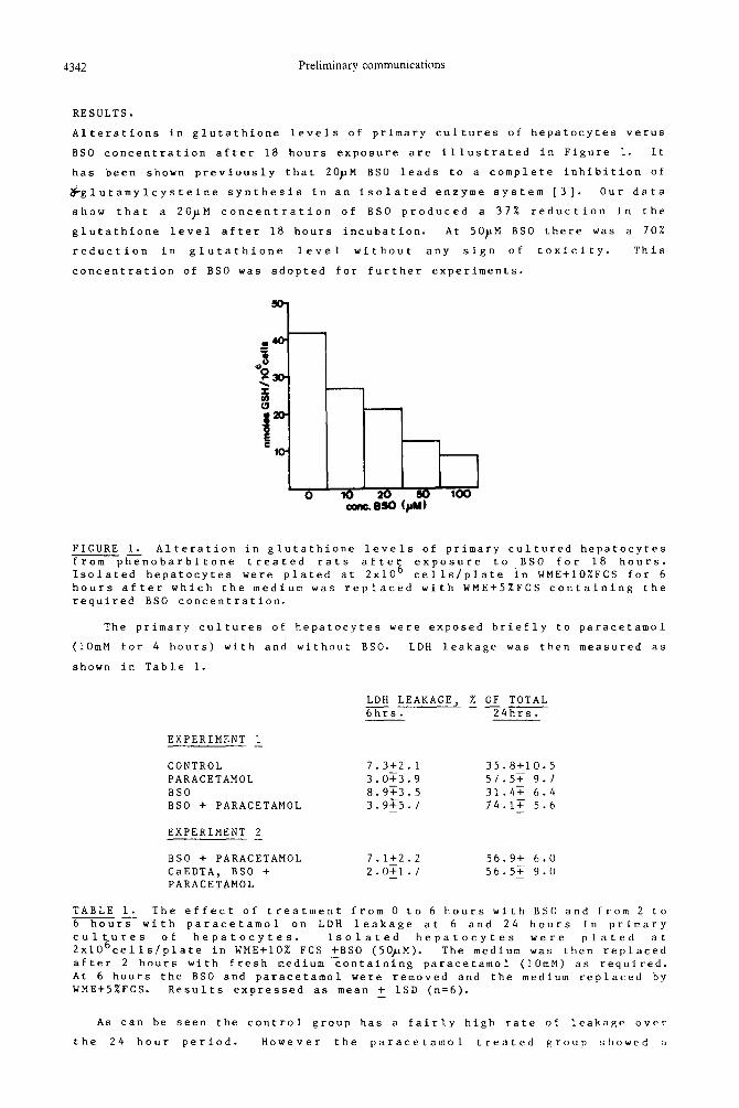
Alterations in glutathione levels of primary cultures of hepatocytes verus
BSO concentration after 18 hours exposure are illustrated in Figure 1. It
has been shown previously that 20pM BSO leads to a complete inhibition of
8glutamylcysteine synthesis in an isolated enzyme system [3]. Our data
show that a 2OpM concentration of BSO produced a 37% reduction in the
glutathione level after 18 hours incubation. At 50pM BSO there was a 70%
reduction in glutathione level without any sign of toxicity. This
concentration of BSO was adopted for further experiments.
FIGURE 1. Alteration in glutathione levels of primary cultured hepatocytes fromph<nobarbitone treated rats after exposure to BSO for 18 hours. Isolated hepatocytes were plated at 2x10 6 cells/plate in WME+lO%FCS for 6 hours after which the medium was replaced with WME+5%FCS containing the required BSO concentration.
The primary cultures of hepatocytes were exposed briefly to paracetamol
(1OmM for 4 hours) with and without BSO. LDH leakage was then measured as
shown in Table 1.
LDH LEAKAGE, % OF TOTAL --- 6hrs. 24hrs.
EXPERIMENT 1 -
CONTROL 7.3+2.1 35.8+10.5 PARACETAMOL 3.oT3.9 57.5+ 9.7
BSO 8.9T3.5 31.4T 6.4 BSO + PARACETAMOL 3.9F5.7 74.lT 5.6 - -
EXPERIMENT 2 -
BSO + PARACETAMOL 7.1+2.2 56.9+ 6.0 CaEDTA, BSO + 2.0T1.7 56.57 9.0 - - PARACETAMOL
TABLE 1. The effect of treatment from 0 to 6 hours with BSO and from 2 to 6rswith paracetamol on LDH leakage at 6 and 24 hours in primary cultures of hepatocytes. Isolated hepatocytes 2x10⁶cells/plate in WME+lO% FCS +BSO (50pM).
were plated at The medium was then replaced
after 2 hours with fresh medium containing paracetamol (IOmM) as required. At 6 hours the BSO and paracetamol were removed and the medium replaced by WME+5%FCS. Results expressed as mean + 1SD (n=6). -
As can be seen the control group has a fairly high rate of leakage over
the 24 hour period. However the paracetamol treated group showed a
Preliminary communications 4343
significantly higher leakage than the control group. The cells differ
markedly in that the paracetamol treated cultures did not form the adherent
syncitial layers found in the control plates (41.
CaEDTA protects suspended hepatocytes from paracetamol injury but we
found that 4mM CaEDTA was unable to arrest the LDH leakage induced by BSO
and paracetamol. In other experiments we have shown that CaEDTA penetrates
suspended hepatocytes but not into cultured cells.
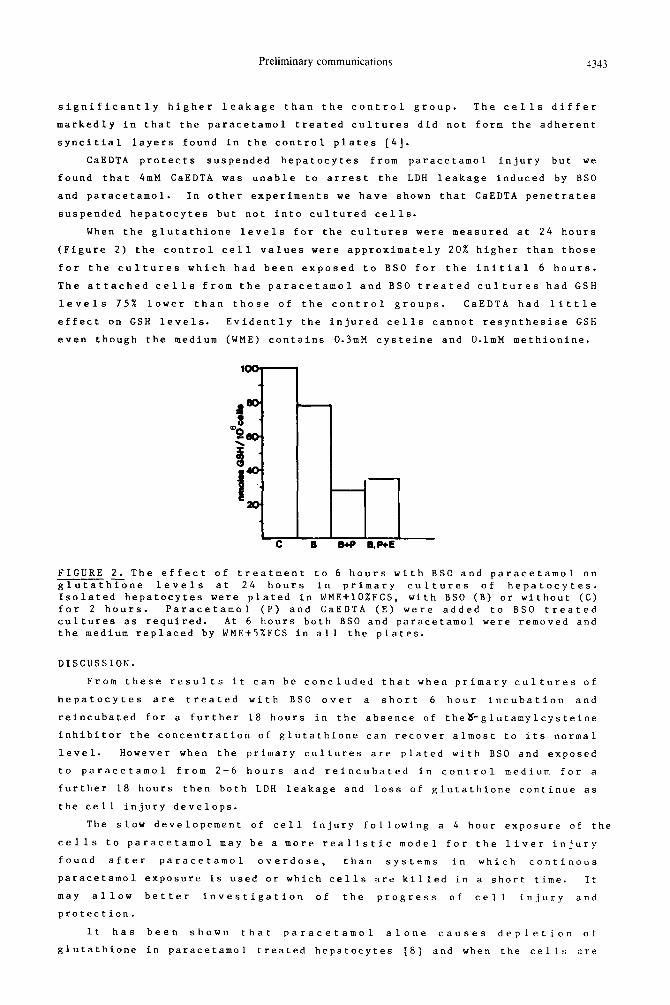
When the glutathione levels for the cultures were measured at 24 hours
(Figure 2) the control cell values were approximately 20% higher than those
for the cultures which had been exposed to BSO for the initial 6 hours.
The attached cells from the paracetamol and BSO treated cultures had GSH
levels 75% lower than those of the control groups. CaEDTA had little
effect on GSH levels. Evidently the injured cells cannot resynthesise GSH
even though the medium (WME) contains 0.3mM cysteine and O.lmM methionine.
FIGURE 2. The effect of treatment to 6 hours with BSO and paracetamol on ahzne levels at 24 hours in primary cultures of hepatocytes. Isolated hepatocytes were plated in WME+lO%FCS, with BSO (B) or without (C) for 2 hours. Paracetamol (P) and CaEDTA (E) were added to BSO treated cultures as required. At 6 hours both BSO and paracetamol were removed and the medium replaced by WME+5%FCS in all the plates.
DISCUSSION.
From these results it can be concluded that when primary cultures of
hepatocytes are treated with BSO over a short 6 hour incubation and
reincubated for a further 18 hours in the absence of the&glutamylcysteine
inhibitor the concentration of glutathione can recover almost to its normal
level. However when the primary cultures are plated with BSO and exposed
to paracetamol from 2-6 hours and reincubated in control medium for a
further 18 hours then both LDH leakage and loss of glutathione continue as
the cell injury develops.
The slow developement of cell injury following a 4 hour exposure of the
cells to paracetamol may be a more realistic model for the liver injury
found after paracetamol overdose, than systems in which continous
paracetamol exposure is used or which cells are killed in a short time. It
may allow better investigation of the progress of cell injury and
protection.
It has been shown that paracetamol alone causes depletion of
glutathione in paracetamol treated hepatocytes [8] and when the cells are
4344 Preliminary communications
incubated in a medium devoid of sulphur amino acids further resynthesis is
prevented. In our model the paracetamol would appear to be reducing the
resting glutathione level and the BSO also prevents resynthesis, causing a
very rapid glutathioine depletion followed by deleterious effects on cell
viability. Although CaEDTA penetrates and protects suspended hepatocytes
against paracetamol toxicity [2] it is unable to protect the hepatocytes
under the circumstances of attached culture where CaEDTA does not penetrate
the cells.
REFERENCES
1) D. Acosta, D.C. Anuforo and R.V. Smith. Toxicol. Appl. Pharmacol 53,
306 6 (1980).
2) D. Beales, D.P. Hue and A.E.M. McLean. Biochem. Pharmacol. 6, 19
(1985).
3) O.W. Griffith and A. Meister J. Biol. Chem.254, 7558 (1979).
4) J.L. Devalia, R.C. Ogilvie and A.E.M. McLean. Biochem. Pharmacol. 2,
3745 (1982).
5) P. Moldeus, J. Hogberg and S. Orrenius. Meth. Enzymol.2, 60 (1978).
6) G.M. Richards. Analyt. Biochem. 51, 36 (1974).
7) O.W. Griffith. Analyt. Biochem. 106, 207 (1980).
8) P. Moldeus. Biochem. Pharmacol. 27, 2859 (1978).