hemolysis and pre-analytical variables · what is hemolysis? the breakage of the red blood cell’s...
TRANSCRIPT
C23
CRI and COLA do not endorse, directly or indirectly, the presentations given at this conference or the products or services provided by the exhibiting vendors. Presentations are intended to be free of bias. The use of any particular product is for demonstration purposes only, and does not imply an endorsement of the product by the presenter or the sponsors of the symposium. © 2017 CRI
Hemolysis and Pre-analytical Variables
Kathleen Finnegan, MS MT(ASCP)SHCM
Clinical Associate Professor and Program Director of the Phlebotomy Training Program, Dept. of Clinical Laboratory Sciences,
Stony Brook University, New York
DESCRIPTION:
Hemolysis results when red blood cells are damaged or destroyed releasing hemoglobin. Hemolyzed specimens can result from patient conditions but most often result from procedural errors in specimen collection and handling. Numerous factors are associated with pre-analytical errors. These errors can compromise specimen integrity and impact patient care.
OBJECTIVES:
At the end of the session, participants will be able to:
Recognize the cause of hemolysis.
Discuss how to prevent hemolysis
Determine how to reduce pre-analytical variables and specimen rejection
Thursday April 6, 2017
Kathleen Finnegan MS MT(ASCP)SH
Clinical Associate Professor
Stony Brook University, New York
Objectives Recognize the cause of hemolysis
Discuss how to prevent hemolysis
Determine how to reduce pre‐analytical variables and specimen rejection
What is Hemolysis? The breakage of the red blood cell’s membrane
Causes the release of hemoglobin and other internal components into the fluid
Visually detected by a pink to red color in the serum or plasma
A common occurrence
Compromises laboratory testing
Hemolysis Can occur from two sources:
In‐vivo usually do to pathologic conditions
Autoimmune Hemolytic Anemia
Transfusion Reaction
Toxins and Poisons
In‐vitro
Improper collection
Specimen processing
Specimen transport

Degree of Hemolysis
Causes of Hemolysis Specimen Collection
Prolonged tourniquet application
Vein size and trauma
Needle size
Under filled tubes
Slow flow of blood into collection tubes
Alcohol preparation
Syringe collection and transfer
Cather Collection
Milking a skin puncture
Causes of Hemolysis Specimen Processing
Vigorous mixing or shaking of the tubes
Not allowing the serum tube to clot properly
Use of applicator sticks to dislodge fibrin
Prolonged contact of serum or plasma with cells
Exposure of excessive heat or cold
Don’t over centrifuge or centrifuge at high speeds
Causes of Hemolysis Specimen Handling
Mechanical trauma during transport of a pneumatic tube system
Do not subject the specimen to significant jarring
Protect the specimen for transport
Don’t expose the specimen to extreme temperatures
Effect of Hemolysis Increase of certain analytes
Interference in the test method
Degree of hemolysis varies with the interference of laboratory results
Quality Specimens = Quality Test Results
Hemolysis
Destruction of red blood cells with the release of hemoglobin
Analytes affected: Potassium Magnesium Bilirubin
Phosphorus LD AST
ALT Ammonia Folate
Coagulation Iron Total Protein
CK Cholesterol Triglycerides
Troponin T Sodium Calcium
Magnesium Haptoglobin Amylase
Effected AnalytesIncrease
Potassium
Magnesium
Iron
LD
Phosphorus
Ammonia
Total Protein
Calcium
Decrease
RBC count
Hemoglobin
Hematocrit
Coagulation Factors
Haptoglobin
Troponin T
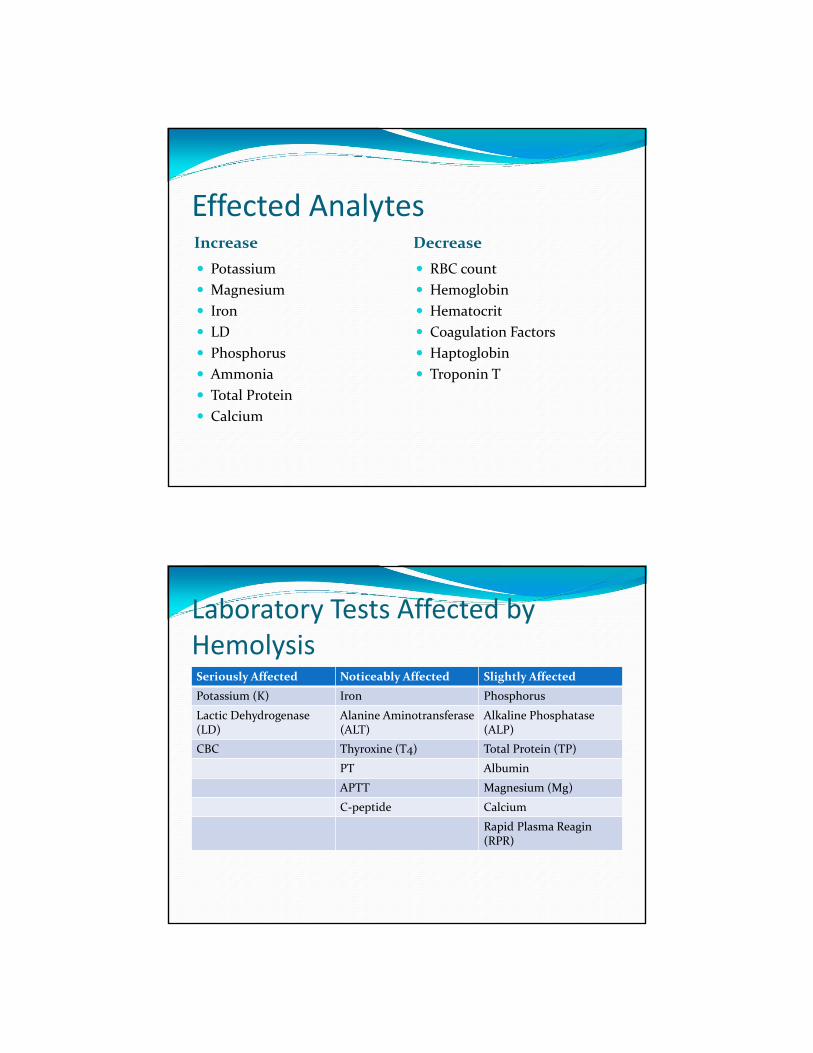
Laboratory Tests Affected by HemolysisSeriously Affected Noticeably Affected Slightly Affected
Potassium (K) Iron Phosphorus
Lactic Dehydrogenase (LD)
Alanine Aminotransferase (ALT)
Alkaline Phosphatase(ALP)
CBC Thyroxine (T4) Total Protein (TP)
PT Albumin
APTT Magnesium (Mg)
C‐peptide Calcium
Rapid Plasma Reagin (RPR)
Hemoconcentration
Intravascular pressure build up, allows analytes to escape through the capillary walls and into the tissues
This results with an concentration of analytes in the circulatory system
Biochemical changes take place in the trapped blood
14
HemoconcentrationPossible Cause and Effects
Leaving the tourniquet on the patient arm longer than two minutes.
Asking or allowing the patient to pump their fist.
Changes in CBC results.
Changes in results of blood chemistries including Iron, Potassium, Magnesium, Calcium, Enzymes and Troponin T tests.
Prolonged Tourniquet Application
Potassium
Magnesium
Ionic Calcium
Albumin/Protein
Decrease in pH
WBC and Hemoglobin
Cholesterol and Triglycerides
Coagulation Factors
Iron
Ammonia
Hemoconcentration And Hemolysis
Hemoconcentration
can
lead to Hemolysis
Ways to Prevent Hemoconcentration Ask the patient to release the fist when blood appears in the first tube
Choose the appropriate vein
Do not allow the patient to continue pumping the fist
Do not massage the area
Do not slap the area
Do not probe or redirect the needle in search of a vein
Release the tourniquet within 1 minute
Rejected SpecimensPreanalytical phase highly susceptible to error
Hemolyzed specimens account for 60% of rejected specimens
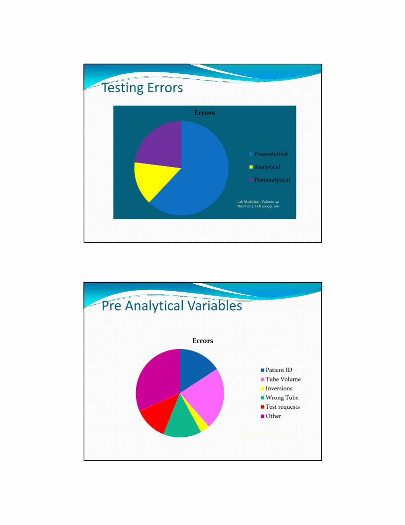
Testing Errors
Errors
Preanalytical
Analytical
Postanalytical
Lab Medicine , Volume 40 Number 2, Feb 2009 p. 106
Pre Analytical Variables
Errors
Patient ID
Tube Volume
Inversions
Wrong Tube
Test requests
Other
Lab Medicine , Volume 40 Number 2, Feb 2009 p. 106
Specimen Rejection Identity discrepancies
Inadequate volume of blood
Hemolyzed specimens
Incorrect tubes
Specimens improperly transported
Anticoagulated specimens that contain clots
Contaminated Specimens
Timed sample drawn at the incorrect time
Importance of Preventing Hemolysis Impacts laboratory tests
Higher rate of rejected specimens
Usually requires repeat collection
Delayed diagnosis
Delayed treatment
Additional discomfort for the patient
Additional cost
Frustration for the laboratory

Phlebotomy Practices
Ordering
Patient Identification
Anticoagulant
Tourniquet
Traumatic Phlebotomy
Tube Volume
Tube Inversion
Pre‐AnalyticalOrdering Collection Collection Processing Transporting
Patient Identification
Labeling
Diet
Exercise
Posture
Additives
Vein Selection
Order of Draw
Cleansing
Tourniquet
Timing
Specimen Volume
Short Draw
Inverting
IV Lines and Line Draws
Mastectomy
Edema
Separation
Centrifugation
Hemolysis
Lipemia
Light
Evaporation
Temperature
Timing
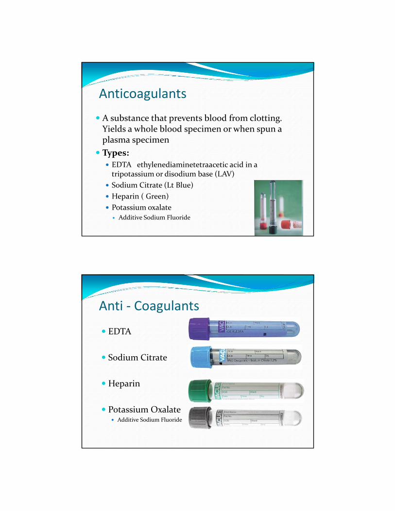
Anticoagulants
A substance that prevents blood from clotting. Yields a whole blood specimen or when spun a plasma specimen
Types:
EDTA ethylenediaminetetraacetic acid in a tripotassium or disodium base (LAV)
Sodium Citrate (Lt Blue)
Heparin ( Green)
Potassium oxalate Additive Sodium Fluoride
Anti ‐ Coagulants
EDTA
Sodium Citrate
Heparin
Potassium Oxalate Additive Sodium Fluoride
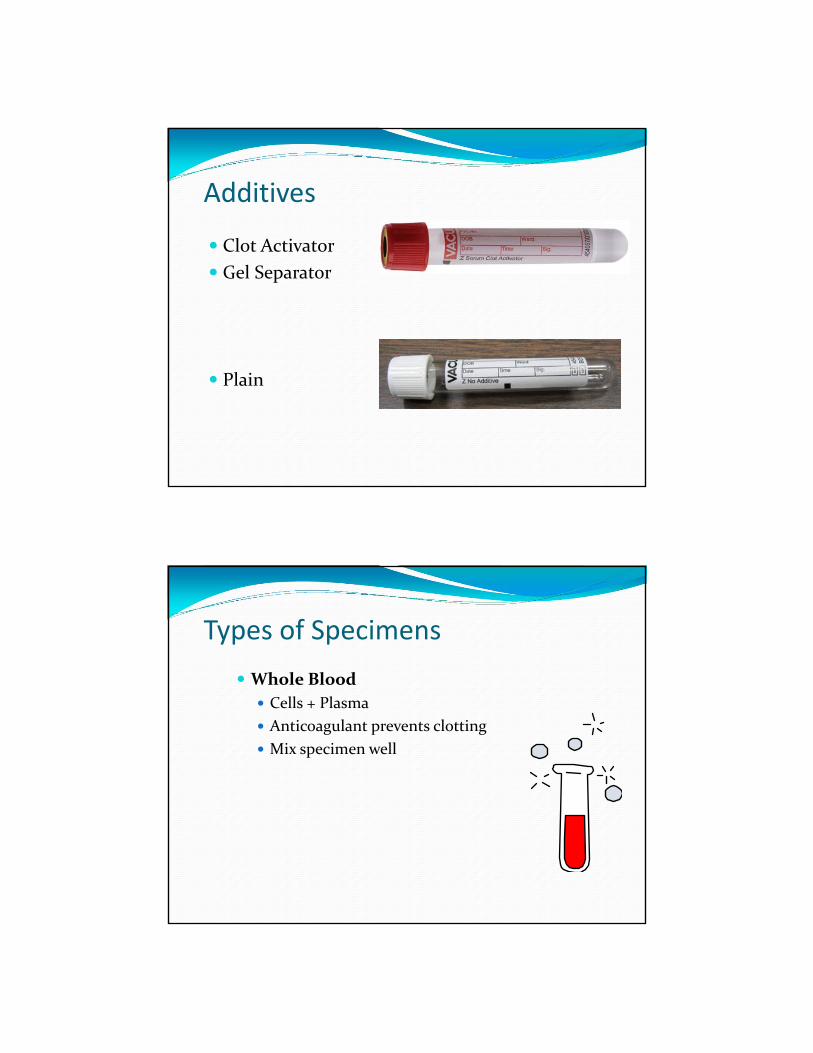
Additives
Clot Activator
Gel Separator
Plain
Types of Specimens
Whole Blood
Cells + Plasma
Anticoagulant prevents clotting
Mix specimen well
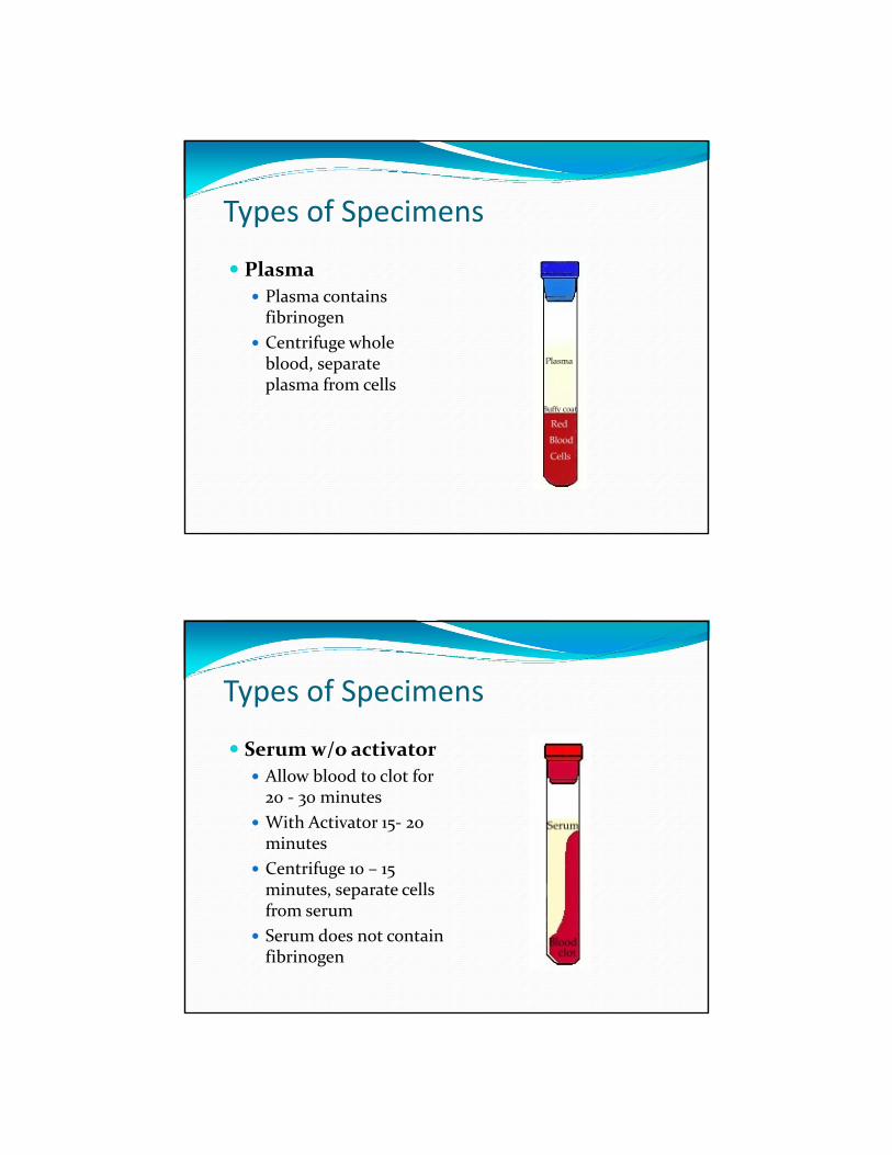
Types of Specimens
Plasma
Plasma contains fibrinogen
Centrifuge whole blood, separate plasma from cells
Types of Specimens
Serum w/o activator
Allow blood to clot for 20 ‐ 30 minutes
With Activator 15‐ 20 minutes
Centrifuge 10 – 15 minutes, separate cells from serum
Serum does not contain fibrinogen
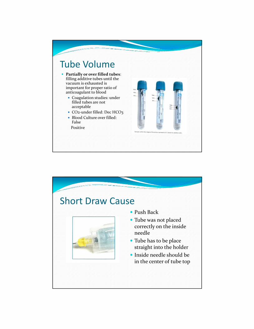
Tube Volume Partially or over filled tubes:
filling additive tubes until the vacuum is exhausted is important for proper ratio of anticoagulant to blood
Coagulation studies: under filled tubes are not acceptable
CO2‐under filled: Dec HCO3
Blood Culture over filled: False
Positive
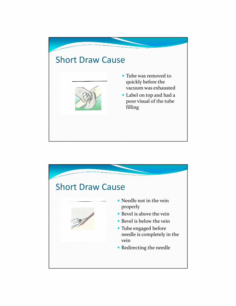
Short Draw Cause Push Back
Tube was not placed correctly on the inside needle
Tube has to be place straight into the holder
Inside needle should be in the center of tube top
Short Draw Cause
Tube was removed to quickly before the vacuum was exhausted
Label on top and had a poor visual of the tube filling
Short Draw Cause
Needle not in the vein properly
Bevel is above the vein
Bevel is below the vein
Tube engaged before needle is completely in the vein
Redirecting the needle
Short Draw Cause
Tiny veins
Removing the tourniquet to soon
Poor circulation
Veins that are hard
Damaged veins
Short Draw Cause
When using a butterfly a “Blank “ or “Discard “tube should be drawn for Coagulation
There is dead space in the butterfly tubing
The vacuum pulls air into the tube and the correct fill is not obtained
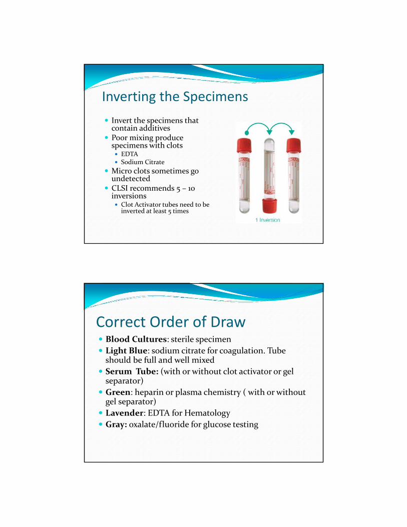
Inverting the Specimens
Invert the specimens that contain additives
Poor mixing produce specimens with clots EDTA Sodium Citrate
Micro clots sometimes go undetected
CLSI recommends 5 – 10 inversions Clot Activator tubes need to be inverted at least 5 times
Correct Order of Draw Blood Cultures: sterile specimen
Light Blue: sodium citrate for coagulation. Tube should be full and well mixed
Serum Tube: (with or without clot activator or gel separator)
Green: heparin or plasma chemistry ( with or without gel separator)
Lavender: EDTA for Hematology
Gray: oxalate/fluoride for glucose testing

Additive Contamination
Tests affected by EDTA contamination
Calcium PT Potassium
APTT Sodium Serum Iron
Tests affected by Heparin
PT aPTT ACT
Tests affected by Potassium Oxalate
Potassium RBC Morphology
Vein Selection
Antecubital Region
Median Cubital
Cephalic
Basilic Volar Venous network (hand)
Basilic is the last choice and should not be used because of possible nerve damage
Specimen Processing
Transportation
Deliver in a timely manner
Processing
Tubes should be in upright at room temperature
Separation
Prolonged contact of cells causes changes
Centrifugation
Specimens should be fully clotted
Specimen Clotting
Serum Specimens in Glass Tubes 20 – 30 minutes
Plastic Tubes 30 – 45 minutes
Clot Activators 15 – 20 minutes
Tubes should be upright at room temperature while clotting
Separation
Plasma Specimens One hour after collection
Serum Specimens Two hours after collection
Prolonged contact with cells Increase: CK, Lactate, LD , Ammonia
Decrease in glucose, Bicarbonate, Acid phosphatase
Plasma/Serum Heparinized plasma is preferred over serum for potassium tests
When blood clots potassium is released from the cells into the serum
Can falsely elevate the potassium results
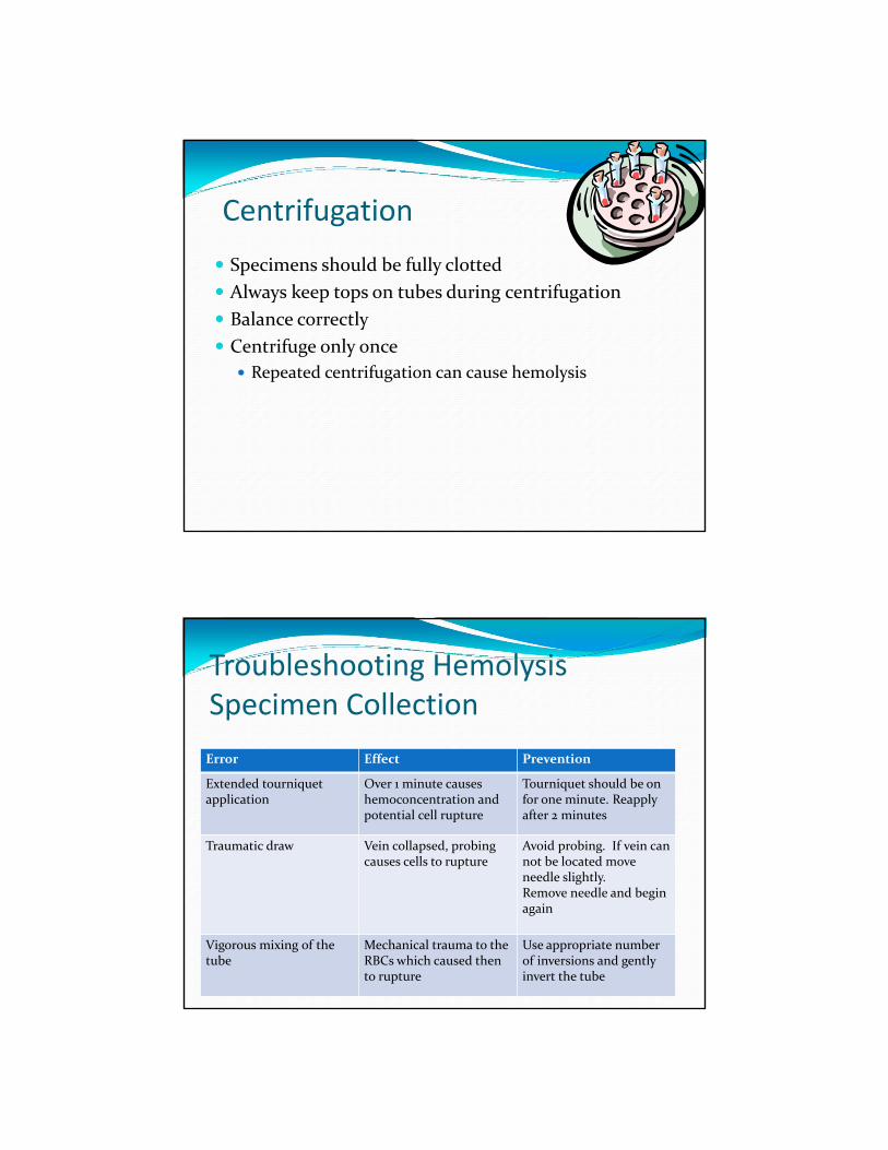
Centrifugation
Specimens should be fully clotted
Always keep tops on tubes during centrifugation
Balance correctly
Centrifuge only once
Repeated centrifugation can cause hemolysis
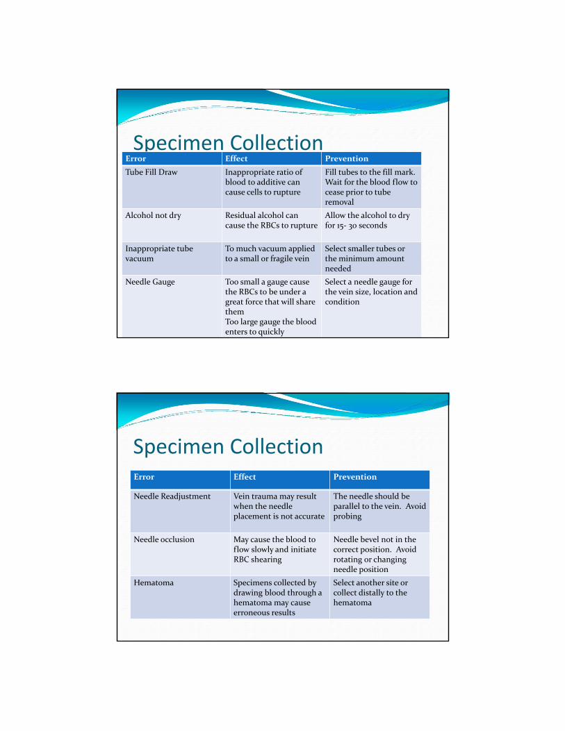
Troubleshooting HemolysisSpecimen Collection
Error Effect Prevention
Extended tourniquet application
Over 1 minute causes hemoconcentration and potential cell rupture
Tourniquet should be on for one minute. Reapply after 2 minutes
Traumatic draw Vein collapsed, probing causes cells to rupture
Avoid probing. If vein can not be located move needle slightly.Remove needle and begin again
Vigorous mixing of the tube
Mechanical trauma to the RBCs which caused then to rupture
Use appropriate number of inversions and gently invert the tube
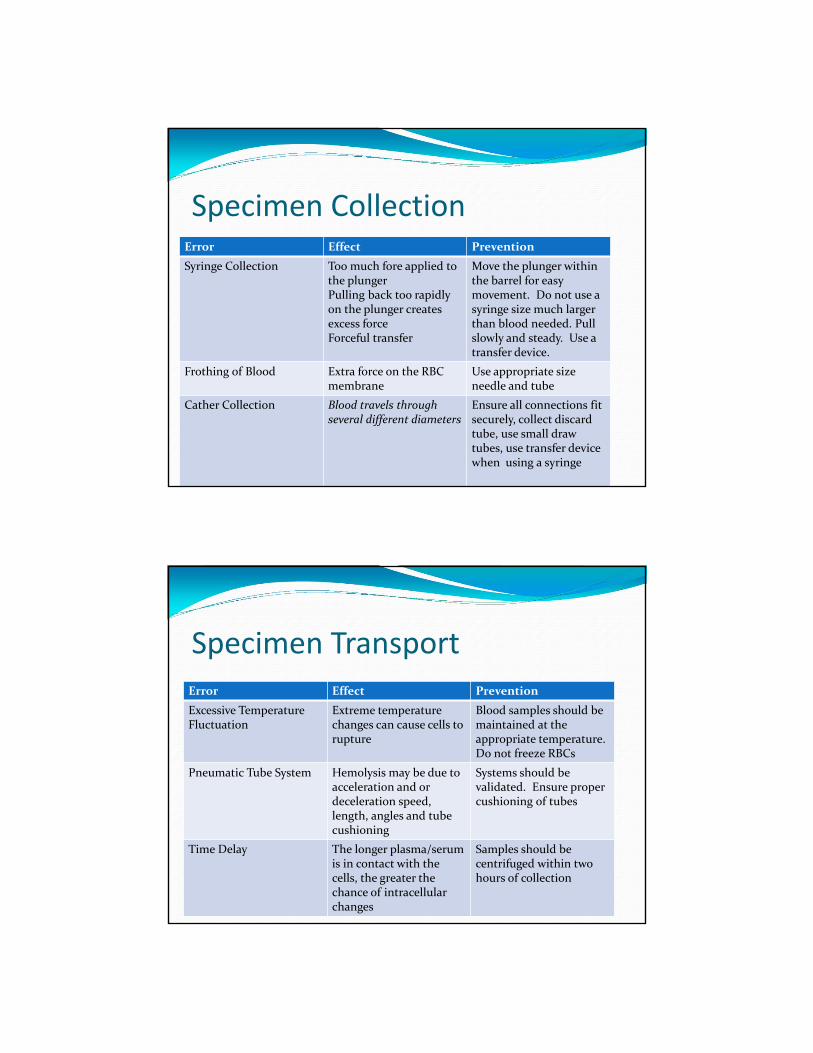
Specimen CollectionError Effect Prevention
Tube Fill Draw Inappropriate ratio of blood to additive can cause cells to rupture
Fill tubes to the fill mark.Wait for the blood flow to cease prior to tube removal
Alcohol not dry Residual alcohol can cause the RBCs to rupture
Allow the alcohol to dry for 15‐ 30 seconds
Inappropriate tube vacuum
To much vacuum applied to a small or fragile vein
Select smaller tubes or the minimum amount needed
Needle Gauge Too small a gauge cause the RBCs to be under a great force that will share themToo large gauge the blood enters to quickly
Select a needle gauge for the vein size, location and condition
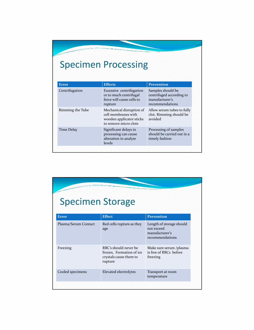
Specimen CollectionError Effect Prevention
Needle Readjustment Vein trauma may result when the needle placement is not accurate
The needle should be parallel to the vein. Avoid probing
Needle occlusion May cause the blood to flow slowly and initiate RBC shearing
Needle bevel not in the correct position. Avoidrotating or changing needle position
Hematoma Specimens collected by drawing blood through a hematoma may cause erroneous results
Select another site or collect distally to the hematoma
Specimen CollectionError Effect Prevention
Syringe Collection Too much fore applied to the plungerPulling back too rapidly on the plunger creates excess forceForceful transfer
Move the plunger within the barrel for easy movement. Do not use a syringe size much larger than blood needed. Pull slowly and steady. Use a transfer device.
Frothing of Blood Extra force on the RBC membrane
Use appropriate size needle and tube
Cather Collection Blood travels through several different diameters
Ensure all connections fit securely, collect discard tube, use small draw tubes, use transfer device when using a syringe
Specimen TransportError Effect Prevention
Excessive Temperature Fluctuation
Extreme temperature changes can cause cells to rupture
Blood samples should be maintained at the appropriate temperature. Do not freeze RBCs
Pneumatic Tube System Hemolysis may be due to acceleration and or deceleration speed, length, angles and tube cushioning
Systems should be validated. Ensure proper cushioning of tubes
Time Delay The longer plasma/serum is in contact with the cells, the greater the chance of intracellular changes
Samples should be centrifuged within two hours of collection
Specimen Processing
Error Effects Prevention
Centrifugation Excessive centrifugationor to much centrifugal force will cause cells to rupture
Samples should be centrifuged according to manufacturer’s recommendations
Rimming the Tube Mechanical disruption of cell membranes with wooden applicator sticks to remove micro clots
Allow serum tubes to fully clot. Rimming should be avoided
Time Delay Significant delays in processing can cause alteration in analyte levels
Processing of samples should be carried out in a timely fashion
Specimen StorageError Effect Prevention
Plasma/Serum Contact Red cells rupture as they age
Length of storage should not exceed manufacturer’s recommendations
Freezing RBC’s should never be frozen, Formation of ice crystals cause them to rupture
Make sure serum /plasma is free of RBCs before freezing
Cooled specimens Elevated electrolytes Transport at room temperature
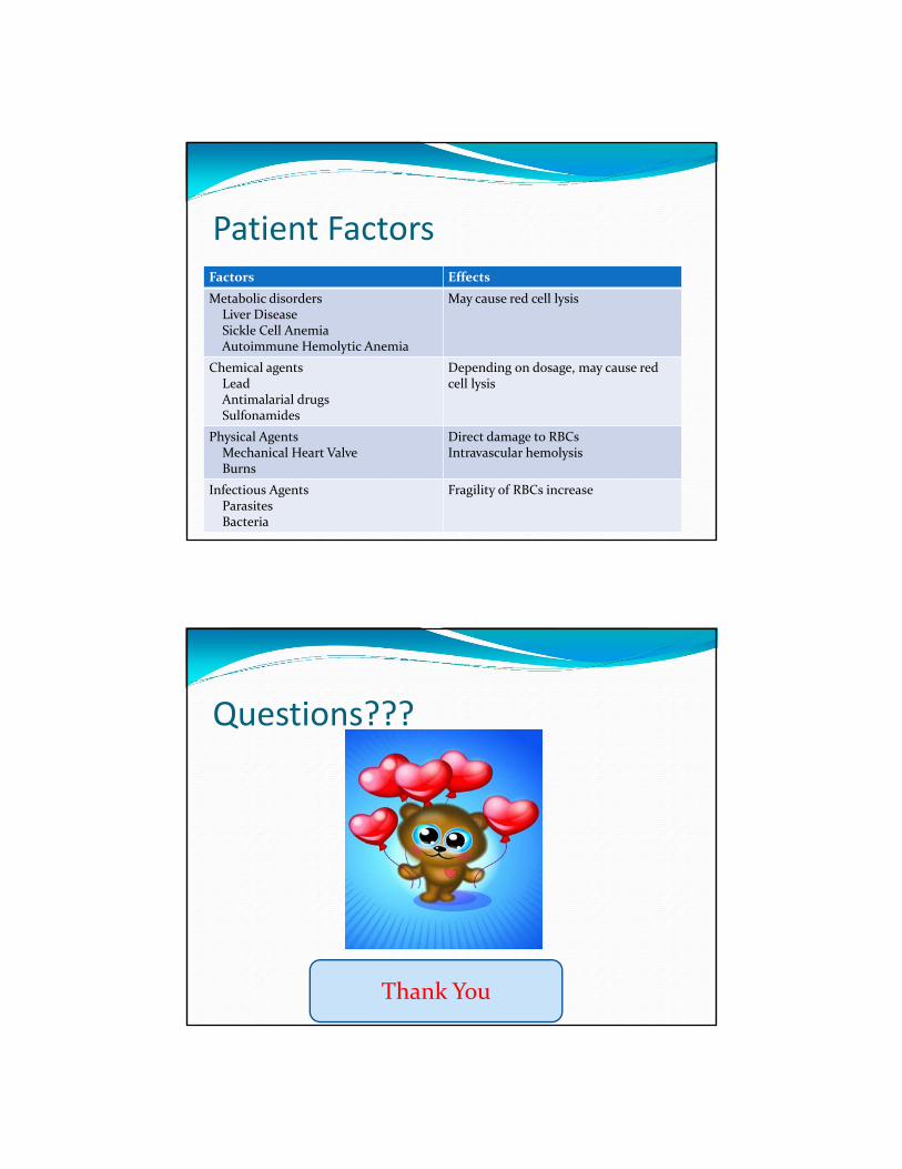
Patient FactorsFactors Effects
Metabolic disordersLiver DiseaseSickle Cell AnemiaAutoimmune Hemolytic Anemia
May cause red cell lysis
Chemical agentsLeadAntimalarial drugsSulfonamides
Depending on dosage, may cause red cell lysis
Physical AgentsMechanical Heart ValveBurns
Direct damage to RBCsIntravascular hemolysis
Infectious AgentsParasitesBacteria
Fragility of RBCs increase
Questions???
Thank You