hematology standard operating procedures (sops)
TRANSCRIPT

Hematology Standard Operating Procedures (SOPs)
دائشة ختبشاث بن د استشف١اث -اإلداسة اعات ستشف١اث -صاسة اظحت
1
Hematology Department
STANDARD OPERATING PROCEDURES
(SOPs)
April 2016

Hematology Standard Operating Procedures (SOPs)
دائشة ختبشاث بن د استشف١اث -اإلداسة اعات ستشف١اث -صاسة اظحت
2
اهداء:
انتحانم انطبة يهنة واىل أرواح شهدائنا خاصة يف شهدائنا األبزاراىل أرواح
انشهد/ راي انسهىت
انشهد/ حساو راضو
وكذنك اىل روح انشيم/ حساو أبى مشانة
روح انشيم/ ادب عهىا و
واىل يزضاا انشيالء/ يهند انشنط
املكزوبىنىج( SOPsوانشيم حمىد اجلزو )وهى عضى فزق اعداد
سال اهلل هلى انشفاء انعاجم ونهشهداء انفزدوص األعهى.
االساة ي سيالت وسيالء اىل كم ي نت اىل هذه املهنة
اءهندي هذا انعم ريشا نهىف

Hematology Standard Operating Procedures (SOPs)
دائشة ختبشاث بن د استشف١اث -اإلداسة اعات ستشف١اث -صاسة اظحت
3
شكز وتقدز
ـــــذ٠ت ــــ اشــــىش اتمــــذ٠شــــــ فش٠مـــ ــــشاث بــــن د استشــــف١اث بدض٠ ك عــــ ختب
ــــ ـــ، الدددور/ يو ف ادددا ايددد الدددافة الرحددد ل اي ال ددد ااألخ ـوـــ ـــ إ األخ الدددور/ يو إ
دـــاص " ـــا لـــذ ـــ دعـــ لســـ١ ـــ ا ـــ اعبدددو الف حدددا ال دددعاا ادددوفا عدددع ال / ددد حع
االاددددد/عكو ادددددعرا ايددددد ادددددببعتا ادددددوفا ا دددددا ا /بددددداا لي ددددد ـــــ إـــــزا اعـــــ وـــــزه
ا لذ دع شسة ف ١ع شاح اداص زا اع . ال / حع اعيقعا
اـــ وـــ ـــ فش٠ـــك ااعـــذاد فش٠ـــك اشا عـــت ـــا بـــز ـــ اتمـــذ٠ش وـــا تمـــذ بدض٠ـــ اشـــىش
ـــزا اع ـــا ـــ ا ـــ ال ـــذ ـــن د ـــشاث ب ـــ ختب ـــا١ ف ـــة اع ـــع اا ـــ ١ ـــزه ا ـــ و
.اإل شاءاثاستشف١اث از٠ س١ى اذس اشائذ ف لطب١ك ز
ؤسســـت اعـــ اطبـــ فســـط١١١ شعـــا٠ت باعـــت إـــوـــزه تمـــذ بدض٠ـــ اشـــىش اعشفـــا
زا اذ١.

Hematology Standard Operating Procedures (SOPs)
دائشة ختبشاث بن د استشف١اث -اإلداسة اعات ستشف١اث -صاسة اظحت
4
بس هللا اشح اشح١
لاي لعا: ) ل اعا فس١ش هللا عى سس اؤ( طذق هللا اعظ١
األ ة األ اث اضالء األواس...
اسال ع١ى سحت هللا بشوال..... أا بعذ
ــ ــ ــذ٠ى ) د١ ــ١ أ٠ ــ ــ داعــ ســشس أ أيــع ب ــت احــذ( ا شق افحطــاث اخبش٠
(Standard Operating Procedure SOPs) اــز ٠تنــ ل١مــاي لفظــ١الي طــشق عــ
افحطاث ات لمذا ختبشاث بن د استشف١اث، ره أ لظ١ اعـ يـا دلـ
ف ١ع اب ذاث اختبشاث بن اذ.
لأو١ـذاي لشسـ١خاي بـذأ اعـ بـشر افش٠ـك ااحـذ ل١ـك لح١ـذ ـشق لأل احا ت إ ـزا اعـ
وافــت اعــ ابــ عــ أســو ع١ــت اــز لتدــ صاسة اظــحت اإلداسة اعاــت ستشــف١اث فــ
اح اع لطعاي شل بت اتحا١ اطب١ت اطتي، بددة اخذاث اطب١ت امذت ف اصاسة
عا. بشى
وا ٠عتبش زا اذ١ األي احذ د١ع ختبشاث بن د استشف١اث اـز سـ١ى ـ بشـ١ ت
ــ أ ــ ااسلمــاء باخــذاث هللا األــش اا٠دــاب عــ ــدة اخــذاث اخبش٠ــت دــ األداء افــ
امذت ا .
ــ ــت ا ٠ســع إا أ ألمــذ بخــاض اشــىش اتم ذ٠ش فش٠ــك عــ دائــشة ختبــشاث بــن د فــ اا٠
استشف١اث ١ع سا ف اداص زا اع ساءي ف اإلعذاد أ اشا عت أ اطباعت اإل شاج.
حا ال عا . عبو الف
اوفا عع ال / حع
Hematology Standard Operating Procedures (SOPs)
دائشة ختبشاث بن د استشف١اث -اإلداسة اعات ستشف١اث -صاسة اظحت
5
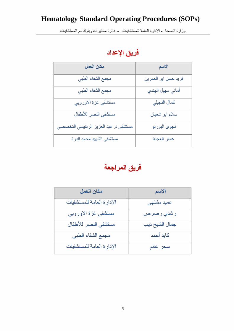
افق اإلعوا ف
اكعت الب االام
اطب اشفاء دع فش٠ذ حس اب اعش٠
اطب اشفاء دع أا س١ اذ
األسبة غضستشف واي اد١
أل فاي ستشف اظش سال اب شعبا
اتخظظ اشت١سد. عبذ اعض٠ض ستشف د ابس
اذسة اش١ذ حذ ستشف عاس اعدت
فافق ال ااجب
االام اكعت الب
ع١ذ شت اإلداسة اعات ستشف١اث
سشذ سطشص ستشف غضة ااسب
أل فاي ستشف اظش اي اش١خ د٠ب
وا٠ذ أحذ دع اشفاء اطب
سحش غا اإلداسة اعات ستشف١اث

Hematology Standard Operating Procedures (SOPs)
دائشة ختبشاث بن د استشف١اث -اإلداسة اعات ستشف١اث -صاسة اظحت
6
Contents
S No Subject SOP NO.
1 ABX Micros ES60 Hematology Analyzer SP 01
2 Sysmex KX-21 Hematology Analyzer SP 02
3 Cell-Dyn 1800 Hematology Analyzer
SP 03
4 CELL DYN 3500-3700 Hematology Analyzer SP 04
5 ACL Automated Coagulation Analyzer (Factor Deficient
Plasma VIII, IX, XI, XII Tests- HemosIL®)
SP 05
6 ACL Automated Coagulation Analyzer
(PT-FIB/APTT Tests- HemosIL®)
SP 06
7 CoaLAB 1000 Automated Coagulation Analyzer SP 07
8 Thrombolyzer compact X
SP 08
9 Prothrombin Time (MANUAL TEST)
SP 09
10 Partial Thromboplastin Time (manual test)
SP 10
11 Bleeding time SP 11
12 Clotting time SP 12
13 Blood film (leishman's stain) SP 13
14 Blood Group (Slide Method) BIOTEC LABORAYORIES
SP 14
15 Erythrocyte Sedimentation Rate (Westergren technique)
SP 15
16 Osmotic Fragility Test SP 16
17 Cerebrospinal Fluid Cellular Examination SP 17
18 Blood Film \ (Giemsa Stain) Coral Clinical system SP 18
19 Reticulocytes Count \ (Brilliant cresyl blue) RAL. Diagnostics SP 19
20 Sickling test (Na-meta-bisulfate method) SP 20

Hematology Standard Operating Procedures (SOPs)
دائشة ختبشاث بن د استشف١اث -اإلداسة اعات ستشف١اث -صاسة اظحت
7
ABX Micros ES60 Hematology Analyzer
SOPs\ HGA \.......H/ Haem /01
Version: …1…………………………
Date effective: …………… …...…...
Copy number: ……………………
Head of department: …………………………
Quality Officer: ……………………………...
Director of : ………………………….............
Purpose & Definition:
The ABX Micros ES60 performs automated blood counts and requires no manual
operations for aspirating blood, dilutions, measuring, calculations, print-outs and computer
transfer of data. It measure the following 18 hematologic parameters: WBC , LYM% ,
LYM# , MON% , MON# , GRA% , GRA# , PDW* , PCT* , RBC , HGB , HCT , MCV ,
MCH , MCHC , RDW , PLT , MPV.
Responsibilities:
Haematology department personal are required to be knowledgeable of this procedure.
New employees are trained and assessed for competence before they can handle
patient sample.
The head of the department must resolve any problem with the process and difficulties
in using this SOP.
Specimen requirements:
About 2-3 ml of venous blood collected into EDTA tubes.
Specimens should be transported at room temperature 18 - 26ºC and can be store in the
refrigerator of 2 - 8ºC up to 6 hours. If stored in a refrigerator, samples should be returned
to room temperature, for approximately 30 minutes, before analysis.
Specimen reception:
Reception of request and sample should be recorded, and record time of reception. Pay
attention to sample identification and labeling of tubes.
Criteria for rejection haematology specimens
1. When the identification is missing /inadequate.
2. Insufficient quantity.
3. Inappropriate container.

Hematology Standard Operating Procedures (SOPs)
دائشة ختبشاث بن د استشف١اث -اإلداسة اعات ستشف١اث -صاسة اظحت
8
4. Inappropriate transport/storage.
5. Unknown duration of delay.
6. Clotted sample.
Equipment & Items required:
Diluent: ABX Minidil LMG (10L).
Cleaner: ABX Miniclean (1L).
Lyse: ABX Minilyse LMG (1L).
Abbreviations:
CBC: Complete blood count.
EDTA: Ethylene diamine tetra acetic acid
WBC: White Blood Cells
LYM%: Lymphocyte percentage
LYM#: Lymphocyte absolute value
MON%: Monocyte percentage
MON#: Monocyte absolute value
GRA%: Granulocyte percentage
GRA#: Granulocyte absolute value
RBC: Red Blood Cells
HGB: Hemoglobin
HCT: Hematocrit
MCV: Mean Corpuscular Volume
MCH: Mean Corpuscular Hemoglobin
MCHC: Mean Corpuscular Hemoglobin Concentration
RDW: Red cell Distribution Width
PLT: Platelets
MPV: Mean Platelet Volume
PDW*: Platelet Distribution Width
PCT*: Plateletcrit
NRBCs: Nucleated Red Blood Cells

Hematology Standard Operating Procedures (SOPs)
دائشة ختبشاث بن د استشف١اث -اإلداسة اعات ستشف١اث -صاسة اظحت
9
Procedure:
1. Check operation of the machine, ensuring it is clean and that all required supplies
are present in sufficient quantities.
2. Switch the instrument on by pressing the ON/OFF switch, located on the back of
the instrument.
3. The instrument performs an initialization phase for the internal electronics. Please
wait.
4. Once the initialization phase is complete, the ABX Micros ES60 OT/CT will
automatically run a startup cycle.
5. If the ABX does not automatically run a startup cycle after the initialization phase
is completed, press "Startup" button in the "Status" area to initiate a startup cycle.
6. Then, the instrument will perform a blank cycle for a reference blank count (an
analysis cycle based on reagents without any blood sample).
7. Check and verify that the reference blank counts do not exceed the following
parameter limits: WBC < 0.3, RBC < 0.02, HGB < 0.3, PLT < 10 then: Press "OK"
button to validate blank results.
8. Perform quality control analysis on 3 levels of control blood material (low, normal
and high) to verify that the instrument is performing within the specified ranges of
the quality control material.
9. Entering patient ID, sample ID, Patient name, etc
10. Follow the indications displayed in the "Sample analysis" dialog box to run the
analysis.
a. Mix the sample gently and thoroughly.
b. Remove the cap from the sample tube.
c. Place the sample beneath the sampling needle.
d. Raise up the tube so that the sampling needle lowers into the blood and
press the manual sample bar.
e. The analysis cycle will begin.
11. When the analysis is completed, the "Sample analysis" dialog box is closed and
results are displayed in the "Result display" menu for print out.
12. Dilute the sample if White blood cell counts ≥100,000 /mm3 and platelet counts
≥1,000,000 /mm3 are outside the linearity specifications of the instrument.

Hematology Standard Operating Procedures (SOPs)
دائشة ختبشاث بن د استشف١اث -اإلداسة اعات ستشف١اث -صاسة اظحت
11
Quality control procedures:
1. At the beginning of each work shift all parameters are tested with blood control.
2. The 3 levels include: Abnormal Low, Normal, Abnormal High
3. Controls are stored at 2-8ºC and brought to room temperature on a roller mixer before
use .
4. Controls are gently inverted many times according to the manufacturer’s instruction
before use.
5. From the RUN screen, press [SPECIMEN TYPE].
6. Use the arrow key on the keyboard to move the cursor to the appropriate QC file (i.e.,
low, normal or high) and press the [QC SPECIMEN] key.
7. Control values must be within three standard deviation, otherwise the measurement has
to be repeated, if the control still out of range:
a. Check operation of the machine, ensuring it is clean and that all required supplies
are present in sufficient quantities.
b. Check reagents for expiration dates and lot numbers. Ensure that all machine
lines are in appropriate receptacle where applicable. If this does not solve the
problem:
Prepare new control(s) and try again.
If the controls are still out, inform your supervisor to check the operator's manual,
or recalibrate instrument and If controls are still out,. Contact Medical Maintenance
where applicable, or servicing engineer.
8. All control data are managed using software that provides graphical reports (Levey-
Jennings graphs, and monthly cumulative histograms).
Linearity:
PARAMETER LINEAR RANGE
WBC (103/mm
3) 0.4 – 106.6
RBC (106/mm
3) 0.2 – 8.1
HGB (g/dl) 0.68 - 26
PLT (103/mm
3) (A) 13 – 2777
PLT (103/mm
3) (B) 13 - 4856
HCT (%) 2.0 - 80

Hematology Standard Operating Procedures (SOPs)
دائشة ختبشاث بن د استشف١اث -اإلداسة اعات ستشف١اث -صاسة اظحت
11
Limitations/ Interfering substance:
Verification of any "Abnormal" test result (including flagged results or results outside their
normal range) is due to the following listed:
WBC White Blood Cells (Leukocytes):
NRBC, Non-lysed Red Cells, Multiple myeloma, Hemolysis, Leukemia, Increased
turbidity, Chemotherapy, Cryoglobulins.
RBC Red Blood Cells (Erythrocytes):
High WBCs, Agglutinated red blood cells, Cold agglutinins.
HGB (Hemoglobin):
Turbidity of the blood sample which may be due to Elevated WBC or Elevated Lipids,
Fetal bloods mixed with maternal bloods may produce a falsely elevated hemoglobin
value.
PLT (Platelets):
Very small erythrocytes, Agglutinated red blood cells, Giant platelets in excessive
numbers, Chemotherapy, Hemolysis, RBC inclusions, Platelet agglutination.
Expected values:
Interpretation of the results:
Certain disease states are defined by an absolute increase or decrease in the number of a
particular type of cell in the bloodstream and many types of anemia.

Hematology Standard Operating Procedures (SOPs)
دائشة ختبشاث بن د استشف١اث -اإلداسة اعات ستشف١اث -صاسة اظحت
12
Reporting result:
According to lab policy.( automated printing or computerized)

Hematology Standard Operating Procedures (SOPs)
دائشة ختبشاث بن د استشف١اث -اإلداسة اعات ستشف١اث -صاسة اظحت
13
Sysmex KX-21 Hematology Analyzer
SOPs\ HGA \.......H/ Haem / 02
Version: 1……………………………
Date effective: …………… …...…...
Copy number: ……………………
Head of department: …………………………
Quality Officer: ……………………………...
Director of : ………………………….............
Purpose/Definition:
The KX-21 performs speedy and accurate analysis of 18 parameters in blood (Whole
WBC, LYM%, MXD%, NEUT% , LYM# , MXD#, NEUT# , RBC count, Hemoglobin,
HCT, MCV, MCH, MCHC, RDW-CV, RDW-SD, PLT, PDW, MPV, P-LCR)
The KX-21 employs three detector blocks and two kinds of reagents for blood analysis.
The WBC count is measured by the WBC detector block using the DC detection method.
The RBC count and platelets are taken by the RBC detector block, also using the DC
detection method. The HGB detector block measures the hemoglobin concentration using
the noncyanide hemoglobin method.
Responsibilities:
Haematology department personal are required to be knowledgeable of this procedure.
New employees are trained and assessed for competence before they can handle
patient sample
The head of the department must resolve any problem with the process and difficulties
in using this SOP.
Specimen requirements:
About 2-3 ml of venous blood collected into EDTA tubes.
Specimens should be transported at room temperature 18 - 26ºC and can be store in the
refrigerator of 2 - 8ºC up to 6 hours.. If stored in a refrigerator, samples should be returned
to room temperature, for approximately 30 minutes, before analysis.
Specimen reception:
Reception of samples should be recorded, and record time of reception. Pay attention to
sample identification and labeling of tubes.
Criteria for rejection haematology specimens

Hematology Standard Operating Procedures (SOPs)
دائشة ختبشاث بن د استشف١اث -اإلداسة اعات ستشف١اث -صاسة اظحت
14
1. When the identification is missing /inadequate.
2. Insufficient quantity
3. Inappropriate container
4. Inappropriate transport/storage
5. Unknown duration of delay
6. Clotted sample
Equipment & Items required:
Sysmex KX -21N analyzer
Reagent Storage Conditions
Sysmex KX-21 diluent 5 - 30ºC
Stromatolyser- KX 21 5 - 30ºC
CellClean (detergent) 1 - 30ºC
Abbreviations:
CBC: Complete blood count.
EDTA: Ethylene diamine tetra acetic acid
WBC: White Blood Cells
LYM%: Lymphocyte percentage
LYM#: Lymphocyte absolute value
MON%: Monocyte percentage
MON#: Monocyte absolute value
GRA%: Granulocyte percentage
GRA#: Granulocyte absolute value
RBC: Red Blood Cells
HGB: Hemoglobin
HCT: Hematocrit
MCV: Mean Corpuscular Volume
MCH: Mean Corpuscular Hemoglobin
MCHC: Mean Corpuscular Hemoglobin Concentration
RDW: Red cell Distribution Width
PLT: Platelets
MPV: Mean Platelet Volume

Hematology Standard Operating Procedures (SOPs)
دائشة ختبشاث بن د استشف١اث -اإلداسة اعات ستشف١اث -صاسة اظحت
15
PDW*: Platelet Distribution Width
PCT*: Plateletcrit
P-LCR: Platelet larger cell ratio
Procedures:
1. Check to see that the reagents needed for the number of the samples to be
processed for the day are available.
2. Turn ON the power switch on the right side of the unit. Self-check, auto rinse, and
background check will be automatically performed, and the "Ready" (ready for
analysis) will appear.
3. When auto rinse and background check are normally completed, "Ready" is
displayed.
4. Perform quality control analysis on 3 levels of control blood material (low, normal
and high) to verify that the instrument is performing within the specified ranges of
the quality control material.
5. If the result of quality control in acceptable range input your blood samples.
6. Input from the panel keyboard.
7. Press [SAMPLE No.] key in the Ready status.
8. Entering patient ID, sample ID, Patient name, etc
9. Press [ENTER] key, This will fix the sample No. and the status becomes ready for
analysis.
10. Mix the sample sufficiently before analysis.
11. Remove the plug while taking care not to allow blood scatter.
12. Set the tube to the sample probe, and in that condition, press the start switch.
13. when the LCD screen displays "Analyzing," remove the tube.
14. After that, the unit executes automatic analysis and displays the result on the LCD
screen.

Hematology Standard Operating Procedures (SOPs)
دائشة ختبشاث بن د استشف١اث -اإلداسة اعات ستشف١اث -صاسة اظحت
16
15. Then the unit turns to the ready status, becoming ready for analysis of the next
samples.
Quality control procedures:
1. At the beginning of each work shift, all parameters are tested with blood control.
2. The 3 levels include: Abnormal Low, Normal, Abnormal High
3. Controls are stored at 2-8ºC and brought to room temperature on a roller mixer
before use .
4. Controls are gently inverted eight times according to the manufacturer’s instruction
before use.
5. From the RUN screen, press [SPECIMEN TYPE].
6. Use the arrow key on the keyboard to move the cursor to the appropriate QC file
(i.e., low, normal or high) and press the [QC SPECIMEN] key.
9. Control values must be within three standard deviations, otherwise the measurement
has to be repeated, if the control still out of range:
a. Check operation of the machine, ensuring it is clean and that all required
supplies are present in sufficient quantities.
b. Check reagents for expiration dates and lot numbers. Ensure that all machine
lines are in appropriate receptacle where applicable, If this does not solve the
problem:
Prepare new control(s) and try again.
If the controls are still out, inform your supervisor to check the operator's manual,
or recalibrate instrument and If controls are still out,. Contact Medical Maintenance
where applicable, or servicing engineer.
7. All control data are managed using software that provides graphical reports (Levey-
Jennings graphs, and monthly cumulative histograms).
8. Dilute the sample if White blood cell counts ≥100,000 /mm3 and platelet counts
≥1,000,000 /mm3 are outside the linearity specifications of the instrument.

Hematology Standard Operating Procedures (SOPs)
دائشة ختبشاث بن د استشف١اث -اإلداسة اعات ستشف١اث -صاسة اظحت
17
Linearity:
Limitations/ Interfering substance:
The following is a list of possible substances that may interfere with the listed parameters.
1. WBC: platelet aggregation, giant platelets, nucleated RBCs, cryoglobulins, lyse-
resistant RBCs in patients with haemoglobinopathies, severe liver disease or neonates.
2. RBC: Cold agglutinins, severe micryocytosis, fragmented RBCs, large numbers of
giant platelets, in vitro haemolysis.
3. HGB: Lipemia, abnormal proteins in blood plasma, severe leukocytes (above
100,000/µl). The effect of abnormal proteins and Lipemia may be removed by plasma
replacement or plasma blank procedures.
4. HCT: Cold agglutinins, leukocytosis (above 100,000/µl), abnormal red cell fragility.
5. PLT: Pseudothrombocytopenia, platelet aggregation, increased micrcrocytosis,
megalocyttic platelets
6. Low sample volume of <1 mL may dilute patient samples with EDTA in the collection
tube giving falsely low results. If a low sample volume is expected, use a pediatric
EDTA tube; fill to the second line and mix well.
7. Dilute the sample if White blood cell counts ≥100,000 /mm3 and platelet counts
≥1,000,000 /mm3 are outside the linearity specifications of the instrument

Hematology Standard Operating Procedures (SOPs)
دائشة ختبشاث بن د استشف١اث -اإلداسة اعات ستشف١اث -صاسة اظحت
18
Expected values:
Interpretation of the results:
Certain disease states are defined by an absolute increase or decrease in the number of a
particular type of cell in the bloodstream and many types of anemia.
Reporting result:
According to lab policy.( automated printing or computerized)

Hematology Standard Operating Procedures (SOPs)
دائشة ختبشاث بن د استشف١اث -اإلداسة اعات ستشف١اث -صاسة اظحت
19
Cell-Dyn 1800 Hematology Analyzer
SOPs\ HGA \.......H\ Haem \ 03
Version: 1……………………………
Date effective: …………… …...…...
Copy number: ……………………
Head of department: …………………………
Quality Officer: ……………………………...
Director of : ………………………….............
Purpose &Definition:
The Cell-Dyn 1800 Hematology Analyzer performs a Complete Blood Count (CBC),
Platelet Count, and a Three-Part Differential. Whole blood is aspirated, diluted, and then
divided into two samples. One sample is used to analyze the red blood cells and platelets
while the second sample is used to analyze the white blood cells and hemoglobin.
Electrical impedance is used to count the white blood cells, red blood cells, and platelets as
they pass through an aperture. As each cell is drawn through the aperture, a change in
electrical resistance occurs generating a voltage pulse. The number of pulses during a cycle
corresponds to the number of cells counted. The amplitude of each pulse is directly
proportional to the cell volume.
Responsibilities:
All hematology department personal are required to be knowledgeable of this
procedure.
The head of the department must resolve any problem with the process and difficulties
in using this SOP.
New employees are trained and assessed for competence before they can handle
patient sample.
Specimen requirements:
Whole blood collected in an EDTA tube.
The instrument aspirates 30 µl of patient sample.
Specimen receptions:
Reception of samples should be recorded, and record time of reception. Pay attention to
sample identification and labeling of tubes.
Criteria for rejection haematology specimens
1. When the identification is missing /inadequate.

Hematology Standard Operating Procedures (SOPs)
دائشة ختبشاث بن د استشف١اث -اإلداسة اعات ستشف١اث -صاسة اظحت
21
2. Insufficient quantity
3. Inappropriate container
4. Inappropriate transport/storage
5. Unknown duration of delay
6. Clotted sample
Equipment & Items Required:
1. Cell-Dyn Diluent, Cell-Dyn Lytic Agent, Cell-Dyn Detergent:
a. Stable at room temperature until the expiration date on the container.
b. Protect from direct sunlight, extreme heat, and freezing during storage.
c. Do not use if reagent has been frozen.
2. Enzymatic Cleaner:
a. Stable at 2-8°C until the expiration date on the container.
b. Do not use if reagent has been frozen.
Abbreviations:
CBC: Complete Blood Count
EDTA: Ethylene diamine tetra acetic acid
AMR: Analytical Measurement Range
Procedures:
1. Check operation of the machine, ensuring it is clean and that all required
supplies are present in sufficient quantities.
2. Switch the instrument on by pressing the ON/OFF switch, located on the back
of the instrument.
3. Press MAIN to return to the MAIN MENU. At the MAIN MENU, enter in the
operator ID and press RUN, next press SPECIMEN TYPE.
The results shall be within the following specifications.
4. If the Open Mode Background count results are acceptable, proceed to Step 4.

Hematology Standard Operating Procedures (SOPs)
دائشة ختبشاث بن د استشف١اث -اإلداسة اعات ستشف١اث -صاسة اظحت
21
5. If the Open Mode Background count results fail, press CLEAR ORIFICE to
clear the orifice. Press MAIN then SPECIAL PROTOCOL then AUTO
CLEAN and put enzymatic cleaner in tube and place the sample probe in the
tube and press RUN. When cleaning is complete, press NORMAL
BACKGROUND and press the Plate.
6. To perform patient testing:
A. Press MAIN to return to the MAIN MENU screen. Enter in the Operator ID
and press RUN. Press SPECIMEN TYPE then press PATIENT
SPECIMEN. Verify that RUN Ready is displayed in the Status Box.
B. Mix the patient sample well and remove the cap.
C. Place the sample probe in the tube so that the end is immersed in the sample
but not resting on the bottom of the tube.
D. Press the Touch Plate to start the run. The Status Box on the RUN menu
indicates the stage of the run.
E. When Remove Specimen is displayed in the Status Box and the probe has
moved up through the wash block remove the sample tube and replace the
tube cap. A beep will indicate that the probe cleaning cycle has begun.
F. After the probe cleaning cycle is complete, the probe will move down into
position for the next sample and the results will be displayed on the screen.
G. If needed, press PRINT REPORT for a hardcopy of the report.
H. Dilute the sample if White blood cell counts ≥100,000 /mm3 and platelet
counts ≥1,000,000 /mm3 are outside the linearity specifications of the
instrument.
Quality control procedure:
1. At the beginning of each work shift, all parameters are tested with blood control.
2. The 3 levels include: Abnormal Low, Normal, Abnormal High
3. Controls are stored at 2-8ºC and brought to room temperature on a roller mixer
before use .
4. Controls are gently inverted many times according to the manufacturer’s instruction
before use.
5. From the RUN screen, press [SPECIMEN TYPE].

Hematology Standard Operating Procedures (SOPs)
دائشة ختبشاث بن د استشف١اث -اإلداسة اعات ستشف١اث -صاسة اظحت
22
6. Use the arrow key on the keyboard to move the cursor to the appropriate QC file
(i.e., low, normal or high) and press the [QC SPECIMEN] key.
7. Control values must be within three standard deviations, otherwise the
measurement has to be repeated. if the control still out of range:
a. Check operation of the machine, ensuring it is clean and that all required
supplies are present in sufficient quantities.
b. Check reagents for expiration dates and lot numbers. Ensure that all
machine lines are in appropriate receptacle where applicable. If this does
not solve the problem:
Prepare new control(s) and try again.
If the controls are still out, inform your supervisor to check the operator's manual,
or recalibrate instrument and If controls are still out,. Contact Medical Maintenance
where applicable, or servicing engineer.
8. All control data are managed using software that provides graphical reports (Levey-
Jennings graphs, and monthly cumulative histograms).
Linearity:
Analytical Measurement Range (Linearity)
Limitations/ Interfering substance:
The following is a list of possible substances that may interfere with the listed parameters.
1. WBC: platelet aggregation, giant platelets, nucleated RBCs, cryoglobulins, lyse-
resistant RBCs in patients with haemoglobinopathies, severe liver disease or neonates.
2. RBC: Cold agglutinins, severe micryocytosis, fragmented RBCs, large numbers of
giant platelets, in vitro haemolysis.
3. Hgb: Lipemia, abnormal proteins in blood plasma, severe leukocytes (above
100,000/µl). The effect of abnormal proteins and Lipemia may be removed by plasma
replacement or plasma blank procedures.

Hematology Standard Operating Procedures (SOPs)
دائشة ختبشاث بن د استشف١اث -اإلداسة اعات ستشف١اث -صاسة اظحت
23
4. Hct: Cold agglutinins, leukocytosis (above 100,000/µl), abnormal red cell fragility.
5. PLT: Pseudothrombocytopenia, platelet aggregation, increased micrcrocytosis,
megalocyttic platelets.
6. Low sample volume of <1 mL may dilute patient samples with EDTA in the collection
tube giving falsely low results. If a low sample volume is expected, use a pediatric
EDTA tube; fill to the second line and mix well.
Expected values:
Interpretation of the result:
Certain disease states are defined by an absolute increase or decrease in the number of a
particular type of cell in the bloodstream and many types of anemia.
Reporting result:
According to lab policy.( automated printing or computerized)

Hematology Standard Operating Procedures (SOPs)
دائشة ختبشاث بن د استشف١اث -اإلداسة اعات ستشف١اث -صاسة اظحت
24
CELL DYN 3500-3700 Hematology Analyzer
SOPs\ HGA \.......H\ Haem \04
Version: 1……………………………
Date effective: …………… …...…...
Copy number: ……………………
Head of department: …………………………
Quality Officer: ……………………………...
Director of : ………………………….............
Purpose & Definition:
The CELL-DYN 3500 system is an automated hematology analyzer that uses electrical
impedance, flow cytometry, laser light scatter, and spectrophotometric technologies to
measure the following 22 hematologic parameters: WBC, RBC, HGB, HCT, MCV, MCH,
MCHC, RDW, PLT, MPV, PDW and PCT. The instrument also determines the percent
WBC values and calculates the absolutes for NEU, LYMPH, MONO, EOS and BASO.
Responsibilities:
Haematology department personal are required to be knowledgeable of this procedure.
New employees are trained and assessed for competence before they can handle
patient sample
The head of the department must resolve any problem with the process and difficulties
in using this SOP.
Specimen requirements:
About 2-3 ml of venous blood collected into EDTA tubes.
Specimens should be transported at room temperature 18 - 26ºC and can be store in the
refrigerator of 2 - 8ºC up to 6 hours.. If stored in a refrigerator, samples should be returned
to room temperature, for approximately 30 minutes, before analysis.
Specimen reception:
Reception of samples should be recorded, and record time of reception. Pay attention to
sample identification and labeling of tubes.
Criteria for rejection haematology specimens
1. When the identification is missing /inadequate.
2. Insufficient quantity
3. Inappropriate container

Hematology Standard Operating Procedures (SOPs)
دائشة ختبشاث بن د استشف١اث -اإلداسة اعات ستشف١اث -صاسة اظحت
25
4. Inappropriate transport/storage
5. Unknown duration of delay
6. Clotted sample
Equipment & Items required:
1. EQUIPMENT:
1.1. CELL-DYN 3500 Analyzer
1.2. Specimen rotator
2. REAGENTS:
2.1. CELL-DYN diluent
2.2. CELL-DYN WIC/HGB lyse
2.3. CELL-DYN sheath reagent
2.4. CELL-DYN detergent
2.5. CELL-DYN control material (low, normal & high).
2.6. CELL-DYN calibrator
2.7. CELL-DYN enzymatic cleaner
NOTE: The diluent, detergent, sheath & lyse reagents are stored at room temperature. The
enzymatic cleaner is stored at 2-8 º C. All reagents are stable until the manufacturer’s
expiration date. Reagents are packaged ready to use.
Abbreviations:
CBC: Complete blood count.
EDTA: Ethylene diamine tetra acetic acid
WBC — White Blood Cell or Leukocyte count
NEU — Neutrophil absolute count
%N — Neutrophil percent
LYM — Lymphocyte absolute count
%L — Lymphocyte percent
MONO — Monocyte absolute count
%M — Monocyte percent
EOS — Eosinophil absolute count
%E — Eosinophil percent
BASO — Basophil absolute count
%B — Basophil percent

Hematology Standard Operating Procedures (SOPs)
دائشة ختبشاث بن د استشف١اث -اإلداسة اعات ستشف١اث -صاسة اظحت
26
RBC — Red Blood Cell or Erythrocyte count
HGB — Hemoglobin concentration
HCT — Hematocrit
MCV — Mean Corpuscular Volume
MCH — Mean Corpuscular Hemoglobin
MCHC — Mean Corpuscular Hemoglobin Concentration
RDW — Red Cell Distribution Width
PLT — Platelet or Thrombocyte count
MPV — Mean Platelet Volume
PDW*— Platelet Distribution Width
PCT* — Plateletcrit
RETIC % — Reticulocyte Percent
RETIC ABS — Reticulocyte Absolute
IRF — Immature Reticulocyte Fraction
WIC: WBC Impedance Count
WOC: WBC Optical Count
Procedures:
1. Check operation of the machine, ensuring it is clean and that all required supplies
are present in sufficient quantities.
2. Switch the instrument on by pressing the ON/OFF switch, located on the back of
the instrument.
3. Perform quality control analysis on 3 levels of control blood material (low, normal
and high) to verify that the instrument is performing within the specified ranges of
the quality control material.
4. Entering patient ID, sample ID, Patient name, etc
5. With the analyzer in the ready mode, select [RUN], [SPECIMEN TYPE] and then
[PATIENT].
6. Enter the following information in the indicated fields:
a. NEXT ID: Manually enter or barcode.
b. PATIENT FIELD: Type in the patient’s first and last name.
c. PARAMETER SET: Enter the number 1.
7. Place the sample under the probe and immerse the probe in the specimen.
Hematology Standard Operating Procedures (SOPs)
دائشة ختبشاث بن د استشف١اث -اإلداسة اعات ستشف١اث -صاسة اظحت
27
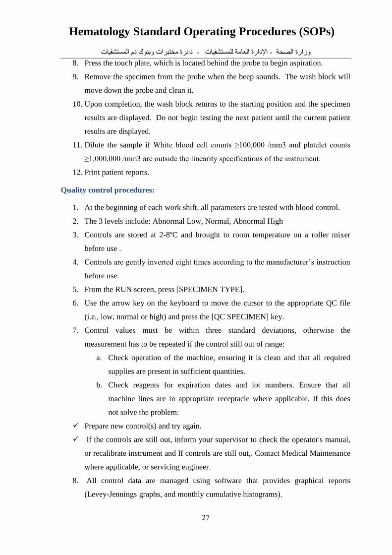
8. Press the touch plate, which is located behind the probe to begin aspiration.
9. Remove the specimen from the probe when the beep sounds. The wash block will
move down the probe and clean it.
10. Upon completion, the wash block returns to the starting position and the specimen
results are displayed. Do not begin testing the next patient until the current patient
results are displayed.
11. Dilute the sample if White blood cell counts ≥100,000 /mm3 and platelet counts
≥1,000,000 /mm3 are outside the linearity specifications of the instrument.
12. Print patient reports.
Quality control procedures:
1. At the beginning of each work shift, all parameters are tested with blood control.
2. The 3 levels include: Abnormal Low, Normal, Abnormal High
3. Controls are stored at 2-8ºC and brought to room temperature on a roller mixer
before use .
4. Controls are gently inverted eight times according to the manufacturer’s instruction
before use.
5. From the RUN screen, press [SPECIMEN TYPE].
6. Use the arrow key on the keyboard to move the cursor to the appropriate QC file
(i.e., low, normal or high) and press the [QC SPECIMEN] key.
7. Control values must be within three standard deviations, otherwise the
measurement has to be repeated if the control still out of range:
a. Check operation of the machine, ensuring it is clean and that all required
supplies are present in sufficient quantities.
b. Check reagents for expiration dates and lot numbers. Ensure that all
machine lines are in appropriate receptacle where applicable. If this does
not solve the problem:
Prepare new control(s) and try again.
If the controls are still out, inform your supervisor to check the operator's manual,
or recalibrate instrument and If controls are still out,. Contact Medical Maintenance
where applicable, or servicing engineer.
8. All control data are managed using software that provides graphical reports
(Levey-Jennings graphs, and monthly cumulative histograms).
Hematology Standard Operating Procedures (SOPs)
دائشة ختبشاث بن د استشف١اث -اإلداسة اعات ستشف١اث -صاسة اظحت
28
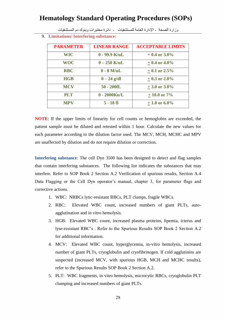
9. Limitations/ Interfering substance:
PARAMETER LINEAR RANGE ACCEPTABLE LIMITS
WIC 0 - 99.9 K/uL + 0.4 or 3.0%
WOC 0 – 250 K/uL + 0.4 or 4.0%
RBC 0 - 8 M/uL + 0.1 or 2.5%
HGB 0 – 24 g/dl + 0.3 or 2.0%
MCV 50 - 200fL + 3.0 or 3.0%
PLT 0 - 2000Ku/L + 10.0 or 7%
MPV 5 - 18 fl + 1.0 or 6.0%
NOTE: If the upper limits of linearity for cell counts or hemoglobin are exceeded, the
patient sample must be diluted and retested within 1 hour. Calculate the new values for
each parameter according to the dilution factor used. The MCV, MCH, MCHC and MPV
are unaffected by dilution and do not require dilution or correction.
Interfering substance: The cell Dyn 3500 has been designed to detect and flag samples
that contain interfering substances. The following list indicates the substances that may
interfere. Refer to SOP Book 2 Section A.2 Verification of spurious results, Section A.4
Data Flagging or the Cell Dyn operator’s manual, chapter 3, for parameter flags and
corrective actions.
1. WBC: NRBCs lytic-resistant RBCs, PLT clumps, fragile WBCs.
2. RBC: Elevated WBC count, increased numbers of giant PLTs, auto-
agglutination and in vitro hemolysis.
3. HGB: Elevated WBC count, increased plasma proteins, lipemia, icterus and
lyse-resistant RBC’s . Refer to the Spurious Results SOP Book 2 Section A.2
for additional information.
4. MCV: Elevated WBC count, hyperglycemia, in-vitro hemolysis, increased
number of giant PLTs, cryoglobulin and cryofibrinogen. If cold agglutinins are
suspected (increased MCV, with spurious HGB, MCH and MCHC results),
refer to the Spurious Results SOP Book 2 Section A.2.
5. PLT: WBC fragments, in vitro hemolysis, microcytic RBCs, cryoglobulin PLT
clumping and increased numbers of giant PLTs.

Hematology Standard Operating Procedures (SOPs)
دائشة ختبشاث بن د استشف١اث -اإلداسة اعات ستشف١اث -صاسة اظحت
29
6. Specimens greater than (>) 24 hours old may cause multiple flags. Do not
perform a differential count unless the slide was made the day the specimen
was collected.
Expected values:
Interpretation of the result:
Certain disease states are defined by an absolute increase or decrease in the number of a
particular type of cell in the bloodstream and many types of anemia.
Reporting result:
According to lab policy.( automated printing or computerized)

Hematology Standard Operating Procedures (SOPs)
دائشة ختبشاث بن د استشف١اث -اإلداسة اعات ستشف١اث -صاسة اظحت
31
ACL Automated Coagulation Analyzer
(Factor Deficient Plasma VIII, IX, XI, XII Tests- HemosIL®)
SOPs\ HGA \.......H\ Haem \05
Version: 1……………………………
Date effective: …………… …...…...
Copy number: ……………………
Head of department: …………………………
Quality Officer: ……………………………...
Director of : ………………………….............
Purpose/Definition:
The ACL analyzer is a fully automated, microcomputer-controlled, nephlometric
microcentrifugal instrument capable of performing the following tests:
PT-FIB (Prothrombin Time and Fibrinogen Level).
APTT (Activated Partial Thromboplastin Time).
PT-FIB/APTT (all three tests run simultaneously).
Single Factors (II, V, VII, VIII, IX, X, XI, XII).
Responsibilities:
Haematology department personal are required to be knowledgeable of this procedure.
New employees are trained and assessed for competence before they can handle
patient sample
The head of the department must resolve any problem with the process and difficulties
in using this SOP.
Specimen Requirements:
Plasma sample, The anticoagulant of choice for coagulation studies is 3.2% Sodium
Citrate (Blue Top Tube).
The standard ratio for citrated specimens is nine (9) parts of blood + one (1) part of
anticoagulant, (9:1 ratio) that is critical for valid results.
Platelet poor plasma required, Set the centrifugation speed to 3,000 rpm and centrifuge
a sample for 10 minutes.
Separate the plasma from the cells within 30 minutes after centrifugation of the sample
and place the plasma sample in a plastic tube.

Hematology Standard Operating Procedures (SOPs)
دائشة ختبشاث بن د استشف١اث -اإلداسة اعات ستشف١اث -صاسة اظحت
31
If the specimen will be tested within two to four hours after centrifugation, it may be
kept at room temperature.
Freeze the specimen if it cannot be processed within four (4) hours after collection.
Plasma can be frozen at (-20ºC) or lower for at (7) days or at (-80ºC) for six (6)
months without loss of most factors.
Thaw rapidly at (37ºC) in a water bath. Remove the plasma sample as soon as it is
thawed and perform the test immediately. Samples are viable for a maximum of (2)
hours at room temperature after they have been thawed .
Plasma samples cannot be re-frozen.
Specimen reception:
1. Prior to performing coagulation testing on patient samples, verify the patient
identification. Any discrepancy must be investigated before processing the
specimen.
2. Check the specimen for clots, visually hemolyzed and lipemic samples.
3. Ensure the specimen is labeled and label a sample cup.
Criteria for rejecting hematology specimen:
When the identification is missing /inadequate.
Clotted specimens.
Lipemic, icteric or hemolyzed plasma samples
Incomplete filling of tube or over-filled samples.
Inappropriate container.
Unknown duration of delay.
Inappropriate transport/storage
Abbreviations:
APTT: Activated Partial Thromboplastin Time
SD: Standard Deviation
Equipment & Items required:
ACL Analyzer
Centrifuge
Sample cups (0.5mL capacity)

Hematology Standard Operating Procedures (SOPs)
دائشة ختبشاث بن د استشف١اث -اإلداسة اعات ستشف١اث -صاسة اظحت
32
Rotors
Plastic test tubes
Reference Solution
Factor deficient plasma VIII
Factor deficient plasma IX
HemosIL APTT reagent
HemosIL Calcium Chloride reagent
HemosIL Normal and Abnormal controls
HemosIL Calibration plasma
HemosIL Factor Diluent
Reagents Preparation:
Factor deficient plasmas VIII, IX : Reconstitute all deficient plasmas with 1.0ml of
deionized water. Swirl gently and let stand for 30 minutes at room temperature. Maintain at
2-8ºc for no more than four hours.
APTT Reagent (SynthAFax): APTT reagent comes ready for use. Each vial must be
mixed by inversion several times before use to assure homogeneity of the reagent.
Calcium Chloride (0.025M): Calcium Chloride comes ready for use. Mix well by
inversion before use.
Calibration Procedure:
1. Dilute the calibration plasma (or pooled plasma) 1+4 with factor diluent according
to the program selected.
2. Place diluted calibration plasma in the pool position of the special (factor assay)
sample tray.
3. Place factor diluent in the Dil position. Place factor deficient plasmas in appropriate
positions of the sample tray.
4. Place the APTT reagent in the reagent reservoir No.2 and the calcium chloride in
the reagent reservoir No.3.
5. Select the factor VIII or IX program and follow the instructions for calibration
displayed on the ACL analyzer video screen.

Hematology Standard Operating Procedures (SOPs)
دائشة ختبشاث بن د استشف١اث -اإلداسة اعات ستشف١اث -صاسة اظحت
33
Assay Procedure:
1. Dilute patient samples (1+4) with factor diluent (100µl sample with 400µl).
2. According to the sensitivity range required, dilute calibration plasma as follows:
a. High curve (1+4) with factor diluent (100µl+400µl factor diluent).
b. Low curve (1+79) with factor diluent (100µl+7.9ml factor diluent).
3. Place pre-diluted samples in the appropriate positions of the sample tray.
4. Place pre-diluted calibration plasma in the pool position of the sample tray.
5. Place factor diluent in the Dil position of the sample tray.
6. Place factor deficient plasmas in the appropriate position of the sample tray.
7. Place the APTT reagent in the reagent reservoir No.2 and the calcium chloride in
the reagent reservoir No.3.
8. Select the single factor program and follow the instructions displayed on the ACL
analyzer video screen.
Quality Control:
Normal and abnormal control will be run on each tests.
Control results must be within the specified (±2SD) limits of the quality control
chart.
If one or more level of control is outside + 3SD, do not report patient results,
Perform troubleshooting action:
check reagent levels and expiration dates.
Repeat the control again, If it is still outside + 3SD:,
reconstitute new controls,
check instrument maintenance/cleaning, and repeat.
If results are still outside limits, notify the Hematology Supervisor
Immediately, Corrective action must be taken before reporting patient results.
Out of range controls will be recorded on the appropriate (normal/abnormal
control) Service Troubleshooting Log along with the corrective action taken.
Limitations/ Interfering Factors:
1. Hemolysis can cause clotting factor activation.
2. Lipemia and icterus interfere with the spectrophotometric measurements resulting
in falsely decreased end point determinations.

Hematology Standard Operating Procedures (SOPs)
دائشة ختبشاث بن د استشف١اث -اإلداسة اعات ستشف١اث -صاسة اظحت
34
Expected values:
Reference ranges: 50-150%.
Interpretations of results:
A Factor VIII deficiency indicates the possible presence of Hemophilia A or von
Willebrand’s disease. Hemophilia A is a sex-linked recessive trait. Hemophilia patients
are classified by the amount of Factor VIII activity measured in their plasma, severe
(<1%), moderate (1-5%) and mild (5-30%). Von Willebrand’s disease is an autosomal
dominant trait exhibiting decreased levels of Factor VIII coagulant activity, affecting
both sexes equally. A differential diagnosis is made based on the results of other
specialized coagulation tests, in conjunction with Factor VIII coagulant activity level.
Factor IX has a decreased activity in a congenital condition known as hemophilia B or
Christmas Disease, which is sex-linked recessive. The most common cause of an
acquired deficiency of blood clotting factors is hepatic dysfunction due to liver cell
damage or non-availability of vitamin K to the liver. Fibrinogen, factors II, VII, IX, X
and possibly factor V are produced in the liver and all except fibrinogen and factor V
require vitamin K for normal synthesis.
Reporting Results:
Factor VIII or IX (Activity) ………………%

Hematology Standard Operating Procedures (SOPs)
دائشة ختبشاث بن د استشف١اث -اإلداسة اعات ستشف١اث -صاسة اظحت
35
ACL Automated Coagulation Analyzer
(PT-FIB/APTT Tests- HemosIL®)
SOPs\ HGA \.......H\ Haem \06
Version: 1……………………………
Date effective: …………… …...…...
Copy number: ……………………
Head of department: …………………………
Quality Officer: ……………………………...
Director of : ………………………….............
Purpose/Definition:
The ACL analyzer is a fully automated, microcomputer-controlled, nephlometric
microcentrifugal instrument capable of performing the following tests:
PT-FIB (Prothrombin Time and Fibrinogen Level).
APTT (Activated Partial Thromboplastin Time).
PT-FIB/APTT (all three tests run simultaneously).
Single Factors (II, V, VII, VIII, IX, X, XI, XII).
Responsibilities:
Haematology department personal are required to be knowledgeable of this procedure.
New employees are trained and assessed for competence before they can handle
patient sample
The head of the department must resolve any problem with the process and difficulties
in using this SOP.
Specimen Requirements:
Plasma sample, The anticoagulant of choice for coagulation studies is 3.2% Sodium
Citrate (Blue Top Tube).
The standard ratio for citrated specimens is nine (9) parts of blood + one (1) part of
anticoagulant, (9:1 ratio) that is critical for valid results.
Platelet poor plasma required, Set the centrifugation speed to 3,000 rpm and centrifuge
a sample for 10 minutes.
Separate the plasma from the cells within 30 minutes after centrifugation of the sample
and place the plasma sample in a plastic tube.

Hematology Standard Operating Procedures (SOPs)
دائشة ختبشاث بن د استشف١اث -اإلداسة اعات ستشف١اث -صاسة اظحت
36
If the specimen will be tested within two to four hours after centrifugation, it may be
kept at room temperature.
Specimen reception:
1. Prior to performing coagulation testing on patient samples, verify the patient
identification. Any discrepancy must be investigated before processing the specimen.
2. Check the specimen for clots, visually hemolyzed and lipemic samples.
3. Ensure the specimen is labeled and label a sample cup.
Criteria for rejecting hematology specimen:
When the identification is missing /inadequate.
Clotted specimens.
Lipemic, icteric or hemolyzed plasma samples
Incomplete filling of tube or over-filled samples.
Inappropriate container.
Unknown duration of delay.
Inappropriate transport/storage
Abbreviations:
PT-FIB: Prothrombin Time and Fibrinogen Level
APTT: Activated Partial Thromboplastin Time
INR: International Normalized Ratio
ISI: International Sensitivity Index
DIC: disseminated intravascular coagulation
VWF: Von Willebrand factor
SD: Standard Deviation
Equipment & Items required:
ACL Analyzer
Centrifuge
Sample cups (0.5mL capacity)
Rotors
Pipettes (1mL capacity)
Plastic test tubes

Hematology Standard Operating Procedures (SOPs)
دائشة ختبشاث بن د استشف١اث -اإلداسة اعات ستشف١اث -صاسة اظحت
37
Reference Solution
HemosIL PT-FIB reagent
HemosIL APTT reagent
HemosIL Calcium Chloride reagent
HemosIL Normal and Abnormal controls
HemosIL Calibration plasma
HemosIL Sample Diluent
Calibration:
Calibration is necessary for every new reagent lot (thromboplastin), new lot of reference
emulsion, when indicated by QC information, after major maintenance or service.
Calibration Procedure:
1. Reconstitute 2 vials of lyophilized Calibration Plasma with one (1) mL of Reagent
Grade Water each, Invert gently to mix. Do not shake . Maintain the calibration
plasma at room temperature for 30 minutes before use, stable for two hours from
reconstitution.
2. Fill one 1 mL sample cup with HemosIL Sample Diluent and place it in the
"DILUENT" position in the sample tray. Fill another 1 mL sample cup with both
vials of reconstituted Calibration Plasma and place it in the ―POOL‖ position on the
sample tray.
3. Put the Reference Solution, Thromboplastin reagent into the appropriate positions
on the instrument.
4. Load a new rotor into the rotor holder.
5. Press PROG to return to the READY menu. Select PT-FIB using the ―↑‖ or ―↓‖ and
press ENTER.
6. The "Check" frame is displayed. Press ―↑‖ to start the calibration cycle.
7. Enter the reference values of all parameter then press ENTER. Press ―↓‖ key to
start the Calibration.
8. At the end of the analysis, the calibration data, to include results and graphics, will
be printed and stored in the memory of the instrument.
Reagents Preparation:
PT-FIB HS reagent:

Hematology Standard Operating Procedures (SOPs)
دائشة ختبشاث بن د استشف١اث -اإلداسة اعات ستشف١اث -صاسة اظحت
38
Reconstitute each vial of thromboplastin reagent with one vial of buffer. Mix by gentle
inversion to ensure complete resuspension (DO NOT SHAKE).
Label the reconstituted reagent vial with the date and initials of tech placing reagent in
use. Maintain at room temperature for (30) minutes before using. A teflon coated
magnetic stir bar must be inserted into the reagent reservoir for continuous mixing
action.
Stability after reconstitution:
8 Hours at 15ºC (on the ACL with continuous stirring), 3 days at 2 to 8ºC (in the
original bottle).
Do not freeze, do not use reagents after the expiration date.
APTT Reagent:
APTT reagent comes ready for use. Each vial must be mixed by inversion several times
before use to assure homogeneity of the reagent.
Calcium Chloride:
Calcium Chloride (0.025 M) comes ready for use. Mix well by inversion before use.
Procedure:
1. From the READY status of the instrument, select the desired test (i.e., PT-FIB, PT-
FIB/APTT or APTT) by using ―↑‖ or ―↓‖ keys and press ENTER.
2. The "Check" frame is displayed.
3. Empty the PT-FIB HS (thromboplastin) vial content into reservoir number 1 on the
instrument. Empty the APTT vial content into reservoir number 2 (APTT) on the
instrument, Empty the Calcium Chloride vial content into reservoir number 3
(CaCl2) on the instrument. Ensure that the Reference Solution volume is sufficient
to perform testing.
4. Press ―↓‖ to continue.
5. Load the sample tray with patient plasma. For PT-FIB/APTT testing, only eight (8)
samples can be programmed to run at one time. For PT-FIB testing, 18 samples
can be programmed and loaded on the sample tray. Calibration plasma loaded in
the pool position of the sample tray.
6. Load a new rotor on the instrument.
7. Press the commands key to start analysis.

Hematology Standard Operating Procedures (SOPs)
دائشة ختبشاث بن د استشف١اث -اإلداسة اعات ستشف١اث -صاسة اظحت
39
8. At the end of analysis the "Results" frame is displayed and results will be printed
out for patient samples. Review the results that are to be filed and certified.
Quality Control:
Normal and abnormal control will be run on each tests.
Control results must be within the specified (±2SD) limits of the quality control
chart.
If one or more level of control is outside + 3SD, do not report patient results,
Perform troubleshooting action:
check reagent levels and expiration dates.
Repeat the control again, If it is still outside + 3SD:,
reconstitute new controls,
check instrument maintenance/cleaning, and repeat.
If results are still outside limits, notify the Hematology Supervisor
Immediately, Corrective action must be taken before reporting patient results.
Out of range controls will be recorded on the appropriate (normal/abnormal
control) Service Troubleshooting Log along with the corrective action taken.
Limitations/ Interfering Factors:
1. Hemolysis can cause clotting factor activation and falsely decrease end point
measurements.
2. Lipemia and icterus interfere with the spectrophotometric measurements
resulting in falsely decreased end point determinations.
3. Contaminated reagents will give inaccurate results.
4. Samples with a fibrinogen levels <25 mg/dl should be suspected of being a
serum sample and rejected.
5. APTT assay results may be affected by many commonly administrated drugs.
Expected values:
PT seconds: 11.5-15.0
PT activity: 70-120%
INR: 0.90-1.15
Therapeutic levels of INR 2.0 – 3.0 target range 2.5
Fibrinogen: (200-400)mg/dl (2.00-4.00) g/l

Hematology Standard Operating Procedures (SOPs)
دائشة ختبشاث بن د استشف١اث -اإلداسة اعات ستشف١اث -صاسة اظحت
41
PTT seconds: lies between (27-35) seconds.
Note: recommendation that each laboratory determines its own normal range according to
the type of reagents.
Interpretation of results:
Interpretation of PT and PTT in patients with a Bleeding or Clotting Syndrome:
Prolonged PT:
Inherited: Factor VII deficiency
Acquired: Vitamin K deficiency, Liver disease, Warfarin use, Factor VII inhibitor
Prolonged APTT:
Inherited: vWF, factor VIII, IX, XI, XII deficiency
Acquired: Heparin use, Inhibitor of vWF, factor VIII, IX, XI, XII,
Antiphospholipid antibodies
Prolonged both PT and APTT:
Inherited: Prothrombin, fibrinogen, factor V, X or combined factor deficiency
Acquired: Severe Liver disease, Disseminated intravascular coagulation (DIC).
Supratherapeutic heparin or warfarin, Combined heparin or warfarin use, Inhibitor
of prothrombin, fibrinogen, factor V, X, Direct thrombin inhibitor.
Reporting Results:
The PT and PTT tests are reported in seconds along with the reference ranges.
PT test results reported in:
Time (Seconds)
Ratio (PT patient/PT control)
Activity Percentage
INR
The ACL Analyzer will automatically calculate the INR value when the ISI value is
entered in the ACL. This value will appear beside the PT result.
Note: The International Sensitivity Index (ISI) is an experimentally derived measurement,
usually provided by the thromboplastin manufacturer "according to package insert".

Hematology Standard Operating Procedures (SOPs)
دائشة ختبشاث بن د استشف١اث -اإلداسة اعات ستشف١اث -صاسة اظحت
41
CoaLAB 1000 Automated Coagulation Analyzer
(PT-FIB/APTT Tests- LABiTec)
SOPs\ HGA \.......H\ Haem \07
Version: 1……………………………
Date effective: …………… …...…...
Copy number: ……………………
Head of department: …………………………
Quality Officer: ……………………………...
Director of : ………………………….............
Purpose/Definition:
The CoaLAB 1000 analyzer, is a fully automated photooptical blood plasma
hemostasis instrument capable for performing a wide range of coagulometric,
chromogenic and immunologic coagulation tests such as Prothrombin time, activated
partial Thromboplastin time, Fibrinogen, special tests such as single factor assays,
Anti-Thrombin III, Protein C, Protein S, C-Reactive Protein, D-Dimer and others
based on the wavelength available in different analyzer types.
Responsibilities:
Haematology department personal are required to be knowledgeable of this procedure.
New employees are trained and assessed for competence before they can handle
patient sample
The head of the department must resolve any problem with the process and difficulties
in using this SOP.
Specimen Requirements:
Plasma sample, The anticoagulant of choice for coagulation studies is 3.2% Sodium
Citrate (Blue Top Tube).
The standard ratio for citrated specimens is nine (9) parts of blood + one (1) part of
anticoagulant, (9:1 ratio) that is critical for valid results.
Platelet poor plasma required, Set the centrifugation speed to 3,000 rpm and centrifuge
a sample for 10 minutes.
Separate the plasma from the cells within 30 minutes after centrifugation of the sample
and place the plasma sample in a plastic tube.
If the specimen will be tested within two to four hours after centrifugation, it may be
kept at room temperature.

Hematology Standard Operating Procedures (SOPs)
دائشة ختبشاث بن د استشف١اث -اإلداسة اعات ستشف١اث -صاسة اظحت
42
Specimen reception:
1. Prior to performing coagulation testing on patient samples, verify the patient
identification. Any discrepancy must be investigated before processing the
specimen.
2. Check the specimen for clots, visually hemolyzed and lipemic samples.
3. Ensure the specimen is labeled and label a sample cup.
Criteria for rejecting hematology specimen:
When the identification is missing /inadequate.
Clotted specimens.
Lipemic, icteric or hemolyzed plasma samples
Incomplete filling of tube or over-filled samples.
Inappropriate container.
Unknown duration of delay.
Inappropriate transport/storage
Abbreviations:
PT-FIB: Prothrombin Time and Fibrinogen Level
APTT: Activated Partial Thromboplastin Time
INR: International Normalized Ratio
ISI: International Sensitivity Index
DIC: disseminated intravascular coagulation
VWF: Von Willebrand factor
Equipment & Items required:
CoaLAB 1000 analyzer.
Centrifuge
Sample-cups (0.5mL capacity)
Cuvette ring contains a 1 x 4 mm stir bar
Pipettes (1mL capacity)
Plastic test tubes
Two containers for rinsing system " Distilled water and waste containers"
Washing solution

Hematology Standard Operating Procedures (SOPs)
دائشة ختبشاث بن د استشف١اث -اإلداسة اعات ستشف١اث -صاسة اظحت
43
Cleaning solution
LABiTec PT reagent
LABiTec APTT reagent
LABiTec Calcium Chloride reagent
LABiTec Fibrinogen reagent
LABiTec Normal and Abnormal controls
Calibration plasma
Calibration:
Before starting a routine run operation:
Check the level of the distilled water container,
Empty the waste water container.
Fill liquid system and flush system.
System Start-up:
Set the power switch on.
The analyzer now runs through a full initialization procedure checking all
internal modules automatically don’t replace reagents or washing solution or c-
ring during initialization.
Once the initialization process is finished the MAIN MENU appears in the
display.
Calibration is necessary for every new reagent lot, when indicated by QC information,
after major maintenance or service.
Use this menu to calibrate a test manually or by automated calibration, to
define or change the calibration parameters for a test.
A calibration is required for test to which the raw values need to be converted
into concentration units/activities.
1. From the Main Menu, press the button Setup to access the Setup Menu.
2. From the Setup Menu, press the button Calibration to access the Calibration
Menu.
3. Select a test for calibration by using the arrow keys first to select the test and
then press the OK button to continue. The calibration for test screen appears.

Hematology Standard Operating Procedures (SOPs)
دائشة ختبشاث بن د استشف١اث -اإلداسة اعات ستشف١اث -صاسة اظحت
44
The analyzer offers two different ways to calibrate a test.
Under Edit Calibration a test can be calibrated manually by editing the
calibration data previously measured. Enter manually the evaluated results
achieved by a normal measurement with dilutions or if available with standard
plasmas or those which have been provided in the package inserts of the
reagent supplier used.
Under Auto Calibration a test will be automatically diluted from a standard and
the measured calibration data will be automatically added into a calibration
curve and memorized for the test.
Procedure:
From the Main Menu, press on the Run Preparation the following display
appears.
Run preparation:
Load C-ring: load a new cuvette ring.
Load reagents: load tests, set reagent vials, re-new reagents, define positions.
Load samples: load patient samples (Routine & STAT), edit patient ID.
Rinsing modes: flush, intensive wash, cleaning, maintenance.
From the Main Menu, press on the Measurement to access the Measurement Menu.
Start the run
During run: add STAT runs, show status, Immediate STOP, show results.
Show results (all, by sample, by test), display curve, print results/graphs.
From the Main Menu, press the button CuvCARD to load C-ring balance.
Quality Control:
Normal and abnormal control will be run on each tests.

Hematology Standard Operating Procedures (SOPs)
دائشة ختبشاث بن د استشف١اث -اإلداسة اعات ستشف١اث -صاسة اظحت
45
Control results must be within the specified (±2SD) limits of the quality control
chart.
If one or more level of control is outside + 3SD, do not report patient results,
Perform troubleshooting action:
check reagent levels and expiration dates.
Repeat the control again, If it is still outside + 3SD:,
reconstitute new controls,
check instrument maintenance/cleaning, and repeat.
If results are still outside limits, notify the Hematology Supervisor
Immediately, Corrective action must be taken before reporting patient results.
Out of range controls will be recorded on the appropriate (normal/abnormal
control) Service Troubleshooting Log along with the corrective action taken.
Limitations/ Interfering Factors:
1. Hemolysis can cause clotting factor activation and falsely decrease end point
measurements.
2. Lipemia and icterus interfere with the spectrophotometric measurements
resulting in falsely decreased end point determinations.
3. Contaminated reagents will give inaccurate results.
4. Samples with a fibrinogen levels <25 mg/dl should be suspected of being a
serum sample and rejected.
5. APTT assay results may be affected by many commonly administrated drugs.
Expected values:
PT seconds: (11.5-15.0)
PT activity: ( 70-120)%
INR: ( 0.90-1.15)
Therapeutic levels of INR (2.0 – 3.0) , target range (2.5)
Fibrinogen: (200-400)mg/dl (2.00-4.00) g/l
PTT seconds: lies between (27-35) seconds.

Hematology Standard Operating Procedures (SOPs)
دائشة ختبشاث بن د استشف١اث -اإلداسة اعات ستشف١اث -صاسة اظحت
46
Interpretation of results:
Interpretation of PT and PTT in patients with a Bleeding or Clotting
Syndrome:
Prolonged PT:
Inherited: Factor VII deficiency
Acquired: Vitamin K deficiency, Liver disease, Warfarin use, Factor VII
inhibitor
Prolonged APTT:
Inherited: vWF, factor VIII, IX, XI, XII deficiency
Acquired: Heparin use, Inhibitor of vWF, factor VIII, IX, XI, XII,
Antiphospholipid antibodies
Prolonged both PT and APTT:
Inherited: Prothrombin, fibrinogen, factor V, X or combined factor deficiency
Acquired: Severe Liver disease, Disseminated intravascular coagulation
(DIC).
Supratherapeutic heparin or warfarin, Combined heparin or warfarin use,
Inhibitor of prothrombin, fibrinogen, factor V, X, Direct thrombin inhibitor.
Reporting Results:
The PT and PTT tests are reported in seconds along with the reference ranges.
PT test results reported in:
Time (Seconds)
Ratio (PTpatient/PTcontrol)
Activity Percentage
INR
The ACL Analyzer will automatically calculate the INR value when the ISI value is
entered in the ACL. This value will appear beside the PT result.
Note: The International Sensitivity Index (ISI) is an experimentally derived
measurement, usually provided by the thromboplastin manufacturer "according to
package insert".

Hematology Standard Operating Procedures (SOPs)
دائشة ختبشاث بن د استشف١اث -اإلداسة اعات ستشف١اث -صاسة اظحت
47
Thrombolyzer compact X
SOPs\ HGA \.......H\ Haem \08
Version: 1……………………………
Date effective: …………… …...…...
Copy number: ……………………
Head of department: …………………………
Quality Officer: ……………………………...
Director of : ………………………….............
Purpose & Definition
Thrombolyzer compact x is a classic, fast and reliable coagulation analyzer, it
performs up to 160 tests/ hour, capable of performing the following tests
PT,PTT,FIBRONGEN , ATIII, TT.
Responsibilities:
Hematology department personal are required to be knowledgeable of this
procedure
The head of the department must resolve any problem with the process and
difficulties in using this sop
New employees are trained and assessed for competence before they can
handle patient sample
Specimen requirements:
Plasma sample, The anticoagulant of choice for coagulation studies is (3.2%)
Sodium Citrate (Blue Top Tube).
The standard ratio for citrated specimens is nine (9) parts of blood + one (1)
part of anticoagulant, (9:1 ratio) that is critical for valid results.
Platelet poor plasma required, Set the centrifugation speed to (3,000) rpm and
centrifuge a sample for (10) minutes.
Separate the plasma from the cells within 30 minutes after centrifugation of the
sample and place the plasma sample in a plastic tube.
If the specimen will be tested within two to four hours after centrifugation, it
may be kept at room temperature.

Hematology Standard Operating Procedures (SOPs)
دائشة ختبشاث بن د استشف١اث -اإلداسة اعات ستشف١اث -صاسة اظحت
48
Specimen reception:
Prior to performing coagulation testing on patient samples, verify the patient
identification. Any discrepancy must be investigated before processing the
specimen.
Check the specimen for clots, visually hemolyzed and lipemic samples.
Ensure the specimen is labeled and label a sample cup.
Criteria for rejecting hematology specimen:
When the identification is missing /inadequate.
Clotted specimens.
Lipemic, icteric or hemolyzed plasma samples
Incomplete filling of tube or over-filled samples.
Inappropriate container.
Unknown duration of delay.
Inappropriate transport/storage
Abbreviations:
PT : Prothrombin Time.
APTT: activated thromboplastin time.
TT: thrombin time.
ATIII : anti thrombin III.
INR: International Normalized Ratio
ISI: International Sensitivity Index
Equipment & Items required:
PT reagent .
PTT reagent.
Cacl2 reagent .
Sample cup.
Cuvette rack segment
Dropper .
Centrifuge.
Probe cleaner solution

Hematology Standard Operating Procedures (SOPs)
دائشة ختبشاث بن د استشف١اث -اإلداسة اعات ستشف١اث -صاسة اظحت
49
Calibration :
1. Form the "calibration" menu's test window choose the test you want to used, then
press (enter).
2. Move the cursor to the "Manual" box, using the (enter) keys, confirming this box
by pressing (enter) the values can be entered.
3. Should you want to use less calibration points for the calibration, change the values
of the last positions to 0 (zero).
4. Having completed the changes to the table, change the Normal, ISI, Min, and Max
values, if necessary.
5. With all changes done, press (Esc). The cursor moves to the "Curve" box (enter).
You can now view the new curve representation. Press (esc) to exit the graph.
6. The cursor moves to the "Valid" box (enter). If you wish to keep the old values,
change the "Valid: Yes" box using the (space) key to "No" before exiting the menu
with (enter. The cursor moves to the "Manual" box (esc).
Procedure :
1. System start-up: set the power switch on.
2. Before starting a routine run operation:
check the level of the distilled water container,
empty the waste water container.
3. The analyzer now runs through a full initialization procedure checking all internal
modules automatically.
4. From the main menu, select patient preparation enter the data on keyboard and
select the desired test (a, b, c, or d) by using ―↑‖ or ―↓‖ keys and press enter.
5. Add enough volume of plasma in sample cup .
6. Put sample cup in the rack sample of thrombolyzer according to their position
7. Press ESC then press F2 to start working the test.
8. Wait for result.
9. Record the result in the result book

Hematology Standard Operating Procedures (SOPs)
دائشة ختبشاث بن د استشف١اث -اإلداسة اعات ستشف١اث -صاسة اظحت
51
Quality Control:
Normal and abnormal control will be run on each tests.
Control results must be within the specified (±2SD) limits of the quality
control chart.
If one or more level of control is outside + 3SD, do not report patient
results,
Perform troubleshooting action:
check reagent levels and expiration dates.
Repeat the control again, If it is still outside + 3SD:,
reconstitute new controls,
check instrument maintenance/cleaning, and repeat.
If results are still outside limits, notify the Hematology Supervisor
Immediately, Corrective action must be taken before reporting patient
results.
Out of range controls will be recorded on the appropriate
(normal/abnormal control) Service Troubleshooting Log along with the
corrective action taken.
Limitations/ Interfering Factors:
1. Hemolysis can cause clotting factor activation and falsely decrease end point
measurements.
2. Lipemia and icterus interfere with the spectrophotometric measurements
resulting in falsely decreased end point determinations.
3. Contaminated reagents will give inaccurate results.
4. Samples with a fibrinogen levels <25 mg/dl should be suspected of being a
serum sample and rejected.
5. APTT assay results may be affected by many commonly administrated drugs.
Expected values:
PT seconds: (11.5-15.0)
INR: (0.90-1.15)
Therapeutic levels of INR (2.0 – 3.0) , target range (2.5)
PTT seconds: lies between (27-35) seconds.

Hematology Standard Operating Procedures (SOPs)
دائشة ختبشاث بن د استشف١اث -اإلداسة اعات ستشف١اث -صاسة اظحت
51
However, this varies widely between laboratories and is dependent upon a
number of variables including whether the test is automated or manual, the
type of activator and the incubation times employed in the test.
Note: recommendation that each laboratory determines its own normal range
Interpretation of the results:
Interpretation of PT and PTT in patients with a Bleeding or Clotting Syndrome
Prolonged PT:
Inherited: Factor VII deficiency
Acquired: Vitamin K deficiency, Liver disease, Warfarin use, Factor VII inhibitor
Prolonged APTT:
Inherited: vWF, factor VIII, IX, XI, XII deficiency
Acquired: Heparin use, Inhibitor of vWF, factor VIII, IX, XI, XII,
Antiphospholipid antibodies
Prolonged both PT and APTT:
Inherited: Prothrombin, fibrinogen, factor V, X or combined factor deficiency
Acquired: Severe Liver disease, Disseminated intravascular coagulation (DIC).
Supratherapeutic heparin or warfarin, Combined heparin or warfarin use, Inhibitor of
prothrombin, fibrinogen, factor V, X, Direct thrombin inhibitor.
Reporting result:
The PT and PTT tests are reported in seconds along with the reference ranges.
PT test results reported in:
Time (Seconds)
INR
The thrombolyzer Analyzer will automatically calculate the INR value when the
ISI value is entered in the thrombolyzer This value will appear beside the PT
result.
Note: The International Sensitivity Index (ISI) is an experimentally derived measurement,
usually provided by the thromboplastin manufacturer "according to package insert"

Hematology Standard Operating Procedures (SOPs)
دائشة ختبشاث بن د استشف١اث -اإلداسة اعات ستشف١اث -صاسة اظحت
52
PROTHROMBIN TIME (MANUAL TEST)
SOPs\ HGA \.......H\ Haem \09
Version: 1……………………………
Date effective: …………… …...…...
Copy number: ……………………
Head of department: …………………………
Quality Officer: ……………………………...
Director of : ………………………….............
Definition & Purpose :
The prothrombin time is the time required for the plasma to clot after an excess of
thromboplastin and an optimal concentration of calcium have been added.
The PT measures functional activity of the extrinsic and common pathways (VII, V,
and X, prothrombin, and fibrinogen).
PT is the most widely used method for monitoring patients receiving oral anticoagulant
as warfarin therapy.
Responsibilities:
Hematology department personal are required to be knowledgeable of this procedure
The head of the department must resolve any problem with the process and difficulties
in using this sop
New employees are trained and assessed for competence before they can handle
patient sample
Specimen Requirements:
Plasma sample, The anticoagulant of choice for coagulation studies is 3.2% Sodium
Citrate (Blue Top Tube).
The standard ratio for citrated specimens is nine (9) parts of blood + one (1) part of
anticoagulant, (9:1 ratio) that is critical for valid results.
Platelet poor plasma required, Set the centrifugation speed to 3,000 rpm and centrifuge
a sample for 10 minutes.
Separate the plasma from the cells within 30 minutes after centrifugation of the sample
and place the plasma sample in a plastic tube.
If the specimen will be tested within two to four hours after centrifugation, it may be
kept at room temperature.
Hematology Standard Operating Procedures (SOPs)
دائشة ختبشاث بن د استشف١اث -اإلداسة اعات ستشف١اث -صاسة اظحت
53
Specimen reception:
1. Prior to performing coagulation testing on patient samples, verify the patient
identification. Any discrepancy must be investigated before processing the
specimen.
2. Check the specimen for clots, visually hemolyzed and lipemic samples.
3. Ensure the specimen is labeled and label a sample cup.
Criteria for rejecting hematology specimen:
When the identification is missing /inadequate.
Clotted specimens.
Hemolyzed plasma samples
Incomplete filling of tube or over-filled samples.
Inappropriate container.
Unknown duration of delay.
Inappropriate transport/storage
Abbreviations:
PT : prothrombin time.
I.N.R : international normalized ratio
ISI : international sensitivity index
DIC : disseminated intravascular coagulation
VWF : Von Willebrand factor
SD : Standard Deviation
Equipment & Items required:
Calcified thromboplastin reagent, Fresh pooled plasma normal control, abnormal control,
Water bath at 37˚C, Tissue paper, Stop watch, Glass tube, Yellow tips , Micro pipette 50-
200 µl.
Procedure:
1. Reconstitute tissue thromboplastin reagent according to instructions. Label the
thromboplastin with the time, date and initials.
2. Pipet 100 µL of plasma into test tubes.
3. Allow at least 1-3 minute for the control and sample to reach 37 o C in water bath.

Hematology Standard Operating Procedures (SOPs)
دائشة ختبشاث بن د استشف١اث -اإلداسة اعات ستشف١اث -صاسة اظحت
54
4. Pipet 200 µL of PT reagent into the tube containing the plasma. Start the stop
watch simultaneously.
5. Mix the tube and leave in the water bath for a minimum of 7-8 seconds. Then
remove, wipe the exterior, tilt back and forth gently until a visible clot is formed.
As the clot forms, the mixture will gelatinize and may turn cloudy.
6. Stop the stop watch immediately when the clot begins to form and record the time
in seconds.
7. Repeat the procedure for the second run of plasma to confirm result and Record the
time.
8. If results are not within required limits, a third run should be performed and
average the two that match within acceptable limits.
Quality control procedures:
The normal control and the abnormal control will be run on each tests. The control results
must be within the specified (±2SD) limits of the quality control chart.
If one or more level of control is outside + 3SD, do not report patient results.
Perform troubleshooting action:
check reagent levels and expiration dates.
Repeat the control again. If it is still outside + 3SD,
reconstitute new controls and reagents,
check water bath temperature, and repeat.
If results are still outside limits, notify the Hematology Supervisor immediately.
Corrective action must taken before reporting patient results.
Out of range controls will be recorded on the appropriate (normal/abnormal
control) Service Troubleshooting Log along with the corrective action taken.
Expected values:
The normal values for the prothrombin time range from 11.5 to 15.0 seconds and it is
different from lab to lab depend on the source of thromboplastin used.
PT seconds: 11.5-15.0.
PT activity: 70-120%
INR: 0.90-1.15
Therapeutic levels of INR 2.0 – 3.0 target range 2.5

Hematology Standard Operating Procedures (SOPs)
دائشة ختبشاث بن د استشف١اث -اإلداسة اعات ستشف١اث -صاسة اظحت
55
Note: recommendation that each laboratory determines its own normal range.
Limitations/ Interfering substance:
1. Associated with specimen (Pre-analytical)
Inappropropriate ratio of anticoagulant to blood
Delay in testing or processing
Inappropriate storage
2. Associated with Reagent (Analytical)
Incorrect preparation of reagents
Use of reagents beyond reconstituted stability time or expiration date
Contaminated reagent.
3. Associated with procedure
Incorrect temperature
Incorrect incubation times.
Incorrect volumes of sample, reagents or both
Also test may be affected by :
Severe diarrhea or vomiting that causes fluid loss and dehydration, this may
increase the INR.
Getting a lot of vitamin k may decrease the INR.
Medication: antibiotics, aspirin, cimetidine.
Interpretation of the results:
Interpretation of PT and PTT in patients with a Bleeding or Clotting Syndrome
Prolonged PT:
Inherited: Factor VII deficiency
Acquired: Vitamin K deficiency, Liver disease, Warfarin use, Factor VII inhibitor
Prolonged APTT:
Inherited: vWF, factor VIII, IX, XI, XII deficiency
Acquired: Heparin use, Inhibitor of vWF, factor VIII, IX, XI, XII,
Antiphospholipid antibodies
Prolonged both PT and APTT:
Inherited: Prothrombin, fibrinogen, factor V, X or combined factor deficiency
Acquired: Severe Liver disease, Disseminated intravascular coagulation (DIC).

Hematology Standard Operating Procedures (SOPs)
دائشة ختبشاث بن د استشف١اث -اإلداسة اعات ستشف١اث -صاسة اظحت
56
Supratherapeutic heparin or warfarin, Combined heparin or warfarin use, Inhibitor of
prothrombin, fibrinogen, factor V, X, Direct thrombin inhibitor.
Reporting Results:
The PT is reported in seconds along with the reference ranges.
PT test results reported in:
Time (Seconds)
Ratio (PT patient/PT control)
Activity Percentage
INR
Note: The International Sensitivity Index (ISI) is an experimentally derived measurement,
usually provided by the thromboplastin manufacturer "according to package insert".

Hematology Standard Operating Procedures (SOPs)
دائشة ختبشاث بن د استشف١اث -اإلداسة اعات ستشف١اث -صاسة اظحت
57
PARTIAL THROMBOPLASTIN TIME (MANUAL TEST)
SOPs\ HGA \.......H\ Haem \10
Version: 1……………………………
Date effective: …………… …...…...
Copy number: ……………………
Head of department: …………………………
Quality Officer: ……………………………...
Director of : ………………………….............
Definition & Purpose :
The activated partial thromboplastin time is the time required for plasma to clot when
maximal surface contact activation, optimal phospholipid, and calcium concentration are
provided. The PTT measures functional activity of the intrinsic and common pathway as
well as screen for coagulation inhibitors such as the lupus-like anticoagulant. APTT is the
most widely used method for monitoring intravenous heparin anticoagulant therapy.
Responsibilities:
Haematology department personal are required to be knowledgeable of this procedure.
New employees are trained and assessed for competence before they can handle
patient sample
The head of the department must resolve any problem with the process and difficulties
in using this SOP.
Specimen Requirements:
Plasma sample, The anticoagulant of choice for coagulation studies is 3.2% Sodium
Citrate (Blue Top Tube).
The standard ratio for citrated specimens is nine (9) parts of blood + one (1) part of
anticoagulant, (9:1 ratio) that is critical for valid results.
Platelet poor plasma required, Set the centrifugation speed to 3,000 rpm and centrifuge
a sample for 10 minutes.
Separate the plasma from the cells within 30 minutes after centrifugation of the sample
and place the plasma sample in a plastic tube.
If the specimen will be tested within two to four hours after centrifugation, it may be
kept at room temperature.

Hematology Standard Operating Procedures (SOPs)
دائشة ختبشاث بن د استشف١اث -اإلداسة اعات ستشف١اث -صاسة اظحت
58
Specimen reception:
1. Prior to performing coagulation testing on patient samples, verify the patient
identification. Any discrepancy must be investigated before processing the
specimen.
2. Check the specimen for clots, visually hemolyzed and lipemic samples.
3. Ensure the specimen is labeled and label a sample cup.
Criteria for rejecting hematology specimen:
When the identification is missing /inadequate.
Clotted specimens.
Hemolyzed plasma samples
Incomplete filling of tube or over-filled samples.
Inappropriate container.
Unknown duration of delay.
Inappropriate transport/storage
Abbreviations:
APTT (activated partial thromboplastin time).
Cacl2 (calcium chloride)
DIC (disseminated intravascular coagulation).
VWF (Von Willebrand factor)
Equipment & Items required:
Partial thromboplastin reagent: consists of phospholipids and a contact activator, Cacl2
(0.025M), Fresh pooled plasma normal control, abnormal control, Water bath at 37˚C,
Tissue paper, Stop watch, Glass tube, Yellow tips , Micro pipette 50-200 µl.

Hematology Standard Operating Procedures (SOPs)
دائشة ختبشاث بن د استشف١اث -اإلداسة اعات ستشف١اث -صاسة اظحت
59
Procedure:
1. Pre-warm a sufficient quantity of 0.025M calcium chloride reagent to 37˚C.
2. Pipette 100 µL of patient plasma into a labeled test tube.
3. Into test tube, add 100 µL of partial thromboplastin reagent.
4. Incubate the patient plasma/partial thromboplastin mixture at 37oC for a minimum
of three (3) minutes (optimum activation of contact factors).
5. Add 100 µL of calcium chloride into the patient plasma / partial thromboplastin
mixture and start the stop watch immediately.
6. After 20 seconds, remove the tube from the water bath. Wipe off the outside of the
tube. Gently tilt the tube back and forth until a visible clot forms.
7. Immediately record the time in seconds.
8. The result must be run in duplicate.
Quality control procedures:
The normal control and the abnormal control will be run on each tests. The control results
must be within the specified (±2SD) limits of the quality control chart.
If one or more level of control is outside + 3SD, do not report patient results.
Perform troubleshooting action:
check reagent levels and expiration dates.
Repeat the control again. If it is still outside + 3SD,:
reconstitute new controls and reagents,
check water bath temperature, and repeat.
If results are still outside limits, notify the Hematology Supervisor immediately.
Corrective action must taken before reporting patient results.
Out of range controls will be recorded on the appropriate (normal/abnormal
control) Service Troubleshooting Log along with the corrective action taken.
Expected values:
PTT reported in seconds: lies between 27-35 seconds. However, this varies widely
between laboratories and is dependent upon a number of variables including whether the
test is automated or manual, the type of activator and the incubation times employed in the
test.
Note: recommendation that each laboratory determines its own normal range.

Hematology Standard Operating Procedures (SOPs)
دائشة ختبشاث بن د استشف١اث -اإلداسة اعات ستشف١اث -صاسة اظحت
61
Limitations/ Interfering Factors:
1. Associated with specimen (Pre-analytical)
Inappropropriate ratio of anticoagulant to blood
Delay in testing or processing
Inappropriate storage
2. Associated with Reagent (Analytical)
Incorrect preparation of reagents
Use of reagents beyond reconstituted stability time or expiration date
Contaminated reagent.
3. Associated with procedure
Incorrect temperature
Incorrect incubation times (↑ incubation time=↓PTT due to contact
activation and > 5min heating will result in loss of heat-labile factor V)
Incorrect volumes of sample, reagents or both
4. Some drugs such as heparin, antihistamine ascorbic acid ,chlorpromazine and
salicylates.
Interpretation of the results:
Interpretation of PT and PTT in patients with a Bleeding or Clotting Syndrome
Prolonged PT
Inherited: Factor VII deficiency
Acquired: Vitamin K deficiency, Liver disease, Warfarin use, Factor VII inhibitor
Prolonged APTT
Inherited: vWF, factor VIII, IX, XI, XII deficiency
Acquired: Heparin use, Inhibitor of vWF, factor VIII, IX, XI, XII,
Antiphospholipid antibodies
Prolonged both PT and APTT
Inherited: Prothrombin, fibrinogen, factor V, X or combined factor deficiency
Acquired: Severe Liver disease, Disseminated intravascular coagulation (DIC).
Supratherapeutic heparin or warfarin, Combined heparin or warfarin use, Inhibitor of
prothrombin, fibrinogen, factor V, X, Direct thrombin inhibitor.
Reporting Results: PTT reported in seconds.

Hematology Standard Operating Procedures (SOPs)
دائشة ختبشاث بن د استشف١اث -اإلداسة اعات ستشف١اث -صاسة اظحت
61
Bleeding time
SOPs\ HGA \.......H\ Haem \11
Version: 1……………………………
Date effective: …………… …...…...
Copy number: ……………………
Head of department: …………………………
Quality Officer: ……………………………...
Director of : ………………………….............
Purpose & Definition:
Bleeding time is defined as the time taken for a standard skin wound to stop
bleeding upon vessel injury , platelets adhere and form a haemostatic platelet plug.
bleeding time measures the ability of these platelets to arrest bleeding and therefore
measures platelets function as well as the integrity of the vessel wall.
Bleeding time: it is a test for :
1. Capillary response to injury.
2. Platelet function: stick to each other and form plug (aggregate), and break and
release thromboplastin.
There are several methods of performing the bleeding time:
1- Duke method
2- IVY method.
Responsibilities:
Haematology department personal are required to be knowledgeable of this procedure
The head of the department must resolve any problem with the process and difficulties
in using this sop
New employees are trained and assessed for competence before they can handle
patient sample
Specimen requirements:
Patient Preparation: Check the patient's history for recent use of drugs that prolong
bleeding time. If the test is being used to identify a suspected bleeding disorder, it should
be postponed and the drugs discontinued. If the test is being used preoperatively to assess
hemostatic function, it should proceed as scheduled.

Hematology Standard Operating Procedures (SOPs)
دائشة ختبشاث بن د استشف١اث -اإلداسة اعات ستشف١اث -صاسة اظحت
62
Specimen reception:
1. Explain to the patient this test is used to measure the time required to form a clot
and stop bleeding.
2. Reassure the patient that, although he may feel some discomfort from the incisions,
the antiseptic, advise the patient that the incisions will leave two small, hairline
scars that should be barely visible when healed.
Equipment & Items required:
Blood pressure cuff, disposable lancet, 70% alcohol, filter paper, bandage, stopwatch.
Abbreviations:
B.T: Bleeding time
Procedures:
Duke method
1. Gently clean the lobe of the ear with cotton wool and alcohol, do not rub. Allow to
dry.
2. Puncture the ear lobe, with the lancet, making the incision 2-4mm deep the blood
should flow freely, without any need to squeeze the ear lobe. Start the stopwatch.
3. After 30 seconds collect the first drop of blood on a corner of the filter-paper
4. Do not touch the skin with the paper.
5. Wait 30 seconds more. Collect the second drop of blood in the same way, a little
further along the strip of paper.
6. Continue to collect one more drop of blood every 30 seconds. The drops become
progressively smaller.
7. When no more blood appears, stop the timer and record the time.
Ivy method
1. Blood pressure cuff is placed on arm and inflated to 40mm Hg
2. With pressure maintained, the arm is cleaned and a lancet is again used to make a
2-4 mm cut on the volar aspect of the forearm, being careful NOT to cut any visible
blood vessels.
3. Filter paper is used to blot blood every 30 seconds until it stops flowing.
4. If bleeding continues for more than 15 minutes, the procedure should be
discontinued.

Hematology Standard Operating Procedures (SOPs)
دائشة ختبشاث بن د استشف١اث -اإلداسة اعات ستشف١اث -صاسة اظحت
63
Limitations/ Interfering substance:
1. If the patient has taken aspirin or aspirin-containing compounds 7 to 10 days prior
to the procedure, the bleeding time may be prolonged.
2. Other medications such as dextran, streptokinase, and streptodornase may also
affect the bleeding time.
3. Results may be affected by an improperly performed puncture. A puncture that is
too shallow, too deep, or in an inappropriate location will adversely affect test
results.
4. The alcohol must be completely dried before making the puncture. If residual
alcohol is on a puncture site, the bleeding time will be erroneously prolonged.
5. If the phlebotomist allows the filter paper to touch the wound, the platelet clot may
be dislodged, causing falsely elevated results.
6. Blood pressure cuff should be maintained exactly at (40mm Hg).
Expected values:
Duke method: 1–3 minutes
Ivy method: 3 to 6 minutes
Interpretation of the results:
A. Abnormal increased results could be due to:
1. Thrombocytopenia, with failure to produce a platelet plug.
2. Poor platelet function
3. Failure of vessel constriction on injury
4. Puncture of larger blood vessel (technical error)
5. Disturbing the clot (technical error)
B. Prolonged bleeding time may indicate the presence of disorders associated with
such as Hodgkin's disease, acute leukemia, disseminated intra vascular coagulation,
hemolytic disease of the newborn, liver disease- end stage and in some cases of
Von Willebrand Disease, Hypofibrinogenaemia, Bernard-Soulier disease and
Glanzmann's thrombasthenia.
C. Decreased BT: Not clinically significant.
Reporting result:
Bleeding time: ………minute :………..sec

Hematology Standard Operating Procedures (SOPs)
دائشة ختبشاث بن د استشف١اث -اإلداسة اعات ستشف١اث -صاسة اظحت
64
Clotting time
SOPs\ HGA \.......H\ Haem \12
Version: 1……………………………
Date effective: …………… …...…...
Copy number: ……………………
Head of department: …………………………
Quality Officer: ……………………………...
Director of : ………………………….............
Purpose & Definition:
It is the time required for blood to clot without the presence of any substance .
The whole blood clotting time is a rough measure of all intrinsic clotting factors in the
absence of tissue factors. Variations are wide and the test sensitivity is limited. Whole
blood, when removed from the vascular system and exposed to a foreign surface, will form
a solid clot. Within limits, the time required for the formation of the solid clot is a measure
of the coagulation system.
There are various methods for determining the clotting time, the most common being
the capillary tube method and slide method.
Responsibilities:
Haematology department personal are required to be knowledgeable of this procedure
The head of the department must resolve any problem with the process and difficulties
in using this sop
New employees are trained and assessed for competence before they can handle patient
sample
Specimen requirements:
1- About 3 drops of blood on a glass slide and examining it for clot formation.
2- About 200 ul blood in capillary tube.
Specimen reception:
Explain to the patient this test is used to measure the time required to form a clot and
explain how the test will be performed.

Hematology Standard Operating Procedures (SOPs)
دائشة ختبشاث بن د استشف١اث -اإلداسة اعات ستشف١اث -صاسة اظحت
65
Equipment & Items required:
Alcohol swab, lancet, glass slide, stopwatch and capillary tubes.
Abbreviations:
C.T: Clotting time
Procedures:
Capillary tube method.
1. Clean the patient finger with alcohol swab and allow to dry.
2. Pricked the finger by lancet, remove the first drop of blood.
3. Squeeze the finger to obtain a larger drop of blood and fill two capillary tubes with
blood.
4. After one minute start breaking small pieces of the capillary tube every 30 second
until a fibrin thread is seen between the two broken ends.
Slide method
1. Clean one finger with the alcohol swab then puncture it with the lancet.
2. Place a large drop of blood on the glass slide. Start the stopwatch.
3. After one minute, dip the lancet all the way into the blood drop then slowly lift the
lancet out.
4. If you see a gel-like thread of fibrin (blood clot protein) the blood has clotted.
5. Record the clotting time.
6. If no piece of fibrin is observed, keep repeating the dipping procedure until the
thread is observed.
Limitations/ Interfering substance:
The test sensitive only in extreme factor deficiencies, and is insensitive to high
doses of heparin.
The following variables tend to decrease the clotting time: rough handling of the
blood specimen, presence of tissue fluids (traumatic venipuncture).

Hematology Standard Operating Procedures (SOPs)
دائشة ختبشاث بن د استشف١اث -اإلداسة اعات ستشف١اث -صاسة اظحت
66
Expected values:
Slide method (2 to 7 ) minutes
Capillary tube method (2 to 8) minutes
Interpretation of the results:
In coagulation disorders like hemophilia, clotting time is prolonged but bleeding
time remains normal.
Clotting time is also prolonged in conditions like vitamin K deficiency, liver
diseases, disseminated intravascular coagulation, overdosage of anticoagulants etc.
Reporting result:
Clotting time ……….minutes : ……… seconds.

Hematology Standard Operating Procedures (SOPs)
دائشة ختبشاث بن د استشف١اث -اإلداسة اعات ستشف١اث -صاسة اظحت
67
Blood film (leishman's stain)
SOPs\ HGA \.......H\ Haem \ 13
Version: 1……………………………
Date effective: …………… …...…...
Copy number: ……………………
Head of department: …………………………
Quality Officer: ……………………………...
Director of : ………………………….............
Purpose & Definition:
Describe the procedure for preparing thin blood smear and staining with leshimans stain
for the purpose of diagnosis of different anemia's, leukemia's, viral and parasitic
infections, in addition associated with inclusion bodies, allergic reactions,
hemoglobinopathies, and platelets disorders
It is based on a methanolic mixture of "polychromed" methylene blue and eosin.
The methanolic stock solution is stable and also serves the purpose of directly fixing
the smear eliminating a prefixing step.
Responsibilities:
Hematology department personal are required to be knowledgeable of this procedure
The head of the department must resolve any problem with the process and difficulties
in using this sop
New employees are trained and assessed for competence before they can handle patient
sample
Specimen requirements:
Freshly collected venous blood in an EDTA container is recommended.
Specimen reception:
Reception of samples should be recorded, and record time of reception. Pay attention to
sample identification and labeling of tubes.
Smears of peripheral blood must be made immediately
Hematology Standard Operating Procedures (SOPs)
دائشة ختبشاث بن د استشف١اث -اإلداسة اعات ستشف١اث -صاسة اظحت
68
Criteria for rejection hematology specimens:
1. When The Identification Is Missing /Inadequate.
2. Insufficient Quantity
3. Inappropriate Container
4. Inappropriate transport/storage
5. Unknown duration of delay
6. Haemolysed and/or clotted sample reject
Equipment & Items required:
Leshimen's stain
Microscope.
Microscopic slides.
Distilled water.
Methanol 96%.
Droppers.
Immersion oil.
Abbreviations:
B. film : Blood film.
RBC : red blood cell.
WBC : white blood cell.
CBC : complete blood count.
EDTA : ethylene diamine tetra acetic acid
RNA : Ribonucleic acid
Procedures:
First preparation of leishman's stock stain :
1. Dissolve 1.5 g of leishman's powder in 1000 ml of methanol.
2. Mix will until the powder dissolve completely & let it to stand for 24 hr.
3. filter the stock stain with filter paper.
Second blood film preparation :
1. Small drop of blood placed at the end of the slide.
2. Using another slide, the blood can be spread to make the smear.
3. Let’s to dry

Hematology Standard Operating Procedures (SOPs)
دائشة ختبشاث بن د استشف١اث -اإلداسة اعات ستشف١اث -صاسة اظحت
69
4. Add one volume of leishman's stain on blood film smear for 1 min
5. Add two volume of distilled water .
6. Mix then wait for 15 min.
7. Wash with tape water.
8. let's to dry and observe under microscope.
Quality control procedures:
A well-made slide will have three distinct regions: a head, body, and tail.
Repeat counts of differential WBC's and RBC's morphology on selected slides on
subsequent days will give an indication of the range in the variation of the results.
(include note on reagent quality).
When a new batch of stain is prepared, decide the best staining time to use, e.g. stain
films made from the same blood at different times, e.g. 5, 7, 10, 12, 15 minutes.
Limitations/ Interfering substance:
Causes of incorrectly stained blood smear (too blue):
1. Buffered water or stain is two alkali.
2. Excessive thickness of the smear.
3. Alkaline residue on the slide.
4. Insufficient washing.
5. Prolonged staining.
Causes of incorrectly stained blood smear (too red):
1. Buffered water or stain is two acid.
2. Acid residue on slide.
Entire smear has a pale stain:
1. Under staining
2. Weak stain.
3. Excessive washing on allowing buffered distilled water to stand on slide.
4. Using worm or hot buffered distilled water for washing slide.
Variations on staining on different areas of the smear:
1. Buffered water was unevenly applied.
2. Acid or alkali residue on slide.
3. Water wasn't properly drained from slide after washing.

Hematology Standard Operating Procedures (SOPs)
دائشة ختبشاث بن د استشف١اث -اإلداسة اعات ستشف١اث -صاسة اظحت
71
Precipitated stain:
1. Lack of through washing.
2. Stain hasn't properly filtered.
Evaporation of alcoholic stain may be due to excessive staining time, slide titled so that
stain runs to one end or to titling stain of slide before washing.
Expected Result:
Erythrocytes stain buff pink to pale bluish-gray.
Leukocytes: neutrophils, monocytes and lymphocytes have a pale blue-
gray cytoplasm and purple nucleus.
Eosinophil have coarse, pink granules in the cytoplasm.
Basophils have coarse, deep blue granules in the cytoplasm.
Platelets have pale blue cytoplasm and diffuse red nuclear material.
Normal RBC’s shape are normocytic, normochromic, no variation in size, no
shape variation , and no inclusion bodies.
Normal WBC's morphology and differential.
Normal platelets size, shape and distribution.
Interpretation of the results:
Red cell according to hemoglobin content : normochromic, hypochromic,
hyperchromic
Variation of red cell size (anisocytosis): normocytic, macrocytic, and
microcytic
Variation of red cell shape (poikilocytosis): normal or abnormal; report the
presence of sickle cells, target cells, spherocytes, etc.
Red cell inclusion: RNA, polychromasia, Howell-Jolly bodies, etc.
Nucleated red blood cells and all types of erythroblasts are abnormal.
Elevated white blood cell count may mean infection.
Decreases in white blood cell count may occur with disease progression or may
indicate bone marrow suppression.
Total lymphocyte count: decrease in absolute lymphocyte count may reflect
bone marrow suppression.

Hematology Standard Operating Procedures (SOPs)
دائشة ختبشاث بن د استشف١اث -اإلداسة اعات ستشف١اث -صاسة اظحت
71
An increase in neutrophils may be due to an acute bacterial infection or
hematological malignancies such as myeloid leukemia.
An increase in eosinophils may be due to a parasitic infection or an allergic
reaction.
An increase in lymphocytes may be due to viral infections or chronic infections
such as tuberculosis or lymphocytic leukemia.
An increase in monocytes is found in hematological malignancies such as
chronic myelomonocytic leukemia and certain bacterial and parasitic infections
(e.g., typhoid fever, malaria).
Reporting result:
RBC's Morphology:…………………………………………………………
WBC's differential:
Neutrophils: ………..%.
Lymphocytes:………%.
Eosinophil’s:……… .%.
Basophils:…………..%.
Blast cells:…………..%.
Platelets morphology & distribution:………………………………………
صاسة اظحت

Hematology Standard Operating Procedures (SOPs)
دائشة ختبشاث بن د استشف١اث -اإلداسة اعات ستشف١اث -صاسة اظحت
72
Blood Group \ (Slide Method) BIOTEC LABORAYORIES
SOPs\ HGA \.......H\ Haem \14
Version: 1……………………………
Date effective: …………… …...…...
Copy number: ……………………
Head of department: …………………………
Quality Officer: ……………………………...
Director of : ………………………….............
Purpose & Definition:
Determine the ABO and Rh group in human blood. ABO and Rh group are determine by
detection the presence or absence of A & B antigen in ABO system and presence or
absence of D antigen in Rh system. Because of its low sensitivity, slide method used only
for preliminary grouping.
Responsibilities:
Hematology department personal are required to be knowledgeable of this procedure.
New employees are trained and assessed for competence before they can handle patient
sample
The Head of the department must resolve any problem with the process and difficulties
in using this SOP.
Specimen requirements:
Blood collected in an EDTA container is recommended.
Specimen reception and rejection:
Reception of samples should be recorded, and record time of reception. It is essential to
pay strict attention to sample identification and labeling of tubes.
Clotted samples, hemolysed samples and samples collected in other anticoagulants than
EDTA are rejected. Samples should be stored at 2-8 Co if not tested immediately.
Equipment & Items required:
Glass slides, Wooden applicator.
Anti-A(blue), Anti-B(yellow) and Anti-D reagents.

Hematology Standard Operating Procedures (SOPs)
دائشة ختبشاث بن د استشف١اث -اإلداسة اعات ستشف١اث -صاسة اظحت
73
Abbreviations:
ABO : blood group system. Rh : rhesus factor.
Procedures:
1. Bring the reagents at room temperature.
2. Mix blood tube gently.
3. Transfer one drop of whole blood for each test to a glass slide.
4. Transfer one drop of each reagent to each blood drop.
5. Mix whole blood & reagent by using wooden applicator.
Quality control procedures:
The reactivity of blood grouping reagents should be confirmed by testing with known
blood group blood samples on each day of use.
Limitations/ Interfering substance:
Drying up of reaction mixture can cause aggregation of cells, giving false positive
results.
Abnormal plasma proteins, cold auto agglutinins, positive direct anti globulin test
and in some cases bacteremia may interfere.
Expected result:
Agglutination indicates a positive result to the corresponding antigen, no agglutination
indicates a negative result to corresponding antigen.
Interpretation of the results:
Anti- A Anti-B Anti-D Blood group
- - + O+
+ - + A+
- + + B+
+ + + AB+
- - - O-
+ - - A-
- + - B-
+ + - AB-
Reporting result:
Blood group: ………… Rh:………………

Hematology Standard Operating Procedures (SOPs)
دائشة ختبشاث بن د استشف١اث -اإلداسة اعات ستشف١اث -صاسة اظحت
74
Erythrocyte Sedimentation Rate (Westergren technique)
SOPs\ HGA \.......H\ Haem \15
Version: 1……………………………
Date effective: …………… …...…...
Copy number: ……………………
Head of department: …………………………
Quality Officer: ……………………………...
Director of : ………………………….............
Purpose/Definition:
The erythrocyte sedimentation rate (ESR) is a non-specific test. It is raised in a wide range
of infectious, inflammatory, degenerative, and malignant conditions associated with
changes in plasma proteins, particularly increases in fibrinogen, immunoglobulins, and
CRP.
When well-mixed venous blood is placed in a vertical tube, erythrocytes will tend to fall
toward the bottom. The length of fall of the top of the column of erythrocytes in a given
interval of time.
Responsibilities:
Hematology department personal are required to be knowledgeable of this procedure.
New employees are trained and assessed for competence before they can handle patient
sample
The head of the department must resolve any problem with the process and difficulties
in using this SOP.
Specimen requirements:
Either venous blood collected directly into sodium citrate and tested within 2 hours, or
EDTA anticoagulated blood diluted in sodium citrate can be used.
If EDTA blood is used and kept refrigerated at 4-8°C, citrate dilution of the blood and
testing can be delayed for up to 6 hours.
Specimen reception:
Samples must be transported as soon as possible after collection at 18–22ºC, and the tests
samples should be analyzed within 2 hours after collection.

Hematology Standard Operating Procedures (SOPs)
دائشة ختبشاث بن د استشف١اث -اإلداسة اعات ستشف١اث -صاسة اظحت
75
Criteria for Rejecting Hematology Specimens:
1. When the identification is missing /Inadequate.
2. Insufficient quantity.
3. Inappropriate container.
4. Inappropriate transport /storage.
5. Unknown duration of delay.
Equipment & Items required:
1. Tri-Sodium citrate, 32 g/l (3.2 % w/v) anticoagulant.
2. Glass Westergren pipettes or when available, disposable plastic Westergren pipettes
can be used.
3. Westergren pipettes measures 300mm in length (plastic pipettes are often shorter) and
are graduated from 0-200mm. The diameter should not be less than 2.55mm.
4. Timer.
Abbreviations:
ESR: Erythrocyte sedimentation rate
CRP: C-reactive protein.
EDTA: Ethylene diamine tetra acetic acid
Procedures:
1. Pipette 0.4 ml of sodium citrate anticoagulant into a small container.
2. Add 1.6 ml of venous blood or EDTA anticoagulated blood and mix well.
3. Remove the cap of the container and place the sample in the ESR stand (make a note of
the number in the patient’s notes).
4. Insert a Westergren pipette and ensure it is positioned vertically.
5. Using a safe suction method, draw the blood to the 0 mark of the Westergren pipette,
avoiding air bubbles.
6. Check that the ESR stand is level by making sure that the bubble in the spirit level is
central. If required, adjust the screws on the bottom of the stand. Re-check that the
pipette is vertical.
7. Set the timer for 1 hour. Ensure the ESR stand and pipette will not be exposed to direct
sunlight.

Hematology Standard Operating Procedures (SOPs)
دائشة ختبشاث بن د استشف١اث -اإلداسة اعات ستشف١اث -صاسة اظحت
76
8. At the end of the hour read the height of clear plasma above the upper margin of the
column of sedimenting cells to the nearest millimeter.
Quality control procedures:
The most practical way of controlling ESR tests is to follow the test method exactly.
Limitations/ Interfering substance:
1. Using the wrong volume of blood to anticoagulant.
2. Blood not sufficiently mixed with anticoagulant.
3. Clots in the blood.
4. Air bubbles at the top of the column.
5. Testing blood samples at the hottest time of the day, or leaving tests in direct
sunlight. Temperatures over 25°C increase sedimentation.
6. Using a pipette, which is, not clean or not dry.
7. Pipette not positioned vertically. Even slight variations from the upright increase
sedimentation.
8. Not checking whether the ESR stand is level on the bench.
9. Placing an ESR stand on the same bench as a centrifuge where vibration will
interfere with sedimentation.
10. Measuring the ESR when a patient is dehydrated.
Expected values:
Men…Up to 15 mm/hour
Women…Up to 20 mm/hour
Elderly…Up to 20 mm/hour
Children…Up to 15 mm/hour
Interpretation of the results:
An elevated ESR may be found in:
Pregnancy (after the third month).
Acute and chronic infections.
Rheumatic fever.
Rheumatoid arthritis.
Myocardial infection.

Hematology Standard Operating Procedures (SOPs)
دائشة ختبشاث بن د استشف١اث -اإلداسة اعات ستشف١اث -صاسة اظحت
77
Nephrosis.
Acute hepatitis.
Menstruation.
Tuberculosis.
Hypothyroidism.
Hyperthyroidism.
Adults over 60 years of age frequently have a slightly higher ESR value due primarily to
decreased concentrations of plasma albumin.
A decreased ESR will be present in:
Polycythemia.
Congestive heart failure.
Hypofibrinogenemia.
The presence of red blood cell abnormalities (poikilocytosis, spherocytes,
and sickle cells).
Reporting result:
ESR: ………mm/1st Hour

Hematology Standard Operating Procedures (SOPs)
دائشة ختبشاث بن د استشف١اث -اإلداسة اعات ستشف١اث -صاسة اظحت
78
Osmotic Fragility Test
SOPs\ HGA \.......H\ Haem \16
Version:1…………………………….
Date effective: ……………………...
Copy number: ………………………
Head of department: ………………………
Quality Officer: ……………………………
Director of : …………………………..........
Definition &Purpose:
Osmotic fragility test is a test to measure red blood cell (RBC) resistance to hemolysis
when exposed to a series of increasingly dilute saline solutions "hypotonic solutions" .
The test is done to evaluate hemolytic anemia especially hereditary spherocytosis and
hemolytic states.
This procedure is employed to help diagnose different types of anemia's, in which the
physical properties of the red blood cell are altered.
Responsibilities:
Hematology department personal are required to be knowledgeable of this procedure.
New employees are trained and assessed for competence before they can handle patient
sample
The head of the department must resolve any problem with the process and difficulties
in using this SOP.
Specimen requirements:
Heparinized venous blood sample (2-3 ml) . The test should be carried out within 2 hours
of collection or 6 hours if kept at 4oC.
Specimen reception:
Reception of samples should be recorded, and record time of reception. Pay attention to
sample identification and labeling of tubes.
Samples must be transported as soon as possible, and the tests samples should be analyzed
within 2 hours after collection.
Criteria for rejection haematology specimens
1. When the identification is missing /inadequate.
2. Insufficient Quantity
3. Inappropriate Container

Hematology Standard Operating Procedures (SOPs)
دائشة ختبشاث بن د استشف١اث -اإلداسة اعات ستشف١اث -صاسة اظحت
79
4. Inappropriate transport/storage
5. Unknown duration of delay
6. Haemolysed and/or clotted sample reject
Equipment & Items required:
Plastic centrifuge tube, Racks, Stopwatch, Parafilm, Automatic pipette 1ml, Automatic
pipette 10 to 50 µL, Stock solutions consist of buffered NaCl 1%, Distilled water,
Centrifuge, Calculator, Yellow tips, Blue tip, Cuvette، Spectrophotometer, Vortex.
Abbreviations:
EDTA: Ethylene diamine tetra acetic acid.
O,D: Optical Density.
HS: (hereditary spherocytosis).
NaCl: (sodium chloride).
Procedures:
Prepare dilutions of buffered NaCl and place in appropriately labeled test tube
Final conc. (%) D.W. (ml) 1.0%NaCl (ml) Test tube
1.00 0.0 10.0 1
0.85 1.5 8.5 2
0.75 2.5 7.5 3
0.65 3.5 6.5 4
0.60 4.0 6.0 5
0.55 4.5 5.5 6
0.50 5.0 5.0 7
0.45 5.50 4.5 8
0.40 6.0 4.0 9
0.35 6.5 3.5 10
0.30 7.0 3.0 11
0.20 8.0 2.0 12
0.10 9.0 1.0 13
0.00 10.0 0.0 14

Hematology Standard Operating Procedures (SOPs)
دائشة ختبشاث بن د استشف١اث -اإلداسة اعات ستشف١اث -صاسة اظحت
81
1. Mix the preceding dilutions well, using Parafilm to cover each test tube while
mixing.
2. Add 0.050 ml of the patients heparinized blood to each of 14 test tubes.
3. Mix each test tube immediately by gentle inversion in vortex.
4. Allow the test tubes to stand at room temperature for 30 minutes.
5. Remix the test tubes gently and centrifuge at 2500 rpm for 5 minutes.
6. Carefully transfer the supernatants to cuvettes and read on Spectrophotometer at
wavelength of 540 nm. Set the optical density at 0.0% hemolysis using the
supernatant in test tube 1,which represent the blank , or 0.0% hemolysis . Test tube
14 represent 100% hemolysis.
7. Calculate the percent of hemolysis for each supernatant and will be reported.
8. The result of the test may be graphed.
Quality control procedures:
Freshly collected normal heparinized sample obtained in the same way as that the patient.
Limitations/ Interfering substance:
This test should be performed immediately because shape change and osmotic
conditions change with time.
Presence of hemolytic organisms in the sample gives invalid results "fragility
increased"
Severe anemia or other conditions with fewer RBCS available for testing.
Recent blood transfusion .
If anticoagulants blood is used for test use only heparin as anticoagulant , in order
to avoid adding more salts to blood such as oxalate , EDTA.
Arise in the temperature at which the tests are carried out "decreased osmotic
fragility".
The fragility of the red cells is increased by a fall in pH.
If hemolysis is present in the 0.85% sodium chloride tube or in the normal control,
the buffered sodium chloride stock and working solutions should be discarded and
re-prepared.
Expected values:
Hemolysis begins 0.45% and complete 0.35%.

Hematology Standard Operating Procedures (SOPs)
دائشة ختبشاث بن د استشف١اث -اإلداسة اعات ستشف١اث -صاسة اظحت
81
Interpretation of the results:
Osmotic fragility decreased in:
Thalassemia.
Iron deficiency anemia.
Sickle cell anemia
After splenectomy, chronic liver disease
Hyponatremia.
Polycythemia Vera.
Osmotic fragility increased in :
Hemolytic anemia
Hereditary spherocytosis.
Acquired spherocytosis
Hypernatremia
And whenever spherocytes are found.
The older red cells are also more fragile.
Reporting results:
Calculate the percent of hemolysis , the percent of hemolysis is calculated as
Percent of hemolysis = O.D. of supernatant
x 100 O.D. supernatant tube #14
The results of the test may then be graphed, with the percent hemolysis plotted on
the ordinate (vertical axis) and the sodium chloride concentration on the (horizontal
axis).

Hematology Standard Operating Procedures (SOPs)
دائشة ختبشاث بن د استشف١اث -اإلداسة اعات ستشف١اث -صاسة اظحت
82
Cerebrospinal Fluid Cellular Examination
SOPs\ HGA \.......H\ Haem \17
Version: 1……………………………
Date effective: …………… …...…...
Copy number: ……………………
Head of department: …………………………
Quality Officer: ……………………………...
Director of : ………………………….............
Purpose & definition :
Cerebrospinal Fluid (CSF) is the product of the secretory activity of the choroid plexus. It
is the third major fluid of the body and supplies nutrients to the nervous tissue, removes
metabolic waste, and protects the brain and spinal cord from trauma. The purpose of a CSF
analysis is to diagnose medical disorders that affect the central nervous system.
Responsibilities:
Haematology department personal are required to be knowledgeable of this procedure.
The head of the department must resolve any problem with the process and difficulties
in using this SOP.
New employees are trained and assessed for competence before they can handle patient
sample.
Specimen requirements
A physician collects CSF by lumbar puncture and always under aseptic conditions.
It is a routine practice to collect three (3) sterile tubes of CSF (1-5 ml per tube) for
analysis.
Tubes should be collected and labeled sequentially by the physician at the time of
collection.
All tubes must be labeled properly and delivered immediately to the following sections:
Tube #1: To Chemistry for protein and glucose or serology study.
Tube #2: To Microbiology for culture and gram stain.
Tube #3: To Hematology for cell count and differential
If only one tube is collected, perform testing in the following order to
preserve specimen and avoid contamination:
1. Microbiology - culture and gram stain.
2. Hematology - cell count and differential.
3. Chemistry

Hematology Standard Operating Procedures (SOPs)
دائشة ختبشاث بن د استشف١اث -اإلداسة اعات ستشف١اث -صاسة اظحت
83
Specimen reception:
Reception of samples should be recorded, and record time of reception. Pay attention to
sample identification and labeling of tubes.
Samples must be transported as soon as possible, and the tests samples should be analyzed
within 1 hours after collection.
Criteria for rejection haematology specimens
1. when the identification is missing /inadequate.
2. insufficient quantity
3. inappropriate container
4. inappropriate transport/storage
5. unknown duration of delay
6. Clotted bloody sample.
Equipment & Items required:
1. Microscope
2. Counting chamber
3. Automatic pipet
4. Yellow tips
5. Coverslips (supplied with the counting chamber)
6. Tubes 2–5ml
7. Turks solution
8. Glass slides
9. Giemsa stain
10. Methanol
11. Normal saline
Abbreviations:
CSF: cerebrospinal fluid

Hematology Standard Operating Procedures (SOPs)
دائشة ختبشاث بن د استشف١اث -اإلداسة اعات ستشف١اث -صاسة اظحت
84
Procedures:
For turbid CSF sample
1. Make a 1: 20 dilution using 0.05 ml of the CSF and 0.95 ml of Turks solution.
2. Pipette into a small tube and mix.
3. put one drop from the diluted sample on the counting chamber.
4. Leave the counting chamber on the bench for 5 minutes to allow the cells to settle.
5. Place the chamber on the microscope stage.
6. Count the cells in the 4 WBC squares' using the 10 x objective.
7. the counted WBC in the 4 squares' multiply with 50.
For clear CSF sample:
1. If undiluted CSF is used, no calculation is necessary; count the 9 WBC squares' and
another random square'.
2. Gives the number per mm3 of CSF.
3. If erythrocytes are present: Make a dilution 1: 2 using 0.1 ml of the CSF and 0.1 ml
of Turks solution.
4. Count the cells in the 10 WBC squares' using the 10 x objective.
5. The counted WBC in the 10 squares' multiply with 2.
Erythrocytes counting:
1. Make a dilution 1: 200 using 0.01 ml of the CSF and 1.99 ml of saline solution.
2. Count the cells in the 5 RBC squares' using the 40 x objective.
3. The counted RBC in the 5 squares' multiply with 10.000.
N.b: to use other dilution the equation can be used:
Number of cells counted x dilution = cells/µl
Number of squares counted x vol. of 1 square (0.1)
For bloody sample use the equation as follow:
WBCs (added) = WBC(blood) x RBC (CSF)
RBC (blood)
True CSF WBCs = WBC CSF – WBC added

Hematology Standard Operating Procedures (SOPs)
دائشة ختبشاث بن د استشف١اث -اإلداسة اعات ستشف١اث -صاسة اظحت
85
Determination of the leukocyte differential leukocyte count:
Clear CSF sample:
1. Centrifuge the CSF at 3000 g for 10 minutes. Pour off the supernatant fluid into another
tube (to be used for other tests).
2. Mix the deposit by tapping the end of the tube, spread on a clean slide and leave to dry.
3. Fix with methanol and stain with a Giemsa stain as described in blood film.
Turbid CSF sample:
1. Pipette one drop of uncentrifuged, mixed CSF on to a slide.
2. Make a thin smear and leave to dry.
3. Fix with methanol and stain with a Giemsa stain as described in blood film section.
Limitations/ Interfering substance
1. Specimens must be well mixed. Failure to mix the specimen can cause invalid
results.
2. Leukocytes may begin to lyse within one (1) hour after collection. Cell counts must
be performed promptly.
Interpretation of the results:
An increased number of leukocytes can be found in:
Bacterial meningitis mostly neutrophils.
Viral meningitis: mostly lymphocytes.
Other microbial infection.
Expected values:
Normal RBC count :( 0 ) cell/μl
Normal WBC count (0-5) cell/μl

Hematology Standard Operating Procedures (SOPs)
دائشة ختبشاث بن د استشف١اث -اإلداسة اعات ستشف١اث -صاسة اظحت
86
Reticulocytes Count \ (Brilliant cresyl blue) RAL. Diagnostics
SOPs\ HGA \.......H\ Haem \18
Version: 1……………………………
Date effective: …………… …...…...
Copy number: ……………………
Head of department: …………………………
Quality Officer: ……………………………...
Director of : ………………………….............
Purpose & Definition:
Describe the procedure for preparing thin blood smear stained with supra-vital dye in order
to visualize reticulocytes.
Reticulocytes are the final immature cells in the red blood cells maturation process, and the
only immature red cells seen normally in peripheral blood, they are released from the bone
marrow and circulate in the peripheral blood to be fully differentiated within 24 hours.
The reticulocyte count is an important diagnostic tool. It is a reflection of the amount of
effective red blood cell production taking place in the bone marrow.
Responsibilities:
Haematology department personal are required to be knowledgeable of this procedure.
The head of the department must resolve any problem with the process and difficulties
in using this SOP.
New employees are trained and assessed for competence before they can handle patient
sample.
Specimen requirements:
Freshly collected venous blood in an EDTA container is recommended.
Specimen reception & rejection :
Reception of samples should be recorded, and record time of reception. Pay attention to
sample identification and labeling of tubes.
Criteria for rejection haematology specimens
1. When the identification is missing /inadequate.
2. Insufficient Quantity
3. Inappropriate Container

Hematology Standard Operating Procedures (SOPs)
دائشة ختبشاث بن د استشف١اث -اإلداسة اعات ستشف١اث -صاسة اظحت
87
4. Inappropriate transport/storage
5. Unknown duration of delay
6. Haemolysed and/or clotted sample reject.
Equipment & Items required:
Microscope.
Microscope slides.
Plastic tube.
Commercially prepared liquid Brilliant cresyl blue .
Immersion oil.
Micropipette .
Abbreviations:
Retics count : Reticulocyte count.
RBCs: Red blood cells.
µl: Microliter.
Procedures:
1. Deliver about 100 µl of stain into tube, add equal volume of patient blood.
2. Mix well & incubate 15 min at 37ºC.
3. At the end of the time, resuspend the red cells by gentle mixing.
4. Dispense one drop of the mixture on slide.
5. Spread the mixture as the blood film, lets dry.
6. Choose the area of field where cells are undistorted and stain is good, using 100X
immersion lens count retics seen per 1000 red cells.
7. Report the result in percentage in the result book.
Result and calculation:
Reticulocyte appears as cell containing dark blue granules or blue network.
Count 1000 RBCs including reticulocytes, the number of reticulocyte is reported as
percentage of the total RBCs .
Retics count
Retics % = ---------------- x 100
1000 RBCs

Hematology Standard Operating Procedures (SOPs)
دائشة ختبشاث بن د استشف١اث -اإلداسة اعات ستشف١اث -صاسة اظحت
88
Quality control procedures:
Two slides should be done for each retics count, final calculated retics % from the two
slides should agree with 15%, if this agreement is not reached, prepare a third slide and
count.
Limitations/ Interfering substance:
1. High glucose levels and the use of heparin can cause reticulocytes to stain poorly.
2. Falsely decreased reticulocyte counts can result from under staining the blood with
new methylene blue. Be sure the stain/blood mixture incubates the full 15 minutes.
3. Thick and thin spreading are improper for counting.
4. Filtration of the stain is necessary when precipitated material is present which can
resemble a reticulocyte.
5. A retractile appearance of erythrocytes should not be confused with reticulocytes.
Retractile bodies are due to poor drying owing to moisture in the air.
6. If no reticulocytes are observed after scanning at least two slides, report ―none seen‖.
Expected value:
Up to 1.5% (15/1000) of total red blood cells in all ages.
Up to 6% (60/1000) of total red blood cells in neonates but return to adult levels in 1-
2 weeks.
Interpretation of the results:
The reticulocyte count is elevated:
1. In patients with hemolytic anemia including G6PD deficiency attacks and
hemolytic diseases of newborn.
2. In those with hemorrhage (acute and chronic).
3. Following treatment of iron-deficiency anemia and the megaloblastic
anemia.
4. In patients with uremia.
5. thalassemia, sideroblastic anemia.
6. Pregnancy.
7. Medications such as levodopa, malarial medications, corticotrophin, and
fever-reducing medications.
The reticulocyte count is decreased in cases of:

Hematology Standard Operating Procedures (SOPs)
دائشة ختبشاث بن د استشف١اث -اإلداسة اعات ستشف١اث -صاسة اظحت
89
1) Aplastic anemia.
2) Aplastic crises of hemolytic anemia .
3) Ineffective erythropoiesis pernicious anemia.
4) Untreated pernicious anemia, megaloblastic anemia and iron deficiency
anemia.
5) Exposure to radiation or radiation therapy.
6) Medications such as azathioprine, chloramphenicol, dactinomycin,
methotrexate, and other chemotherapy medications.
Reticulocytopenia in the presence of a suggested hemolytic anemia may often make
diagnosis difficult. The diagnosis of a hemolytic anemia can be made because the
combination of both hemolysis and reticulocytopenia results in a rapidly falling
hemoglobin and hematocrit.
Reporting result:
Reticulocytes count : ………….. % .

Hematology Standard Operating Procedures (SOPs)
دائشة ختبشاث بن د استشف١اث -اإلداسة اعات ستشف١اث -صاسة اظحت
91
Blood Film \ (Giemsa Stain) Coral Clinical system
SOPs\ HGA \.......H\ Haem \19
Version:1……………………………..
Date effective: 01/02/2015………......
Copy number:1………………………
Head of department: …………………………
Quality Officer: ……………………………...
Director of : ………………………….............
Purpose & Definition:
Describe the procedure for preparing thin blood smear and staining with Giemsa stain for
the purpose of diagnosis of different anemia's, leukemia's, viral and parasitic infections, in
addition associated with inclusion bodies, allergic reactions, hemoglobinopathies, and
platelets disorders. Blood film is a visible blood picture that confirms CBC reports and
explains them in order to detect and identify abnormal cells.
Responsibilities:
Haematology department personal are required to be knowledgeable of this procedure.
The head of the department must resolve any problem with the process and difficulties
in using this SOP.
New employees are trained and assessed for competence before they can handle patient
sample.
Specimen requirements:
Freshly collected venous blood in an EDTA container is recommended.
Specimen reception and rejection:
Reception of samples should be recorded, and record time of reception. Pay attention to
sample identification and labeling of tubes.
Smears of peripheral blood must be made immediately
Criteria for rejection haematology specimens
1. When the identification is missing /inadequate.
2. Insufficient Quantity
3. Inappropriate Container
4. Inappropriate transport/storage
5. Unknown duration of delay

Hematology Standard Operating Procedures (SOPs)
دائشة ختبشاث بن د استشف١اث -اإلداسة اعات ستشف١اث -صاسة اظحت
91
6. Haemolysed and/or clotted sample reject.
Equipment & Items required:
Microscope.
Microscopic slides.
Distilled water.
Methanol 96%.
Droppers.
Immersion oil.
Giemsa stain.
Abbreviations:
B. film : Blood film.
RBC : Red blood cell.
WBC : White blood cell.
CBC : Complete blood count.
Hb : Hemoglobin.
EDTA : Ethylene diamine tetra acetic acid.
Procedures:
1. Small drop of blood placed at the end of the slide.
2. Using another slide, the blood can be spread to make the smear.
3. Let’s dry.
4. Fix blood film with methanol for 2 to 3 min.
5. Wash the slide with distilled water & let the blood film to dry.
6. Dissolve one volume of commercially prepared Giemsa stain + nine volume of
distilled water.
7. Cover the slide with working stain for 15 min. then wash with water.
8. Observe the normal & abnormal morphology of WBC & RBC & record the
result in the result book.

Hematology Standard Operating Procedures (SOPs)
دائشة ختبشاث بن د استشف١اث -اإلداسة اعات ستشف١اث -صاسة اظحت
92
Quality control procedures:
A well-made slide will have three distinct regions: a head, body, and tail.
Repeat counts of differential WBCs and RBCs morphology on selected slides on
subsequent days will give an indication of the range in the variation of the results. (include
note on reagent quality).
Limitations/ Interfering substance:
A ragged spreader or one that is not smooth can ruin a slide.
Make sure the drop of blood is not too big or too small.
Hold the spreader at the correct angle .
Do not use a glass slide that is dirty or oily.
Films made from blood that had been standing for more than 6 hours affects
the quality of staining.
Unfiltered stain gives bad staining.
Long fixation time may give a wrong view of aberrant RBC’s.
Expected Result:
Erythrocytes stain buff pink to pale bluish-gray.
Leukocytes : neutrophils, monocytes and lymphocytes have a pale blue-
gray cytoplasm and purple nucleus. Eosinophil have coarse, pink granules in
the cytoplasm. Basophils have coarse, deep blue granules in the cytoplasm.
Platelets have pale blue cytoplasm and diffuse red nuclear material.
Normal RBC’s shape are normocytic, normochromic, no variation in size, no
shape variation , and no inclusion bodies.
Normal WBCs morphology.
Normal WBCs differential:
Neutrophils: 45% - 65%.
Lymphocytes: 35 – 55%.
Eosinophil’s: up to 3%.
Basophils: 0 – 1%.
Normal platelets size, shape and distribution.

Hematology Standard Operating Procedures (SOPs)
دائشة ختبشاث بن د استشف١اث -اإلداسة اعات ستشف١اث -صاسة اظحت
93
Interpretation of the results:
Hb content (red cells color): normochromic, hypochromic, hyperchromic.
Red cell size: normocytic, macrocytic, and microcytic.
Anisocytosis: variation in RBCs size.
Red cell shape: normal or abnormal; report the presence of sickle cells, target cells,
spherocytes, etc.
Poikilocytosis: variation in RBCs shape.
Red cell inclusion: RNA, polychromasia, punctate basophilia, Howell-Jolly bodies,
etc.
Report the presence of parasites such as sporazoa, nematodes, and trypanosomes as
well as bacteria such as spirochetes. .
Nucleated red blood cells and all types of erythroblasts are abnormal.
Elevated white blood cell count may mean infection.
Decreases in white blood cell count may occur with disease progression or may
indicate bone marrow suppression.
Total lymphocyte count: a decrease in absolute lymphocyte count may reflect bone
marrow suppression.
An increase in neutrophils may be due to an acute bacterial infection or hematological
malignancies such as myeloid leukemia.
An increase in eosinophil’s may be due to a parasitic infection or an allergic reaction.
An increase in lymphocytes may be due to viral infections or chronic infections such
as tuberculosis or lymphocytic leukemia.
An increase in monocytes is found in hematological malignancies such as chronic
myelomonocytic leukemia and certain bacterial and parasitic infections (e.g., typhoid
fever, malaria).

Hematology Standard Operating Procedures (SOPs)
دائشة ختبشاث بن د استشف١اث -اإلداسة اعات ستشف١اث -صاسة اظحت
94
Reporting result:
RBCs Morphology:…………………………………………………………
WBCs differential: …………………………………………………………
Neutrophils: ………..%.
Lymphocytes:………%.
Eosinophil’s:……… .%.
Basophils:…………..%.
Blast cells:…………..%.
Platelets morphology & distribution:………………………………………
-اإلداسة اعات ستشف١اث -صاسة اظحت

Hematology Standard Operating Procedures (SOPs)
دائشة ختبشاث بن د استشف١اث -اإلداسة اعات ستشف١اث -صاسة اظحت
95
Sickling test (Na-meta-bisulfate method)
SOPs\ HGA \.......H\ Haem \20
Version: 1……………………………
Date effective: …………… …...…...
Copy number: ……………………
Head of department: …………………………
Quality Officer: ……………………………...
Director of : ………………………….............
Purpose& definition :
The purpose of this test is to detect sickle cell disorder (Anemia or Trait). Sickle cell
anemia is caused by an abnormal form of hemoglobin known as hemoglobin-S, which
tends to precipitate in a way that the red cell takes the sickling shape.
Whole blood is mixed with a sodium metabisulfite reagent on a slide. If the red cells
contain an abnormal hemoglobin, they will become sickle-shaped (or half-moon shape).
The reagent sodium metabisulfite removes oxygen from the cells, allowing sickling to take
place.
Responsibilities:
Hematology department personal are required to be knowledgeable of this procedure
The head of the department must resolve any problem with the process and
difficulties in using this SOP
New employees are trained and assessed for competence before they can handle
patient sample
Specimen requirements:
Whole blood using EDTA or heparin as anticoagulant sample.
Specimen reception
Reception of samples should be recorded, and record time of reception. Pay attention to
sample identification and labeling of tubes.
Samples must be transported as soon as possible, and the tests samples should be analyzed
within 2 hours after collection.

Hematology Standard Operating Procedures (SOPs)
دائشة ختبشاث بن د استشف١اث -اإلداسة اعات ستشف١اث -صاسة اظحت
96
Criteria for rejection hematology specimens
1. When The Identification Is Missing /Inadequate.
2. Insufficient Quantity
3. Inappropriate Container
4. Inappropriate transport/storage
5. Unknown duration of delay
6. Haemolysed and/or clotted sample reject
Equipment & Items required:
Distilled water & plastic tubes
Na-meta-bisulfate powder
slide and cover slide
petroleum jelly
microscope
Abbreviations:
Na: sodium
D.W: distill water
Procedures:
First preparation of Na-meta-bisulfate
1. prepare 2% of Na-meta-bisulfate solution
2. dissolve 2 gram of Na-meta-bisulfate in 100ml D.W
3. This solution, if stored at 3º or 4ºC remains effective for about one week.
4. Stable for 8 hr at room temperature
Second sickle cell test
1. Place a small drop of capillary blood in the centre of a slide.
2. Add an equal drop of the fresh sodium metabisulfite solution.
3. Mix carefully and cover with a cover slide with petroleum jelly , make sure that no
air bubbles form.
4. Place the slide in a wet chamber.
5. Examine under the microscope after 15 minutes using the 40 x objective.
6. The test is negative if the red cells remain round.
7. The test is positive if the cells become sickle-shaped, or banana-shaped.

Hematology Standard Operating Procedures (SOPs)
دائشة ختبشاث بن د استشف١اث -اإلداسة اعات ستشف١اث -صاسة اظحت
97
Quality control procedures:
Its advantage to run positive control each time this test is performed
Limitations/ Interfering substance
1. This test should not performed on infant less than six months old
2. Sickling of red blood cells is maximum at 37°c and decrease as the temperature
lowers
3. Sever anemia will cause false negative result
4. Low Hb cause false negative : the use of packed cells will overcome this problem
5. Specimen with 25% Hb F present may cause false negative.
6. Blood from patients with polycythemia ,multiple myleoma ,dysglobulinemia
may cause false positives
7. False-negative results may occur if:
— outdated reagents are used.
— concentrations of hemoglobin S are low.
— patients have moderate or severe anemia.
8. In all cases where abnormalities are indicated or suspected electrophoretic
confirmation is recommended
Interpretation of the results:
Negative result
The erythrocytes remain round
If the test is negative, re-examine the slide after a further 30
minutes, then after 2 hours and after 24 hours.
Positive result
The erythrocytes become sickle-shaped or banana-shaped
often with spikes
It is important to examine several parts of the preparation,
as sickling can occur more quickly in one part than in another.
Do not mistake normal erythrocytes lying on their side
created cells for sickle cells.
Reporting result:
Positive for sickle cell test.
Negative for sickle cell test.

Hematology Standard Operating Procedures (SOPs)
دائشة ختبشاث بن د استشف١اث -اإلداسة اعات ستشف١اث -صاسة اظحت
98
References:
1. Alvin H. Schmaier, Hillard M. Lazarus. Concise Guide to Hematology. John Wiley
& Sons. 2011
2. Bernadette F. Rodak, George A. Fritsma, Kathryn Doig. Hematology: Clinical
Principles and Applications. Elsevier Health Sciences. 2015
3. Elaine Keohane, Larry Smith, Jeanine Walenga. Rodak's. Hematology: Clinical
Principles and Applications 5th ed Elsevier Health Sciences. 2016
4. Martha Roper. Blood Cells and Hematology . Hayle Medical. 2015
5. Shirlyn B. McKenzie, J. Lynne Williams, Ph.D., Kristin Landis-Piwowar. Clinical
Laboratory Hematology. Pearson. 2014
6. World Health Organisation. Guidelines on Standard Operating Procedures for
Haematology. WHO 2000
7. World Health Organisation. Manual of Basic Techniques for a Health Laboratory
2nd ed. WHO 2003