hematology - doctor2016.jumedicine.com · iron def. anemia anemia of chr. dis. thalassemia mcv low...
TRANSCRIPT

Hematology New- 2016
Edited by: Fareed Halteh
Anemia
- Anemia is defined as the reduction in one or more of the major RBC measurements: Hb,
PCV or RBC count
- Anemia thresholds:
o Women: 12
o Men: 13
o Pregnant: 11
- Causes of anemia:
o Decreased production
o Blood loss
o Hemolysis
- Any anemia history should include:
o Bleeding history
o Systemic illness
o Dietary history
o Family history
o Surgical history
o Drug history
- Anemia syndrome (due to tissue hypoxia)
o Dizziness
o Fatigue
o Shortness of breath
o Headaches
o Palpitations
- any exam of anemic patient should include:
o liver and spleen exam
o signs of systemic disease
- blood parameters:
o MCV = PCV/# RBC 88±8
o MCH = Hb/#RBC 28±2
o MCHC MCH/MCV 34±2
- Corrected reticulocytes count: actual PCV/Normal PCV x reticulocyte correction factor
- Serum iron: amount of iron bound to transferrrin
- TIBC: amount needed to bind all transferrin
- Percent saturation: amount of transferrin bound to iron expressed as a percentage
- Ferritin: amount of iron in the stores

Iron def. anemia Anemia of chr. Dis. thalassemia
MCV Low Normal/low low
Serum iron Low low Normal/high
TIBC High low normal
% saturation Low low Normal/high
Ferritin Low Normal/high Normal/high
- Ferritin is one of the best markers of iron deficiency anemia
- RDW: RBC distribution width; it measure variation in RBC volume, it ranges from
11/5% to 14.5%
- Follow up for IDA:
o CBC every 3 months
o Ferritin every 3 months
- Pathogenesis of anemia of chronic disease:
o Decrease erythropoietin production
o Suppression of erythroid progenitors
o Blockade of reticulo-endothelial iron release
- Anemia is not a final diagnosis
- Hb electrophoresis does not give good results unless IDA is corrected
- Rule of 3:
o Hb x 3 = PCV
o #RBC x 3 = Hb
- Clues to macrocytic anemia:
o Large beefy tongue
o Associated autoimmune diseases such as vitilligo, TIDM, and autoimmune
thyroid disease.
o Neurological symptoms are more common with B12 deficiency (compared to
folate deficiency)
o Pernicious anemia is an autoimmune disease that is the end result of atrophic
body gastritis
o Positive parietal cell and intrinsic factor antibodies
o The schilling test: test used to diagnose pernicious anemia
o Causes of macrocytic anemia:
B12 deficiency
Folate deficiency
Chronic PPI use
Ileal disease or resection
o Folate can correct B12 deficiency hematologically but not neurologically
o Complications: subacute combined degeneration of spinal cord
o Treatment:
No blood transfusion
Vitamin B12 injection daily for 7 days then monthly for life

Thyroid function and DM monitoring
o Response to treatment:
Megaloblastic changes disappear in 2 days
Fall of serum LDH in 2 days
Reticulocytosis in 3-4 days
Rise in Hb concentration in 10 days and normalization in 10 weeks
o During early treatment, watch out for severe hypokalmia
- Myelodysplastic syndrome:
o a spectrum of heterogenous myeloid clonal disorders characterized by:
Ineffective hematopoeisis
Dysmorphic cells
Pancytopenia
Frequent progression to AML
o Increase in MCV and splenomegaly: think of MOS
o Peak incidence occurs at age 60
o 50% have cytogenic abnormality; most commonly deletion 5q
o IPSS: international prognostic scoring system. It depends on:
% of BM blasts
Karyotype
Cytopenia
o The lesser the IPSS score, the better the prognosis
o Survival ranges between 6 months and 6 years.
o WHO classification based prognostic scoring system (WPSS): here, transfusion
requirement is added as a prognostic variable
o Treatment:
Best supportive care including iron chelation
Hemopoetic growth factor
Immunomodulatory drugs
Chemotherapy
Stem cell therapy
- Hemolytic anemia:
o Clues:
Jaundice
Increased LDH
Indirect bilirubenemia
Polycythemia
Supravital stain
Erythroid hyperplasia in bone marrow
o Spherocytosis:
Hereditary spherocytosis

Autoimmune hemolytic anemia
o If the RBC lifespan is >20 days, there will be no symptoms:
o It can be classified into:
Congenital:
Membrane defects such as hereditary spherocytosis
Enzymopathies in cases of G6PD and PK deficiencies
Hemoglobinpathies: thalassemia and sickle cell anemia
Acquired:
Immune mediated
Non-immune mediated
o A different classification:
Extravascular hemolysis: ingested by reticuloendothelial cells in the liver
and spleen
Intravascular:
Very toxic metabolites
Decreased serum haptoglobin
Hemoglobinurea and hemosidenuria
o Consequences of hemolytic anemia:
Splenomegaly
Gallstones (small and multiple)
Dark urine
Increased folate requirement
Aplastic crisis due to parvovirus B19
o Warm autoimmune hemolytic anemia:
Causes extravascular hemolysis
IgG mediated
Positive Coomb’s test
Etiology:
Primary: 45%
Secondary: 40%:
o Lymphoproliferative disease
o Connective tissue disease
o Infections
o Drugs (especially methyldopa)
MCV: normal to high
Treatment:
Prednisone 1mg/kg/day for two weeks then taper
Rituximab
IVIG
o Cold autoimmune hemolytic anemia:
Rare
Signs and symptoms exacerbated by cold
IgM mediated
Associated with mycoplasma infection
Therapy is ineffective
It is more severe than the warm type because it is intravascular.
It is caused by:
Mechanical damage: microangiopathic hemolytic anemia
Chemical damage
Infection
Transfusion reaction
Differential diagnosis of microangiopathic hemolytic anemia:
TTP
HUS
DIC
Pre-eclempsia/HELLP
Vasculitis
Malignant hypertension
o Congenital hemolytic anemias:
G6PD deficiency:
Ranges from asymptomatic to severe intravascular hemolysis
Triggers:
o Drugs: primaquine, sulphamide antibiotics, sulfur
containing drugs, Henna in infants.
o Infections
Mediterranean and African (A-) are the most clinically significant
Enzyme activity is scarcely detectable in the Mediterranean type,
but is normal in the African type
X=linked caused by single point mutations
G6PD Mediterranean is caused by 563 CT
If there is red urine, think of hemolysis
Hereditary spherocytosis:
Autosomal dominant
Clinical severity is highly variable
Presents with gallbladder stones
No consensus for splenectomy indications
Increased osmotic fragility
-ve DAT
Mutation in ankyrin

Mutation in spectrin
Sickle cell:
Autosomal recessive
Point mutation in beta globin gene (GluVal)
Common in blacks
Hb electrophoresis confirms the diagnosis and distinguished
between SS, AS, and other variants
Consequences:
o Chronic hemolytic anemia
o Increased susceptibility to infections
o Vaso-occlusive crisis: most common complication
Organs susceptible to vascular injury:
o Lung
o Brain
o Ankle
o Penis
Crises:
o Vaso-occlusive crisis
o Aplastic crisis
o Sequestration crisis
Predisposing factors:
o Hypoxia
o Cold
o Acidosis
o Stress
o Fever
o Infection
o Dehydration
50% of vaso-occlusive pain occurs in the lumbar spine.
Management of painful events:
o Use hypotonic fluid and limit volume to avoid
overhydration
o Treat any underlying illness
o Opioids (pethidine is not recommended)
o Blood transfusion is indicated in uncomplicated pain
episode
Prevention of pain episodes: Hydroxyurea: increases fetal
hemoglobin. Side effects: leukopenia
Pain episodes last 5-7 days
Avascular necrosis of the hip occurs in 33%
May have abnormal finger shape
Acute chest syndrome:
o Emergency
o Can lead to death
o Multifactorial: rib infarcts, pulmonary fat embolism, anf
infection
o 6% mortality rate
o Treatment:
Incentive spirometry
Treat possible infection
Bronchodilators and oxygen
RBC transfusion
Indications for transfusion in sickle cell patients:
o Stroke
o Acute chest syndrome
o Aplastic crisis preoperative treatment
o Splenic sequestration
o Symptomatic anemia
- Thalssemia:
o Beta thalassemia: chromosome 11
o (B) normal, (B+) mutated with some activity, (B
0) mutated with no activity
o Features:
Bossing
Expansion of bone marrow
Hair on end sign
Stunted growth
Iron overload: heart, liver, endocrine gland, and skin
o Treatment:
Blood transfusions (more than sickle cell patients)
Iron chelation (deferroxamine, oral deferasirox)
Allo-bone marrow transplant (curative)
Diagnosis by Hb electrophoresis: increase HgA2
- Aplastic anemia:
o Severe life threatening syndrome
o Characterized by peripheral pancytopenia and accompanied hypocellular bone
marrow
o Etiology:
Acquired:
Idiopathic: most cases
Drugs: chloamphenicol
Chemicals
Infections: infectious mononucleosis
Congenital:
Fanconi anemia
Familial aplastic anemia
o Features:
Anemia syndrome
Neutropenia syndrome
Thromboccytopenia sydrome
No splenomegaly
o Treatment:
Remove causative agent
Supportive:
Treat infections
Treat bleeding
Transfusion
Immune-suppressants
Bone marrow transplant in patients <50
Delay transfusion due to possible graft Vs host disease
Bleeding disorders
- Extrinsic pathway: tissue factor increases the activity of factor VII
- Intrinsic pathway: factor XII XI IX
- Common pathway: factor X V II (thrombin)
- Factor XIII stabilizes fibrin
- Factor VII can be activated by factor IX
- Gamma carboxylase is dependent on vitamin L
- Warfarin blocks vitamin K dependent factors
- PT extrinsic pathway
- PTT intrinsic pathway
- Thrombin time (TT) common pathway
- Hypocalcemia does not cause bleeding; very low levels of calcium are enough
- BT (bleeding time) VWD and thrombocytopenia
- Prolonged bleeding time does not predict excess surgical blood loss
- The most important thing before a surgery is a good history
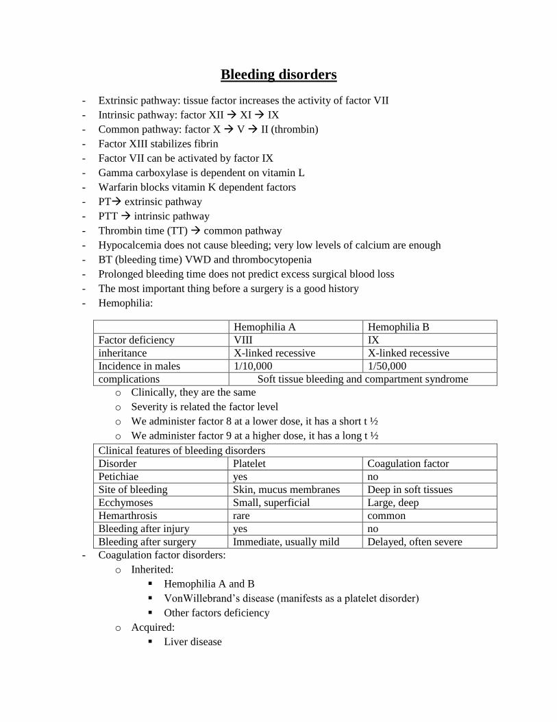
- Hemophilia:
Hemophilia A Hemophilia B
Factor deficiency VIII IX
inheritance X-linked recessive X-linked recessive
Incidence in males 1/10,000 1/50,000
complications Soft tissue bleeding and compartment syndrome
o Clinically, they are the same
o Severity is related the factor level
o We administer factor 8 at a lower dose, it has a short t ½
o We administer factor 9 at a higher dose, it has a long t ½
Clinical features of bleeding disorders
Disorder Platelet Coagulation factor
Petichiae yes no
Site of bleeding Skin, mucus membranes Deep in soft tissues
Ecchymoses Small, superficial Large, deep
Hemarthrosis rare common
Bleeding after injury yes no
Bleeding after surgery Immediate, usually mild Delayed, often severe
- Coagulation factor disorders:
o Inherited:
Hemophilia A and B
VonWillebrand’s disease (manifests as a platelet disorder)
Other factors deficiency
o Acquired:
Liver disease
Vitamin K deficiency or warfarin overdose
DIC
- F8 gene on chromosome X
- F8 intron 22 inversion is responsible for 45% of cases of hemophilia A
- Severity is related to factor level
o <1%: severe spontaneous bleeding
o 1-5%: moderate bleeding with mild injury
o 5-25% mild bleeding with surgery or trauma
- Management of hemophilia A
o Treat acute attacks with factor replacement
o Analgesics
o Evacuate for synovectomy (chemical, surgical)
o Long term prophylaxis
o Education, genetic counseling
o Screen for inhibitor twice yearly since therapy is different
o FVIII: recombinant or plasma derived
o Complications of therapy (formation of inhibitors)
10-15% of severe hemophilia A patients
1-2% of hemophilia B patients
- VonWillebrand’s disease:
o Labs:
Bleeding time: increased, normally below 10
PTT: increased
Factor VIIIc decreased, reduced because vWF is needed to carry it
vWFAg: decreased
INR: normal
Platelets: normal
Clot retraction: normal; used to exclude Glanzmann thromb.
o vWFactor:
synthesized in endothelium and megakaryocytes
forms large multimer
carries factor VIII
anchors platelet to subendothelium
bridge between platelets
o vWD
autosomal dominant
incidence: 1/10,000
causes mucocutaneous bleeding, but may manifest like hemophilia A
o lifespan of factor VIII is reduced from 12-20 hours to <2 hours
o Types:

Type 1: partial quantitative deficiency (most common)
Type 2: qualitative
Type 2A
Type 2B:
o Here only the large multimers are absent
o Association with hyperaggregation. Here, we also have
thrombocytopenia, so we cannot give DDAVP.
Type 3: total quantitative deficiency
vWF assay 1 2 3
vWF antigen decreased normal Decreased
vWF activity decreased decreased Decreased0
Multimer analysis normal Normal/abnormal Absent
o Acquired vonWillbrand syndromes:
Immune mediated
Proteoloysis
o Treatment:
Cryoprecipitate: fibrinogen, factor VIII, and vWF
DDAVP )vasopressin, antidiuretic hormone)
Stimulates vWF secretion from endothelium
Used for mild type 1
Factor VIII concentrate (Humate P): used for types 2 and 3
- DIC:
o Mechanism is through systemic activation of coagulation which leads to:
Intravascular deposition of fibrin which leads to thrombosis of small
vessels with organ failure
Depletion of platelets and coagulation factors which leads to bleeding
o Circulatory thrombin is responsible for the consumption of all the factors
o Increased PTT, PT, TT, and increased dimmers.
o Increased fibrin degradation products
o Schistocytes
o Decreased fibrinogen, decreased platelets, and increased BT
o Triggers:
Sepsis
Trauma
Malignancy
Obstetric complications
Vascular disorders
Toxins
Immunological disorders
o These triggers work by:
Release of tissue factor or thromboplastic substances into the circulation
Widespread injury to endothelial cells
o Treatment:
Treat the underlying cause
Platelet transfusion
Fresh frozen plasma
Coagulation inhibitor concentrate (antithrombin)
Anticoagulation with heparin
Monitor PT, PTT, DD, fibrinogen degradation products, and platelet count
- Thrombophilia workup:
o Mutations: methylhydrofolate reductase (the most common)
o Factors:
Factor V laden
Protein C, S
Antithrombin 3 (most severe)
Factor VIII
Antiphospholipid antibody
- Glanzmann throbasthenia
o Defect of platelet aggregation
o Life-long mucosal bleeding
o Ovarian bleeding bleeding in closed spaces
o Treatment is supportive (transfusion)
o Labs:
Normal platelet count and morphology
Prolonged bleeding time
Absent or impaired clot retraction
No aggregation with physiological aggregating agent (light doesn’t pass
through the plasma mixture). These agents include ADP, thrombin, and
collagen
Absent or reduced GPIIb-IIIa
Normal PT, PTT, and TT
o Common in Jordan
o Autosomal recessive
o no binding of fibrinogen
Platelet disorders
- Types:
o Quantitative:
Abnormal districution
Dilution effect
Decreased production
Increased destruction
o Qualitative:
Inherited:
Defects of platelet adhesion: Bernard Soulier disease, von
Willbrand disease
Defects of platelet secretion
Defects of platelet aggregation (thrombastenia)
Acquired:
Medications (aspirin, NSAID’s)
CKD
Cardiopulmonary bypass
- Platelet transfusion complications:
o Transfusion reaction:
Higher than in RBC transfusions
Bacterial contamination
o Platelet transfusion refractoriness:
Allo-immune
Non-immune:
Microangiopathic hemolytic anemia
Coagulopathy
Splenic sequestration
Fever and infection
Medications: vancomycin, interferons
- ITP (AKA ATP)
o Increased platelet destruction mediated by autoantibodies
o Characterized by decreased production of platelets despite increased
megakaryocytes in bone marrow
o Treatment:
50,000 platelet count is considered the safe cutoff value; therefore,
treatment depends on platelet count:
> 50,000: no symptoms, no treatment
50,000: if the patient is not bleeding, no treatment. If the patient is
bleeding administer steroids, IVIG, or antiD
<20,000: if the patient is not bleeding, administer steroids. If the
patient is bleeding, administer steroids, IVIG, antiD and admit.
Curative therapy:
Splenectomy
Rituximab
Rescue therapy:
High dose steroids
IVIG or anti-D
Chronic therapy: many agents including thrombopoeitin agonists
Steroids increased platelet count by increased apoptotic death of
autoantibody producing lymphocytes and down regulation of macrophage
activity responsible for platelet destruction
IVIG increases the platelets by overwhelming the reticuloendothelial
system. It interferes with platelet destruction
Anit-D: is an Ig directed against the D antigen of RH blood group system,
it raises platelet count by saturation macrophage Fc receptor with anti-D
coated RBC’s
Follow up for secondary causes of ITP such as SLE and
lymphoproliverative neoplasms.
If female, monitor during pregnancy and delivery. Make sure to provide
adequate post-delivery care and avoid using forceps for delivery
o Flashback:
Thrombocytopenia associated with shortened survival:
Immune mediated thrombocytopenia:
o ITP
o TTP
o Heparin induced thrombocytopenia (HIT)
o Drug induced thrombocytopenia
Non-immune destruction of platelets:
o DIC
o Sepsis
Multifactorial thrombocytopenia:
o Hospital associated
o Cancer associated
o Thrombocytopenia:
Associated with bleeding:
ITP
Drug induced
Associated with thrombosis:
TTP

DIC
Trosseau’s syndrome
HIT
o Heparin induced thrombocytopenia:
Suspected in:
Normal platelet count prior to heparin with decline to <100,000 or
reduction of platelet count by 50%
Onset of thrombocytopenia by day 14
Any new thrombotic event while on heparin
Skin inflammation or necrosis at heparin injection site
Exclusion of other causes of thrombocytopenia
Outcome in HIT patients:
New thrombosis in up to 50%
Amputation in 10%
Death in 10-20%
6 principles of treatment in HIT:
2 do’s
o Stop heparin
o Start new anticoagulant: donnaparoid, lepirudin, or
argatroban
2 don’t
o No warfarin until substantial platelet count recovery
o No platelet transfusion
2 diagnostics:
o Labs for HIT
o Duplex for lower limb
o TTP:
Pentad of findings:
Fever
Neurologic changes
Renal impairment
Thrombocytopenia (<20,000)
Microangiopathic hemolytic anemia (schistocytes), Hgb <10, and
lab findings of hemolysis
Other findings:
Severe deficiency of ADAM-TS13
PT, PTT,TT are normal (unlike DIC)
MRI may show leukoencephalopathy or brain infarcts

ADAM-TS13 is vWF protease; its deficiency causes ultra large
multimer production which predisposes to thrombus formations
Differential: HUS; however, in HUS ADAM-TS13 is normal
Treatment:
Initial treatment: plasma exchange (plasmapheresis) daily
Relapse: plasmapheresis + rituximab (anti CD20)
Other treatment:
o Vincristin
o Splenectomy
o Steroids
o Aspirin
Monitor LDH, platelets, clinical status, and ADAM-TS13
LDH correlates with disease activity
o Veno-thrombo embolism (VTE):
Causes:
Genetic
Environmental
Triggers
Risk factors:
Stasis
Hypercoagulability
Endothelial damage
Prophylaxis:
Pharmacological prophylaxis reduces DVT and PE by 50-65%
Bleeding risk is rare
HIT 2.4% with unfractionated heparin, 0.06% with LMWH
Prophylaxis reduces VTE’s burden
Homozygous factor V laiden patients have a very high risk for developing
VTE (20-30%)
Importance of VTE:
Preventable
Life-threatening
Long term complications
Common
Costly
The burden of VTE:
DVT:
o 40% develop post thrombotic syndrome
o 30% develop PE:
3% death
5% pulmonary hypertension
Patients >45 years of age are at a greater risk for VTE
Post DVT syndrome:
Pain (aching and cramping)
Heaviness
Itching
Swelling
Varicose veins
Brownish skin discoloration
Ulcers
Treatment:
Unfractionated heparin
LMWH
Overlap of heparin and warfarin
Other medications:
Thrombolytic therapy
Thrombectomy
IVC filter
Embolectomy
Duration of treatment is individualized
Heparin’s side effects:
HIT (early and late)
Bleeding
Hypersensitivity
Osteoporosis
Increased thyroxin
Dermatologic (alopecia)
Metabolic (hypokalemia, hyponatremia, and hypertriglyceremia)
Heparin’s antidote: protamine sulfate
LMWH antidote: factor X + fresh blood
Warfarin:
Plasma concentration peaks 2-8 hours after oral dose
99% bound to albumin
T 1/2: 25-60 hours
Inhibits vitamin K dependent factors: prothrombin, factor VII, IX,
and X.
Inhibits protein C and S
The 1st factors to decrease after warfarin administration are factor
VII and protein C
It takes 3-5 days for warfarin to start working; we usually bridge
the patients using heparin
Warfarin resistance (>20 mg per day with subtherapeutic INR)
Non-compliance
Lab errors
Excessive vitamin K intake
Mutations (rare)
Warfarin sensitivity: (<2mg per day with high INR)
15% of Caucasians
Cytocrome p450 polymorphism that decreases the rate of
metabolism
Side effects of warfarin:
Bleeding (treated with vitamin K or fresh frozen plasma)
Birth defects and abortion
Skin necrosis
Blood transfusion
- ABO system:
o O antigen is made of H substance
o A antigen is made of H substance + N-acetylgalactosamine
o B antigen is made of H substance and galactose
- Blood types, antibodies and antigens:
o A: A antigen on RBC, serum anti B
o B: B antigen on RBC, serum anti A
o AB: A and B antigen on RBC, no serum antibodies
o O: no antigens on RBC, serum anti A and anti B
- O plasma is not a common donor because it has anti-A and anti B while O RBC is a
common donor
- Blood donor criteria:
o Age (17-65)
o Weight >50
o Contact with infection
o General health
o Specific illness
- Whole blood donation (500 mL); then it can be centrifuged:
o 200 mL of packed RBC
o Platelets with plasma (can be centrifuged)
Platelet concentrate (50 mL): 5 days shelf life
Plasma (fresh frozen): 250 mL; one year shelf life
- Leukodepletion:
o Universal leukodepletion introduced in 1999 to reduce the risk of vCJD
transmission by blood
o Other benefits: less febrile reaction, less allo0immunization, less GVHD, and less
CMV
- Blood donation testing:
o Microbiology markers
o Blood grouping and screening for high titer antibodies
o Quality monitoring
- Washed RBCs:
o Prevents hemolysis and anaphylaxis
o For PNH patients and IgA deficient patients
- Irradiated RBCs:
o Prevents GVHD
o For immune-deficient patients
- RBCs shelf life:
o With citrate: 28 days

o With adenine: 42 days
- Transfusion reaction:
o Acute:
Immunologic:
Hemolytic
Febrile
Allergic
TRALI
Non-immunologic:
Circulatory overload
Hemolytic
Air embolism
Metabolic
o Delayed (>24 hours)
Immunologic:
Allo-immunization (HLA)
o Hemolytic
o Post transfusion purpura
o Graft Vs Host disease (GVHD)
o Immunedulation
Non-immunogenic:
o Iron overload
o Viral infections
o Other infections
o Protocol for all transfusion reactions:
Stop transfusions immediately
Maintain IV access with 0.9% NaCl
Check blood components for patient’s ID
Notify blood bank
Send blood sample and urine to blood bank
Keep blood unit in case culture becomes necessary
Support patient as necessary
o Transfusion transmitted disease:
HIV: 1/500,000
Hep C: 1/600,000
Hep B: 1/500,000
CMV: 50% of donors are sero-positive
Bacteria: 1/250 with platelet transfusion
o Platelet transfusion:
Platelet concentrate (random donors)

Pheresis platelets (single donor)
o Target levels:
Bone marrow suppressed patients >20,000
Bleeding/surgical patients >50,000
o Platelet transfusion complications:
Higher incidence than in RBC transfusions
Related to length of storage, leukocytes, or RBC mismatch
Bacterial contamination
o Patients with frequent platelet transfusions become refractory to transfusion
because:
Allo-immune destruction of platelets (HLA antigen)
Non-immune refractoriness:
Microangiopathic hemolytic anemia
Coagulopathy
Splenic sequestration
Fever and infection
Medications (amphotericin, vancomycin, ATG, and interferones)
- Fresh frozen plasma:
o Content: plasma with low factor V and VIII
o Indications:
Coagulation deficiencies (liver disease and trauma)
DIC
Warfarin reversal
Factor VII and XI deficiencies
o Dose: 10-15 mL/kg
- TRALI:
o Transfusion related acute lung injury
o Not rare, but underdiagnosed
o Potentially fatal
o Presents as pulmonary edema
o Occurs within 1-4 hours of starting the transfusion
o Clinical features:
Acute respiratory distress
Fever with chills
Non-productive cough
Cyanosis
Hypotension
Chest pain
Chest X-ray shows bilateral pulmonary infiltrates in the hilar region
o Pathogenesis:
Classical theory (immune TRALI)
Donor’s antibodies reacts with patient’s neutrophils
Neutrophils sequestrate in pulmonary vasculature
Cytokine and components are liberated
Damage to endothelium leading to pulmonary edema
Two-hit theory (non-immune TRALI)
Predisposing condition (sepsis, surgery, trauma, or malignancy)
Pulmonary endothelial activation and neutrophil sequestrations
Lipids and WBCs antibodies activate neutrophils which causes
endothelial damage
o TRALI management:
Non-specific
Largely supportive
Respiratory support with O2 and mechanical ventilation
Steroids
o Note: females with previous pregnancy are not allowed to donate blood because
all females produce antibodies against their husbands’ and babies’ antigens
Leukemias
- CLL:
o The most common adult leukemia
o Clues for diagnosis:
Elderly >50
Hypoglobinemia (IgA deficiencies to increased lymphocytes)
Autoimmune hemolysis (DAT positive)
CD19, CD 20
Mostly asymptomatic
Uncontrolled proliferation of mature defective B lymphocytes
o Clinical presentation:
Lymphocytosis:
Morphologically mature
Immunologically immature
Accumulation in blood, lymphatics, and bone marrow
Enlarged lymph nodes
Splenectomy
Hypogammaglobinemia: mucosal infections
o Approach:
Decide the type of lymphocyte T Vs B
Determine the stage (Rai Vs Binet systems)
Cytogenetics
Decide therapy, prognosis, and follow-up
o Staging (Rai/Binet systems)
Early: 10 year median survival
Intermediate: 5-7 years median survival
Advanced: 1-3 years median survival
o It is a heterogenous disease:
o Prognostic factors:
Lymphocytosis
Lymph node involvement
Organomegaly
Anemia
Thrombocytopenia
Lymphocyte doubling time:
>1 year: good
<1 year bad prognosis
VH gene mutation:
Unmutated: rapid progression

Mutated: slow progression
Surrogate markers ZAP70 and CD38 carry a bad prognosis
Loss of P53 carries the worst prognosis
o Treatment criteria:
Symptomatic: if the patient is asymptomatic, wait until B cell symptoms
appear
Decline in Hb or Platelets
Lymphadenopathy
Hepatosplenomegaly
Recurrent infections
o Treatment:
Rituximab- antiCD20
Chemoimmunotherapy
Chlorambucil
- CML
o Clonal expansion of hematopooetic stem cells possessing a reciprocal
translocation between chromosome 9 and 22 (Philadelphia chromosome)
o Fusion of BCR region on chromosome 22 with ABL gene from chromosome 9
o Has 3 phases:
Chronic
Accelerated
Blas crisis
o Incidence is 1.5/100,000
o Middle age (40-60)
o Accounts for 20% of adult leukemias
o Symptoms:
Insidious onset, accidental discovery
Fatigue, malaise, weight loss
Symptoms due to splenomegaly
Infections, thrombosis, bleeding
Gout
o Physical examination:
Mild to moderate splenomegaly
Mild hepatomegaly
Rare to find lymphadenopathy except in terminal stages
o Labs:
Elevated WBC’s
Elevated platelets
Normochromic, normocytic anemia
Basophilia
The cytogenic hallmark t(9:22) in 95% of patients
Accelerated phase:
Basophilia
Thrombocytopenia
Blasts between 10-20%
Blastic phase:
Blasts >20%
Hyposegmented neutrophils (Petger-Het anomaly)
Worsening of symptoms heralds progression (fever, weight loss, decreased
response to treatment, and bone pain)
o Treatment:
If not treated, converts into AML
Aims:
Reduce WBC: hematologic
Reduce gout
Target the molecular cause
Modalities:
Imatinib:
o a targeted treatment; competitive inhibition of adenosine
triphosphate binding site of the ABL kinase
o 95% of patients achieved complete hematologic remission
o 60% of patients achieved major cytogenic remission within
few months
o Side effects:
Main side effect is fluid retention, nauseam muscle
cramps, diarrhea, and skin rashes
Myleosuppression is the most common
hematological side effect
Stem cell transplant: the only definitive therapy
Others:
o Gamma interferons
o Chemotherapy
o 2nd
generation of tyrosine kinase inhibitors for failure or
relapse
o Bone marrow transplant for crisis
Response to treatment:
We cannot detect any response beyond 5log (1012
-107)
PCR is the most accurate
Mechanism of resistance to treatment:
Gene amplification
Mutation at the kinase site
Enhanced expression of multi-drug exporter proteins
Alternative signaling pathways
- AML:
o Clues:
Adult
Auer bodies
DIC – M3
No TdT markers
Blast with or without leukocytosis. The form with leukocytosis is the most
common
o Common manifestations:
Anemia
Thrombocytopenia
Neutropenia
Extramedullary infiltration: lymph nodes, skin, CNS
Hyperviscosity associated with neurological symptoms
Release of metabolites: DIC, gout, ARF
o Classification:
FAB: French-American-British classification; it is a morphological
classification
WHO classification
Cytogenetic
o Prognosis based on cytogenetics:
Favorable: t(15,17), PML-PARA (M3), t(8;21), inv(16), t(16;16)
Intermittent: t(9;11)
Unfavorable: t(6;9), inv(3)/t(3,3), d(7), complex karyotype
o Promyelocytic leukemia (M3)
Associated t(15;17) involving the retinoic acid receptor (RAR) gene
Good prognosis
Commonly associated with DIC
Prominent Auer bodies
o Treatment:
In general: correct Hb before chemotherapy, treated with anthracyclin and
RCA
M3:
Tretinoin (all trans retinoic acid (ATRA)); an oral drug that
induces the differentiation of leukemic cells bearing the t(15,17). It
is not effective in other forms of AML.

Acute M3 patients are responsive to cytarabine and daunorubcin,
but about 10% of patients treated with these drugs die from DIC
induced by the release of granule components by dying tumor
cells.
Tretinoin:
No DIC
Causes rretinoic acid syndrome(ATRA syndrome):
o In the first three weeks of treatment
o Characterized by fever, dyspnea, chest pain, pulmonary
infiltrates, effusion and hypoxia
o Treatment: steroids, chemotherapy, supportive measures
o Mortality rate: 10%
Other side effects:
o Nasal stuffiness
o Dry, red skin
o Transient increase in ALT, AST, bilirubin and
triglycerides. They rarely require any attention during
treatment
- ALL:
o Clues:
Young
Pancytopenia and bone marrow failure
Immature B cells
Positive TdT markers
Blast acute
Positive periodic acid-Schiff stain (due to glycogen rich vacuoles), but
negative peroxidase and negative non-specific esterase
Can present with acute leukemia syndrome
o Classifications:
Morphological (FAB)
L1 75%
L2 20%
L3 5%
Immunological classification:
B lineage (80%)
o Pro-B: CD19, TdT
o Common: CD19, TdT, CD10
o Pre-B: CD19, TdT, CD10, cyIg (cytoplasm Ig)
o Mature B: CD19, TdT, CD10, cyIg, smIg (surface Ig)
T lineage:

o Pre-T: CD7, TdT
o Mature T: CD7, TdT, CD2
Molecular abnormalities with prognostic importance:
Better prognosis:
o Normal karyotype
o Hyperdiploidy
Poor prognosis:
o t(8;14)
o t(4;11)
very poor prognosis:
o t(9;22); Philadelphia chromosome
o Risk classification in ALL:
Standard risk
High risk
Very high risk
o High risk ALL:
Pre-T
Pro-B
Age >35
WBC >30 in B-ALL; >100 in T-ALL
o Treatment:
Determinant:
Risk qualification
Immunophenotype of leukemic cells
Age and biological condition
Goal of treatment
Remission induction treatment in ALL:
Anti-neoplastic treatment:
o Drugs: steroid, vincristine, asparginase, cyclophosphamide
o Duration: 4-8 weeks
o 1-2 courses
CNS prophylaxis: via methotrexate intrathecally
Supportive care
Treatment of complications
Post remission therapy in standard risk ALL:
Maintenance: 6-mercatopurine, methtroxate
Intensification treatment periodically
CNS prophylaxis
Post remission therapy in high risk ALL:
Intensification treatment
Hematopoietic stem cell transplant
Treatment results:
Complete remission in 80-85% of adults, and 95-99% of children
Leukemia free survival in 30-40% of adults and 70-80% of
children
Splenomegaly is unusual in acute leukemias
- Acute leukemias (ABCDEF)
o Acute
o Blast predominance
o Children
o Drastic course
o Elderly
o Fever
- Chronic leukemias:
o Mature predominance
o Middle age
o Less drastic course
o Usually no fever
- Summary of treatment:
o ALL: vincristin, prednisone, laspraginase, anthracyclin
o AML: anthracyclin, cytarabin
o Acute pro-myelocytic leukemia: all trans retinoic acid
o CLL: no treatment if asymptomatic; clorambucil and rituximab
o CML: imatinib, gamma intereferon
o Hodgkin (IA, IB): radiotherapy
Lymphomas
- Common features:
o Painless lymph node enlargement
o B-symptoms (fever, night sweats, weight loss)
o Compression symptoms secondary to enlarged lymph nodes
o Extra-nodal involvement
o Needs lymph node biopsy for diagnosis
o Each have different histology types
o Both have similar staging systems
- Non-Hodgkin lymphoma (NHL):
o Each type of lymphoma can be viewed as a lymphocyte arrested at a certain stage
of development and transformed into a malignant cell
o 85% are of a B-cell origin
o 15%: T-cell or null all
o Etiology:
Idiopathic: most common
Immune suppression:
Congenital (Wiskott Aldrich)
Organ transplant (cyclosporine)
AIDS
Aging
DNA repair defects:
Ataxia telangectasia
Xeroderma pigmentosa
Chronic inflammation and antigenic stimulation:
Helicobacter pylori- stomach
Chalmydia psittaci – ocular adnexia
Sjogren’s syndrome
Viral causes:
EBV and Burkitt lymphoma
HTLV-1 and T-cell leukemia
HTLV-V and cutaneous T cell lymphoma
Hepatitis C
o Diagnosis:
Chromosome changes:
T(14:18) in follicular lymphoma (bcl oncogene)
T(8:14) and others in Burkitt lymphoma (c-myc oncogene)
T(11:14) in mantle cell lymphoma (cyclin D1 gene)
o Staging:

Ann Arbor
Same for NHL and HD
I: 1 lymph node region or structure
II: >1 lymph node region or structure; same side on diaphragm
III: both sides of diaphragm
IV: extra nodal sites, diffuse
A: no systemic symptoms other than pruritis
B: presence of B cell symptoms
E: extra nodal extension
Revised European American lymphoma classification:
Indolent: follicular
Aggressive
Very aggressive (Burkitt, lymphoblastic lymphoma)
o Frequency of NHL subtypes in adults:
30% diffuse large B-cell
20% follicular
o Prognostic factors in non-Hodgkin’s:
Adverse factors: age >60, stage III and IV
High serum LDH: indicating high turnover
Performance status (ECOG 2 or more)
More than one extra-nodal site involved
o Treatment options in advanced indolent lymphoma:
Observation only
Radiotherapy at the site of the problem
Systemic chemotherapy:
Oral agents: chlorambucil and prednisone
IV agents: CHOP, COP-R, FC-R
Anti-CD20: rituximab
Stem cell or bone marrow transplant
o Treatment options for aggressive lymphoma:
Potentially curable
Disseminate through blood stream: early
Must use systemic chemotherapy:
CHOP-R 8 cycles
CHOP-R 3 cycles followed by radiotherapy
Bone marrow transplant in some cases
CHOP-R: cycophosphamide, Hydroxydaunirubcin, vincrystin, prednisone,
Rituximab
Intrathecal chemotherapy for AIDS and CNS involvement
Radiotherapy for spinal cord compression and bulky disease
- Hodgkin disease:
o With appropriate treatment about 85% of patients with Hodgkin’s disease are
curable
o Treatment based on stage:
IA, IB: radiotherapy
IIA: chemotherapy + radiotherapy
IIB, IIIA, IIIB, IVA, IVB: chemotherapy with or without radiotherapy
o Chemotherapy (ABVD)
Adriamycin
Bleomycin
Vincristin
Dacarbazine
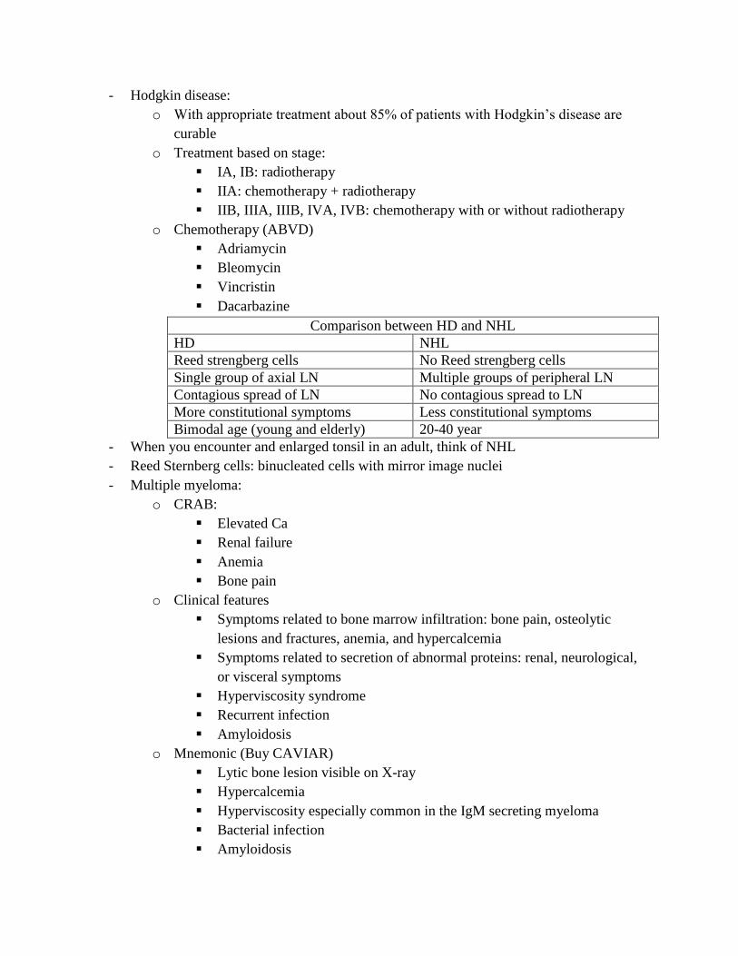
Comparison between HD and NHL
HD NHL
Reed strengberg cells No Reed strengberg cells
Single group of axial LN Multiple groups of peripheral LN
Contagious spread of LN No contagious spread to LN
More constitutional symptoms Less constitutional symptoms
Bimodal age (young and elderly) 20-40 year
- When you encounter and enlarged tonsil in an adult, think of NHL
- Reed Sternberg cells: binucleated cells with mirror image nuclei
- Multiple myeloma:
o CRAB:
Elevated Ca
Renal failure
Anemia
Bone pain
o Clinical features
Symptoms related to bone marrow infiltration: bone pain, osteolytic
lesions and fractures, anemia, and hypercalcemia
Symptoms related to secretion of abnormal proteins: renal, neurological,
or visceral symptoms
Hyperviscosity syndrome
Recurrent infection
Amyloidosis
o Mnemonic (Buy CAVIAR)
Lytic bone lesion visible on X-ray
Hypercalcemia
Hyperviscosity especially common in the IgM secreting myeloma
Bacterial infection
Amyloidosis

Renal failure: occurs in 50% of patients because most of the light chains
are toxic to the tubules
o Work-up:
CBC and blood film: roloux formation
ESR, Ca, creatinine
Albumin
Bone marrow biopsy and aspirate
Serum proteins and electrophoresis and immune-fixation
Skeletal survey: plain X-ray better than a bone scan because lytic lesions
do not show well on a bone scan
Quantitative immunoglobulins
Bence Jones protein
o Durie-Salmon staging system for multiple myeloma disease burden (tumor load)
Stage I:
Hb >10
Normal bone or solitary plasmacytoma
Low immunoglobulin spike (M-component)
o IgG < 5, IgA <3
o Bence Jone’s protein <4g/24 hours
Stage II:
IIA: normal renal function (Cr <2)
IIB: abnormal renal function (Cr >2)
Stage III:
Hb <8.5
Serum Ca >12
Multiple lytic bone lesions on X-ray
High M component
o IgG >7, IgA >5
o Bence-Jone’s protein >12g/24 hours
o International staging system:
I: good prognosis:
Serum albumin >3.5 g/dL
Serum B2 microglobulin <3.5 mg/dL
II: between I and III
III: B2 microglobulin >5 mg/dL
o Treatment:
Standard chemotherapy:
Dexa and thalidomide
Dexa and Bartezomib (Velcade)
Melphalane and prednisone for elderly

High dose chemotherapy:
Bone marrow transplant
Peripheral stem cell transplant
Myeloproliferative neoplasms
- Myeloid malignancies:
o EML
o AML
o Polycythemia rubra vera (PRV)
o Essential thrombocytopenia (ET)
o Myelofibrosis (MF)
- PRV, ET, and MF: compose the chronic myeloproliferative disorders (CMPN)
- Common features of CMPN:
o Each has specific diagnostic criteria, but they share some characteristics
o Increased number of one or more myeloid cells
o Splenomegaly
o Hypercatabolism: weight loss and gout (AML)
o Clonal marrow hyperplasia without dysplasia
o Predispose to evolve into AML
o Generalized pruritis (after bathing)
o Unusual thrombosis (Budd Chiari syndrome)
- Polycythemia rubra vera:
o Clinical features:
Palpable spleen
Enlarged liver
JAKII mutation
Elevated leukocyte alkaline phosphatase (LAP)
Bone marrow shows erythroid hyperplasia and increased number of
megakaryocytes
EPO is not diagnostic but suggestive
10% converts into AML
o Diagnostic tools:
JAKII mutation
Normal or decreased erythropoietin
Increased RBC with normal saturation
o Mutations in CMPN (due to activation of STAT3/5)
Gain of function in JAKII, MPL, CBL
Loss of function in LNK and NF1
o JAKII:
Gain of function presents in:
95% of PRV
23-57% of ET
43-57% of cases of MF

o Risk classification:
Low risk:
Age <60
No previous thrombosis
High risk:
Age >60
Previous thrombosis
o Diagnostic criteria for PRV (you need A1 + A2 with one more A criteria or 2
more B criteria)
A criteria:
A1: raised RBC mass
A2: Normal O2 saturation and EPO
A3: palpable spleen
A4: no BCR-ABL fusion (absent Philadelphia chromosome)
B criteria:
B1: thrombocytosis >400 x 109
B2: neutrophilia: >10 x109
B3: radiological splenomegaly
Endogenous erythroid colonies
o Treatment of PRV:
Phlebotomy (Hct <45%)
Low dose aspirin
Hydroxyurea or interferon gamma
Busulphan in elederly
Manage CVS risk factors
Allopuranol
Increased water intake
- Treatment of ET:
o Hydroxyurea
o Aspirin if microvascular disturbance
o Manage cardiovascular risk
- Myelofibrosis:
o Teardrop cells
o Bone marrow shows hypercellularity with grade II fibrosis