hematology case conference - hemepathreviewhemepathreview.com/hemeconferences/10-07-03.pdf ·...
TRANSCRIPT
Hematology Case
Conference
10/7/03
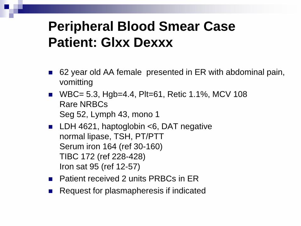
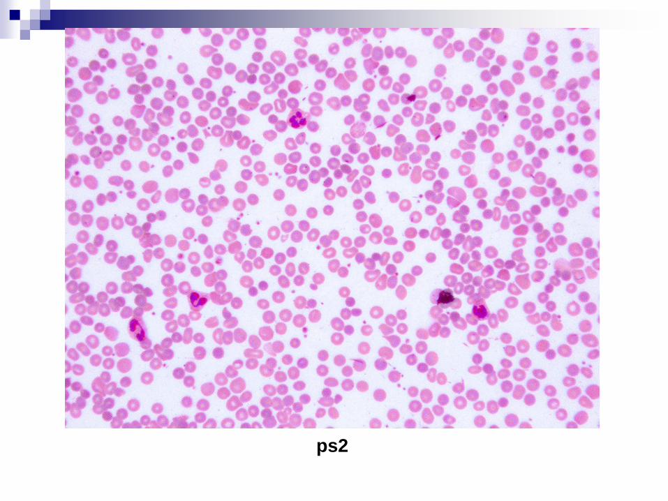
Peripheral Blood Smear Case
Patient: Glxx Dexxx
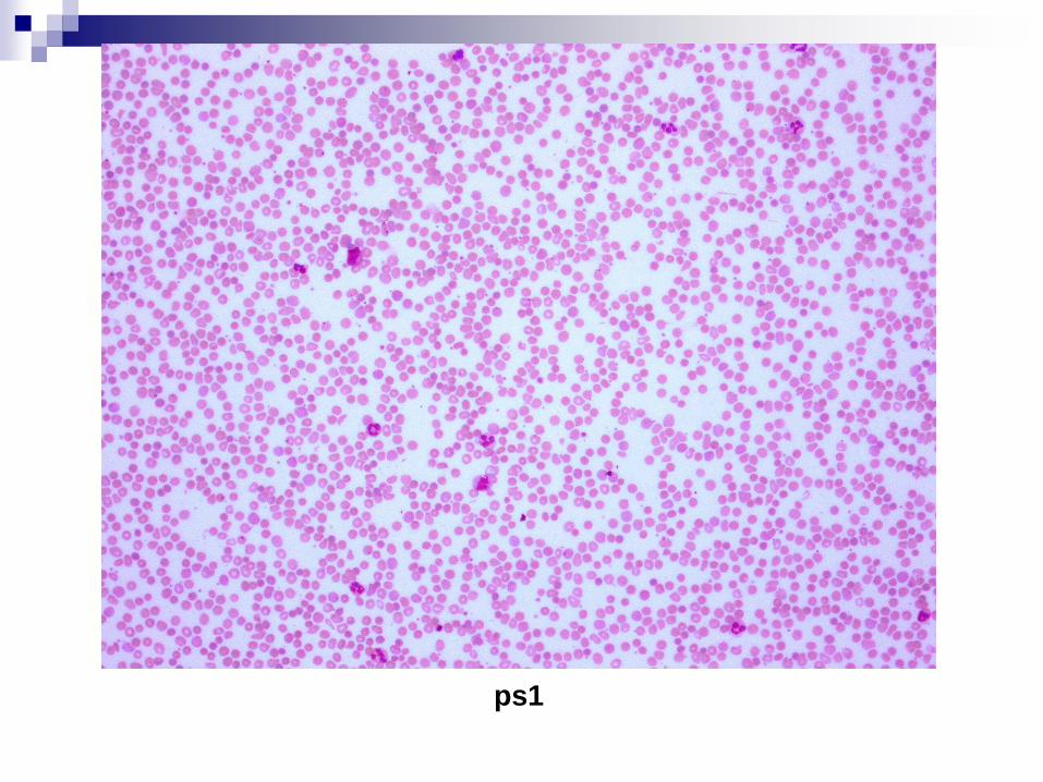
62 year old AA female presented in ER with abdominal pain,
vomitting
WBC= 5.3, Hgb=4.4, Plt=61, Retic 1.1%, MCV 108
Rare NRBCs
Seg 52, Lymph 43, mono 1
LDH 4621, haptoglobin <6, DAT negative
normal lipase, TSH, PT/PTT
Serum iron 164 (ref 30-160)
TIBC 172 (ref 228-428)
Iron sat 95 (ref 12-57)
Patient received 2 units PRBCs in ER
Request for plasmapheresis if indicated
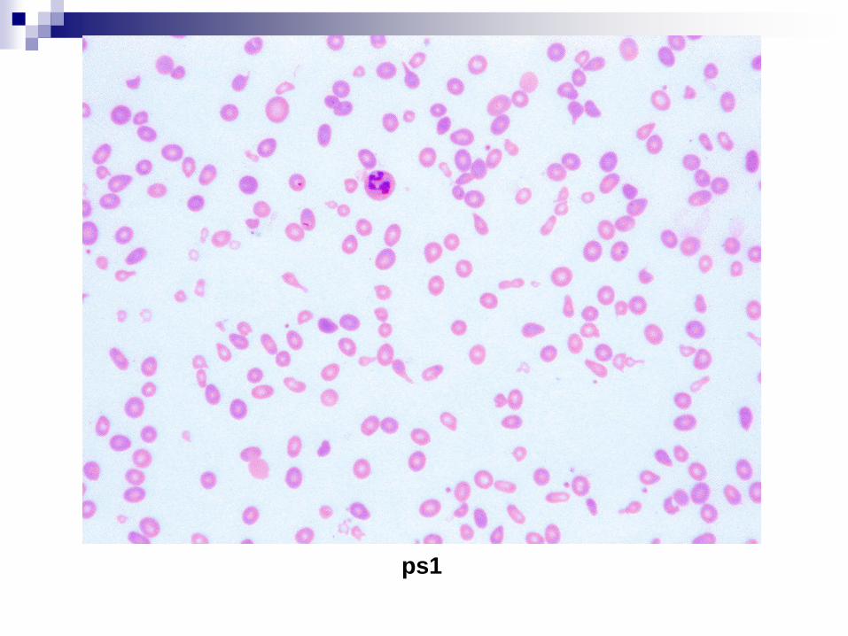
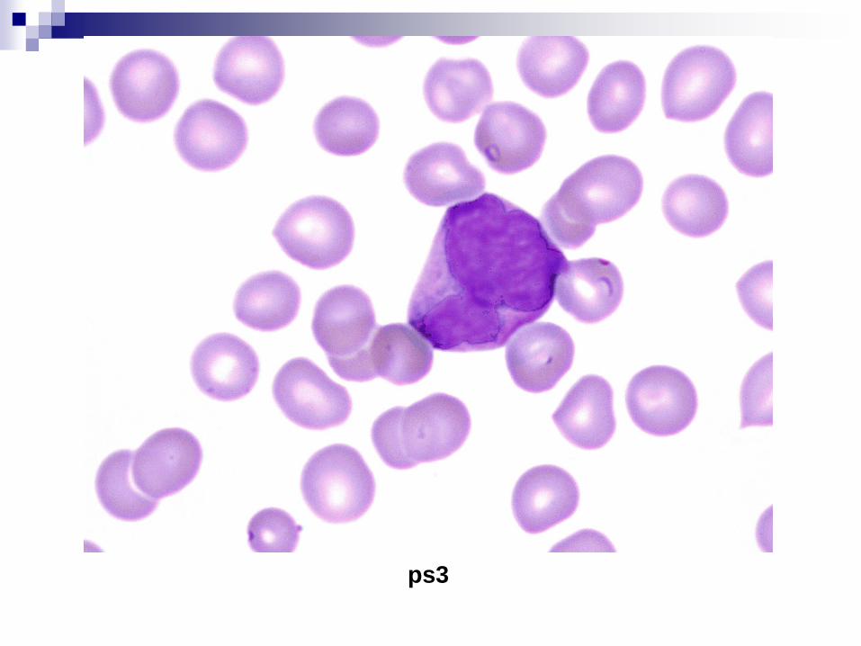
ps1
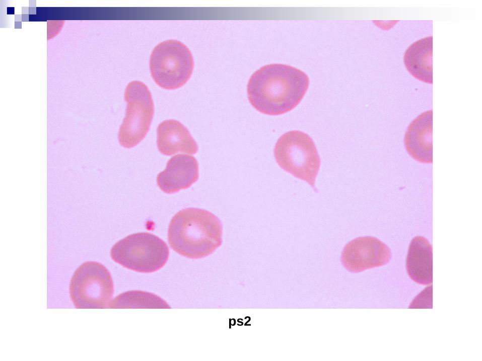
ps2

ps3
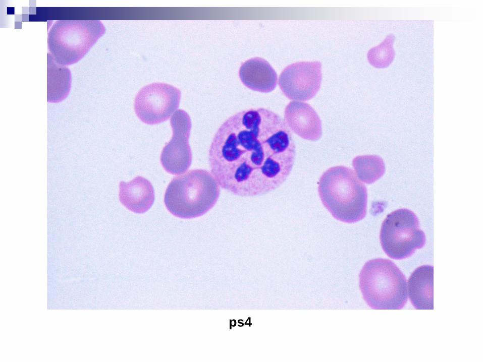
ps4
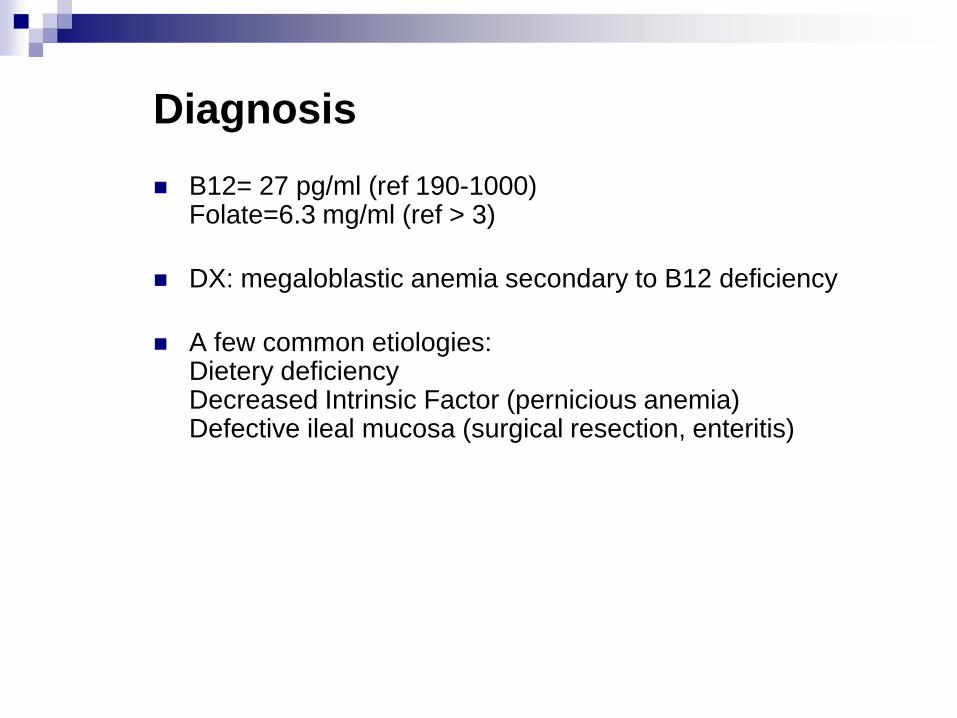
Diagnosis
B12= 27 pg/ml (ref 190-1000) Folate=6.3 mg/ml (ref > 3)
DX: megaloblastic anemia secondary to B12 deficiency
A few common etiologies: Dietery deficiency Decreased Intrinsic Factor (pernicious anemia) Defective ileal mucosa (surgical resection, enteritis)
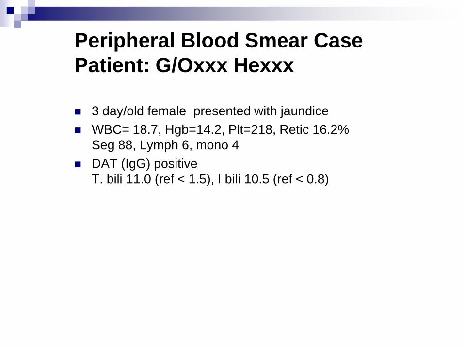
Peripheral Blood Smear Case
Patient: G/Oxxx Hexxx
3 day/old female presented with jaundice
WBC= 18.7, Hgb=14.2, Plt=218, Retic 16.2%
Seg 88, Lymph 6, mono 4
DAT (IgG) positive
T. bili 11.0 (ref < 1.5), I bili 10.5 (ref < 0.8)
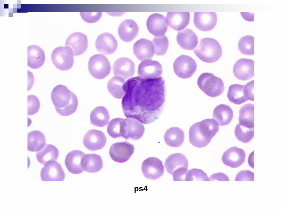
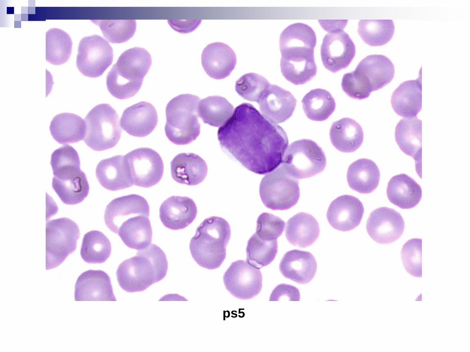
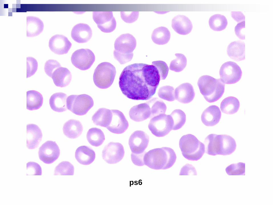
ps1
ps2
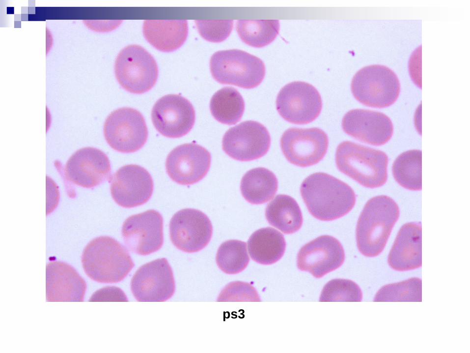
ps3
ps4

ps5
ps6

Diagnosis
Patient blood group B pos
Mother O pos, negative antibody screen
Dx: ABO hemolytic disease of the newborn HDN
(fetal-maternal incompatibility)
ABO HDN
Common but usually not severe
Mother Gr O, fetus Gr A, B, or AB Can occur in 1 st pregnancy
Secreated A or B substance crosses placenta-> production of maternal IgG isotype of Anti-A or Anti-B-> Anti-A or Anti-B IgG cross placenta to attach to the fetal RBCs
Poor avidity, typically occurs 3-4 days after delivery
Sequestration of IgG-sensitized RBCs by spleen and liver-> hepatospelomegaly, extravascular hemolysis
Tx: phototherapy to oxidize bilirubin in the infant’s skin
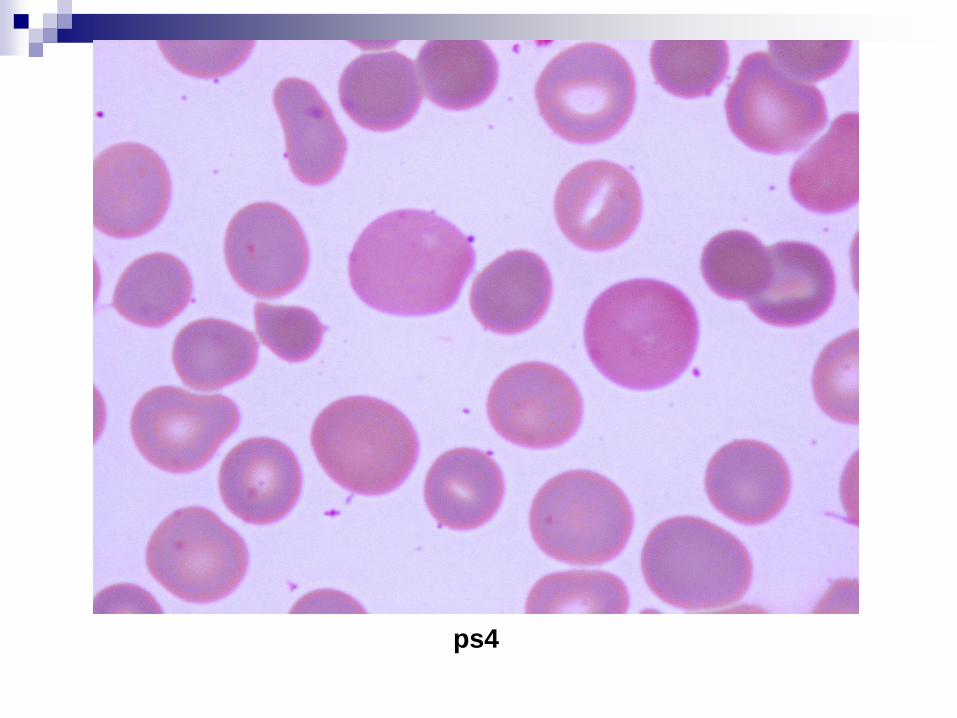
Peripheral Blood Smear Case
Patient: Hexxx Jixxx
28 year old male presented at MNW with H/A, recent hx of
brusing. Patient developed intracranial hemorrhage and was
transferred to Hermann only after a few hours at MNW
No significant past medical hx
WBC= 21.4, Hgb=11.5, Plt=6
Abnormal DIC panel
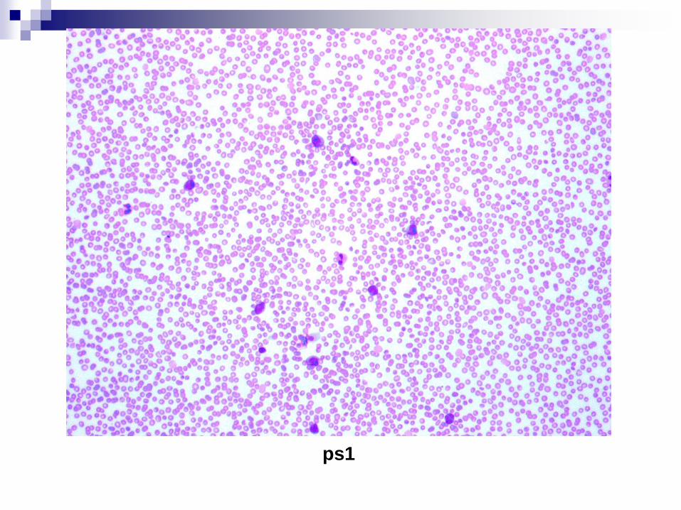
ps1
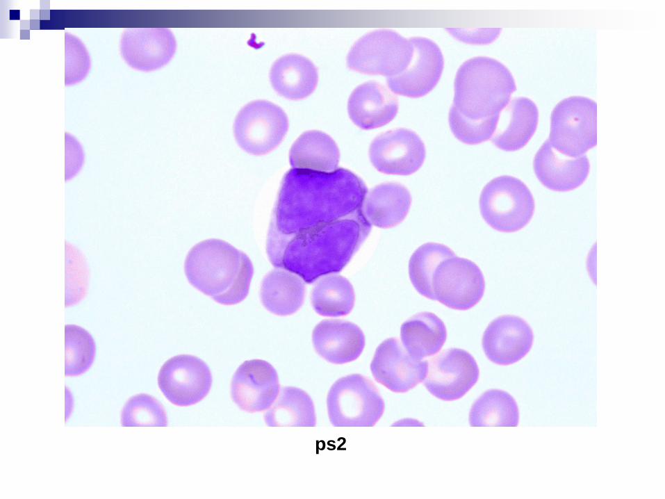
ps2
ps3
ps4
ps5
ps6
ps7
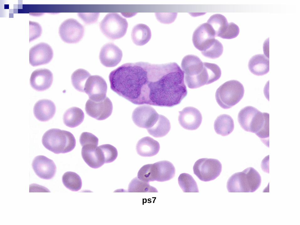
Diagnosis
Acute promyelocytic leukemia, hypogranular variant
Peripheral blood was submitted for flow cytometry
immunophenotyping and cytogentics but were subsequently
cancelled
Acute promyelocytic leukemia,
hypogranular variant Small granules in most leukemic promyelocytes (also known
as “microgranular variant”)
Predominant promyelocytes with bilobed nuclei (“apple-core” nuclei)
Peripheral blood typically shows increase in leukocytes with may leukemic promyelocytes, a few blasts
Immunophenotypes: (+) CD13, CD33 (-) HLA-DR, CD34
High risk of DIC
Tx: all trans-retinoic acid (ATRA) + anthracycline
Prognosis: relatively favorable after initial risk of DIC, ~ AML with t(8;21) or inv(16)
Acute promyelocytic leukemia, Genetics
Translocation t(15; 17) retinoic acid receptor alpha (RARα) gene on 17q12 PML gene on 15q22
Normally, RARα protein forms heterodimers with retinoid X receptor protein (RXR) -> activate transcription (differentiation)
With t(15; 17), PML-RARα fusion protein binds to PML and RXR proteins-> repress transcription (differentiation)
Other translocations (resistant to all-trans retinoic acid): t(5;17) t(11, 17)
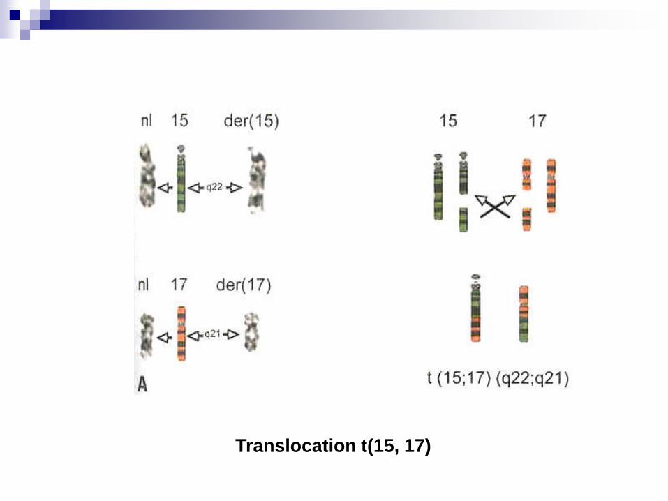
Translocation t(15, 17)
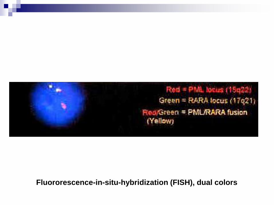
Fluororescence-in-situ-hybridization (FISH), dual colors
DIC in Acute promyelocytic leukemia,
possible mechanisms
High level of annexin II on APL cells-> accelerate formation of
plasmin by t-PA-> fibrinolysis
APL cells contain tissue factor -> activate extrinsic pathway
APL cells activate factor X directly with a cysteine proteinase (~solid tumors)
APL cells have large amount of Interleukin-1-> increase synthesis of tissue factor by endothelial cells
DIC worsens with lysis of APL cells. ATRA promotes differentiation of promyelocytes to mature forms and alleviate DIC
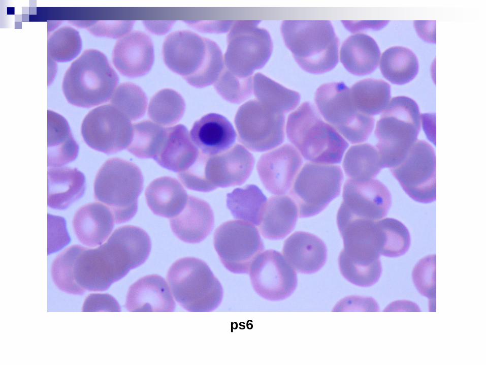
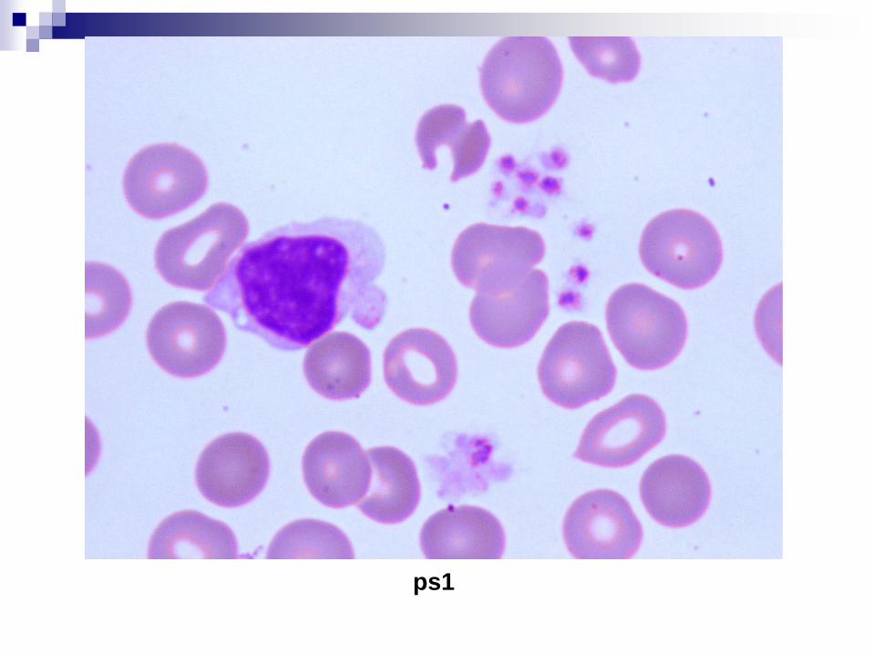
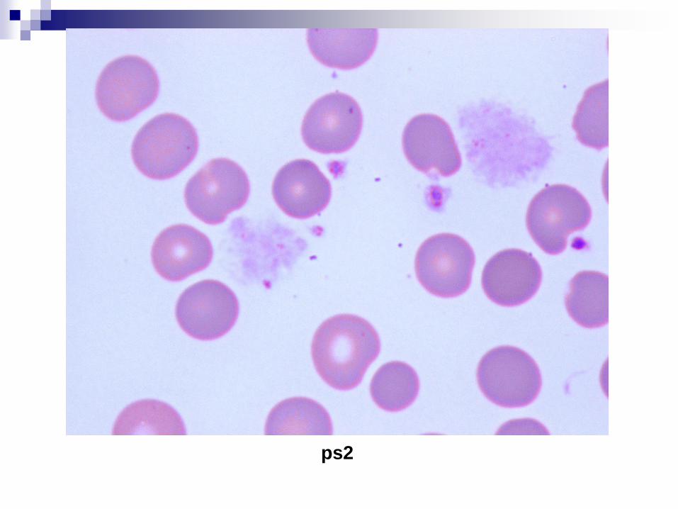
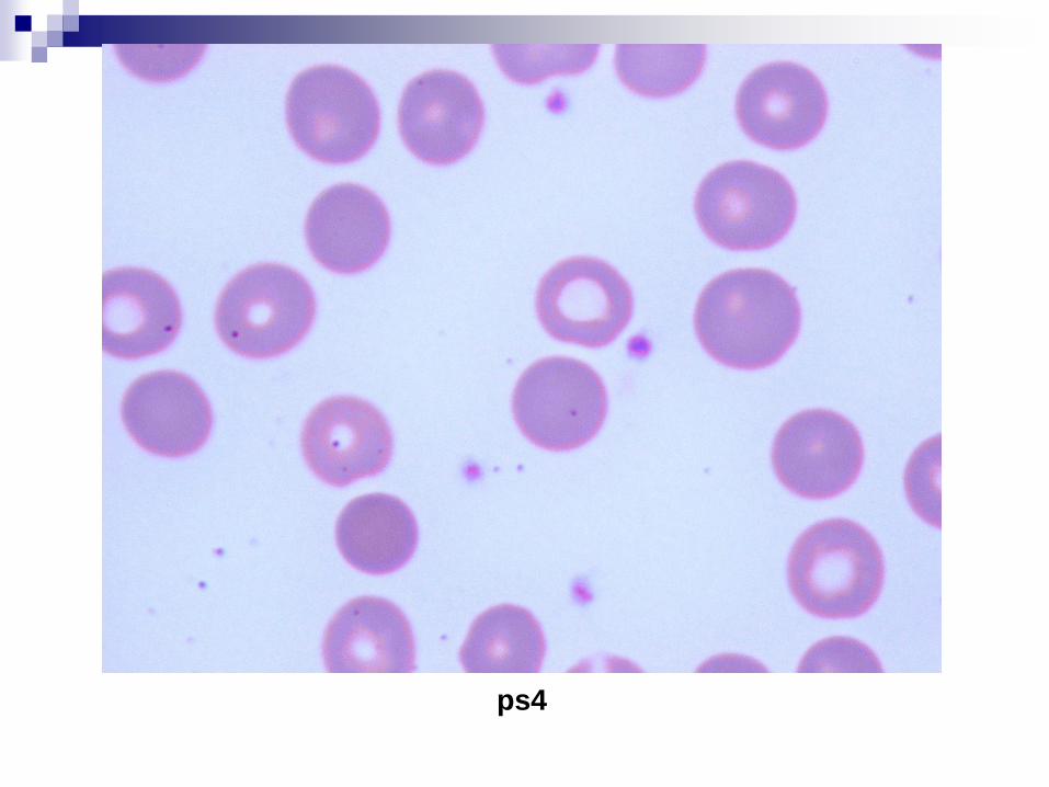
Peripheral Blood Smear Case
Patient: Dxx Mcxxx
66 year old male with Cardiology service
WBC= 24.6, Hgb=8.1, Plt=76
Seg 59, Lymph 28, mono 8
ps1
ps2

ps3
ps4
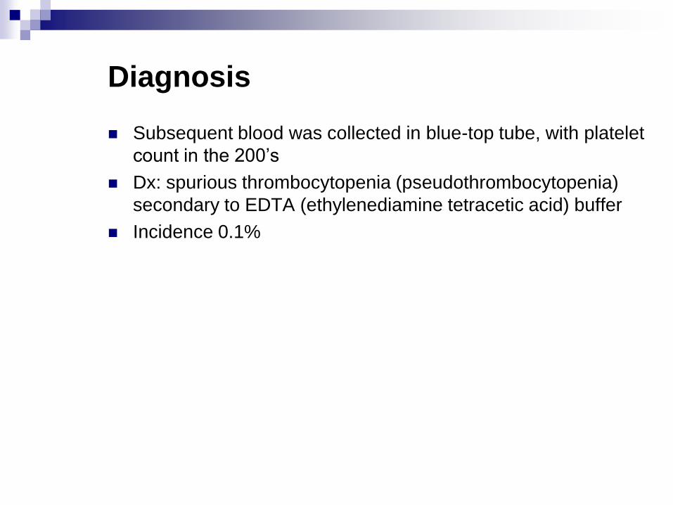
Diagnosis
Subsequent blood was collected in blue-top tube, with platelet
count in the 200’s
Dx: spurious thrombocytopenia (pseudothrombocytopenia)
secondary to EDTA (ethylenediamine tetracetic acid) buffer
Incidence 0.1%
Peripheral Blood Smear Case
Patient: Joxxx Nexxx
87 year old male with pelvic fracture, admitted through trauma
service
WBC= 22.3, Hgb=12.0, Plt=384
Seg 30, Lymph 63, mono 4

ps1

ps2
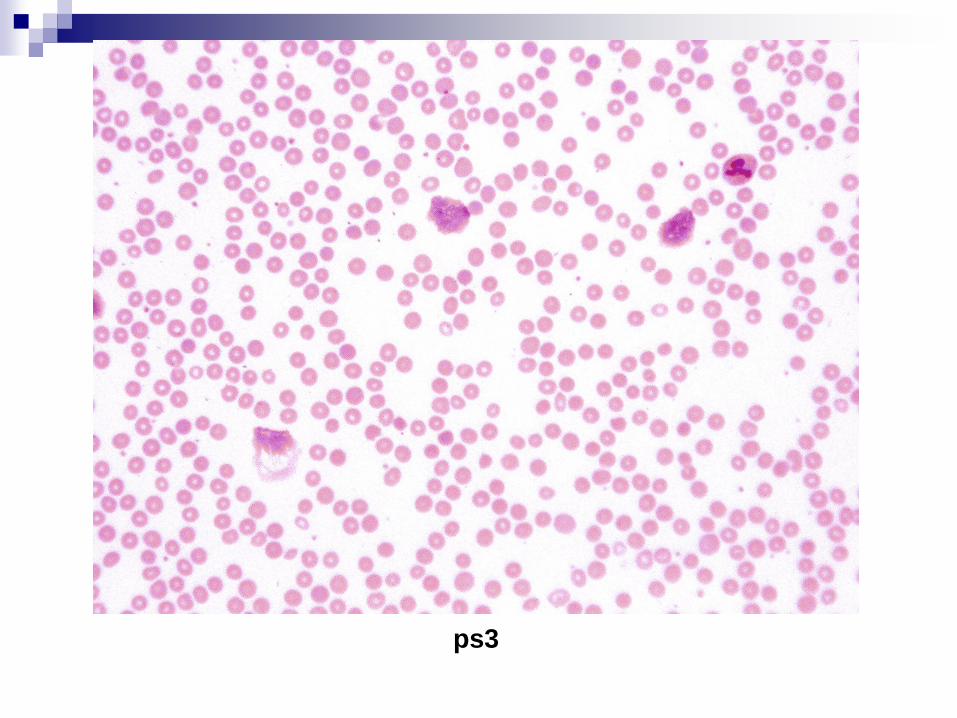
ps3
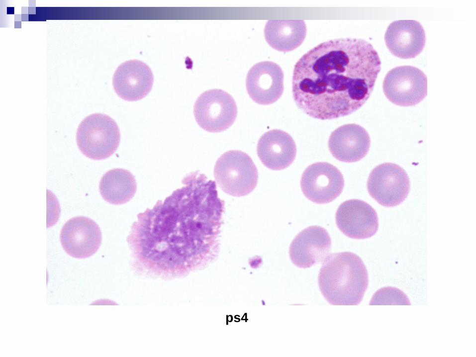
ps4
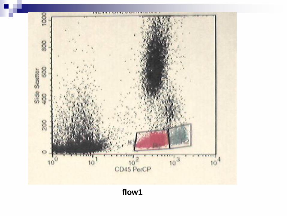
flow1
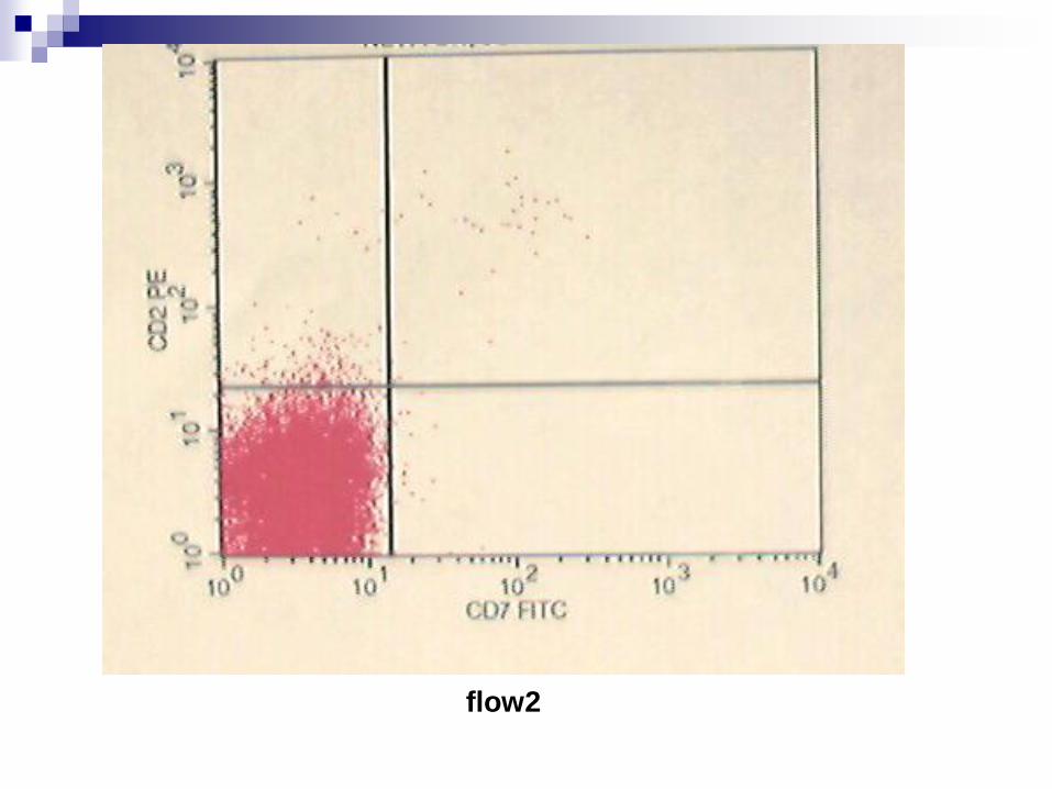
flow2
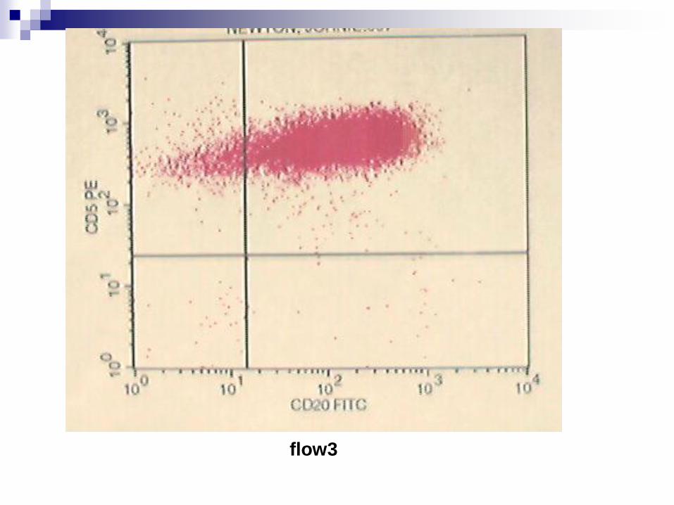
flow3
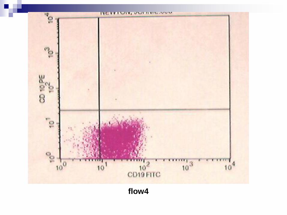
flow4
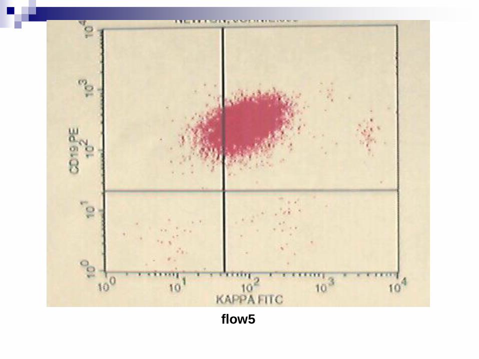
flow5
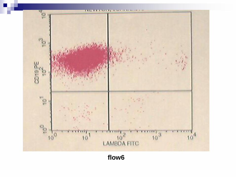
flow6
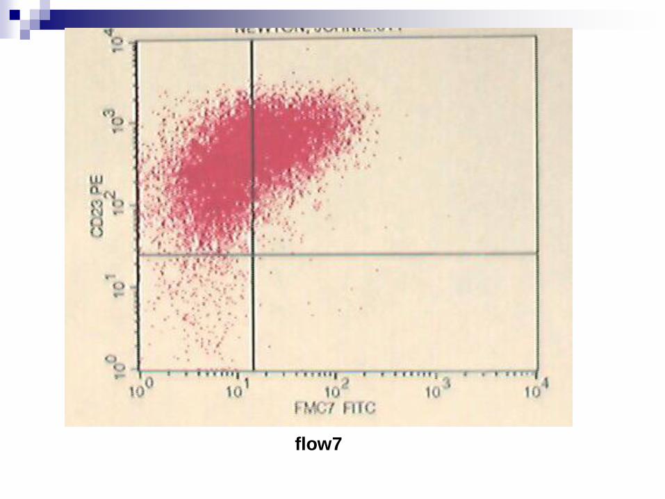
flow7
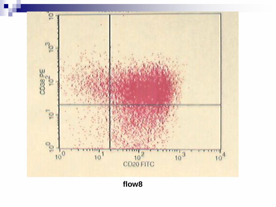
flow8
Diagnosis
Flow cytometry results: B-cells that are (+) CD5, CD19, CD20, CD23, Kappa light-chain restriction (-) CD10
Chronic lymphocytic leukemia (CLL)