guidelines for safe work practices in human and animal medical
TRANSCRIPT
Supplement / Vol. 61 January 6, 2012
U.S. Department of Health and Human ServicesCenters for Disease Control and Prevention
Morbidity and Mortality Weekly Report
Guidelines for Safe Work Practices in Human and Animal Medical Diagnostic Laboratories
Recommendations of a CDC-convened, Biosafety Blue Ribbon Panel
Supplement
The MMWR series of publications is published by the Office of Surveillance, Epidemiology, and Laboratory Services, Centers for Disease Control and Prevention (CDC), U.S. Department of Health and Human Services, Atlanta, GA 30333.Suggested citation: Centers for Disease Control and Prevention. [Article title]. MMWR 2011;60(Suppl):[inclusive page numbers].
Centers for Disease Control and PreventionThomas R. Frieden, MD, MPH, Director
Harold W. Jaffe, MD, MA, Associate Director for ScienceJames W. Stephens, PhD, Director, Office of Science Quality
Stephen B. Thacker, MD, MSc, Deputy Director for Surveillance, Epidemiology, and Laboratory ServicesStephanie Zaza, MD, MPH, Director, Epidemiology and Analysis Program Office
MMWR Editorial and Production StaffRonald L. Moolenaar, MD, MPH, Editor, MMWR SeriesChristine G. Casey, MD, Deputy Editor, MMWR Series
Teresa F. Rutledge, Managing Editor, MMWR SeriesDavid C. Johnson, Lead Technical Writer-Editor
Lynne McIntyre, Project Editor
Martha F. Boyd, Lead Visual Information SpecialistMaureen A. Leahy, Julia C. Martinroe, Stephen R. Spriggs, Terraye M. Starr
Visual Information SpecialistsQuang M. Doan, MBA, Phyllis H. King
Information Technology SpecialistsMMWR Editorial Board
William L. Roper, MD, MPH, Chapel Hill, NC, ChairmanVirginia A. Caine, MD, Indianapolis, IN
Matthew L. Boulton, MD, MPH, Ann Arbor, MIJonathan E. Fielding, MD, MPH, MBA, Los Angeles, CA
David W. Fleming, MD, Seattle, WAWilliam E. Halperin, MD, DrPH, MPH, Newark, NJ
King K. Holmes, MD, PhD, Seattle, WADeborah Holtzman, PhD, Atlanta, GATimothy F. Jones, MD, Nashville, TNDennis G. Maki, MD, Madison, WI
Patricia Quinlisk, MD, MPH, Des Moines, IAPatrick L. Remington, MD, MPH, Madison, WI
Barbara K. Rimer, DrPH, Chapel Hill, NCJohn V. Rullan, MD, MPH, San Juan, PR
William Schaffner, MD, Nashville, TNAnne Schuchat, MD, Atlanta, GA
Dixie E. Snider, MD, MPH, Atlanta, GAJohn W. Ward, MD, Atlanta, GA
CONTENTS
1. Introduction: A Culture of Safety for Diagnostic Laboratories .........2
2. Biological Risk Assessment and Biosafety Guidelines .........................7
3. Fundamental Safety Practices in Diagnostic Laboratories ............. 13
4. Tuberculosis Laboratory .............................................................................. 34
5. Autopsy/Necropsy, Surgical Pathology ................................................. 38
6. Parasitology Laboratory .............................................................................. 47
7. Mycology Laboratory ................................................................................... 52
8. Virology Laboratory ...................................................................................... 55
9. Chemistry Laboratory .................................................................................. 66
10. Hematology and Phlebotomy Laboratory ......................................... 68
11. Blood Bank ..................................................................................................... 72
12. Veterinary Diagnostic Laboratory ......................................................... 74
13. Storing, Packaging, and Shipping Infectious Substances............. 80
14. Emergency Procedures and Responsibilities .................................... 87
15. Biosafety Education .................................................................................... 91
16. Continuous Quality Improvement ........................................................ 94
References ............................................................................................................. 95
Appendix ............................................................................................................102
MMWR / January 6, 2012 / Vol. 61 1
Supplement
Guidelines for Safe Work Practices in Human and Animal Medical Diagnostic Laboratories
Recommendations of a CDC-convened, Biosafety Blue Ribbon Panel
Prepared byJ. Michael Miller, PhD1
Rex Astles, PhD2 Timothy Baszler, DVM, PhD3
Kimberle Chapin, MD4 Roberta Carey, PhD1
Lynne Garcia, MS5 Larry Gray, PhD6
Davise Larone, PhD7
Michael Pentella, PhD8
Anne Pollock, MT1
Daniel S. Shapiro, MD9
Elizabeth Weirich, MS1
Danny Wiedbrauk, PhD101National Center for Emerging and Zoonotic Infectious Diseases, CDC
2Laboratory Science, Policy and Practice Program Office, CDC3College of Veterinary Medicine, Washington State University, Pullman, WA
4Lifespan Academic Medical Centers, Providence, RI5LSG and Associates, Santa Monica, CA
6TriHealth Laboratories, Cincinnati, OH7Weill Medical College of Cornell University, New York, NY
8University of Iowa Hygienic Laboratory, Iowa City, IA9Lahey Clinic, Burlington, MA
10Warde Medical Laboratory, Ann Arbor, MI
Summary
Prevention of injuries and occupational infections in U.S. laboratories has been a concern for many years. CDC and the National Institutes of Health addressed the topic in their publication Biosafety in Microbiological and Biomedical Laboratories, now in its 5th edition (BMBL-5). BMBL-5, however, was not designed to address the day-to-day operations of diagnostic laboratories in human and animal medicine. In 2008, CDC convened a Blue Ribbon Panel of laboratory representatives from a variety of agencies, laboratory organizations, and facilities to review laboratory biosafety in diagnostic laboratories. The members of this panel recom-mended that biosafety guidelines be developed to address the unique operational needs of the diagnostic laboratory community and that they be science based and made available broadly. These guidelines promote a culture of safety and include recommendations that supplement BMBL-5 by addressing the unique needs of the diagnostic laboratory. They are not requirements but recommenda-tions that represent current science and sound judgment that can foster a safe working environment for all laboratorians.
Throughout these guidelines, quality laboratory science is reinforced by a common-sense approach to biosafety in day-to-day activities. Because many of the same diagnostic techniques are used in human and animal diagnostic laboratories, the text is presented with this in mind. All functions of the human and animal diagnostic laboratory — microbiology, chemistry, hematol-ogy, and pathology with autopsy and necropsy guidance — are addressed. A specific section for veterinary diagnostic laboratories addresses the veterinary issues not shared by other human laboratory departments. Recommendations for all laboratories include use of Class IIA2 biological safety cabinets that are inspected annually; frequent hand washing; use of appropriate disinfectants, including 1:10 dilutions of household bleach; dependence on risk assessments for many activities; development of written safety
protocols that address the risks of chemicals in the laboratory; the need for negative airflow into the laboratory; areas of the laboratory in which use of gloves is optional or is recommended; and the national need for a central site for surveillance and nonpunitive reporting of laboratory incidents/exposures, injuries, and infections.
The material in this report originated in the National Center for Emerging and Zoonotic Infectious Diseases, Beth P. Bell, MD, MPH, Director. Corresponding preparer: J. Michael Miller, PhD, Microbiology Technical Services, LLC, Dunwoody, GA 30338. Telephone: 678-428-6319; Fax: 770-396-0955; E-mail: [email protected].
2 MMWR / January 6, 2012 / Vol. 61
Supplement
1. Introduction: A Culture of Safety for Diagnostic Laboratories
This report offers guidance and recommends biosafety practices specifically for human and animal clinical diagnostic laboratories and is intended to supplement the 5th edition of Biosafety in Microbiological and Biomedical Laboratories (BMBL-5), developed by CDC and the National Institutes of Health (1). This document was written not to replace existing biosafety guidelines, but to 1) improve the safety of activities in clinical diagnostic laboratories, 2) encourage laboratory workers to think about safety issues they might not previously have considered or addressed, and 3) encourage laboratorians to create and foster a culture of safety in their laboratories. Should any of the guidelines provided herein conflict with federal, state, or local laws or regulatory requirements, the laboratorian should defer to the federal, state, or local require-ments. This culture of safety is also supported by the Clinical and Laboratory Standards Institute (2). Work in a diagnostic laboratory entails safety considerations beyond the biological component; therefore, these guidelines also address a few of the more important day-to-day safety issues that affect labo-ratorians in settings where biological safety is a major focus.
According to the U.S. Bureau of Labor Statistics, in 2008, approximately 328,000 medical laboratory technicians and technologists worked in human diagnostic laboratories in the United States. An estimated 500,000 persons in all professions work in human and animal diagnostic laboratories. Any of these workers who have chronic medical conditions or receive immunosuppressive therapy would be at increased risk for a laboratory-acquired infection (LAI) after a laboratory exposure. Precise risk for infection after exposure is unknown because determining the source or the mode of transmission often is difficult. No national surveillance system is available.
LAIs and exposures have been reported since early in the 20th century, but only in the 1970s were sufficient data available to attempt quantitative assessments of risk. Recent MMWR reports (3–11) have indicated that bacteria account for >40% of infections, with >37 species reported as etiologic agents in LAIs; however, other microbes are often implicated. Hepatitis B has been the most frequent laboratory-acquired viral infection, with a rate of 3.5–4.6 cases per 1000 workers, which is two to four times that of the general population. Any laboratorian who collects or handles tubes of blood is vulnerable (12).
Early surveys of LAIs found that laboratory personnel were three to nine times more likely than the general population to become infected with Mycobacterium tuberculosis (13,14). In a 1986 survey of approximately 4000 workers in 54 public health and 165 hospital laboratories in the United States, 3.5/1000 employee infections occurred in hospital laboratories,
and 1.4/1000 employee infections occurred in public health laboratories (15). In a 1994–1995 survey of 25,000 laboratory workers from 397 clinical laboratories in the United Kingdom, the overall rate of LAI was 18/100,000 employees (16).
In a 2005 CDC study of bacterial meningitis in U.S. labo-ratorians, Neisseria meningitidis accounted for a substantial number of LAIs. The attack rate of this organism in the general population was 13/100,000 persons. The attack rate in the gen-eral population aged 30–59 years (the estimated age range of the average laboratorian) was 0.3 per 100,000. The attack rate for microbiologists (aged 30–59 years) was 20/100,000 (17).
LAIs have also included fungal and parasitic infections. The most common agents of laboratory-acquired fungal infections are the dimorphic fungi Blastomyces, Histoplasma, and Coccidioides (18,19); most reported infections were caused by inhalation of conidia. Reported parasite-associated LAIs were caused primarily by Leishmania, Plasmodium, Toxoplasma, Chagas disease organ-ism, and other trypanosomes (20). Of the 52 cases of laboratory-acquired malaria, 56% were vector borne (from mosquitoes used in research, not clinical laboratories). Most infected health-care workers acquired infection from needle sticks during preparation of blood smears or while drawing blood.
In clinical chemistry laboratories, data from 17 New York hospitals listed needle puncture (103 cases), acid or alkali spills (46), glass cuts (44), splash in eye (19), and bruises and cuts (45) as the most frequent exposures (21). Needle puncture, glass cuts, splash in eye, and bruises and cuts have the highest potential for infection from microbes.
In the hematology laboratory, the major causes of injuries are likely to be exposure to blood and body fluids; needle sticks, aerosols from centrifuge or removal of tube stoppers, tube breakage; or contaminated gloves (22). In non-microbiology sections of the diagnostic laboratory, the primary mistake may be assuming that a given specimen contains no infec-tious agents and then working with little attention to risk for infection. This scenario can be particularly problematic in laboratories developing new technologies, such as molecular and biochemical technologies, and in point-of-care diagnos-tics performed by staff unaccustomed to testing that requires biosafety considerations and use of barrier techniques such as personal protective equipment.
1.1. Methods The risks and causes of LAIs have been documented. However,
there is a dearth of evidence-based research and publications focused on biosafety; particularly missing are studies documenting safe practices in the day-to-day operations of diagnostic laboratories.
MMWR / January 6, 2012 / Vol. 61 3
Supplement
In 2008, CDC convened a Blue Ribbon Panel of laboratory representatives from a variety of agencies, laboratory organiza-tions, and facilities to review laboratory biosafety in diagnostic laboratories. Members of the panel were either selected by the invited national laboratory organization they represented or were invited by CDC because of their roles in biosafety at the national level. The organizations participating in the panel represented the majority of laboratory technologists in the United States. In addition, some members of the panel were representatives of the biosafety community. The Blue Ribbon Panel recommended that biosafety guidelines be developed to address the unique operational needs of the diagnostic labo-ratory community and that they be science based and made available broadly.
Panel members reviewed the guidelines that were developed and synthesized by the writing team. Official endorsements by the organizations they represented were not required, although each representative was required to submit written approval of the recommendations. Edits and comments from each participant were carefully considered and incorporated where appropriate. The guidelines provided herein are synthesized and supported from systematic reviews of peer-reviewed publica-tions of evidence-based data from which recommendations could be made, justifying common-sense approaches that should be articulated, and where safe procedures have been described and proven. Because of the lack of evidence-based research in much of the current literature on biosafety practices, no attempt was made to weight the evidence and resulting recommendations (i.e., strong or weak). In the absence of supporting evidence-based research and documentation, some recommendations are based on expert opinion by international experts in the field of microbiology and must be appropriately applied until evidence-based research can substantiate their validity. The authors reviewed and approved their own sections and also evaluated how their topics accurately reflected and supported the goals of the entire document.
Each section of recommendations was reviewed both within CDC and by the relevant national organizations whose mem-bers would embrace these guidelines. These included the College of American Pathologists, Greater New York Hospital Association Regional Laboratory Task Force, American Society for Microbiology, American Clinical Laboratory Association, Association of Public Health Laboratories, American Society for Clinical Laboratory Science, American Society for Clinical Pathology, American Biological Safety Association, American Association of Veterinary Laboratory Diagnosticians, and indi-vidual physicians and subject matter experts. Future research in biosafety practices in the laboratory will contribute to further recommendations and will substantiate others as well as provide opportunities to revise this document.
1.2. RiskPersons working in clinical diagnostic laboratories are
exposed to many risks (1). Whether the patients are humans or animals and whether laboratorians work in microbiology or elsewhere in the laboratory, the human and animal diag-nostic laboratory is a challenging environment. The more that laboratorians become aware of and adhere to recommended, science-based safety precautions, the lower the risk. The goal of a safety program is to lower the risk to as close as possible to zero, although zero risk is as yet unattainable as long as patient specimens and live organisms are manipulated. Protection of laboratorians, coworkers, patients, families, and the environ-ment is the greatest safety concern.
1.3. Laboratory ExposuresLaboratory exposures occur more often than is generally
suspected. Other laboratory incidents such as minor scrapes or cuts, insignificant spills, or unrecognized aerosols occur even more frequently and might not cause an exposure that results in an LAI. In this report, “laboratory exposures” refer to events that put employees at risk for an LAI and events that result in actual acquisition of LAIs. Except for report-ing requirements imposed by CDC’s Select Agent Program, which deals with handling of specific, potentially hazardous biological agents and toxins, no national surveillance system is in place to which medical laboratory exposures and subsequent work-related infections are reported. Increased attention has been focused on laboratory biosafety and biosecurity since 2001 but has been largely limited to precautions required for agents of bioterrorism. Other laboratory exposures and LAIs continue to occur, almost always because of a breakdown of established safety protocols. Because of the lack of an official surveillance mechanism for reporting LAIs and because of the fear of punitive action by an oversight agency if injuries are reported, the data needed to determine the extent and cause of LAIs are unavailable. In addition, there is a dearth of science-based insights on prevention of LAIs.
The Blue Ribbon Panel recognizes the need for a voluntary, nonpunitive surveillance and reporting system with the poten-tial for anonymity to be implemented in the United States. Such a system would allow for reporting and evaluation of all LAIs and would potentially lead to training and interventions to facilitate a negligible incidence rate.
1.4. Routes of Laboratory InfectionThe five most predominant routes of LAIs are• parenteral inoculationswith syringe needles or other
contaminated sharps;• spillsandsplashesontoskinandmucousmembranes;
4 MMWR / January 6, 2012 / Vol. 61
Supplement
• ingestionorexposurethroughmouthpipettingortouch-ing mouth or eyes with fingers or contaminated objects;
• animalbitesandscratches(researchlaboratoriesoractivi-ties); and
• inhalationofinfectiousaerosols(1).The first four routes are relatively easy to detect, but they account
for <20% of all reported LAIs (23,24). No distinguishable exposure events were identified in approximately 80% of LAIs reported before 1978 (24–26). In many cases, the only association was that the infected person worked with a microbiological agent or was in the vicinity of a person handling a microbiological agent. The inability to identify a specific event was also reported in a more recent study (27), which found that the probable sources of LAIs were apparent in only 50% of cases. These data suggest that unsuspected infectious aerosols can play a large role in LAIs (1,23,24,28).
1.5. A Culture of SafetyThe concept of a “culture of safety,” as described in this report,
encourages all human and animal diagnostic laboratories to promote an organizational culture of systematic assessment of all work processes and procedures to identify associated risks and implement plans to mitigate those risks. In addition to the often unknown biohazard risk associated with handling diagnostic specimens, each section of the diagnostic laboratory has proce-dures and processes for handling known infectious agents that convey excessive risk for exposure and possible infection and/or occupational injury. These risks typically are associated with design flaws or lack of or inadequacy of safety procedures and training (1,2). In addition, the day-to-day operations of a human or animal diagnostic laboratory differ markedly from those of an academic or research laboratory and require different biosafety guidelines; these differences prompted the focus of this report on medical laboratory communities, their occupational risks, potential for exposure, and opportunities to mitigate those risks.
Successful establishment of a culture of safety requires that laboratory safety become an integral and apparent prior-ity to the organization, embraced first and foremost by top management and with the concomitant infrastructure sup-port required to foster safe behaviors among its employees (29–31). As required by the Clinical Laboratory Improvement Amendments, the College of American Pathologists, and other accrediting agencies, a laboratory director needs to assume the responsibility for• establishingandenforcingapolicyforacultureofsafety
within the laboratory;• identifying asmanyhazards as possible and specifying
practices and procedures that will minimize or eliminate those hazards;
• ensuringthatallpersonnelareinstructedinandengagedin performing risk assessments and demonstrating that
they can identify laboratory hazards in their individual work environments;
• ensuring that all personnel are trained and competentin the standard practices and techniques that minimize identified workplace hazards;
• providinganavenueforpersonneltoidentifyhazardsandto present risk-mitigation strategies to management; and
• educatingcliniciansandnursesaboutsafespecimenpro-curement and transport to ensure their safety and that of the laboratory personnel who receive the clinical samples.
1.6. Laboratory Design and Architectural Planning for Microbiology
Laboratory design is fundamental to the safety of labora-tory workers, hospital staff, and patients. The Clinical and Laboratory Standards Institute document, Laboratory Design; Approved Guideline (32), discusses laboratory design in detail. Because remediating poorly designed laboratory workspace is difficult, or even impossible, design warrants careful planning and consideration of safety issues. The following are sugges-tions to consider in the design or renovation of the diagnostic laboratory. Although there is no national standard requirement for an amount of space per person working in the laboratory, 300–350 sq. ft/person within a laboratory department is a reasonable figure to provide a safe work area. Ideally, allow a minimum 5-foot space between the worker (at a laboratory chair) and any object behind the worker to provide reasonable maneuverability.• Designoptions for themicrobiology laboratory should
include an enclosed component of the overall laboratory, separated by closable doors from other laboratory sections. Although not required, directional inward airflow from the main laboratory into the microbiology laboratory is also recommended in newly constructed diagnostic labo-ratories. If the facility is an open design and has no drop ceiling, the microbiology laboratory can have clear glass or Plexiglas walls, which give an appearance of openness but provide a floor-to-ceiling safety barrier from possible aerosol exposures. If a drop ceiling is in place, the clear wall needs to penetrate the deck beyond the ceiling to seal the area. In a previously constructed laboratory without directional room air, the continual operation of biologi-cal safety cabinets (BSCs) is encouraged to provide some direction to potential aerosols.
• Directionalairisencouragedtoprovidezonesofcontain-ment that proceed with increasing negative pressure toward work spaces in which higher-risk laboratory procedures are conducted. Air handling systems within the micro-biology laboratory suite must be able to be adjusted and
MMWR / January 6, 2012 / Vol. 61 5
Supplement
balanced with directional airflow from the corridor into the microbiology laboratory and from the general micro-biology laboratory into separate and enclosed tuberculosis, mycology, and virology specialty laboratories.
• Formicrobiologylaboratories,itiscriticalthatthesupervi-sor and laboratory director, along with a biosafety profes-sional, provide input regarding the special needs of a new laboratory facility. Access into the microbiology section must be restricted to staff only. The microbiology section must have a decontamination facility or have a medical waste contract in place, and it must provide a sink for hand washing. Hands-free sinks (foot-pedal operated) are required for biosafety level (BSL)-3 facilities and are recommended for BSL-2 facilities. Bench-tops must be constructed of impervious materials; laminate materials can delaminate and become difficult to disinfect. For BSCs that vent to the outside, air handling should be planned carefully to ensure that the air is vented to the outside after filtration and that the outside vents are placed away from the facility’s air intake units. For laboratories that contain multiple classes of BSCs, the hazards that are permitted to be manipulated within the specific unit need to be clearly indicated (by label) to staff (1). The general human and animal microbiology laboratory should be BSL-2.
• IfnoBSL-3 facilities are available,BSL-2plusnegativeairflow and use of respiratory precautions may be used for some agents, provided a risk assessment has been conducted.
• For human laboratories, the separate tuberculosis andvirology laboratories that manipulate cultures for iden-tification and characterization would ideally meet BSL-3 requirements. For animal diagnostic virology laboratories in which most manipulated viruses are not human patho-gens, the practice is to meet BSL-2 requirements unless a risk analysis indicates a high probability that an agent in a specimen needs BSL-3 containment. Risk assessments should be performed on each facility to include consider-ation of the specific risks encountered in each laboratory.
• Thereceivingandset-upareasinmicrobiologylaboratoriesshould be designed with sufficient space to accommodate the greatest number of specimens anticipated. This area needs a Class IIA2 BSC, a sink for hand washing, and an emergency eye wash station. Telephone jacks, computer jacks, and electri-cal outlets should be built into the module (Use of wireless technologies can reduce the need for telephone or computer wiring in each module.) along with refrigerator space for one or two side-by-side glass-front refrigerators or one double refrigerator to enable easy access by the set-up staff.
• Thegenerallaboratoryshouldcontainsit-downworkspacesdesigned with adequate space for a computer at each station.
Work benches that have storage shelves above the center of the bench might be preferred; these would provide space for supplies without cluttering the work area. Storage shelves need a 1-cm (1/2-inch) lip to ensure chemicals cannot slide off a shelf. Under-shelf lighting is best to illuminate the work area. For convenience, electrical outlets are recommended at each work station, along with telephone and computer jacks. Gas burners are no longer universally recommended.
• Ifpossible,locatecarbondioxideandanaerobicgastanksoutside the actual laboratory (preferably shielded or even installed outside the walls of the building). Placing the tanks outside the laboratory or the building in a locked area will allow easy access for exchange of tanks. Where appropriate, lines that connect gas tanks to specific areas of the laboratory should be made of synthetic tubing to allow future moving if necessary. Accommodations need to be made for daily reading of the gauges in the labora-tory unless alarms can be installed. Gas tanks should be individually secured (29).
• Ifwastewillbedecontaminatedon-sitebeforedisposal,thelaboratory must have an autoclave large enough to handle its needs. Locate the autoclave in a well-ventilated area, or ensure it is exhausted through a capture hood above it. Ideally, the mycobacteriology laboratory will have its own autoclave. Double-door autoclaves can be installed so that one side opens into the mycobacteriology labora-tory and the other side opens into a disposal area used by the laboratory for disposing of other waste. Validation of the autoclave cycles for effective decontamination of the projected loads is recommended in addition to a regular maintenance and quality-assurance program.
• Optimally,thediagnosticlaboratorywouldplanfor— a general microbiology laboratory area able to be closed
off from the main laboratory, i.e., from other laboratory disciplines;
— separate mycobacteriology, virology, and mycology rooms (under negative pressure relative to the general laboratory with a Class IIA2 BSC) with telephone and computer jacks;
— adequate space or separate rooms for quality control testing, receipt of supplies, and record storage; and
— an extra room for future expansion to offer more services, e.g., molecular or virology testing. The room might need to be renovated to accommodate a Class IIA2 BSC, directional air flow, telephone jacks, and communication devices such as intercoms. The tele-phone jacks and communication devices should be in all such rooms.
6 MMWR / January 6, 2012 / Vol. 61
Supplement
• Ensure that current and futuremicrobiology space isdesigned for an adequate number of blood culture instru-ments, automated identification instruments, automated enzyme immunoassays, nucleic acid extraction and testing platforms, and pipetting instruments; refrigerators; auto-mated Gram stainers; automated plate streakers; BSCs; freezers; and additional computer stations for optional use. Some identification instruments require at least 8 feet of footprint space for the unit, printer, and modules. If the laboratory will provide the service, it should plan for a medium-sized anaerobe chamber, about 6 feet of footprint. Risk assessments must include evaluation of the infectious aerosols that might be produced by automated procedural equipment to determine whether containment ventilation is recommended.
• The availability of board-certified laboratory specialistsin the laboratory is as important to a medical facility as highly trained, board-certified medical specialists and surgeons. Patients deserve no less if laboratory results are used to guide patient care. Additionally, diplomates of the American Board of Medical Microbiology or the American Board of Medical Laboratory Immunology or equivalent specialists in leadership positions are valuable assets to laboratories that receive and manipulate microbes. Using their skills as laboratory director or as consultant is invaluable and highly recommended. Also, technology specialists should be recruited and retained, particularly in microbiology where interpretive judgment is critical to specimen analysis and ultimately directly affects patient care and outcome.
MMWR / January 6, 2012 / Vol. 61 7
Supplement
2. Biological Risk Assessment and Biosafety Guidelines
2.1. Risk AssessmentThe laboratory director is ultimately responsible for iden-
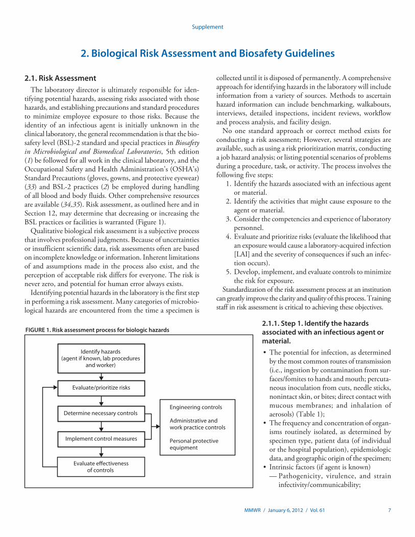
tifying potential hazards, assessing risks associated with those hazards, and establishing precautions and standard procedures to minimize employee exposure to those risks. Because the identity of an infectious agent is initially unknown in the clinical laboratory, the general recommendation is that the bio-safety level (BSL)-2 standard and special practices in Biosafety in Microbiological and Biomedical Laboratories, 5th edition (1) be followed for all work in the clinical laboratory, and the Occupational Safety and Health Administration’s (OSHA’s) Standard Precautions (gloves, gowns, and protective eyewear) (33) and BSL-2 practices (2) be employed during handling of all blood and body fluids. Other comprehensive resources are available (34,35). Risk assessment, as outlined here and in Section 12, may determine that decreasing or increasing the BSL practices or facilities is warranted (Figure 1).
Qualitative biological risk assessment is a subjective process that involves professional judgments. Because of uncertainties or insufficient scientific data, risk assessments often are based on incomplete knowledge or information. Inherent limitations of and assumptions made in the process also exist, and the perception of acceptable risk differs for everyone. The risk is never zero, and potential for human error always exists.
Identifying potential hazards in the laboratory is the first step in performing a risk assessment. Many categories of microbio-logical hazards are encountered from the time a specimen is
collected until it is disposed of permanently. A comprehensive approach for identifying hazards in the laboratory will include information from a variety of sources. Methods to ascertain hazard information can include benchmarking, walkabouts, interviews, detailed inspections, incident reviews, workflow and process analysis, and facility design.
No one standard approach or correct method exists for conducting a risk assessment; However, several strategies are available, such as using a risk prioritization matrix, conducting a job hazard analysis; or listing potential scenarios of problems during a procedure, task, or activity. The process involves the following five steps:
1. Identify the hazards associated with an infectious agent or material.
2. Identify the activities that might cause exposure to the agent or material.
3. Consider the competencies and experience of laboratory personnel.
4. Evaluate and prioritize risks (evaluate the likelihood that an exposure would cause a laboratory-acquired infection [LAI] and the severity of consequences if such an infec-tion occurs).
5. Develop, implement, and evaluate controls to minimize the risk for exposure.
Standardization of the risk assessment process at an institution can greatly improve the clarity and quality of this process. Training staff in risk assessment is critical to achieving these objectives.
FIGURE 1. Risk assessment process for biologic hazards
Identify hazards (agent if known, lab procedures
and worker)
Evaluate/prioritize risks
Determine necessary controls Engineering controls
Administrative and work practice controls
Personal protective equipment
Implement control measures
Evaluate e�ectivenessof controls
2.1.1. Step 1. Identify the hazards associated with an infectious agent or material.• Thepotentialforinfection,asdetermined
by the most common routes of transmission (i.e., ingestion by contamination from sur-faces/fomites to hands and mouth; percuta-neous inoculation from cuts, needle sticks, nonintact skin, or bites; direct contact with mucous membranes; and inhalation of aerosols) (Table 1);
• Thefrequencyandconcentrationoforgan-isms routinely isolated, as determined by specimen type, patient data (of individual or the hospital population), epidemiologic data, and geographic origin of the specimen;
• Intrinsicfactors(ifagentisknown)— Pathogenicity, virulence, and strain
infectivity/communicability;
8 MMWR / January 6, 2012 / Vol. 61
Supplement
— Mode of transmission (mode of laboratory transmission may differ from natural transmission);
— Infectious dose (the number of microorganisms required to initiate infection can vary greatly with the specific organism, patient, and route of exposure);
— Form (stage) of the agent (e.g., presence or absence of cell wall, spore versus vegetation, conidia versus hyphae for mycotic agents);
— Invasiveness of agent (ability to produce certain enzymes); and
— Resistance to antibiotics.• Indicatorsofpossiblehigh-riskpathogensthatmayrequire
continuation of work in a biological safety cabinet (BSC), such as— Slowly growing, tiny colonies at 24–48 hours with
Gram stain showing gram-negative rods or gram-negative coccobacilli;
— Slow growth in blood culture bottles (i.e., positive at ≥48 hours), with Gram stain showing small gram-negative rods or gram-negative coccobacilli;
— Growth only on chocolate agar;— Rapid growth of flat, nonpigmented, irregular colonies
with comma projections and ground-glass appearance;— Gram stain showing boxcar-shaped, gram-positive rods
with or without spores.
2.1.2. Step 2. Identify activities that might cause exposure to the agent or material.• Thefacility(e.g.,BSL-2,BSL-3,openfloorplan[more
risk] versus separate areas or rooms for specific activities
[less risk], sufficient space versus crowded space, workflow, equipment present);
• The equipment (e.g., in the case of uncertifiedBSCs,cracked centrifuge tubes, improperly maintained auto-claves, overfilled sharps containers, Bunsen burners);
• Potentialforgeneratingaerosolsanddroplets. Aerosols can be generated from most routine laboratory
procedures but often are undetectable. The following pro-cedures have been associated with generation of infectious aerosols.— Manipulating needles, syringes and sharps
º Subculturing positive blood culture bottles, making smears
º Expelling air from tubes or bottlesº Withdrawing needles from stoppersº Separating needles from syringesº Aspirating and transferring body fluidsº Harvesting tissues
— Manipulating inoculation needles, loops, and pipettesº Flaming loopsº Cooling loops in culture mediaº Subculturing and streaking culture mediaº Expelling last drop from a pipette (including
Eppendorff pipettes)— Manipulating specimens and cultures
º Centrifugationº Setting up cultures, inoculating media
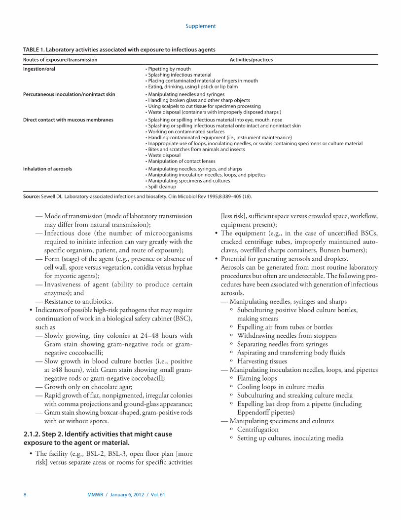
TABLE 1. Laboratory activities associated with exposure to infectious agents
Routes of exposure/transmission Activities/practices
Ingestion/oral •Pipettingbymouth•Splashinginfectiousmaterial•Placingcontaminatedmaterialorfingersinmouth•Eating,drinking,usinglipstickorlipbalm
Percutaneous inoculation/nonintact skin •Manipulatingneedlesandsyringes•Handlingbrokenglassandothersharpobjects•Usingscalpelstocuttissueforspecimenprocessing•Wastedisposal(containerswithimproperlydisposedsharps)
Direct contact with mucous membranes •Splashingorspillinginfectiousmaterialintoeye,mouth,nose•Splashingorspillinginfectiousmaterialontointactandnonintactskin•Workingoncontaminatedsurfaces•Handlingcontaminatedequipment(i.e.,instrumentmaintenance)•Inappropriateuseofloops,inoculatingneedles,orswabscontainingspecimensorculturematerial•Bitesandscratchesfromanimalsandinsects•Wastedisposal•Manipulationofcontactlenses
Inhalation of aerosols •Manipulatingneedles,syringes,andsharps•Manipulatinginoculationneedles,loops,andpipettes•Manipulatingspecimensandcultures•Spillcleanup
Source: SewellDL.Laboratory-associatedinfectionsandbiosafety.ClinMicobiolRev1995;8:389–405(18).
MMWR / January 6, 2012 / Vol. 61 9
Supplement
º Mixing, blending, grinding, shaking, sonicating, and vortexing specimens or cultures
º Pouring, splitting, or decanting liquid specimensº Removing caps or swabs from culture containers,
opening lyophilized cultures, opening cryotubesº Spilling infectious materialº Filtering specimens under vacuumº Preparing isolates for automated identification/
susceptibility testingº Preparing smears, performing heat fixing, staining
slidesº Performing catalase testº Performing serology, rapid antigen tests, wet
preps, and slide agglutinationsº Throwing contaminated items into biohazardous
wasteº Cleaning up spills
• Useofanimals;• Useofsharps;• Productionoflargevolumesorconcentrationsofpotential
pathogens;• Improperlyusedormaintainedequipment; Examples of possible hazards are decreased dexterity or
reaction time for workers wearing gloves, reduced ability to breathe when wearing N95 respirators, or improperly fitting personal protective equipment (PPE).
•Workingaloneinthelaboratory. No inherent biologic danger exists to a person work-
ing alone in the laboratory; however, the supervisor is responsible for knowing if and when a person is assigned to work alone. Because assigning a person to work alone is a facility-specific decision, a risk assessment should be conducted that accounts for all safety considerations, including type of work, physical safety, laboratory security, emergency response, potential exposure or injury, and other laboratory-specific issues.
2.1.3. Step 3. Consider the competencies and experience of laboratory personnel.• Age (younger or inexperienced employeesmight be at
higher risk);• Genetic predisposition and nutritional deficiencies,
immune/medical status (e.g., underlying illness, receipt of immunosuppressive drugs, chronic respiratory conditions, pregnancy, nonintact skin, allergies, receipt of medication known to reduce dexterity or reaction time);
• Education,training,experience,competence;• Stress,fatigue,mentalstatus,excessiveworkload;• Perception,attitude,adherencetosafetyprecautions;and• Themostcommonroutesofexposureorentryintothe
body (i.e., skin, mucous membranes, lungs, and mouth) (Table 1).
2.1.4. Step 4. Evaluate and prioritize risks.Risks are evaluated according to the likelihood of occurrence
and severity of consequences (Table 2).• Likelihoodofoccurrence
— Almost certain: expected to occur— Likely: could happen sometime— Moderate: could happen but not likely— Unlikely: could happen but rare— Rare: could happen, but probably never will
• Severityofconsequences Consequences may depend on duration and frequency of
exposure and on availability of vaccine and appropriate treatment. Following are examples of consequences for individual workers.— Colonization leading to a carrier state— Asymptomatic infection— Toxicity, oncogenicity, allergenicity— Infection, acute or chronic— Illness, medical treatment— Disease and sequelae— Death
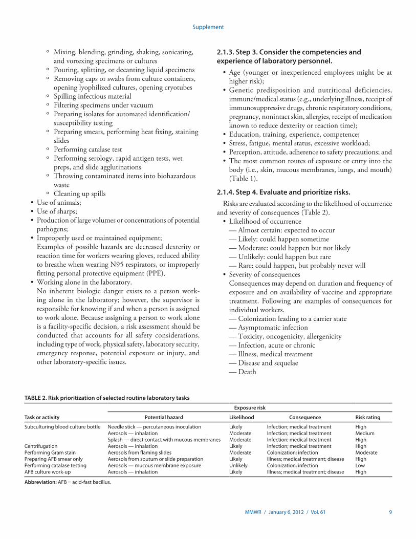
TABLE 2. Risk prioritization of selected routine laboratory tasks
Task or activity
Exposure risk
Potential hazard Likelihood Consequence Risk rating
Subculturing blood culture bottle Needle stick — percutaneous inoculation Likely Infection;medicaltreatment HighAerosols — inhalation Moderate Infection;medicaltreatment MediumSplash—directcontactwithmucousmembranes Moderate Infection;medicaltreatment High
Centrifugation Aerosols — inhalation Likely Infection;medicaltreatment HighPerforming Gram stain Aerosols from flaming slides Moderate Colonization;infection ModeratePreparing AFB smear only Aerosols from sputum or slide preparation Likely Illness;medicaltreatment;disease HighPerforming catalase testing Aerosols — mucous membrane exposure Unlikely Colonization;infection LowAFBculturework-up Aerosols — inhalation Likely Illness;medicaltreatment;disease High
Abbreviation:AFB=acid-fastbacillus.
10 MMWR / January 6, 2012 / Vol. 61
Supplement
2.1.5. Step 5. Develop, implement, and evaluate controls to minimize the risk for exposure.• Engineeringcontrols If possible, first isolate and contain the hazard at its source.
— Primary containment: BSC, sharps containers, cen-trifuge safety cups, splash guards, safer sharps (e.g., autoretracting needle/syringe combinations, disposable scalpels), and pipette aids
— Secondary containment: building design features (e.g., directional airflow or negative air pressure, hand wash-ing sinks, closed doors, double door entry)
• Administrativeandworkpracticecontrols— Strict adherence to standard and special microbiological
practices (1)— Adherence to signs and standard operating procedures— Frequently washing hands— Wearing PPE only in the work area— Minimizing aerosols— Prohibiting eating, drinking, smoking, chewing gum— Limiting use of needles and sharps, and banning recap-
ping of needles— Minimizing splatter (e.g., by using lab “diapers” on
bench surfaces, covering tubes with gauze when opening)— Monitoring appropriate use of housekeeping, decon-
tamination, and disposal procedures
— Implementing “clean” to “dirty” work flow— Following recommendations for medical surveillance
and occupational health, immunizations, incident reporting, first aid, postexposure prophylaxis
— Training— Implementing emergency response procedures
• PPE(asalastresortinprovidingabarriertothehazard)— Gloves for handling all potentially contaminated mate-
rials, containers, equipment, or surfaces— Face protection (face shields, splash goggles worn with
masks, masks with built-in eye shield) if BSCs or splash guards are not available. Face protection, however, does not adequately replace a BSC. At BSL-2 and above, a BSC or similar containment device is required for procedures with splash or aerosol potential (Table 3).
— Laboratory coats and gowns to prevent exposure of street clothing, and gloves or bandages to protect nonintact skin
— Additional respiratory protection if warranted by risk assessment
• Jobsafetyanalysis One way to initiate a risk assessment is to conduct a job
safety analysis for procedures, tasks, or activities performed at each workstation or specific laboratory by listing the steps involved in a specific protocol and the hazards
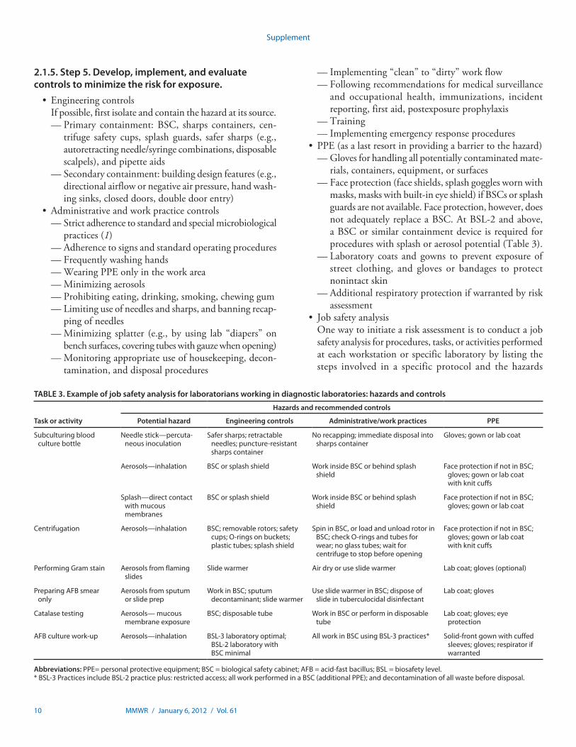
TABLE 3. Example of job safety analysis for laboratorians working in diagnostic laboratories: hazards and controls
Task or activity
Hazards and recommended controls
Potential hazard Engineering controls Administrative/work practices PPE
Subculturing blood culture bottle
Needle stick—percuta-neous inoculation
Safersharps;retractableneedles;puncture-resistantsharps container
Norecapping;immediatedisposalintosharps container
Gloves;gownorlabcoat
Aerosols—inhalation BSC or splash shield Work inside BSC or behind splash shield
FaceprotectionifnotinBSC;gloves;gownorlabcoatwithknitcuffs
Splash—direct contact withmucousmembranes
BSC or splash shield Work inside BSC or behind splash shield
FaceprotectionifnotinBSC;gloves;gownorlabcoat
Centrifugation Aerosols—inhalation BSC;removablerotors;safetycups;O-ringsonbuckets;plastictubes;splashshield
Spin in BSC, or load and unload rotor in BSC;checkO-ringsandtubesforwear;noglasstubes;waitforcentrifuge to stop before opening
FaceprotectionifnotinBSC;gloves;gownorlabcoatwithknitcuffs
Performing Gram stain Aerosols from flaming slides
Slidewarmer Airdryoruseslidewarmer Labcoat;gloves(optional)
Preparing AFB smear only
Aerosols from sputum or slide prep
WorkinBSC;sputumdecontaminant;slidewarmer
UseslidewarmerinBSC;disposeofslide in tuberculocidal disinfectant
Labcoat;gloves
Catalase testing Aerosols— mucous membrane exposure
BSC;disposabletube Work in BSC or perform in disposable tube
Labcoat;gloves;eyeprotection
AFBculturework-up Aerosols—inhalation BSL-3laboratoryoptimal;BSL-2laboratorywithBSC minimal
AllworkinBSCusingBSL-3practices* Solid-frontgownwithcuffedsleeves;gloves;respiratorifwarranted
Abbreviations: PPE=personalprotectiveequipment;BSC=biologicalsafetycabinet;AFB=acid-fastbacillus;BSL=biosafetylevel.*BSL-3PracticesincludeBSL-2practiceplus:restrictedaccess;allworkperformedinaBSC(additionalPPE);anddecontaminationofallwastebeforedisposal.
MMWR / January 6, 2012 / Vol. 61 11
Supplement
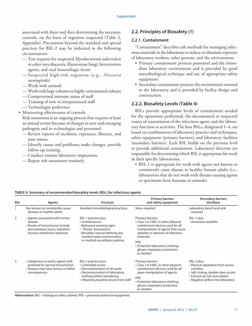
TABLE 4. Summary of recommended biosafety levels (BSL) for infectious agents
BSL Agents PracticesPrimary barriers
and safety equipmentSecondary barriers
(facilities)
1 Notknowntoconsistentlycausediseases in healthy adults
Standard microbiological practices Nonerequired Laboratory bench and sink required
2 •Agentsassociatedwithhumandisease
•Routesoftransmissionincludepercutaneousinjury,ingestion,mucous membrane exposure
BSL-1practiceplus:•Limitedaccess•Biohazardwarningsigns•“Sharps”precautions•Biosafetymanualdefininganyneededwastecontaminationor medical surveillance policies
Primary barriers:•ClassIorIIBSCorotherphysical
containment devices used for all manipulations of agents that cause splashes or aerosols of infectious materials
PPE:•Protectivelaboratoryclothing;gloves;respiratoryprotectionas needed
BSL-1plus:•Autoclaveavailable
3 •Indigenousorexoticagentswithpotential for aerosol transmission
•Diseasemayhaveseriousorlethalconsequences
BSL-2practiceplus:•Controlledaccess•Decontaminationofallwaste•Decontaminationoflaboratory
clothing before laundering•Obtainingbaselineserumfromstaff
Primary barriers:•ClassIorIIBSCorotherphysical
containment devices used for all open manipulation of agents
PPE:•Protectivelaboratoryclothing;gloves;respiratoryprotectionas needed
BSL-2plus:•Physicalseparationfromaccess
corridors•Self-closing,double-dooraccess•Exhaustairnotrecirculated•Negativeairflowintolaboratory
Abbreviation: BSC=biologicalsafetycabinet;PPE=personalprotectiveequipment.
associated with them and then determining the necessary controls, on the basis of organism suspected (Table 3, Appendix). Precautions beyond the standard and special practices for BSL-2 may be indicated in the following circumstances:— Test requests for suspected Mycobacterium tuberculosis
or other mycobacteria, filamentous fungi, bioterrorism agents, and viral hemorrhagic fevers
— Suspected high-risk organism (e.g., Neisseria meningitidis)
— Work with animals— Work with large volumes or highly concentrated cultures— Compromised immune status of staff— Training of new or inexperienced staff— Technologist preference
•Monitoringeffectivenessofcontrols Risk assessment is an ongoing process that requires at least
an annual review because of changes in new and emerging pathogens and in technologies and personnel.— Review reports of incidents, exposures, illnesses, and
near-misses.— Identify causes and problems; make changes, provide
follow-up training.— Conduct routine laboratory inspections.— Repeat risk assessment routinely.
2.2. Principles of Biosafety (1)
2.2.1. Containment“Containment” describes safe methods for managing infec-
tious materials in the laboratory to reduce or eliminate exposure of laboratory workers, other persons, and the environment.• Primarycontainmentprotectspersonnelandtheimme-
diate laboratory environment and is provided by good microbiological technique and use of appropriate safety equipment.
• Secondarycontainmentprotectstheenvironmentexternalto the laboratory and is provided by facility design and construction.
2.2.2. Biosafety Levels (Table 4)BSLs provide appropriate levels of containment needed
for the operations performed, the documented or suspected routes of transmission of the infectious agent, and the labora-tory function or activities. The four BSLs, designated 1–4, are based on combinations of laboratory practice and techniques, safety equipment (primary barriers), and laboratory facilities (secondary barriers). Each BSL builds on the previous level to provide additional containment. Laboratory directors are responsible for determining which BSL is appropriate for work in their specific laboratories.• BSL-1isappropriateforworkwithagentsnotknownto
consistently cause disease in healthy human adults (i.e., laboratories that do not work with disease-causing agents or specimens from humans or animals).
12 MMWR / January 6, 2012 / Vol. 61
Supplement
• BSL-2isappropriateforhandlingmoderate-riskagentsthat cause human disease of varying severity by ingestion or by percutaneous or mucous membrane exposure (i.e., human and animal clinical diagnostic laboratories).
• BSL-3isappropriateforworkwithindigenousorexoticagents that have a known potential for aerosol transmis-sion and for agents that can cause serious and potentially fatal infections (e.g., tuberculosis laboratories).
• BSL-4isreservedforworkwithexoticagentsthatposeahigh individual risk for life-threatening disease by infec-tious aerosols and for which no treatment is available (e.g., laboratories working with Ebola, Marburg, and pox viruses). These high-containment laboratories have complex and advanced facility requirements.
2.3. Material Safety Data Sheets for Organisms and Chemicals
Material Safety Data Sheets (MSDS) for chemicals are avail-able from the manufacturer, supplier, or an official Internet site. The Division of Occupational Health and Safety, National Institutes of Health, has promulgated guidelines for handling genetically manipulated organisms and has additional instruc-tions for accessing MSDS (http://dohs.ors.od.nih.gov/mate-rial_safety_data_main.htm).
2.4. Biosafety Manual• The laboratorydirector is responsible forensuringthat
a laboratory-specific biosafety manual is developed, adopted, annually reviewed, and accessible to all laboratory
personnel. All laboratory employees must read this manual, and the director must maintain records of personnel who have read it.
• Themanualshouldbereviewedandupdatedannuallyandwhenever procedures or policies change. Annual training in biosafety practices is recommended for all personnel who access the laboratory. Recommended topics include the following.— Institutional and laboratory safety policies— Management, supervisor, and personnel responsibilities— Regulations and recommended guidelines— Routes of exposure in the laboratory— Risk assessment and reporting of exposures— Biosafety principles and practices— Standard precautions for safe handling of infectious
materials— Standard operating procedures— Hazard communication and biohazard signs— Engineering controls— Administrative and work practice controls— PPE— When and how to work in a BSC— Transport of biohazardous materials— Emergency procedures— Decontamination and disposal of biohazardous waste— Training program and documentation— Medical surveillance and exposure evaluation
procedures
MMWR / January 6, 2012 / Vol. 61 13
Supplement
Many safety procedures, guidelines, and principles apply to all sections of the diagnostic laboratory. The recommenda-tions presented in this section represent a broad view of safety throughout the laboratory. More detailed recommendations can be found in Biosafety in Microbiological and Biomedical Laboratories (BMBL-5) and in the World Health Organization’s Laboratory Biosafety Manual (1,36).
Hospitals, clinical laboratories, state and local health depart-ments, CDC, and the American Society for Microbiology have established and/or published guidelines to follow when suspected agents of bioterrorism have been or could be released in the community. However, routine clinical laboratory testing may provide the first evidence of an unexpected bioterrorism event. Routine clinical specimens also may harbor unusual or exotic infectious agents that are dangerous to amplify in culture. These agents are often difficult to identify, and the routine bench technologist might continue work on the culture by passage, repeated staining, nucleic acid testing, neutraliza-tion, and other methods. This continued workup places the technologist and others in the laboratory at risk for infection. Ideally, these specimens are not to be processed or tested in the routine laboratory, and they can be removed from the testing stream if the suspected agent is known. Relationships with the state public health laboratory, and subsequently with the Laboratory Response Network, are critical in this effort.
Once the testing process has begun, the bench technologist must have clear and concise instructions about when to seek assistance from the laboratory supervisor and/or director.
3.1. Specimen Receiving and Log-In/Setup Station•Microbiologyspecimensaretobereceivedinuncontami-
nated containers that are intact and are consistent with laboratory specimen collection policy.
• Use of pneumatic tubes for transport of specimens isacceptable for most specimens but might be contraindi-cated for specimens without sealed caps, such as urine cups; these are to be delivered by hand (see 3.1.6). Adopt specific standard operating procedures (SOPs) in the event that irreplaceable specimens are considered for transportation using these systems.
• Ideally,allspecimensinabiosafetylevel(BSL)2orhigherfacility are to be processed in a biological safety cabinet (BSC) adhering to safe BSC practices. If a BSC is unavail-able in the laboratory, the laboratorian processing intake specimens must wear a laboratory coat and gloves, employ
an effective splash shield, and continue to follow universal precautions. Additional precautions may be necessary if warranted by site-specific risk assessments.
• Limittheuseofa4-foot-wideBSCforinoculatingplatesand preparing smears to one employee at a time, wear-ing appropriate personal protective equipment (PPE). Six-foot-wide BSCs may accommodate additional test-ing equipment or materials. Check the manufacturer’s recommendations before allowing two employees to work simultaneously in the larger cabinet, and then allow only after a risk assessment.
•MinimalPPEforthegeneralsetupareaisgownandgloves.In microbiology, a surgical-type mask is recommended, but optional if the BSC is used. For mycobacteriology and virology laboratories where organism manipulation is conducted, workers should wear a fit tested N95 res-pirator or select other appropriate respiratory protection, as indicated by the risk assessment. An N95 respirator is usually not required for biocontainment levels up through BSL-2, although it provides a higher degree of protection than a surgical mask. Safe BSC practices are to be adhered to at all times. Mycobacterial, fungal, viral, and molecular specimens may require specific additional safeguards.
3.1.1. Leaking containers• Submitspecimenstothelaboratoryintransportbagsthat
isolate the patient requisition from specimens; always limit bags to one patient to prevent misidentification and cross-contamination.
• Requestanewspecimenifacontainerisbrokenorhasspilled its contents. These containers are unacceptable for culture because the contents may have become contami-nated. Document the incident, and notify the supervisor if an exposure occurred.
• Visually examine containers for leaks upon arrival andbefore placing on rockers, in centrifuges, in racks, in closed-tube sampling (cap-piercing probe) systems, in automated aliquot stations or automated slide preparation systems, or on conveyor belts.
• Trackanddocumentallincidentsofcrackedtubes,loosecaps, and leaking containers. Increases in documented events may indicate the need to clarify or strengthen speci-men acceptance policies or improve specimen collection or transportation practices, or they might identify defective container lot numbers.
3. Fundamental Safety Practices in Diagnostic Laboratories
14 MMWR / January 6, 2012 / Vol. 61
Supplement
• Considerallsputumcontainersascomingfrompatientswith tuberculosis or pneumonia, and handle with care. External contamination caused by inappropriate lid closure can contaminate the gloves of the laboratorian and all contents of the BSC. If the specimen is leaking or contaminated, consider rejecting it and requesting another specimen if feasible. Change and discard gloves after disinfection and cleanup. (A 1:10 bleach solution or appropriate disinfectant is recommended.) Document the external contamination for reporting purposes.
• Considerallbloodculturebottlesascomingfrompatientspotentially infected (e.g., with human immunodeficiency virus [HIV] or hepatitis), and handle appropriately. If any concern exists about external contamination, carefully disinfect the outside of the tubes or bottles before insert-ing them into the blood culture instruments. Change and discard gloves after cleanup and decontamination of the immediate area. Document the external contamination for reporting purposes.
• Leakingstoolcontainerscanbeahazardtothetechnolo-gist, could contaminate the laboratory, or could present an opportunity for specimen comingling and/or con-tamination that could produce a false result. These should be rejected, if feasible, and a new specimen requested. Otherwise, disinfect the outside of the container before culturing the contents, and change and discard gloves before proceeding. Document the external contamination for reporting purposes.
• Viral specimenswith damaged or leaking containersmay need to be discarded before opening. Contact the supervisor for instructions on whether or not to continue processing, and be prepared to notify the submitter and request another specimen.
3.1.2. Visible contamination of the outside of containers• Considerallspecimencontainersaspotentiallycontaminated.• Donotrelyonvisibleexternalcontaminationtoconfirm
the potential source of contamination.•Wipeoffvisiblecontaminationbyusingatowelorgauze
pad moistened with acceptable decontaminant, such as a 1:10 dilution of household bleach, or use the established laboratory disinfectant. Ensure label and bar code are not obscured before advancing specimen for analysis.
3.1.3. Loose caps• Alwaysgraspthetubeoroutsideofthespecimencontainer,
not the stopper or cap, when picking up tubes or specimen containers to prevent spills and breakage.
• Ensuretopsaretightlysecuredonallspecimencontainers,blood-collection tubes, and sample tubes before advancing for analysis or storage.
3.1.4. Operational procedures• Ensure that specimen placement, specimen flow, and
bench operational workflow are unidirectional (i.e., from clean areas to dirty areas) and uniform for all operators to maximize effective use of engineering controls.
• DetermineappropriatePPEonthebasisofdocumentedrisk and hazard assessments of all the operations performed at each bench. Try to incorporate engineering controls and PPE information in the same location in all procedure manuals, and clearly post the information for each opera-tion carried out at the bench.
• Ensure that workstation proceduremanuals includeinstructions for the organization of all instruments, materi-als, and supplies in each area as well as instructions for any cleaning and disinfection and the frequency of cleaning and disinfection for all surfaces and instruments.
• Supervisorsaretoroutinelyinspectforcleanlinessofthebench.
• Havewrittenprocedures for nonlaboratory operations,e.g., technical instrument maintenance, in-house or contracted maintenance, emergency response, housekeep-ing, and construction and utility operations, to mitigate exposures associated with assigned operational tasks.— Write nonlaboratory operation procedures for non-
laboratory service providers with their input and consultation.
— Document the training and assess the competency of service providers and bench operators for all written nonlaboratory operational bench procedures.
• Generalbacteriologystainsmayconstitutebothachemicalor biological hazard.— Gram stain. Crystal violet, methylene blue, potassium
iodide, and ethanol are all irritants; crystal violet is also car-cinogenic and somewhat toxic; ethanol is a hepatotoxin.
— Other risks associated with Gram stain procedures include handling live organisms, the potential for creating aerosols, and the potential for skin and envi-ronmental contamination.
— Eye protection (safety glasses or chemical splash goggles) and disposable gloves are recommended during staining or preparing stains. Gloves provide protection from the live organisms as smears are prepared and provide protection from unintentional exposure to stain.
— Place contaminated waste in a biohazard bag for dis-posal. Use biohazard bags only once and then discard them. Never wash or reuse them.
MMWR / January 6, 2012 / Vol. 61 15
Supplement
— For all other stains, including fluorescent conjugates, refer to the Material Safety Data Sheets associated with each stain or chemical.
• Equipment decontamination. Examine equipment contami-nated with blood or other potentially infectious materials before servicing or shipping, and decontaminate as nec-essary. Contact the manufacturer for decontamination process.
• If decontamination of equipment or portions of suchequipment is not feasible, do the following.— Label the equipment with a biohazard symbol and a
second label specifically identifying which portions remain contaminated.
— Convey this information to all affected employees and servicing representatives before handling, servicing, or shipping so that appropriate precautions will be taken.
3.1.5. Manual removal of sealed caps; specimen aliquotting and pipetting• Alwaysremovecapsbehindabench-fixedsplashshield,or
wear additional PPE appropriate to protect from splashes and aerosols.
• Placeagauzepadoverthecap,andthenslowlypryorpushthe cap off with an away-from-body motion. Never reuse a gauze pad; doing so might contribute to cross-contam-ination. Several manufacturers market safety devices to help remove caps from tubes and to break open ampoules (e.g., Current Technologies Saf De-Cap [Fisher Health Care, Houston, TX] and the Pluggo [LPG Consulting, Inc., Wood River, IL]).
• Use automated or semiautomated pipettes and safetytransfer devices.
3.1.6. Pneumatic tube systems• EstablishSOPsforuseanddecontaminationofthepneu-
matic tube system (PTS).• BreakageorleakageofspecimenstransportedusingaPTS
risks contamination of the transport system itself.• BaselimitationsonuseofthePTSonacompleterisk/
hazard assessment. Limit specimen size, volume, weight, and container types sent through the tube system, if war-ranted. This applies particularly to cytology specimens and certain types of urine containers.
• PlaceallspecimenssentthroughaPTSinasealedzip-lockbag.• Testbags,andensuretheyareleakproofunderthecondi-
tions in the PTS.• Protect requisition forms by a separate pouch, or
enclose them in a separate secondary bag to prevent contamination.
• Azip-lockbagmustcontainspecimensfromonlyonepatient.
• Place absorbentwaddingbetweenpatient bags to helpabsorb spills and minimize contamination to the outside of the carrier.
• Handlecontaminatedpneumatictubecarriersinaccor-dance with standard precautions.
• Disinfectcontaminatedcarrierswithbleachsolutionorother disinfectant following the protocol recommended by the manufacturer and approved by the hospital’s infection control committee if the system is in use in a hospital.
•WeargloveswhenopeningPTScarrierscontainingpatientspecimens.
• Decontaminatetheoutsideoftubecarriersbeforereturn-ing them to patient-care areas. Decontaminate the inside of the carrier if a leak occurs in the specimen container.
• Establish a facility hotline for immediately reportingproblems with the PTS.
• EstablishanemergencyPTSshutdownplan, includingroles and responsibilities; include implementation of an alternative specimen transport plan.
• Develop a system to track and analyze incidents ofimproperly closed carriers, cracked tubes, loose caps, and leaking containers. Increases in documented events may indicate the need to clarify or strengthen PTS-use poli-cies or improve specimen collection practices, and could identify defective carriers and/or container lot numbers.
• PrepareSOPsforbothlaboratoryoperatorsandthenonlab-oratory service providers with their input and consultation.
• Documenttrainingandcompetencyassessmentofserviceproviders and bench operators for PTS maintenance and decontamination procedures. Documented training and assessment of competency will include knowledge of the risks associated with using a PTS and the precautions to be taken to control those risks.
3.2. Personal Precautions.If engineering controls are in place to prevent splashes or
sprays, the requirement for PPE can be modified on the basis of a risk assessment and evidence of the effectiveness of the engineering control to prevent exposure from splashes or sprays. Examples of engineering controls include use of a BSC, having sealed safety cups or heads in centrifuges, and negative air flow into the laboratory.
3.2.1. Work at the open bench• Becausenotwoworkstationsareidentical,writtenpro-
cedures for each clinical laboratory workstation must include specific work practices and work practice controls to mitigate potential exposures.
• Installadedicatedhandwashingsinkwithhotwaterineachwork area for use after contamination of hands or gloves with
16 MMWR / January 6, 2012 / Vol. 61
Supplement
blood or other potentially infectious materials. Employees cannot rely solely on a sink in a rest room for washing their hands after work in a technical area. Frequent hand wash-ing is essential. Supply each workstation with alcohol hand rub to facilitate frequent hand cleaning, and with absorbent work pads to contain accidental spills. Make safety glasses, splash shield, respiratory protection, and gloves available for use and when determined necessary by the type of isolate, as described in BMBL-5 (1).
• Inthegeneralmicrobiologylaboratory,masksanddispos-able gloves are not required in the open laboratory but may be voluntarily used. If gloves are used, they can easily become contaminated during routine use; therefore, gloves are not to be washed and reused. Discard gloves, and don a new pair when leaving the workstation.
• Splashguardsatworkstationsarerecommendedduringwork at the blood culture bench or at any station at which the potential for splashing exists.
• Notifynearbyworkersandthesupervisorifasplashorspill occurs, regardless of how small.
• Sniffingofbacterialculturesgrowingonartificialmedia(todetect characteristic odors supposedly emitted by certain bacteria) is a potentially unsafe laboratory practice that has been associated with multiple types of LAI.
(http://www.cdc.gov/mmwr/preview/mmwrhtml/mm5342a3.htm,
http://www.cdc.gov/mmwr/preview/mmwrhtml/mm5702a2.htm,
http://www.cdc.gov/mmwr/preview/mmwrhtml/mm5702a3.htm, and
http://www.cdc.gov/mmwr/preview/mmwrhtml/mm5532a1.htm)
CDC continues to recommend that sniffing culture plates should be prohibited. Isolates of small gram-negative or gram-variable rods (e.g., gram-negative coccobacilli) should be manipulated within a BSC.• Donotuseopenflameburnersanywhereinthelaboratory.
Use disposable loops and needles or use electric incinera-tors for metal wire devices.
• Locate disinfectant-containing discard containers andsharps containers within easy reach of the work station.
• Useprotectivecovers for computerkeyboardsatwork-stations; covers need to be easily cleanable and routinely disinfected along with the bench top, at least at the end of the work shift.
• Placebloodculturebottlesbehindasafetysplashshieldorin a BSC when tapping with a needle. Gram-negative coc-cobacilli from blood culture bottles are to be handled within a BSC. Laboratories without the ability to determine or rule out Brucella or Francisella (gram-negative coccobacilli)
should consider directly shipping these isolates to a reference laboratory and not try to isolate and identify them.
• Urineremainingfromcultureactivitiescanbediscardeddown the sink drain or into the sanitary sewer.
• Discardfecesandotherspecimenssuchasbodyfluidsandrespiratory specimens remaining from culture activities with medical waste, and autoclave if warranted by risk assessment.
• DiscardtissueremainingfromcultureactivitiesofBSL-3infectious agents into medical waste, and autoclave it.
3.2.2. Personal protective equipmentEngineering controls (2.1.5. Step 5) should always be the
first line of defense to minimize exposures.PPE includes a variety of items, such as gloves, laboratory
coats, gowns, shoe covers, boots, respirators, face shields, safety glasses, and goggles, that are designed to protect the laboratory worker from exposure to physical, biological, and chemical hazards. Distributing PPE to each employee as needed helps to ensure access to appropriate PPE.
PPE is often used in combination with BSCs and other devices that contain the agents or materials being handled. In some situations where working in a BSC is impractical, PPE, including splash shields, may form the primary barrier between personnel and hazardous materials (1). (See Section 3.1).
The Occupational Safety and Health Administration (OSHA) defines PPE as “appropriate” if it does not permit blood or other potentially infectious materials to pass through or reach the employee’s street clothes, undergarments, skin, eyes, mouth, or other mucous membranes under normal conditions of use (33).• SourcesforPPEstandards
— American Society for Testing and Materials (ASTM [now known as ASTM International]) — laboratory coats, hand protection (disposable gloves).
— American National Standards Institute (ANSI) Z87.1-2003 (or earlier ANSI consensus standards) (USA Standard for Occupational and Educational Eye and Face Protection) — eye and face protection.
— Food and Drug Administration (FDA) — hand protec-tion (gloves).
— OSHA–appropriate use of PPE, hand protection, employee training.
• Laboratorycoats— Protective laboratory coats, gowns, or uniforms are
recommended to prevent contamination of personal clothing. Remove protective clothing before leaving for nonlaboratory areas (e.g., cafeteria, break room, administrative offices). Dispose of single-use protec-tive clothing with other contaminated waste or deposit reusable clothing for laundering by the institution.
MMWR / January 6, 2012 / Vol. 61 17
Supplement
— Do not take laboratory clothing and other PPE home for laundering or other uses. The employer must pro-vide laundry service for reusable protective laboratory coats, gowns, uniforms, or scrubs that are potentially or visibly contaminated with blood or other potentially infectious materials at no cost to the employee.
• Handprotection— No ANSI standard exists for gloves, but ASTM standards
for disposable gloves are based on the specific type of mate-rial with which the glove is made. FDA has indicated that patient examination gloves used during patient care and vascular access procedures meet its adulteration require-ments and have a 510(k) medical device registration with this agency. OSHA recommends that selection be based on the tasks performed and the performance and construc-tion characteristics of the glove material. Disposable gloves must be made available in a variety of sizes to ensure that employees are able to select the size that best fits their hands. Provide disposable gloves made of different materials (e.g., nitrile, chloroprene) for employees who have skin sensitiv-ity to either the type of glove material or the accelerants or other chemicals used in the glove manufacturing process.
— Evaluate the employee medical history for evidence of a latex allergy if latex gloves are used in the laboratory.
— Using the hazard assessment for a given operation, laboratory management or an assigned safety officer or safety team should select the most appropriate glove for the task and establish how long it can be worn.
— Before purchasing gloves, laboratory management or an assigned safety officer or safety team should request docu-mentation from the manufacturer that the gloves meet the appropriate test standard(s) for the hazard(s) anticipated.
• Eyeandfaceprotection— Eye and face protection (goggles, mask, face shield, or
other splatter guard) must be used whenever a splash or spray event could occur. This includes opening containers and pipetting, manipulating, aliquoting, or testing specimens, cultures, biological agents, or other hazardous materials outside the BSC.
— If eye and face protection becomes contaminated, these devices must either be decontaminated before reuse or disposed of with other contaminated laboratory waste.
— Neither eyeglasses nor contact lenses are considered PPE. Laboratory workers who wear contact lenses must use face protection as described above. For those who need corrected vision, wear prescription safety glasses with side shields in the laboratory. In a chemical splash, contact lenses can intensify eye damage because the lens will hold the chemical against the eye for a longer period.
— Surgical masks are not respiratory PPE.
• Employeetraining— Employers are required by OSHA to train employees
to know at least the following (37).º When PPE is necessaryº What PPE is necessaryº How to properly put on, take off, adjust, and wear
PPEº Limitations of PPEº Proper care, maintenance, useful life, and disposal
of PPE
3.3. Biological Safety Cabinet• TheClassII-A1orII-A2BSCisbestsuitedandrecom-
mended for the diagnostic laboratory (Table 5) (1). An overview and summary of the different classes and types of BSCs is available in Appendix A of BMBL-5 (1).
• Everydiagnosticmicrobiology laboratoryneeds one ormore BSCs as a primary means of containment for working safely with infectious organisms. The College of American Pathologists requires a BSC in the microbiology laboratory. The lack of a BSC is a Phase II deficiency for microbiology departments that handle specimens or organisms consid-ered contagious by airborne routes. The three basic types of BSCs are designated as Class I, Class II, and Class III.— The Class I cabinet is similar to a chemical fume hood
and is usually hard-ducted to the building exhaust sys-tem. It protects personnel and the room environment but is not designed to protect the product inside the cabinet. The Class I BSC could be used in the general laboratory setup area as a second choice of cabinet.
— For most diagnostic laboratories where volatile chemi-cals and toxins will not be manipulated within the cabinet, the Class II-A2 BSC would be appropriate and easiest to install without a hard duct to the outside. This cabinet can be used at the specimen-processing station; in the mycobacteriology, mycology, and virol-ogy laboratories; and in chemistry and hematology if needed. Air can be recirculated back into the room through high-efficiency particulate air (HEPA) filters with little risk if the cabinet is maintained properly and certified annually. The A-1 or A-2 BSC in the mycobacteriology laboratory is also an option with a thimble connection to a building exhaust duct and annual certification. Never hard-duct the Class A BSC to the building exhaust system because building airflow patterns cannot be matched to the cabinet. HEPA fil-ters remove at least 99.97% of 0.3-µm particles, which include all bacteria, viruses, and spores and particles or droplets containing these organisms.
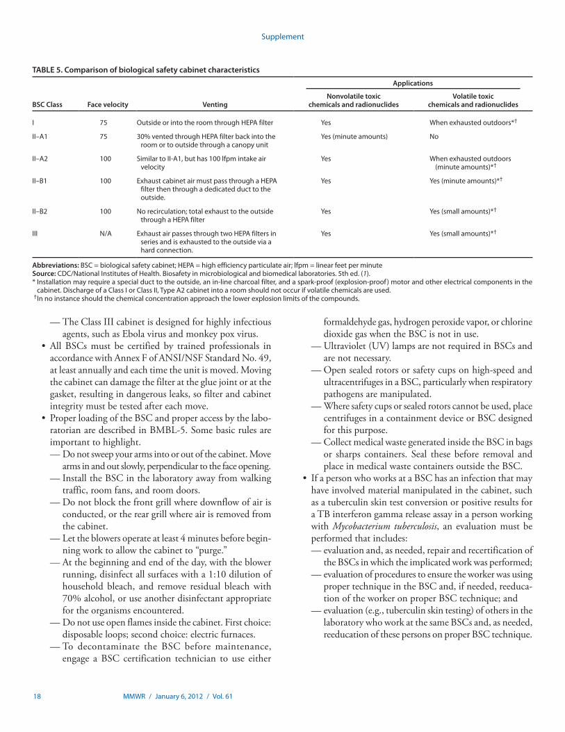
18 MMWR / January 6, 2012 / Vol. 61
Supplement
— The Class III cabinet is designed for highly infectious agents, such as Ebola virus and monkey pox virus.
• AllBSCsmust be certified by trained professionals inaccordance with Annex F of ANSI/NSF Standard No. 49, at least annually and each time the unit is moved. Moving the cabinet can damage the filter at the glue joint or at the gasket, resulting in dangerous leaks, so filter and cabinet integrity must be tested after each move.
• ProperloadingoftheBSCandproperaccessbythelabo-ratorian are described in BMBL-5. Some basic rules are important to highlight.— Do not sweep your arms into or out of the cabinet. Move
arms in and out slowly, perpendicular to the face opening.— Install the BSC in the laboratory away from walking
traffic, room fans, and room doors.— Do not block the front grill where downflow of air is
conducted, or the rear grill where air is removed from the cabinet.
— Let the blowers operate at least 4 minutes before begin-ning work to allow the cabinet to “purge.”
— At the beginning and end of the day, with the blower running, disinfect all surfaces with a 1:10 dilution of household bleach, and remove residual bleach with 70% alcohol, or use another disinfectant appropriate for the organisms encountered.
— Do not use open flames inside the cabinet. First choice: disposable loops; second choice: electric furnaces.
— To decontaminate the BSC before maintenance, engage a BSC certification technician to use either
formaldehyde gas, hydrogen peroxide vapor, or chlorine dioxide gas when the BSC is not in use.
— Ultraviolet (UV) lamps are not required in BSCs and are not necessary.
— Open sealed rotors or safety cups on high-speed and ultracentrifuges in a BSC, particularly when respiratory pathogens are manipulated.
— Where safety cups or sealed rotors cannot be used, place centrifuges in a containment device or BSC designed for this purpose.
— Collect medical waste generated inside the BSC in bags or sharps containers. Seal these before removal and place in medical waste containers outside the BSC.
• IfapersonwhoworksataBSChasaninfectionthatmayhave involved material manipulated in the cabinet, such as a tuberculin skin test conversion or positive results for a TB interferon gamma release assay in a person working with Mycobacterium tuberculosis, an evaluation must be performed that includes: — evaluation and, as needed, repair and recertification of
the BSCs in which the implicated work was performed;— evaluation of procedures to ensure the worker was using
proper technique in the BSC and, if needed, reeduca-tion of the worker on proper BSC technique; and
— evaluation (e.g., tuberculin skin testing) of others in the laboratory who work at the same BSCs and, as needed, reeducation of these persons on proper BSC technique.
TABLE 5. Comparison of biological safety cabinet characteristics
BSC Class Face velocity Venting
Applications
Nonvolatile toxic chemicals and radionuclides
Volatile toxic chemicals and radionuclides
I 75 OutsideorintotheroomthroughHEPAfilter Yes Whenexhaustedoutdoors*†
II–A1 75 30% vented through HEPA filter back into the room or to outside through a canopy unit
Yes(minuteamounts) No
II–A2 100 SimilartoII-A1,buthas100lfpmintakeairvelocity
Yes When exhausted outdoors (minuteamounts)*†
II–B1 100 Exhaust cabinet air must pass through a HEPA filter then through a dedicated duct to the outside.
Yes Yes(minuteamounts)*†
II–B2 100 Norecirculation;totalexhausttotheoutsidethrough a HEPA filter
Yes Yes(smallamounts)*†
III N/A ExhaustairpassesthroughtwoHEPAfiltersinseries and is exhausted to the outside via a hard connection.
Yes Yes(smallamounts)*†
Abbreviations: BSC=biologicalsafetycabinet;HEPA=highefficiencyparticulateair;lfpm=linearfeetperminuteSource: CDC/National Institutes of Health. Biosafety in microbiological and biomedical laboratories. 5th ed. (1).*Installationmayrequireaspecialducttotheoutside,anin-linecharcoalfilter,andaspark-proof(explosion-proof)motorandotherelectricalcomponentsinthe
cabinet. Discharge of a Class I or Class II, Type A2 cabinet into a room should not occur if volatile chemicals are used. † Innoinstanceshouldthechemicalconcentrationapproachthelowerexplosionlimitsofthecompounds.
MMWR / January 6, 2012 / Vol. 61 19
Supplement
3.4. Disinfection
3.4.1. Good work practices• Regardlessofthemethod,thepurposeofdecontamination
is to protect the laboratory worker, the environment, and any person who enters the laboratory or who handles labo-ratory materials that have been carried out of the laboratory. For detailed information see BMBL-5 Appendix A (1).
• Instructionsfordisinfectingalaboratoryworkbenchareto be a part of each SOP and must include what PPE to wear, how to clean surfaces, what disinfectant to use, and how to dispose of cleaning materials. Contact time is a critical and necessary part of the instructions. Post the instructions in the bench area for easy reference.
• Routinelycleanenvironmentalsurfacesbeforesettingupwork areas and again before leaving work areas.
• Cleananyitem(e.g.,timer,pen,telephone,thermometer)touched with used gloves.
• Donotusealcoholsoralcohol-basedsolutionsalonetodis-infect surface areas. These evaporate readily, which substan-tially decreases efficacy. Use disinfectants recommended for environmental surfaces, such as Environmental Protection Agency (EPA)–registered disinfectants effective against hepatitis B virus, HIV, and other bloodborne pathogens, or use a 1:10 dilution of household bleach. EPA environ-mental disinfectant product registration information is available at http://www.epa.gov/oppad001/chemregindex.htm.
• Reservesterilantsandhigh-leveldisinfectantsclearedbyFDA for processing reusable medical devices. FDA has identified manufacturers, active ingredients and contact conditions for these products. FDA-cleared sterilants and high-level disinfectants lists are available at http://www.fda.gov/cdrh/ode/germlab.html.
• Cleanbenchsurfaces,stationaryracks,claytiles,rockers,slide staining racks, water/heating baths and all trays when-ever a spill occurs. Clean all surfaces at the end of each shift.
• Useofdisposablelinersmayreducecleaningintervalsoftheequipment but does not replace the need to clean surface areas or equipment. Clean the underlying bench surface whenever the liner is discarded. The liner must be disinfected or discarded at the end of each shift or if contaminated.
• Disposable, flexible, polyethylene film–backed,nonskidhighly absorbent surface liners are available commercially and help to prevent soak-through of most solutions, includ-ing dyes and corrosive chemicals. Always discard with medi-cal waste after contamination and at the end of the shift.
• Allowdriedbloodorbodyfluidatleast20minutes’contactwith the laboratory-specified decontaminating solution to allow permeation and easy removal (1). Never use a knife
or other instrument to scrape dried blood or body fluid from surface areas; doing so can cause percutaneous injury or generate aerosols.
3.4.2. Bleach solutions (sodium hypochlorite) (38)• Hypochloritesolutionsareclassifiedasirritantsandcor-
rosives. Undiluted bleach solution is corrosive to stainless steel, and thorough rinsing must follow its use in the BSC and stainless steel sinks to remove the residue. Do not autoclave bleach solutions.
• Nevermixdifferentchlorinesolutionsorstorethemwithcleaning products containing ammonia, ammonium chlo-ride, or phosphoric acid. Combining these chemicals could result in release of chlorine gas, which can cause nausea, eye irritation, tearing, headache, and shortness of breath. These symptoms may last for several hours. A worker exposed to an unpleasantly strong odor after mixing of a chlorine solution with a cleaning product should leave the room or area immediately and remain out of the area until the fumes have cleared completely (see Section 9.1).
• Tobeaneffectivedisinfectant,workingbleachsolutionsmust contain >0.5% but <2% sodium hypochlorite. Hypochlorite concentration in household bleach var-ies by manufacturer. Many household bleach solutions contain 5.25% sodium hypochlorite, and a 1:10 dilution (5,000 ppm Cl) will produce a 0.53% hypochlorite solution. Use of bleach solutions with lower hypochlorite concentra-tions might not provide the proper level of disinfection. Each day, prepare a fresh 1:10 household bleach solution.
3.5. Waste ManagementA clinical laboratory must establish a waste management plan.• Aspartofanon-sitewastemanagementplan,therespon-
sibilities of the laboratory management or the designated safety officer or safety team are to — establish a waste-reduction or minimization program;— identify and define all categories of waste generated by
the laboratory;— for each category of waste generated, determine appli-
cability of federal, state, and local regulations, including how that category of waste will be segregated, packaged, labeled/color-coded, stored, transported, and tracked within the laboratory, outside the laboratory, and outside the facility to comply with the applicable regulations;
— segregate all regulated waste to prevent access by the public or clients;
— establish a system for reporting and responding to all issues or problems regarding medical waste manage-ment; and
20 MMWR / January 6, 2012 / Vol. 61
Supplement
— establish treatment and disposal processes (39). Disposal of regulated waste must be by a company meeting state and local licensure requirements.
3.5.1. Decontamination of medical waste before transport and disposal• “Infectiousmedicalwaste”isdefinedaswastecapableof
transmitting disease. “Regulated medical waste” is any waste contaminated with substantial amounts of blood or blood products in liquid or semiliquid form or with contaminated sharps. It is considered to confer a higher level of risk, thus warranting regulatory provisions by state or local authorities.
• Clinicallaboratoriesmustdeterminethefederal,state,andlocal laws governing their organization’s regulated medical waste and ensure that the organization is in compliance with those laws. State and/or local regulations may require— permits or registration numbers to generate medical waste;— development and implementation of a waste manage-
ment plan; and/or— specific recordkeeping compliance.
• Statedepartmentsofenvironmentalservices(orequivalent)are an excellent resource for assistance in complying with state and local medical-waste laws. To find state laws gov-erning medical waste, visit http://www.epa.gov/epawaste/nonhaz/industrial/medical/programs.htm. Choose the state, then look under the “Primary Materials–Cases, Codes and Regulations.” Search the state’s “Administrative Codes” or “Statutes” for information about waste management. (Some states use other terms for “infectious medical waste,” such as “regulated medical waste” or “special waste.”)
• OSHA.29CFRPart1910.1030,OccupationalExposureto Bloodborne Pathogens, provides minimal requirements for labeling and packaging of blood and body fluids when transported or outside a laboratory. Information may be obtained from the local OSHA office or online (33).
• Laboratorymanagementmust ensure that employeesunderstand these laws and ensure regulated medical waste is not mixed with nonmedical waste in a facility.
• Documentcompletionofemployeetrainingandcompe-tency assessment for— constructing and properly labeling containers for medi-
cal waste that require assembly before their use;— disposing of medical waste in properly labeled containers;— use of appropriate supplies, e.g., containers, appropriate
plastic bags, labeling; and— following all federal, state, and local regulations regard-
ing waste management, i.e., handling of medical waste, immediate disposal of medical waste, storage of medical waste, transportation of medical waste, which includes
any required Department of Transportation labeling (e.g., the word “Biohazard” and the universal biohazard symbol) of transport containers, and final disposal of medical waste.
3.5.2. Management of discarded cultures and stocks• Thelaboratory’sbiosafetylevelmustbeconsideredwhen
discarding cultures and stocks of infectious agents.• Discardedculturesandstocksoforganismshandledunder
BLS-3 physical containment (e.g., M. tuberculosis) are to be collected and sealed in containers that are closed, leakproof, and posted with the universal biohazard symbol and the word “Biohazard.” The containers subsequently need to be autoclaved on-site. Use of other on-site medical waste treatment technologies can be considered if these technologies sterilize the organisms, if they have been properly validated, and if they are recognized as medical waste treatment technologies by the appropriate state environmental regulatory agency.
• Decontaminatediscardedculturesandstocksoforganismshandled at BSL-2. If this process is done on-site but remote from the microbiology department, place the discarded cultures and stocks into durable, leakproof containers that are secured when they are moved. Decontamination may be done by a medical waste treatment contractor’s facility if the waste is placed into medical waste shipping contain-ers and packaged in accordance with applicable regulatory standards. To determine whether these activities can be done in a manner that minimizes possible exposures, conduct a risk assessment. The assessment will determine whether these wastes can be safely managed off-site or should be managed on-site.
3.5.3. Discarding a select agent• Clinicalordiagnostic laboratories andother entities that
have identified select agents or toxins contained in a speci-men presented for diagnosis or verification are required by regulation (7 CFR 331, 9 CFR 121, and 42 CFR 73) to report the identification within 7 calendar days to the Animal and Plant Health Inspection Service (APHIS) of the U.S. Department of Agriculture or to CDC. In addition, these laboratories or entities are required to report the identifica-tion of select agents and toxins from samples received for proficiency testing within 90 days after receipt of the sample.
• Disposal of cultures containing identified select agentssuch as Brucella spp., Coccidioides immitis, or Yersinia pestis, whether identified in the local facility or by a reference laboratory, falls under the Select Agent Rule (40). The APHIS/CDC Form 4, “Report of the Identification of a Select Agent or Toxin,” is used by clinical or diagnostic
MMWR / January 6, 2012 / Vol. 61 21
Supplement
laboratories and other entities to notify APHIS or CDC of the identification of a select agent or toxin as the result of diagnosis, verification, or proficiency testing, and of the final disposition of that agent or toxin. No further report-ing is necessary if the isolate is destroyed within 7 days after identification or shipped to a registered laboratory and CDC is notified of the disposition of the isolate.
• Aselectagentortoxincanbedestroyedbyon-siteautoclav-ing. If a medical waste contractor is used for the facility, the cultures containing the identified agent or toxin must first be inactivated by completely immersing the open culture containers in a fresh 1:10 bleach solution overnight before discarding them into medical waste. If the medical waste contractor is registered with the Select Agent Program, the live cultures may be formally transferred to the contrac-tor by using APHIS/CDC Form 2, “Request to Transfer Select Agents and Toxins.” Details on the select agent rule and its impact on clinical laboratories can be found in the Clinical Microbiology Newsletter, April 15, 2006 (41).
3.5.4. Autoclave safety• Gravitydisplacementsteamsterilizers(autoclaves)arefre-
quently used in microbiology (including virology) laborato-ries. Autoclaves generate substantial heat and pressure, and all users must understand and respect the associated risks.
• Personnelwhooperatetheautoclavemustbetrainedtopackage, load, and label materials to be autoclaved in accordance with the procedures used to validate the ster-ilization cycle of the unit. They must also receive training in emergency procedures.
• Donottouchthesidesorbackofolderautoclaves;theyhave little or no heat shielding and may cause burns.
• Donotstackorstorecombustiblematerials(e.g.,cardboard,plastic materials) or flammable liquids next to the autoclave.
• Neverautoclavematerialsthatcontaintoxicagents,corrosives(e.g., acids, bases, phenol), solvents or volatiles (e.g., ethanol, methanol, acetone, chloroform), or radioactive materials.
• Placeallbiomedicalwastetobeautoclavedinanapproved,biohazard-labeled autoclave bag before autoclaving. Not all red or orange bags are capable of being autoclaved. Bags selected for use in autoclaving waste must be specifically manufactured for this purpose. Use only bags designated as appropriate for use in autoclaves when autoclaving medical waste.
• Placeallsharps(e.g.,needles,scalpels,pipettes,orbrokenglass) into an approved, leak-resistant, labeled, and rigid sharps container before sterilizing.
•Whendecontaminatingabagofdrygoods,suchasbenchpaper or paper gowns, place 100 mL of water into the autoclave bag to facilitate steam production within the bag.
• Donotoverfillbagsortheautoclaveunit;thismightresultin inadequate steam circulation, which could interfere with the sterilization process.
• Closeautoclavebagslooselywithtwisttiesorothermeansthat allow steam inside.
• Placebagsontostainlesssteelorpolypropylenetraysforautoclaving. Do not place bags directly into the autoclave.
• Always allow an autoclaveunit to coolbeforeopening.Stand back and open the door slowly to allow the excess steam to escape. Allow the contents to cool before handling. Always use thick, elbow-length, heat-resistant, liquid-impervious gloves to remove hot items from the autoclave.
• Afterautoclaving,checktheautoclaveindicatortapetobesure the bars are black. If the indicator tape is not activated, resterilize the load.
• Atleastweekly,useabiologicalindicatorsuchasBacillus stearothermophilus spore strips (or equivalent) to ensure the autoclave is performing properly. Establish and follow a regular maintenance schedule for this equipment that evaluates seals, drains, and other critical aspects.
3.6. Dry Ice
3.6.1. General informationUnder certain circumstances, dry ice can be an explosion
hazard. Dry ice is solidified carbon dioxide (CO2) and it is extremely cold (-109° F [-79° C]). Unlike water-ice, dry ice sublimates (changes directly from solid to gas) as it warms, releasing CO2 gas. CO2 vapor is considerably heavier than air; in confined, poorly ventilated spaces, it can displace air, causing asphyxiation.• Avoiddryicecontactwithskinandeyes.Dryicecancause
severe frostbite within seconds of direct contact.• Neverplacedryiceintoglassorsealedcontainers.Storage
in a sealed container can cause the container to rupture or explode from overpressurization.
• Neverhandledryicewithbarehands.Alwayswearinsu-lated gloves and safety glasses. Use of laboratory coats is also recommended. Use tongs to handle blocks of dry ice. Use scoops to move pelletized dry ice.
• Donotputdryiceintothemouthorotherwiseingestit.If ingested, dry ice can cause severe internal injury. Never put dry ice in beverages to cool them.
•Whentransportingdryice,placethecontainerinthetrunkof the car or truck bed, and leave the car windows open for fresh air circulation. Never leave dry ice in a parked passenger vehicle. Sublimation of dry ice in a closed pas-senger vehicle can result in accumulation of dangerous concentrations of asphyxiating CO2 vapor. When opening
22 MMWR / January 6, 2012 / Vol. 61
Supplement
a closed cargo area containing dry ice, allow the closed space to ventilate for 5 minutes before entering.
• Donotplacedryicedirectlyonbenchtops,tile,laminatedcountertops, or ceramic sinks. Use an insulating barrier such as double-thickness cardboard or wood. Dry ice can destroy the bonding agent holding the tile or laminated material in place. Dry ice can also cause bench tops and ceramic sinks to crack.
3.6.2. Disposal of dry ice• Allowthedryicetosublimateorevaporatetotheatmo-
sphere in a well-ventilated area where CO2 vapor cannot build up.
• Donotdisposeofdryiceinsewers,sinks,ortoilets.Theextreme cold can fracture ceramic fixtures or crack poly-vinyl chloride (PVC) piping. If flushed down plumbing, the gas buildup can cause an explosion.
• Donotplacedryiceintrashcansorsimilarcontainers.The extreme cold and resulting condensation can destroy these receptacles.
3.7. Electrical Safety• Electricalhazardscanbecategorizedintotwomaintypes:
those that can result in an electrical shock and those that can cause fires and/or explosions.
• Electricalshockscanbeavoidedbyensuringthatequip-ment and electrical cords and plugs are in good repair, grounded outlets are used, and ground-fault interrupt outlets or circuit breakers are used near sinks, eyewashes, emergency showers, or other water sources.
• Donotoverloadelectricalcircuits.Minimizeoreliminatethe use of multi-outlet power strips. When power strips are necessary, the safety office of the facility or a licensed electrician must approve their use.
• Disconnectequipmentattachedtohigh-voltageorhigh-amperage power sources from the source, or provide a lockout device on the breaker box to prevent circuit activation before maintenance is performed.
• Becauseelectricaldevicescangeneratesparks,donotusethem near flammable or volatile gases or liquids.
• Neverplaceflammableliquidsinahouseholdrefrigerator.Thespark generated by the door-activated light switch can ignite fumes trapped in the unit, causing an explosion and fire.
• Specializedrefrigeratorsmustbeusedwhenstoringchemi-cals that have explosion potential.
3.8. Gases in the Laboratory: Compressed Gas Cylinders
Compressed CO2 cylinders are often used to provide gases for CO2 incubators; the risks associated with these incubators are minimal as long as the room is well ventilated.
3.8.1. Hazards• Gascylindersposethreemajorsafetyhazards:
— Gas cylinders are heavy; thus, a falling cylinder can cause injury.
— The valve attached to the cylinder is relatively fragile compared with the cylinder; if the valve is broken off, the cylinder can become a dangerous projectile.
— Faulty valves or regulators can leak, allowing toxic or flammable gases to enter the room.
• In the electronmicroscopy laboratory,nitrogen is usedto bring vacuum chambers to atmospheric pressure, and critical point driers use CO2 as a transitional fluid in the drying process for scanning electron microscopy specimens.
• Argon isused insputtercoaters,andsome laboratoriescarry out plasma ashing of biological specimens, which requires oxygen (42).
3.8.2. Minimizing hazardsMany of these potential hazards can be minimized by adop-
tion of safe handling practices.• Cylindersmust be securely anchored to thewallwith
chains or straps to prevent falling. Cylinders <18 inches tall may be secured in approved stands or wall brackets.
•Wheninstallinganewcylinder,leavetheprotectivevalvecap in place until the cylinder is secured. Replace the pro-tective valve cap before the straps or chains are removed from the cylinder.
• Specialregulatorsandthreadingaredesignedforeachgastype. Do not try to force the threads or use the wrong regulator on a tank.
• Regulators are normally suppliedwith instructions forroutine maintenance and periodic checking to ensure safe operation. Follow these instructions and checks carefully.
• Alwaysusespeciallydesignedcylindercartswhenmovingcylinders. Cylinders must be secured to the cart and the valve covers must be attached when moving them. They are not to be dragged, rolled, or physically carried. Do not pick cylinders up by the cap.
3.9. Liquid Gases (Cryogens)Cryogenic liquids are liquefied gases that have a normal
boiling point below -238°F (-150°C). Liquid nitrogen is used in the microbiology laboratory to freeze and preserve cells and virus stocks. The electron microscopy laboratory, frozen section
MMWR / January 6, 2012 / Vol. 61 23
Supplement
suites, and grossing stations for surgical pathology frequently use liquid nitrogen; some laboratories also use liquid helium. The principal hazards associated with handling cryogenic fluids include cold contact burns and freezing, asphyxiation, explosion, and material embrittlement.
3.9.1. Cold contact burns and freezing• Liquidnitrogenisdangerouslycold(-320°F[-196°C]),and
skin contact with either the liquid or gas phase can imme-diately cause frostbite. At -450°F (-268°C), liquid helium is dangerous and cold enough to solidify atmospheric air.
• Alwaysweareyeprotection(faceshieldoversafetygoggles).The eyes are extremely sensitive to freezing, and liquid nitrogen or liquid nitrogen vapors can cause eye damage.
• Donotallowanyunprotectedskintocontactuninsulatedpiping, hoses, tongs, spargers, specimen box storage racks, or other metal objects because these become extremely cold when exposed to liquid nitrogen. Skin will stick to the metal, tearing the flesh when one attempts to withdraw from it.
•Whenfillingcryogenicdewars,wearlong-sleevedshirtsorlaboratory coats, long trousers (preferably without cuffs which could trap the liquid), closed shoes (never sandals or open shoes), and insulated cryogloves labeled as appropriate for use with cryogenic liquids. Do not tuck pant legs into shoes or boots; doing so could direct liquid into the foot coverings and trap the cryogenic liquid against the skin.
•Wearloose-fittingthermalgloveswithelbow-lengthcuffswhen filling dewars. Ensure that gloves are loose enough to be thrown off quickly if they contact the liquid.
• Neverplaceglovedhandsintoliquidnitrogenorintotheliquid nitrogen stream when filling dewars. Gloves are not rated for this type of exposure. Insulated gloves are designed to provide short-term protection during handling of hoses or dispensers and during incidental contact with the liquid. Use special cryogenic liquid tongs when retriev-ing items from liquid nitrogen.
• Liquidnitrogenconfersahighriskofsplattering;jetsofliquid nitrogen can be generated when canes, canisters, and other objects that are at much higher temperatures are placed into liquid nitrogen. These activities can present a freezing hazard.
• Do not insert a hollow tube into the liquid nitrogenbecause liquefied gas may spurt from the tube.
3.9.2. Asphyxiation hazards• Althoughnitrogenisnontoxicandinert,itcanactasan
asphyxiant by displacing the oxygen in the air to levels below that required to support life. Inhalation of nitrogen in excessive amounts can cause dizziness, nausea, vomiting, loss of consciousness, and death without warning.
•Whenliquidcryogensareexpelledintotheatmosphereatroom temperature, they evaporate and expand to 700–800 times their liquid volume. Even small amounts of liquid can displace large amounts of oxygen gas and decrease the oxygen content of the atmosphere below a safe level (23,38,42,43).
• Donotstoredewarsornitrogencontainersinaconfinedspace. The venting gas could displace enough oxygen to become a hazard.
• Ifenclosedspacesmustbeused,installoxygenmonitors.Train personnel to leave the area immediately if the alarm sounds. The alarm must be audible both inside and outside the room to prevent anyone from entering the room.
3.9.3. Explosion hazards• Liquid gases, even those considered inert, can present
explosion hazards.• Heat flux into thecryogen isunavoidable regardlessof
insulation quality. Cryogenic fluids have small latent heats and will expand 700–800 times as they warm to room temperature. Therefore, even a small heat input can create large pressure increases within the vessel.
• Dewarsmustbemovedcarefully.Sloshingliquidintowarmerregions of the container can cause sharp pressure rises.
• Donotdrop,tip,orrollcontainersontheirsides;doingso could damage the vessel and/or cause a sharp increase in internal pressure.
• Cryogenic containers are equippedwith pressure reliefdevices designed to control the internal pressure. Cryogenic containers will periodically vent gases. This is normal. Do not plug, remove, or tamper with any pressure relief device.
• Ventsmustbeprotectedagainsticingandplugging.Whenall vents are closed, the expanding gas can cause an explo-sion. Vents must be maintained open at all times.
• Alwaysusespecialultralow-temperaturecontainerstoholdliquid nitrogen. Never place liquid nitrogen into domestic thermos flasks because they are not designed to withstand the large and rapid temperature changes that occur when liquid nitrogen is placed in the vessel (42,43).
• Fillliquidnitrogendewarsslowlytominimizetheinternalstresses of cooling. Excessive stress could damage the vessel and cause it to fail.
• Liquidheliumiscoldenoughtosolidifyatmosphericair.Only helium is to be introduced or allowed to enter the helium volume of a liquid helium dewar. Take precautions to prevent air from back-diffusing into the helium volume.
• Liquidnitrogen and liquidheliumhaveboilingpointsbelow that of liquid oxygen, and they can condense oxy-gen from the atmosphere. Repeated replenishment of the system can cause oxygen to accumulate as an unwanted
24 MMWR / January 6, 2012 / Vol. 61
Supplement
contaminant. Similar oxygen enrichment can occur where condensed air accumulates on the exterior of cryogenic piping. An explosion could occur if this oxygen-rich liquid is allowed to soak insulating or other materials that are not compatible with oxygen. In addition, some oils can form an explosive mixture when combined with liquid oxygen.
3.9.4. Cryotube explosions• PPEincludesanANSI-specification,impact-resistantface
shield, heavy gloves, and a buttoned laboratory coat during removal of cryotubes and ampoules from nitrogen tanks.
• Cryotubesandglassampoulesusedforfreezingcellsandviruses can explode without warning when removed from cryogenic storage. These tube explosions are presumed to be caused by entry of liquid nitrogen into the tube through minute cracks; as the tube thaws, the rapidly expanding gas causes the tube to explode, scattering the contents of the tube (23).
•Wheneverpossible,storeampoulesinthegaseousphaserather than submerging in the liquid nitrogen of the cryo-genic dewar. An imperfectly sealed ampoule will pick up less nitrogen in the gaseous phase.
• Nitrogenoutgassingfromanimperfectlysealedvialwillsometimes produce a hissing noise before the vial explodes. The absence of hissing does not mean the vial is safe. Place cryotubes and ampoules onto gauze or paper toweling in an autoclavable, heavy-walled container immediately after removal from the nitrogen tank, and close the lid of the heavy-walled container quickly. If an explosion occurs, autoclave the entire vessel.
3.9.5. Embrittlement• Neverpourcryogenicliquidsdownthedrain.Laboratory
plumbing is one of many ordinary materials that become brittle at cryogenic temperatures and easily fracture.
•Woodandotherporousmaterialsmaytrapoxygenatlowtemperatures and will explode when subjected to mechani-cal shock (42).
3.9.6. Infectious disease hazards• Liquidnitrogencanbecomecontaminatedwhenampoules
are broken in the dewar, and contaminants can be preserved in the nitrogen (23). These potentially infectious contami-nants can contaminate other vials in the dewar and generate an infectious aerosol as the liquid nitrogen evaporates.
• Plasticcryotubesratedforliquidnitrogentemperaturesare recommended for liquid nitrogen storage because they appear to be sturdier than glass ampoules and are less likely to break in the nitrogen.
3.10. Slip, Trip, and Fall HazardsSlips, trips, and falls can cause a laboratory worker to drop or
spill vessels containing infectious agents or dangerous chemi-cals. They can also lead to skin punctures and abrasions that make laboratory workers more vulnerable to LAIs.
Good housekeeping is the most fundamental means for reducing slips, trips, and falls. Without good housekeeping, any other preventive measures (e.g., installation of sophisticated flooring, specialty footwear, or training on techniques of walk-ing and safe falling) will never be fully effective.
3.10.1. Slips• Commoncausesof laboratory slips includewetoroily
surfaces; loose, unanchored rugs or mats; and flooring or other walking surfaces that do not have some degree of traction in all areas.
•Waterontheflooristhemajorsliphazard.Removeanywater on the floor promptly.
• Paraffinfromtissuemountingandcuttingcanaccumulatein tissue processing areas and can make the floor slick despite regular cleaning unless special floor care measures are taken.
•Mineraloils,mountingfluids,stainlesssteelcleaners,andother laboratory chemicals and/or reagents create slip hazards if they get on the floor. Clean up with soap and water as soon as such spills are discovered.
• Donotusealcoholstocleanfloors;alcoholswilldissolvefloor wax, creating areas with different degrees of traction.
•Mats canpresent a sliphazard if theyarenotproperlyanchored to the floor.
•Walkingonpaper,cardboard,orpackagingmaterialscanpresent a slip hazard.
3.10.2. Trips• Commoncausesoftrippingincludeobstructedview,poor
lighting, clutter in the walkway, mats or other items in the walkway, uncovered cables, open drawers or cabinets, and uneven walking surfaces. Permeable mats and rugs are not recommended in microbiology, except as noted later in this section.
• Keepdrawersandcabinetsclosedexceptwhentheyarebeing accessed.
• Clutteranditemsthatprotrudefromkneeholespacescaninjure workers as they move down aisles. Keep clutter to a minimum. Make sure that boxes and other items do not protrude into aisles.
• Donotruncordsorcablesacrossaislesorotherwalkways.• Thesafetyofficerorlaboratorymanagementmustassess
the use of ergonomic antifatigue mats in other sections of the laboratory before employing in a specific laboratory
MMWR / January 6, 2012 / Vol. 61 25
Supplement
area. Concerns to be aware of before using such mats include the following:— These mats are somewhat thick, and the raised surface
presents a trip hazard.— The mats are obstructions for carts and chairs and may
cause them to tip.— They make spill cleanup difficult.— They make cleaning and disinfecting the floors difficult
for the custodial staff.— They may also place custodial staff at risk if they pick
up or move mats that have been inadvertently contami-nated with chemicals or infectious agents.
— Liquids will often wick under the mat, hiding potential contamination problems.
— Mats present a trip/fall hazard that could impede egress from the laboratory in an emergency.
3.11. Ultralow-Temperature FreezersWear thermally resistant gloves and a laboratory coat when
handling items stored at ultralow temperatures. Specimens stored at ultralow temperatures are extremely cold [-70°C to -85°C]), and paradoxically, direct contact with the skin can cause severe burns.
3.12. Ultraviolet light• Short-waveUVlighthashadseveralapplicationsinthe
laboratory, including use in fluorescent microscopes, as a terminal disinfectant in some type 1 water systems, and for visualizing nucleic acid bands in ethidium bromide–stained gels.
• Exposuretoshort-waveUVlighthasbeenlinkedtoskincancers, corneal scarring, and skin burns. These effects can result from direct or reflected UV light exposure (44).
• Do not use UV lights for decontaminating BSCs.Organisms in cracks, shadows, and on the underside of equipment are not affected by UV light treatment. In addition, the radiation and ozone produced by these lights will attack plastic and rubber items in and around the BSC, shortening their lifespan. This exposure can affect mechanical pipette calibrations and other sensitive equipment functions.
• IfUVlightsmustbeusedforotherreasonsinBSCs,pro-vide a means to monitor them throughout their life with intensity sensors. Calibrated UVC sensors are a reliable and cost-effective way to monitor UVC radiation levels in BSCs. Monitor these lights because germicidal UV lights have an expected life of about 9,000 hours.
• GermicidalUVirradiationforlongerthan15minutesiscounterproductive because it produces no additional ger-micidal benefit and it accelerates equipment degradation.
• TheUV lampmust never be onwhile an operator isworking in the cabinet. Not all protective eyewear will protect laboratory workers from deleterious UV light exposure. Make sure the protective eyewear is rated for UVC protection.
•WearUVsafetyglasseswhenperforming routine lampmaintenance or when potential exists for direct or indirect (reflected light) exposure.
•Weargloves, long-sleeved laboratorycoat, and full-faceshield when working with UV view boxes lacking protec-tive filter shields.
• InareaswhereUVlightisused,displayplacardsstating“Caution, Ultraviolet Light, Wear Protective Eyewear.”
3.13. Vacuum devicesVacuum-assisted filtration devices and side-arm suction
flasks are used routinely in the general laboratory, whereas the electron microscopy laboratory uses vacuum-assisted evapo-rators, freeze-driers, freeze-fracture, and sputter coater units. Vacuum-assisted devices present implosion hazards and risk aerosol generation.
3.13.1. Implosion safety• Implosions can occur when the pressure differential
exceeds the specifications of the vessel.• Implosionscanscattersharpglassdebrisinalldirections
and seriously injure anyone in the vicinity (42,43). They will also disperse any infectious agents that are present in the vessel.
• Heavy-walled,side-armsuctionflasksaregenerallyratedto withstand a pressure differential of one atmosphere (14.7 PSI); house vacuum systems or vacuum pumps that provide pressure differentials exceeding that level must be regulated with an in-line pressure regulator.
• Cracks,chips,andscratchesinvacuumflasksandbelljarscan weaken the glass and cause an implosion even when the proper differentials are provided by pressure regulators.
• Caremustbe takentopreventdamagetobell jarsandsuction flasks caused by excessive wear or impact with hard objects.
• Implosionguardsmadeofplasticmeshorplasticboxeshave been used with suction flasks to contain glass pieces if the vessel fails. When infectious agents or blood or blood products are being handled, the use of plastic flasks is strongly recommended.
3.13.2. Aerosol generation• Vacuum-assisted aspiration traps consist of one or two
suction flasks plumbed together in series with an in-line
26 MMWR / January 6, 2012 / Vol. 61
Supplement
HEPA filter (e.g., Vacushield Vent Device, Pall Life Sciences, Port Washington NY, or equivalent device) to prevent contamination of the vacuum pump or house vacuum system (1).
•Whenusingadedicatedvacuumpump,manylaboratoriesalso include a suction flask containing coarse Drierite (W.A. Hammond Drierite Co., Ltd, Xenia, OH) or an equivalent desiccant to remove moisture from the air, thereby protecting the pump. Aspiration traps are used in virology to remove culture media from tubes, shell vials, and other vessels before refeeding or other cell manipula-tions. Aspiration systems are also used in enzyme-linked immunoassay (ELISA) plate washers.
• Allthesedevicesgenerateaerosolsbyagitatingthefluidand placing the fluid surface under reduced pressure (23). Aerosols can deposit infectious agents on the immediate surfaces, and finer aerosols can be inhaled.
3.13.3. Aerosol protection measures• UseaspirationdevicesinaBSCtocontainanyaerosols.• Operatorsare towearadisposable laboratorycoatand
gloves to protect themselves from infectious droplets.•When a culture aspiration is complete, allow theBSC
blower to run for 5 minutes to purge any airborne aerosols; decontaminate the work surfaces in the normal manner.
• Replacethein-lineHEPAfiltersevery6monthsorwhenthey become wet or noticeably blocked.
3.13.4. Disposal of liquid wastes from vacuum-assisted aspiration traps• Neverpourinfectiouswastesdownthesink.• Decontaminateliquidwastesfromaspirationtrapswith
bleach before disposal.•Whenusinganaspirationtrapattachedtoanindividual
vacuum pump, laboratories usually pass the vapors through an activated charcoal trap to protect the pump from chlo-rine vapor corrosion.
• Avarietyofsuctiontrapconfigurationsispossible,andtheultimate configuration will depend upon workflow and individual laboratory practice. The following procedures apply to all configurations.— Change vacuum flasks when they are three-fourths
full to prevent overfilling. Some laboratories prefer to mark the maximum fill volume on the flask and add a sufficient volume of bleach at the beginning of the day to produce a 1:10 bleach solution when the aspirated fluids reach the maximum fill mark.
— Disinfect the hose by aspirating 10–50 mL of a freshly made bleach solution into the trap. Lift the hose to
allow all the bleach to enter the trap. Wait 20 minutes, then remove the trap from the BSC.
— Once decontaminated, the fluid is considered noninfec-tious and may be poured down the sanitary sewer.
Note for the virology laboratory: Bleach will reduce the phe-nol red dye in cell culture media, and the solution will go from red to colorless. If this color change does not occur, the fluid has not been decontaminated and sufficient bleach must be added to decontaminate the vessel.
3.14. Biological Hazards
3.14.1. Punctures and cutsSkin punctures and cuts can directly introduce an infec-
tious agent into the body and can provide a route whereby a secondary agent can enter.• Needlesticks(45)
— Clinical laboratories must establish a needlestick and sharps injury prevention program.
— Limit the use of needles and syringes to procedures for which there are no alternative methods. Needlestick injuries occur most often when needles are returned to their protective sheathes after use.
— Do not resheathe needles. If resheathing is absolutely required, the procedure must utilize a needle resheathing device to minimize injury and accidental inoculation.
— Do not use needle-cutting devices because they can produce infectious aerosols. After use, place needles and syringes in leak- and puncture-resistant containers appropriately labeled with the word “Biohazard” and the universal biohazard symbol for decontamination and disposal.
— Do not bend, shear, recap, or remove needles from disposable syringes, or otherwise manipulate by hand before disposal.
— Microtome/cryostat blades used to cut frozen sections are another potential sharp that must be handled carefully. Wear cut-resistant gloves during disassembly of the poten-tially contaminated blade for cleaning and disinfection.
• Breakage— Never pick up broken glass with gloved or bare hands.
Use forceps, disposable plastic scoops, tongs or hemo-stats to pick up broken glass; dispose of the broken glass into a sharps container. Place a broom or hand brush and dustpan in various laboratories or in the utility closet for picking up noncontaminated glassware.
— Do not use broken or chipped glassware. Discard it in the appropriate sharps container labeled for broken glassware.
— When handling broken containers with spilled infec-tious substances, adhere to the following guidelines (1).
MMWR / January 6, 2012 / Vol. 61 27
Supplement
º Wear appropriate gloves for this procedure (based on risk assessment and protection needed).
º Cover the broken container and spilled infectious substance with a cloth or with paper towels.
º For the routine BSL-2 laboratory, pour a disinfectant or a fresh 1:10 household bleach solution over the covered area and leave for a minimum of 20 minutes. It would take 23 minutes to clear the air of airborne M. tuberculosis from a spill at 99% removal efficiency if the room had 12 room air changes per hour, and 35 minutes for this removal with 99.9% efficiency (46). Given the variability of the number of room air changes per hour in diagnostic laboratories, the wait time has to be carefully evaluated.
º The cloth or paper towels and the broken material should be cleared away into biohazard sharps receptacles. Fragments of glass are to be handled with forceps, not gloved hands. (Using wadded up tape with forceps facilitates this procedure). Small HEPA vacuum cleaners are also available for removal of fine glass particulates.
• Iflaboratoryformsorotherprintedorwrittenmatteriscontaminated, the information on the forms or written matter is to be copied onto another form and the original discarded into the biohazard waste container.
• Pasteurpipettes— Whenever possible, substitute plastic or evaluate the
procedure to determine if a newer or better technique is now available.
— Both the top and the bottom of a Pasteur pipette can cause puncture wounds.
— Before handling a glass Pasteur pipette, examine the top of the pipette to see if it is broken or cracked. Broken pipettes can produce puncture wounds.
— When seating glass Pasteur pipettes into suction lines, hold the pipette at the top and do not allow your hand to extend below the tip. Improper technique while seating the pipette can produce puncture wounds if the hand slips or the pipette breaks.
— Dispose of used Pasteur pipettes in leak- and puncture-resistant containers. In most locations, contaminated Pasteur pipettes are considered sharps and must be disposed of as such.
• Othersharpdevices— Knives, scissors, and tissue homogenizers are frequently
used to dissociate tissue specimens before testing. These items must be handled carefully in order to prevent cuts and skin punctures that could injure or inoculate laboratory workers with infectious materials.
— Pointed forceps are often used for fine dissociation and for removing coverslips from shell vials. These forceps can puncture the unwary user, causing injury and/or infection.
— Glass slides can break and puncture skin.— Culture tubes and shell vials can crack or shatter if caps
are forced. The resulting shards can easily penetrate latex gloves and skin.
— The lip of glass vessels may be chipped, and these chipped surfaces can cut unwary laboratory workers. Discard chipped glassware at the earliest opportunity.
• Sharpsdisposal— Carefully place used disposable needles, syringes, scal-
pels, blades, pipettes, and similar objects into properly labeled leak- and puncture-resistant containers made for disposal. Most authorities require needles and syringes to be disposed of in such sharps containers, whether used or not.
— Locate sharps disposal containers in or near the area where the sharps are used in order to prevent environ-mental contamination and injuries associated with accumulating sharps at the point of generation and moving sharps from one place to another.
— Replace sharps containers that are two-thirds to three-fourths full. Sharps containers must close securely for transport to decontamination areas. Injuries can occur when laboratory personnel try to forcibly close full containers. Overfilled containers can pop open again, creating a hazard for other workers.
— Place nondisposable sharps into a covered leak-resistant, hard-walled container for transport to a processing area for decontamination, preferably by autoclaving (1).
— Place materials to be decontaminated off-site into a medical waste shipping container, and secure for transport in accordance with applicable state, local and federal regulations (1).
— Place clean, uncontaminated sharps (e.g., clean broken glassware, chipped clean pipettes) into rigid, puncture-resistant containers for disposal in the normal trash stream. Tape containers shut to prevent accidental opening and potential injuries.
— Never place sharp items directly into the regular trash. They could injure custodial or other staff members when the trash bags are removed from rigid trash containers.
3.14.2. Ingestion and contact with infectious agents• Refrainfromtouchingeyes,nose,mouth,andlipswhile
in the laboratory.• Donotplacepens,pencils,safetyglasses,orotherlabora-
tory items in the mouth or against the lips.• Donotstorefoodorbeveragesforhumanconsumption
in the laboratory.
28 MMWR / January 6, 2012 / Vol. 61
Supplement
•Mouth pipetting is prohibited;mechanical pipettingdevices must be used.
• Eating,drinking,smoking,handlingcontactlenses,andapplying cosmetics are not permitted in the laboratory.
•Wash hands afterworkingwith potentially hazardousmaterials and before leaving the laboratory. The laboratory must have a sink for handwashing, preferably located near the laboratory exit.
• Glovesmustbeworntoprotecthands fromexposure tohazardous materials. In the molecular biology area, gloves also protect the specimen from nucleases that are on the skin.— Change gloves when they are contaminated, integrity
has been compromised, or when otherwise necessary.— Remove gloves and wash hands when work with haz-
ardous materials has been completed and before leaving the laboratory.
— Do not wash or reuse disposable gloves.— Never touch your face, mouth, eyes, or other mucous
membranes when wearing gloves in the laboratory.— Because gloves worn in the diagnostic laboratory are
considered potentially contaminated, place them into biohazard disposal containers when discarding.
— Remove gloves when answering the telephone or using common equipment like computers.
• Topreventcontaminationofunglovedhands,designthelaboratory so that it can be easily cleaned.— Decontaminate work surfaces with an appropriate
disinfectant after completion of work and after any spill or splash of potentially infectious material (see Section 3.4, Disinfection).
— Bench tops must be impervious to water and resistant to heat, organic solvents, acids, alkalis, and other chemicals.
— Chairs used in laboratory work must be covered with a nonporous material that is easily cleaned. Uncovered cloth chairs are inappropriate.
• Telephonesareapotentialvehiclefortransferringinfec-tious agents to the face and mucous membranes, and they should be used with this in mind.— Never pick up or dial a telephone with gloved hands.— Disinfect telephones regularly with disinfectants.
(Alcohols do not inactivate nonenveloped viruses or destroy DNA.)
— Use the hands-free or speaker phone features whenever possible to avoid touching the telephone handset to the face.
• Neverbringbriefcases,purses,backpacks,books,maga-zines, and other personal items into the laboratory. These items are difficult to disinfect.
3.14.3. Spills and splashes onto skin and mucous membranes• Itistheresponsibilityofalllaboratoryworkerstoperform
all procedures in a manner that minimizes the creation of splashes and aerosols.
• Allsplashestotheeyemustbeflushedforaminimumof15 minutes. If a laboratory worker wearing contact lenses receives a splash to the eye, the eye must be flushed with water, the lens removed, and the eye flushed again. Discard disposable contact lenses. Disinfect reusable contact lenses before returning them to the eye. Consultation with the manufacturer may be warranted.
• Specimencontainers,culturetubes,shellvials,andothercylindrical vessels used in the laboratory are easily tipped and could roll when placed on the bench top. Glass vessels can break if dropped. Secure these vessels in racks whenever possible to prevent opportunities for breakage.
3.14.4. Aerosols and dropletsAny procedure that imparts energy to a microbial suspension
can produce infectious aerosols (1,23). Procedures and equip-ment frequently associated with aerosol production include pipetting, mixing with a pipette or a vortex mixer, and use of blenders, centrifugation, and ultrasonic devices (sonicators) (1,23,47). These procedures and equipment generate respirable particles that remain airborne for protracted periods. When inhaled, these tiny particles can be retained in the lungs.
These procedures and equipment also generate larger drop-lets that can contain larger quantities of infectious agents. The larger droplets settle out of the air rapidly, contaminating, work surfaces as well as the gloved hands and possibly the mucous membranes of persons performing the procedure.
Respirable particles are relatively small and do not vary widely in size distribution. In contrast, hand and surface contamina-tion is substantial and varies widely (1,48). The potential risk from exposure to larger-size droplets requires as much attention in a risk assessment as the risk from respirable particles.• Pipetting Pipettes and pipetting processes can be an appreciable
source of infectious aerosols and environmental con-tamination. Therefore, it is prudent to wear gloves, eye protection, and a laboratory coat with knit cuffs when pipetting and to perform pipetting operations in a BSC. The following guidelines are categorized into those for serologic or mechanical pipettes.— Serologic pipettes. When the last drop of fluid is forcibly
expelled out of the pipette tip, small and large droplet aerosols are formed that can contaminate the hands and the environment (23). To minimize aerosol generation, place the pipette tip against the inside wall of tubes,
MMWR / January 6, 2012 / Vol. 61 29
Supplement
flasks, or other vessels, and gently expel the last drops of fluid.
— When dispersing cell clumps, virologists frequently draw fluids into and out of the pipette to homogenize specimens and cell suspensions. A substantial amount of aerosolization can occur during this process, espe-cially when the fluid is forcibly expelled from the pipette tip. Aerosols are generated in a similar manner during “pipette mixing” of culture dilutions. Closed-cap vortex mixing is the preferred method for this type of mixing. If pipette mixing is required, keep the pipette tip below the surface of the fluid and do not eject the entire fluid volume from the pipette. This will reduce aerosolization and bubble formation.
— Vigorous pipetting (rapid aspiration of fluid into the pipette) can generate aerosols within pipettes. Some of the aerosols will be trapped by the cotton plug at the proximal end of the pipette. However, some aerosols can travel through the cotton plug and contaminate the pipetting device. Certain mechanical pipetting devices have HEPA filters that minimize contamination of the handset. Replace these filters regularly and whenever they become wet. Decontaminate pipette bulbs regu-larly and whenever they become contaminated.
— Mechanical pipettes. Hand-held mechanical pipetting devices are used for enzyme immunoassay (EIA) testing, molecular diagnostics, and other activities that require precision.
— In molecular diagnostics, pipette contamination is the most frequent cause of false-positive results (49). Pipette contamination can occur from aerosols, from touching the outside of the pipette to a contaminated surface, and from contaminating the inside of the pipette during the pipetting process.
— Expelling the last remaining fluid in the tip will result in droplet splatter and aerosol formation. These aerosols and droplets can contaminate the other samples and the environment. Most mechanical pipetting devices have two stops on the plunger — the “To Deliver” stop and the “Expel” stop. Pipette volumes are calibrated for accuracy at the “To Deliver” stop, and there is no need to expel the final amount of fluid to preserve pipetting accuracy.
— Touch pipette tips to the inside of the well or tube before pressing the delivery plunger. Never direct the pipetting stream into the middle of the well because this will cause splashing and contamination.
— Care must be exercised when ejecting used tips into discard containers because the remaining fluid can splash and splatter widely.
— The outside of the pipette barrel can become contaminated through splatter, aerosols, or by touching the barrel to con-taminated objects. Do not extend the barrel of the pipette into a reagent, sample or discard container. If normal-length tips cannot reach the fluid in the tube, use extended-reach pipette tips to prevent barrel contamination.
— Disinfect mechanical pipettes regularly following the manufacturer’s instructions or with a 1:10 household bleach dilution followed by 70% alcohol to remove as much bleach as possible.
— Use of aerosol-resistant pipette tips can substantially reduce nucleic acid contamination inside the pipette. Aerosol-resistant tips contain a hydrophobic micropo-rous filter that is bonded onto the walls of the pipette tip. The microporous filter traps aerosols before they can contaminate the barrel of the pipette. These filters can also prevent contamination of the specimen when a contaminated pipette is inadvertently used (49).
— When an accidental falling drop from a pipette tip encounters a hard surface, it generates aerosols and a series of small droplets, some of which may be large enough to fall and repeat the process (50). Greater contamination ensues when drops fall a greater distance onto a hard surface.
— Many laboratories use commercial plastic-backed bench paper in BSCs and on laboratory work benches to contain or absorb contamination from falling drops.
— When faced with the inevitability of a falling drop, it is best to lower the tip of the pipette and allow the drop to fall a short distance onto an absorbent towel. This procedure will minimize the kinetic energy of the drop and its capacity to splatter.
• Tubesandothervessels— Thin films sometimes form in the neck of culture
tubes, shell vials, microcentrifuge tubes, specimen vials, and other containers. Breaking or popping this film produces aerosols and microdroplet splatter that can contain infectious agents, nucleic acids, or other potential contaminants (23).
— Containers with thin films in the neck are to be recapped and centrifuged whenever possible to disrupt the film or cause it to merge with the fluid in the vessel.
— If centrifugation is not possible (e.g., with culture flasks), place gauze or another absorbent material over the opening and insert a pipette into the flask to dis-rupt the film. Dispose of the pipette and the absorbent material with other contaminated materials.
30 MMWR / January 6, 2012 / Vol. 61
Supplement
— Microcentrifuge and other plug-topped tubes will often produce aerosols and splatter when opened. Screw-cap microcentrifuge tubes can reduce this risk.
— To minimize the amount of fluid on the cap, subject microcentrifuge tubes to a quick “pulse” centrifugation before they are opened.
— Open microcentrifuge tubes in a BSC whenever possible.— When opening plug-seal microcentrifuge tubes, cover
the top of the tube with absorbent material (e.g., alcohol-moistened gauze) to catch any splatter that might occur. Dispose of the absorbent material with other contaminated materials.
• Lyophilizedmaterials,serumvialsandampoulesOpening vials of freeze-dried (lyophilized) material can be
hazardous because these fine dry powders are easily dispersed into the atmosphere when air rushes into the evacuated vessel (23). The following procedure may be used to safely open a serum vial containing lyophilized material.
— Move the vial and the suggested diluent (water or medium as appropriate) to a BSC.
— Wear gloves and laboratory coat when opening lyophi-lized vials.
— Use a hemostat to remove the aluminum crimp from the vial. Discard the crimping material into the sharps container.
— Cover the stopper with a moistened gauze pad, and care-fully lift the edge of the stopper and allow air to slowly enter the vial. Do not disturb the contents of the vial.
— Once the vacuum has been released, remove the stopper completely and place it upside down on absorbent paper.
— Add the appropriate amount of diluent to the vial using a sterile pipette.
— Replace the stopper and allow the vial contents to hydrate for several minutes.
— Discard the gauze, stopper, and absorbent paper with other contaminated materials.
— Using a pipette, transfer the contents of the vial to an appropriate container.
— Discard the original vial with other contaminated materials.
— Needle and syringe methods for removing infectious agents from serum vials are not recommended because they can contaminate the environment and because they provide opportunities for needlestick injuries. Use forceps, not needles, to remove serum separator tubes that are stuck in centrifuge carriers.
• Glassampoules— Once opened, glass ampoules can present a risk for cuts
and punctures.
— Safety ampoule breakers can prevent injuries by cover-ing the ampoule during the breaking process.
— For ampoules containing infectious materials, cover the score line with gauze moistened with disinfectant; then break as usual using the safety ampoule breaker.
— Place the ampoule breaker into a beaker containing a 1:10 bleach solution after removing the ampoule.
3.15. Ultrasonic Devices• Ultrasonicdevicesareprincipallyusedtolysebacteriaand
viruses and to clean glassware and laboratory equipment. • Usethelowesteffectivepowersettingtominimizeaerosol
generation.• Coverbathsonicatorswhilethedeviceisinuse.• Articlesdestinedforultrasoniccleaningmustbeproperly
decontaminated before cleaning to prevent aerosolization of infectious agents (23).
• Alwaysconductorganismlysisandhomogenizationpro-cedures in closed containers.
• Changebathfluidsfrequentlytopreventaerosolizationofbacterial and fungal contaminants present in the bath.
3.16. Clean versus Dirty Areas of the LaboratoryIn the microbiology laboratory, all the technical work areas
of the department are considered dirty. The same concepts of demarcation and separation of molecular testing areas that are described in this section can be used to establish clean and dirty areas in other parts of the diagnostic laboratory.
3.16.1. Clean areas•Weardifferentcolorlaboratorycoatsincleananddirty
areas of the laboratory (have them available at entrance to clean areas), or require no laboratory coats in clean areas.
• Decontaminate reusablematerials anddevices (e.g., tele-phone, clocks, computers, tissue boxes, work books) brought into the clean area unless they are known to be new, and immediately apply laboratory-designated, color-coded tape.
• Avisual reminderonsmallobjects suchasworkbooks,tissue boxes, and pens can easily identify items located to a clean area.
• Demarcateseparationofdirtyandcleanfloorareaswithtape (tape must stand up to floor cleaning) to clearly denote clean/dirty area boundaries.
• Developapolicyforcleaningandmaintainingcleanareas.• Trainallpersonnel(includingservicepersonnel)regarding
how to identify and maintain clean areas and to recognize the significance of the demarcation tape and other means of area identification.
• Documenttrainingandassesscompetencyinuseofandmaintaining clean areas.
MMWR / January 6, 2012 / Vol. 61 31
Supplement
3.16.2. OfficesOffices (e.g., of supervisors and laboratory director) that
open into the clinical laboratory represent hybrid areas within the laboratory. These offices are not typically designed or main-tained in a manner that allows for easy or efficient disinfection.• Keepasupplyofhanddisinfectantgelinallofficeand
work areas and use the gel frequently.• Componentsofofficesthatshouldremaincleanbutmay
be overlooked include— laboratory documents, reports, and records; small
equipment; pens; procedure manuals and other items that have been in the laboratory and could have been handled with gloved hands;
— carpets and chairs that are difficult to disinfect;— books, journals, and other reference materials that can
be taken into the laboratory or taken for use outside the laboratory;
— personal items (e.g., photographs, awards, briefcases, coats, boots, backpacks, purses, personal electronic devices) that are difficult to disinfect and would not be allowed in the general laboratory; and
— food items.• Designating office areas as “clean”does not necessarily
make or keep them uncontaminated, especially when potentially contaminated items are brought into the office and reference materials and documents move freely between the office and laboratory. The following procedures can help reduce the risk of contamination in laboratory office areas.— Never bring specimens, cultures, proficiency samples,
and similar items into office areas.— Remove PPE before entering the offices, and wash
hands before entering these areas.— Establish a dedicated and protected clean area for per-
sonal items (e.g., purses, briefcases, and similar items).— Disinfect desks and personal workspaces, telephones,
and computer keyboards in office areas regularly.— Refrain from touching eyes, nose, mouth, and lips while
in office areas.— Do not place pens, pencils, eyeglass bows, or other
items in the mouth or against the lips.— Do not apply or permit cosmetics in office areas.— Do not store food in the office.— Wash hands after working in the office and before enter-
ing common areas such as rest rooms, administrative areas, cafeteria, and the library.
— Avoid clutter in office areas as much as possible. Boxes, papers, and other items make the office difficult to clean and decontaminate.
— Laboratory directors and supervisors are responsible for assessing the exposure risks associated with use of laboratory documents and reference materials in the dirty areas of the laboratory and developing use policies to minimize those risks.
3.16.3. Dirty areas• Allareasoftheworkinglaboratory—includingallequip-
ment, keyboards, waste, and surfaces — are considered “dirty” areas.
• Nostandardsarecurrentlyavailablethatdescribeoper-ating procedures within dirty areas of the laboratory. Laboratorians must be vigilant in recognizing the potential or risk of transmitting an etiologic agent by touching items in these areas.
3.17. InstrumentationWhether automated or manual, procedures with the poten-
tial for producing specimen aerosols and droplets (e.g., stopper removal, vortexing, opening or piercing evacuated tubes, using automatic sample dispensers) require PPE and engineering controls designed to prevent exposure to infectious agents.
3.17.1. Water baths and water (humidification) pans in CO2 incubators• Cleanregularlyevenifdisinfectantsareaddedtothewater.• Toreducebioburden,adddisinfectantsuchasapheno-
lic detergent, fungicides, or algaecides, to the water as needed. Avoid using sodium azide to prevent growth of microorganisms because it forms explosive compounds with certain metals.
• Raisethetemperatureto90°Corhigherfor30minutesonce a week for decontamination purposes.
• Immediatelycleanafteraspillorbreakage.•WaterbathsandhumidificationpansinCO2 incubators
can harbor bacteria, algae, and fungi that become aero-solized when the water bath lid or incubator doors are opened. These aerosols can contaminate cultures and the environment.
• Empty and cleanwater baths andhumidificationpansregularly to minimize organism buildup and the produc-tion of biofilms that are notoriously difficult to remove.
3.17.2. Centrifuges and cytocentrifuges• Centrifugescanbeextremelydangerousinstrumentsifnot
properly cleaned, maintained and operated. Laboratory staff must be trained in centrifuge operation and the hazards associated with centrifugation.
• Current regulationsgoverning themanufactureof cen-trifuges ensure that operators are safeguarded against some potential injuries and exposures by the fitting of
32 MMWR / January 6, 2012 / Vol. 61
Supplement
lid locks that prevent opening of the lid while the rotor is still spinning, imbalance detectors, and devices to pre-vent rotor overspeed, and that construction materials can withstand any rotor failure. Older centrifuges without these safeguards must be operated with extreme caution, and laboratories should have documented risk assessments and operating manuals that specifically provide operating instructions to mitigate these specific hazards.
• Eachparticularcentrifugetypemusthaveanoperationmanual.
• Operatorsaretohavedocumentedtrainingandcompe-tency assessments on each type of centrifuge they operate. Documented instruction for each centrifuge type includes proper instrument startup and shutdown, emergency pro-cedures and shutdown, balancing of tubes, use of safety cups and covers, rotor and container selection, require-ments for high-speed and ultracentrifuges, and container fill-height limitations.
• Operateallhigh-speedandultracentrifugesonastable,resonance-free surface (floor, bench top, or heavy table) with at least 6-inch clearance at the sides and 4 inches at the rear of the centrifuge.
• InBSL-2orhigherareas,rotorsneedaerosolcontainment(“O-rings”) and gasketed safety cups.
• LoadandunloadrotorsinaBSC,particularlyinvirologyand mycobacteriology sections.
•Manufacturerinstructionsforuseandcareofcentrifuges,and especially rotors, are to be strictly enforced to prevent the serious hazards and potential exposures associated with rotor failure. Store rotors in a dedicated clean space and in an environment specified by the manufacturer.
• Cleancentrifugesattheendofeachshiftandimmediatelyafter a spill.
• Neveroperatecentrifugeswithvisiblespillsofbloodorbody fluid present.
• Rotorsneedannualstresstestingandacompletecertifiedanalysis; most centrifuge manufacturers offer this service.
•Maintain a complete and comprehensive rotor log forevery high-speed and ultracentrifuge rotor to include all user names, run dates, durations, speeds, total rotor revolutions, and any notes on rotor condition.
• Retirerotorsafterthemanufacturers’recommendedrevo-lutions or years of service, whichever comes first, except where an annual stress test (from Magnaflux [Glenview, IL] or other professionally recognized analysis) proves an absence of structural flaws. Long-term budgetary planning for this event is important.
• Duringnormaloperations,airissuesfromcentrifugationventilation ports at high speeds, and any infectious particles present in the airflow will disperse rapidly and widely (47).
• Tubebreakageduringcentrifugationpresentsthegreatestrisk for contamination because large aerosol clouds are produced. Occult contamination can occur when centri-fuging tubes without gasketed safety caps.
• Theairflowrushingaroundthetubescancreateaventurieffect that can draw fluids from the threads of screw-capped tubes. The high-velocity airflow can also aerosol-ize dried or liquid materials that might be present on the outside of the tube.
• Consistentuseofgasketedcentrifugesafetycupsandsealedrotors can substantially reduce the risks associated with centrifuging infectious or potentially infectious materials.
• Centrifugationequipmentmustbeproperlymaintainedtoprevent malfunctions and aerosols within the centrifuge.
• Provideacentrifugespillkitcontainingadisinfectantcom-patible with the centrifuge materials, puncture-resistant gloves, tweezers or forceps, cotton, hemostats, broom, hand brush, and dustpan.
• Ifaspecimentubebreakswithintheplasticscrew-cappedcanister or bucket in a centrifuge, take the following steps.— Turn the motor off and allow time for aerosols to settle
before opening the centrifuge.— Remove the canister and place in a BSC.— Notify a supervisor or senior person in charge and other
colleagues working in the area.— While wearing protective clothing, open the canister
under the BSC.— Pour a 1:10 dilution of bleach or a noncorrosive disin-
fectant into the canister to decontaminate all surfaces; let the canister soak in bleach or disinfectant solution for 20 minutes. Clean the canister thoroughly.
— Do not pick up broken glass with gloved hands. Use forceps or cotton held in forceps, or tongs or hemostats, and dispose into a biosafety sharps container.
— Discard all nonsharp contaminated materials from canister into a red biohazard bag for biohazard waste disposal.
— Swab or wipe unbroken capped tubes with the same disin-fectant; then swab or wipe again, wash with water, and dry.
— All materials used during the cleanup must be treated as infectious waste.
Note: If the specimen tube breaks in a centrifuge that does not have individual canisters but does have a bio-hazard cover and sealed rotor, follow the manufacturer’s instructions for cleaning and decontamination.
3.17.3. Automated analyzers• Automated analyzers frequently have added features to
help reduce operator exposures, but these do not totally eliminate the potential for exposure. A common feature in newer systems is closed system sampling.
MMWR / January 6, 2012 / Vol. 61 33
Supplement
• Sampleprobesthatmovequicklyordeliverfluidrapidlymight generate aerosols and droplets.
• Always use instruments according tomanufacturerinstructions.
• Ensureinstrumentsafetyshieldsandcontainmentdevicesare in place at time of use.
• Limit the amount of handmovementnear the sampleprobe and liquid-level sensors.
•Wearglovesandusegauzepadswithimpermeableplasticcoating on one side on instruments for which the operator is required to wipe sample probes after sampling.
• Newer instruments have automatic probewash cycles,eliminating this source of exposure.
• Handlesampletraysandsampleplateswithcaution,andcover them when not being sampled to prevent spillage.
• Fill sample cups and aliquot tubes usingmechanicaldevices; never decant them.
• Effluentsofclinicalanalyzersaretobeconsideredcon-taminated, and disposal must comply with state and local regulations.
• Followmanufacturerinstructionsforroutinecleaningandtrouble-shooting specimen spills on or within an instru-ment, including the appropriate PPE and type of cleaning solution to be used.
•Whenmanufacturer instructions do not include spillcontainment and cleanup instructions, collaborate with the manufacturer to develop an SOP that will effectively protect the operator and maintain and extend the instru-ment’s operational life.
• Safetyguidelinesforcellsortershavebeenpublished(51). Consider adding bleach to the waste receptacle so that a full receptacle would contain about 10% bleach.
3.17.4. Vacuum-assisted aspiration devices (See Section 3.13.)
3.17.5. ELISA plate washers in microbiology• ELISAplatewasherscancreateaerosolsanddropletsby
agitating the fluid and placing the fluid surface under reduced pressure. Large-particle droplets generated by the washing and aspiration process can deposit infectious agents on the immediate surfaces, and finer aerosols can travel greater distances and can be inhaled.
• HandleELISAplateswithglovesatalltimes,andconsiderthem to be contaminated.
• DisinfectELISAplatewashersandtheareaaroundthewasher each day of use.
•Wheneverpossible,placeaerosolcontainmentcoversoverELISA plate washers to minimize aerosol contamination of laboratory workers and the environment.
3.17.6. Identification, blood culture, and PCR instrumentsBacterial identification and antimicrobial susceptibility
instruments, blood culture instruments, PCR instruments, and other laboratory instruments and devices are to be cleaned or disinfected according to the manufacturer’s directions or recommendations. The routine and emergency cleaning procedure for each instrument must be a part of the safety component of the procedure manual.
3.18. Rapid Tests (Kits)•Whetherarapidtestisconductedinthelaboratoryorat
the point of care, used testing kits are considered contami-nated and should be disposed of appropriately.
• Limittheuseofrapidtestingkitstoaspecificareaofthelaboratory to maximize efficiency of environmental con-trols that can prevent aerosol exposures when manipulating reagents, samples, and control organisms.
• Disposable,flexible,polyethylenefilm–backed,nonskid,highlyabsorbent surface liners are recommended to contain spills and minimize contamination of test kit materials and boxes.
•Wipetheoutsideoftestkitswithappropriatelaboratorydisinfectant before returning them to the storage area.
3.19. Unidirectional Work Flow and Separation of Work Areas• Thereagentpreparationareaisthecleanestarea,followed
by the specimen preparation area, and finally the product detection area.
• Leavetransportableitems(e.g.,pens,tape,scissors,gloveboxes) in each designated area.
• Changelaboratorycoatsandglovesandwashhandsbeforeentering each area.
• Followthisemergencyresponseprocedureifapotentiallyinfectious aerosol release occurs outside a BSC.— All persons must immediately vacate the laboratory
unit where the spill occurred.— Exposed persons are to be referred for medical advice
and evaluation.— Inform the laboratory supervisor and biosafety officer
of the situation immediately.— No one is to enter the room for at least 30 minutes to
allow aerosols to be carried away and heavier particles to settle.
— If the laboratory does not have a central air exhaust system, entrance is to be further delayed (e.g., up to 24 hours).
— Post signs indicating that entry is forbidden.— After the appropriate time, proceed with decontamina-
tion, supervised by the biosafety officer. Appropriate PPE must be worn, which may include respirators.
34 MMWR / January 6, 2012 / Vol. 61
Supplement
4. Tuberculosis Laboratory
Tuberculosis (TB) resulting from exposure to infectious aerosols remains a major risk for laboratorians. There is no safe level of exposure since exposure to as few as 1–10 organisms can cause disease. An estimated 8%–30% of laboratorians may experience tuberculin conversions (52). To reduce exposures to Mycobacterium tuberculosis, a hierarchy of controls must be employed, including safe work practices, use of containment equipment, and specially designed laboratory facilities (1). Tuberculosis laboratories need to be separate and isolated from the main microbiology laboratory. Develop all policies and practices related to safety using a risk assessment process that is documented in the laboratory’s biosafety manual.• Itisthelaboratorydirector’sresponsibilitytoensurethat
every new employee receives safety training that includes proper and safe handling practices, use of safety equip-ment such as the biological safety cabinet (BSC), personal protective equipment (PPE), decontamination procedures, spill cleanup, use of the autoclave, waste disposal, knowl-edge of tuberculosis disease symptoms, and reporting illnesses and exposures.
•Mostimportantly,adherencetobiosafetypracticesmustbe monitored and an annual competency assessment be completed.
• Employeesandlaboratorymanagementmustbefamiliarwith the engineering components of the TB laboratory, such as how many air exchanges occur per hour, how negative pressure is measured, whether doors automatically lock, and how the intercom works in the suite.
4.1. Specimen Receiving and Log-In/Setup StationIn most clinical laboratories specimens are first received in
the main microbiology laboratory (biosafety level [BSL]-2), where they are logged in and processed for other bacteriologic testing. The specimens submitted for TB analysis are moved to the TB laboratory for further processing specific for TB.
4.1.1. Specimen receiving in the main microbiology laboratory• Awidevarietyofspecimensarereceivedfortuberculosis
testing, including sputum, urine, tissue, cerebrospinal fluid, and gastric washings.
• Procedures on clinical specimens that do not producean aerosol can be performed in a BSL-2 laboratory. Propogation and culture manipulation are performed in the BSL-3 laboratory.
• AllclinicalsamplessubmittedspecificallyforTBtestingmust be handled by persons using PPE consisting of a labo-ratory coat and gloves, and the work must be conducted in a BSC (53) that is certified at least annually.
• BeforeopeninganyTBspecimencontainer,regardlessofthepresence of visible contamination, disinfect the outside by wiping it with gauze soaked in a tuberculocidal disinfectant.
•Movethespecimenstothetuberculosislaboratory,whereall procedures for TB specimen decontamination, culture propagation, and subsequent manipulation of the cultures are performed in BSL-3 facilities and with the use of con-tainment equipment and practices and respiratory protec-tion (1).The BSL-3 facility must be properly maintained, and the door to the laboratory kept closed.
• Retrofitting aBSL-2 facility to accommodate aBSL-3laboratory is not an option for some facilities that must test for M. tuberculosis. Biosafety in Microbiological and Biomedical Laboratories (BMBL-5) (1) has removed the language suggesting that BSL-3 procedures could be done in a BSL-2 laboratory when working with M. tuberculosis if a BSC were used and the air exhausted to the outside of the building. It is recommended that this alternative be used for laboratories without a BSL-3 facility only if three conditions can be documented:1) a risk assessment determines that work with M. tuberculosis can be con-ducted safely in a separate, closed BSL-2 laboratory using BSL-3 practices and procedures; 2) exhaust air from the laboratory room is vented to the outside of the building; and 3) the laboratory director approves the practice. If any of these conditions cannot be met, a BSL-3 facility is recommended for culture manipulation.
4.1.2. Specimen receiving in other laboratory sections•Whenprocessingspecimenswithasuspicionoftubercu-
losis in a surgical pathology suite wear an N95 particulate respirator during frozen sectioning. Do not use propellant to flash-freeze tissue.
•Whenperformingautopsyprocedures,bonesawsmusthave a vacuum attachment to minimize dispersal of bone dust. If tuberculosis is suspected, wear an N95 particulate respirator or powered air-purifying respirator (PAPR) dur-ing the procedure, and do not remove it until sufficient time has elapsed after the procedure for effective removal of airborne particles by the ventilation system, as indicated in the laboratory biosafety manual.
MMWR / January 6, 2012 / Vol. 61 35
Supplement
4.1.3. Leaking containers• Collect specimens for processing into a leakproof con-
tainer, and transport the specimens to the laboratory in a sealable leakproof plastic bag. An appropriate container ensures that handling of the specimen can begin without external contamination.
• ThetransportbagisopenedinsideaBSCtoguardagainstthe creation of aerosols, spray and splatter.
4.1.4. Visible contamination on the outside of container• Specimensthatleakduringtransportmustberejectedand
a new specimen requested. Before opening a transport bag, observe the specimen for leakage.
• If the outside of the container is grossly contaminatedwith the contents of the container, reject the specimen, document the rejection, and request another specimen.
•Whenexaminationoftheexteriorofthespecimencon-tainer demonstrates minor or superficial contamination, clean the exterior with an appropriate disinfectant before further handling.
• Openaspecimencontainercarefullybecausesplashingorsplattering may contaminate the outside of the container.
• Wipetheexteriorofthecontainerwithgauzesoakedinatuberculocidal disinfectant after removing and replacing caps.
4.2. Stains and DisposalPrepare smears in a BSC because aerosols, droplets and splat-
ters can be generated. Unstained smears may contain viable tubercle bacilli and are to be handled with caution.
4.2.1. Gram stainSpecimens submitted for routine cultures, especially sputum
and other respiratory specimens, may contain tubercle bacilli and must be handled with care regardless of whether or not acid-fast bacillus (AFB) cultures were ordered.
4.2.2. Acid-fast stains — Kinyoun, Ziehl-Neelsen, auromine–rhodomine (fluorescent)• BeforeremovingsmearsfromtheBSC,heat-fixtheslideon
an electric slide warmer with the temperature set between 149° and 167°F (65° and 75°C) for 2 hours. Monitor and record the temperature of the slide warmer each day of use. Even after heat-fixing, the slide may contain viable tubercle bacilli and should be treated as contaminated.— For laboratories that do not process AFB cultures but
wish to make a direct smear, the smear can be made in the BSL-2 laboratory.
— Use of a slide-warming tray rather than a flame is recommended for fixation of slides. Liquefaction and concentration of sputa for acid-fast staining may be
conducted safely on the open bench by first treating the specimen in a BSC with an equal volume of 5% sodium hypochlorite solution (undiluted household bleach) and waiting 15 minutes before processing (1).
4.3. Culture Reading and Acceptable Activities at the Open Bench• Only those activities that are solely observational and
do not risk creation of aerosols can be performed at the open bench. Any manipulation of colonies of growth is performed within the BSC (54). Only closed, non-glass containers of culture with the outside of the container properly disinfected can be brought out of the BSC for spectrophotometer or other observational readings. However, the preference is that all work with cultures be conducted inside a certified BSC.
• Proceduresmustbeinplacetoaddressthepossibilityofculture breakage (See Section 4.7, Spill Cleanup).Viable cultures must be transported securely using racks, safety carriers and/or carts to prevent breakage.
•Worksurfacesaretobedecontaminatedeachdaytestingis performed in the laboratory.
4.4. Personal Precautions and Work PracticesPrecautions and work practices are selected with regard to
the potential quantity of tubercule bacilli encountered in the procedure being performed. Hence, specimens have a lower concentration than a culture, in which the number of organ-isms is amplified. Because aerosols are generated whenever energy is imparted into the specimen, all protocols in the TB laboratory are evaluated through the risk assessment process for the potential to generate aerosols. Common aerosol-generating procedures are pouring liquid cultures and supernatant fluids, using fixed-volume automatic pipetters, and mixing liquid cultures with a pipette.• Laboratorians who handle specimens in which
M. tuberculosis is a suspected pathogen and/or perform diagnostic testing for M. tuberculosis must undergo at least annual testing for tuberculosis infection. This can be accomplished by a tuberculin skin test (TST) or interferon gamma release assay (IGRA). If the TST is performed, a two-step process is used upon hiring, and followed thereaf-ter by a one-step TST. More frequent screening for TB may be necessary if a laboratory incident with risk of exposure to tuberculosis or a documented conversion occurs. Do not place a TST if the laboratorian has a history of either BCG vaccine or previous positive TST, in which case, an IGRA would be performed.
• Personnelmustbeawarethatcertainchangesinhealth,e.g., receiving chemotherapy, may place them at increased risk for tuberculosis if exposure occurs.
36 MMWR / January 6, 2012 / Vol. 61
Supplement
4.4.1. Personal protective equipment• Asolid-front,disposablegownwithsnug(knit)cuffsis
routinely used as protection against sprays and splatter.• Glovesaretobewornatalltimeswhenworkinginthe
BSL-3 facility and must be long enough to externally overlap the sleeves of the gown. In the BSL-2 environment, wearing of gloves is dependent on the laboratory’s routine practice that is guided by a risk assessment.— In general, gloves are worn whenever there is reasonable
risk of contamination of skin from spray, splatter or droplets during aerosol-generating procedures. Gloves are used starting with the initial work of observing the outside of the container for external contamination.
— Gloves are not required when observing cultures outside the BSL-3 environment.
• Asroutineworkpractice,thelaboratorianshouldremoveall outer protective clothing when leaving the laboratory. Regardless of whether gloves are worn, thorough washing of hands after completion of procedures is required.
4.4.2. Respiratory protection•Wearingarespirator,suchasN95,ishighlyrecommended
for protecting the laboratorian when processing and manipulating specimens or TB cultures. Surgical masks are less effective because they are designed to contain aerosols expelled by the user, not to protect from aerosols. Personnel must be medically evaluated and fit tested before using an N95 respirator. If a person cannot be successfully fit tested for an N95 respirator (e.g., a person with facial hair), an acceptable alternative is a PAPR.
• NoBSCis100%effective.Failuresdooccurandrespira-tors provide added protection.
• PersonnelworkingintheTBlaboratorymustadheretothefacility’s respiratory protection program meeting OSHA requirements (55).— The components of the respiratory protection program
are a written standard operating procedure, training, storage of the respirator if it is to be reused, inspec-tion of the respirator before use, medical review, and program evaluation.
— Eligibility to participate in the respirator program includes a medical review and a pulmonary function test.
— Before use, the respirator must be fit tested to deter-mine the size of respirator that best fits the worker and ensures a tight seal to the face.
— The annual fit testing is an opportunity for personnel to demonstrate proper donning of the respirator.
• ThePPErequirementsforlaboratorypersonnelmustalsobefollowed by outside service technicians. Do not permit servic-ing, cleaning, or checking of equipment in a BSL-3 facility
unless a trained technical or professional person is present to ensure that adequate safety precautions are followed.
• PPEwornintheBSL-3istoberemovedbeforeexitingthe laboratory. Hands are always thoroughly washed after removal of PPE. Likewise, PPE worn in the BSL-2 must be removed before exiting the laboratory. Laboratory coats used while working in the laboratory are never worn outside the laboratory.
4.5. Disinfection• AdisinfectantfortheTBlaboratoryisselectedonthebasis
of its tuberculocidal activity and categorized as intermedi-ate activity level (1). Compounds commonly selected are phenolics, iodophors, chlorine compounds, or alcohols. The killing time of germicides is never instantaneous, and exposure times and matrix of contaminated material must be considered when choosing an appropriate disinfectant.
• DailydisinfectionofallsurfacesintheTBlaboratoryisrequired because M. tuberculosis is very resistant to drying and can survive for long periods on solid surfaces.
• Agooddisinfectionpracticeistosoakagauzepadorpapertowel in disinfectant and place it on the work surface inside the BSC while processing specimens.
•When decanting fluids in theBSC, use a splashproofcontainer. Disinfectant must be added to a splashproof container before use. If the splashproof container has a funnel, rinse it with disinfectant after use.
• Usealoopincineratordeviceoranalcoholsandflasktoremove large clumps of organisms from wire loops or spades.
4.6. Decontamination and Disposal of Laboratory Waste• Provideanautoclave inthemycobacteriology laboratory
so that generated waste can be sterilized before being trans-ported from the laboratory. Adhere to the scheduled quality control and maintenance procedures for the autoclave.
• Ifanautoclaveisnotavailableorforitemsthatcannotbeautoclaved, all waste from the mycobacteriology labora-tory must be securely contained in leakproof containers. Package waste so that the outside of the container can be disinfected before it leaves the laboratory.
• Chemicallydisinfectwastematerialsbeforeremovingthemfrom the BSC.
4.7. Spill Cleanup• Theresponseintheeventofaspilldependsontheamount
of aerosols produced. The decision to follow a minimal aerosol or major aerosol spill response procedure is made in conjunction with the supervisor and the safety officer and in accordance with the biosafety manual (Table 6).
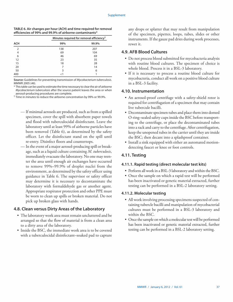
MMWR / January 6, 2012 / Vol. 61 37
Supplement
— If minimal aerosols are produced, such as from a spilled specimen, cover the spill with absorbent paper towels and flood with tuberculocidal disinfectant. Leave the laboratory until at least 99% of airborne particles have been removed (Table 6), as determined by the safety officer. Let the disinfectant stand on the spill until re-entry. Disinfect floors and countertops.
— In the event of a major aerosol-producing spill or break-age, such as a liquid culture containing M. tuberculosis, immediately evacuate the laboratory. No one may reen-ter the area until enough air exchanges have occurred to remove 99%–99.9% of droplet nuclei from the environment, as determined by the safety officer using guidance in Table 6. The supervisor or safety officer may determine it is necessary to decontaminate the laboratory with formaldehyde gas or another agent. Appropriate respirator protection and other PPE must be worn to clean up spills or broken material. Do not pick up broken glass with hands.
4.8. Clean versus Dirty Areas of the Laboratory• Thelaboratoryworkareamustremainunclutteredandbe
arranged so that the flow of material is from a clean area to a dirty area of the laboratory.
• InsidetheBSC,theimmediateworkareaistobecoveredwith a tuberculocidal disinfectant–soaked pad to capture
any drops or splatter that may result from manipulation of the specimen, pipettes, loops, tubes, slides or other instruments. If the gauze pad dries during work processes, rewet it.
4.9. AFB Blood Cultures• Donotprocessbloodsubmittedformycobacteriaanalysis
with routine blood cultures. The specimen of choice is whole blood. Process it in a BSL-3 laboratory.
• If it isnecessary toprocess a routinebloodculture formycobacteria, conduct all work on a positive blood culture in a BSL-3 facility.
4.10. Instrumentation• Anaerosol-proofcentrifugewithasafety-shieldrotoris
required for centrifugation of a specimen that may contain live tubercule bacilli.
• DecontaminatespecimentubesandplacethemintodomedO-ring–sealed safety cups inside the BSC before transport-ing to the centrifuge, or place the decontaminated tubes into a rack and carry to the centrifuge. After centrifugation, keep the unopened tubes in the carrier until they are inside the BSC; then decant into a splashproof container.
• Installasinkequippedwitheitheranautomatedmotion-detecting faucet or knee or foot controls.
4.11. Testing
4.11.1. Rapid testing (direct molecular test kits)• PerformallworkinaBSL-3laboratoryandwithintheBSC.• Oncethesampleonwhicharapidtestwillbeperformed
has been inactivated or genetic material extracted, further testing can be performed in a BSL-2 laboratory setting.
4.11.2. Molecular testing• Allworkinvolvingprocessingspecimenssuspectedofcon-
taining tubercle bacilli and manipulation of mycobacterial cultures must be performed in a BSL-3 laboratory and within the BSC.
• Oncethesampleonwhichamoleculartestwillbeperformedhas been inactivated or genetic material extracted, further testing can be performed in a BSL-2 laboratory setting.
TABLE 6. Air changes per hour (ACH) and time required for removal efficiencies of 99% and 99.9% of airborne contaminants*
ACH
Minutes required for removal efficiency†
99% 99.9%
2 138 207 4 69 104 6 46 69 12 23 35 15 18 28 20 7 14 50 3 6400 <1 1
Source: Guidelines for preventing transmission of Mycobacterium tuberculosis. MMWR2005(46).*Thistablecanbeusedtoestimatethetimenecessarytocleartheairofairborne
Mycobacterium tuberculosisafterthesourcepatientleavestheareaorwhenaerosol-producingproceduresarecomplete.
† Time in minutes to reduce the airborne concentration by 99% or 99.9%.
38 MMWR / January 6, 2012 / Vol. 61
Supplement
5. Autopsy/Necropsy, Surgical Pathology
• Autopsy (human cadaver examination). The infectious and hazardous risks to the laboratory worker performing an autopsy are higher than those for any other health-care professionals because of the procedures used, the popula-tion being assessed, and performance of work in an open area. Use of scalpels, saws, and needles as well as exposure to sharp objects within the body, bone fragments, fractured metal, and/or needles, can result in cuts and percutane-ous injuries. Manipulation of large organs that results in body fluid and blood splashes, and use of instrumentation such as hoses and saws create aerosols in an open area that can result in inhalation, direct contact, or contact with contaminated items in the environment. Those involved in the autopsy directly as well as others in the room are at risk for exposures. Other potential safety risks include exposure to chemicals such as formalin, therapeutic radiation beads, and retained electrical hardware. An alert and well-trained worker, good facility design, optimally fitting and user-friendly personal protective equipment (PPE), appropriate surgical procedures during manipula-tion of the body and dissected material, and subsequent disinfection and sterilization procedures are all critical in minimizing biosafety risk during autopsy and embalming.
•Necropsy (animal cadaver examination). The risk of labo-ratory-acquired infection is very different when working with human cadavers (where infectious agents in essentially all the cadavers are infectious to humans) versus animal cadavers (where infectious agents in most cadavers are not human pathogens). Regardless, animal cadavers can harbor zoonotic agents, and risk assessment to determine whether zoonotic infectious agents may be present in a cadaver, as outlined in Section 12, is critically important for establish-ing appropriate animal necropsy biosafety procedures. The guidelines in this section are combined biosafety best prac-tices for both human autopsy and human surgical pathology and animal necropsy and veterinary surgical pathology. When necessary, biosafety guidelines specific for human or animal diagnostic laboratory settings are highlighted.
5.1. Autopsy/Necropsy–Associated InfectionsThe source of most laboratory-acquired infections and
hazardous exposures that occur during autopsy/necropsy is unknown, and all autopsies and necropsies are to be considered risky (1,56).• Human autopsy facilitieswould function safest at bio-
safety level 3 (BSL-3) for optimal protection of those involved directly with the autopsy and for personnel in
the surrounding area (1,56–58). If a BSL-3 facility is not available, autopsies can be performed using the barrier precautions of BSL-2 plus the negative airflow and respira-tory precautions of BSL-3 (56).
• AnimalnecropsyfacilitiescanfunctionatBSL-2withanoption for BSL-3 practices when warranted by a case-by-case risk assessment (considering, for example availability of Class II biological safety cabinet (BSC), downdraft necropsy tables, and appropriate PPE, such as eye and face protection). Only if a risk assessment indicates a high probability for the presence of a high-consequence livestock pathogen (USDA livestock select agent or toxin [See Section 12.1]) would BSL-3 facilities be required.
• TheMedicalDirector,orinthecaseofanimalnecropsy,the attending pathologist, is responsible for risk assessment and for consideration of limited autopsy/necropsy proce-dures and subsequent acceptable risk level to personnel before each autopsy/necropsy (1,2,56,59)
• Selectastaffmembertobetrainedinsafetyprocedures,and give this person oversight of safety procedures and risk analysis in the pathology suite.
5.1.1. Bloodborne pathogensHuman-health–care workers involved in performance of
autopsies are at high risk for occupationally acquired blood-borne pathogens because of both the injuries sustained and the population undergoing autopsy. Transmission risk is highest per exposure for hepatitis B virus, then hepatitis C virus and human immunodeficiency virus, respectively. These infec-tions have been documented from autopsies as well as during embalming (1,2,56,60–62).
5.1.2. Other infectionsSpecific data for other bloodborne pathogens, such as cyto-
megalovirus, are lacking, but infectious transmission is possible and risk may be higher especially for pregnant (serologically negative) or immunocompromised workers. Assess persons at higher risk for infection on a case-by-case basis and allow them to consent to participating in the autopsy only after being counseled (2,63).
5.1.3. Infectious aerosolsAutopsies/necropsies of cadavers with suspected zoonotic
agents generate potentially infectious aerosols. Although Mycobacterium tuberculosis is the prototypical pathogen most noted to be transmitted by aerosolization, persons who had meningococcemia, anthrax, rickettsiosis and legionellosis are
MMWR / January 6, 2012 / Vol. 61 39
Supplement
other examples. Manipulation of infectious tissue can result in both airborne particles in a size (<5 µm) that floats on air currents for extended periods and can subsequently reach the pulmonary alveoli and small-droplet particles (>5 µm) that settle more quickly. Contamination may occur from fluid-aspi-rating hoses, from spraying the cadaver, and from oscillating saws. The aerosols created stay within the autopsy area and can result in subsequent contact with mouth and eyes, inhalation, or ingestion and can contaminate inanimate surfaces such as computers, telephones and camera equipment (56,57).
5.1.4. Organisms that require additional safety practices• No cases of autopsy-acquiredCreutzfeldt-Jakobdisease
(CJD) have been documented. However, because the prion infectious particle cannot be rendered noninfectious by nor-mal decontamination and sterilization methods, enhanced precautions are mandatory. Transmissibility of the prion is retained in formalin-fixed paraffin blocks (64–67).
• Theonlynaturalanimalpriondiseasewithknownzoo-notic infection potential is classical bovine spongiform encephalopathy (BSE). Necropsy guidelines for cattle with suspected BSE are published elsewhere and focus primarily on avoiding skin puncture, reducing splashes onto mucous membranes, decontaminating facilities and equipment, and disposing of carcasses (68).
5.1.5. Other biosafety exposures• Cyanide,metallic phosphides and organophosphate
pesticides— Specific precautions are required and may include use
of a fume hood or class II type B2 BSC that is ducted to the outside, fume respirators, limiting the autopsy/necropsy, and limiting the time of exposure (56).
• Diagnosticradioactivebeadsortherapeuticscans— Consult the radiation safety officer of record for recom-
mended appropriate measures for limiting the exposure of radiation, transferring the body, and postexposure testing of personnel.
• Electrical and other hardware hazards (pacemakers,indwelling catheters)— Deactivate pacemakers before the autopsy continues.
Discharge of electrical current is possible when defi-brillators are present. If hardware is present, it should be noted and then removed so as not to cause cutting injuries during the autopsy.
5.1.6. Reporting to the morticianReport known bloodborne pathogens or other suspected
aerosolization danger to the mortician and others potentially
handling the body to limit subsequent transmissions that may occur during transport or embalming (69).
5.1.7. Necropsy remains of animalsDispose of animal cadavers with potential zoonotic infectious
agents by appropriate decontamination (e.g., incineration, alkaline digestion or other methods), and do not return them to animal owners for private burial.
5.2. The Autopsy/Necropsy Suite
5.2.1. Inspect the body/carcass• Search for implanted items retained after death.These
are to be noted and clamped or covered before transport to reduce body fluids oozing from the body. Clean the body of visible bloody/body fluids. Cover the autopsy table with a plastic sheet to retain the majority of fluids, or alternatively, use tables with drains so that fluids may be collected in buckets or floor drains.
• Appropriatelydisposeoffluidsandtissuesfromnecropsyof animals with suspected zoonotic agents using methods that provide adequate decontamination, depending upon the specific suspected infectious agent (e.g., incineration, rendering, composting).
5.2.2. Safety guidelines for the suite• Usetheuniversalbiohazardsymboltomarktheautopsy
suite as a biohazard area at the entrance.• Secureaccesstotheautopsysuite,andgrantaccessonlyto
those personnel trained in the biosafety procedures specific to this area.
• ProtectvacuumhoseswithliquiddisinfectanttrapsandHEPA filters or their equivalent.
• Use hand sawswhenever possible to reduce aerosols.Moisten bone before cutting. The pathologist may choose to use oscillating bone saws with a vacuum attachment; use these in a closed area if possible. These vacuum attach-ments are difficult to keep clean and are to be handled as a potential risk of infection. Cover any subsequent jagged edges of exposed bone with towels. When cutting the skull during autopsy when prions are suspected, bag the head.
• Donot leaveusedneedlesonthetable.Donotdetachor resheathe needles. Discard the whole unit into sharps containers. Make sure that sharps containers are available in the work area, and inspect them periodically to ensure that they are never more than two-thirds full. Seal off and replace them when they reach this level.
• Limitthenumberofpersonnelworkingonthehumanbody at any given time to the prosector and/or physician and circulator. Allow only one person to cut at a given time. The same number limitation should apply to small
40 MMWR / January 6, 2012 / Vol. 61
Supplement
animal necropsy. Large animal necropsy generally requires multiple prosectors working together in a way that will avoid accidental lacerations.
• Preparemultiplescalpelsbeforeautopsysobladechangeswhile hands are slippery and contaminated can be avoided. Alternatively, change outer gloves before changing blades. Use blunt-ended scissors when possible instead of scalpels, and use a magnet to pick instruments from the table if they become slippery.
• Donotpasssharpobjectssuchasscalpelsorscissorstoanother person. Place them on the table for another person to pick up.
• Placespecimencontainers(e.g.,bloodculturebottles)ona clean surface for inoculation. Use a rack if possible. Do not hold in the hands while inoculating.
• Examinationoforgansinthebodyandeviscerationtech-nique must be considered so as to limit exposure to blood, body fluids and cuts.
• Forunfixedtissuethatwillberemovedfromtheautopsytable, do the following.— Place on a tray or in a bucket to avoid splashing or
dripping fluids.— After examination, cutting, and/or photography, return
the tissue to the autopsy table to be replaced in the body and/or fixed.
— Place specimens that will be submitted for culture or other laboratory tests in a primary container that is sur-face decontaminated on the outside and then placed into a secondary leakproof container and labeled as biohazard.
— Large organs will have to be removed and cut into multiple sections (breadloaved) so that adequate per-meation of the tissue for fixation will occur.
— Unfixed tissue that will not be returned to the body is considered biohazard waste and is to be kept to a minimum and subsequently disposed of in a manner that will allow appropriate decontamination.
— For autopsy, either suture or staple the body closed. Hold skin flaps with forceps, not hands, when suturing.
— Review of any unfixed tissue requires use of the same PPE as that used in the autopsy.
• Usehands-freeorfoot-activatedrecordingdevicesduringdictation, and a hands-free speaker phone to minimize contamination of inanimate surfaces.
• Provideahands-freesinkattheexitforwashing.• Provideaneyewashstationandshower(2).
5.3. Chemicals (Formaldehyde)Formaldehyde (3.7%–4.0%) used for specimen preserva-
tion is the most common toxic chemical to which autopsy workers are exposed. The chemical is volatile and toxic and
causes irritation to the eyes, mucous membranes, and skin and is associated with increased risk for all cancers. Occupational Safety and Health Administration (OSHA) regulations specify an exposure limit of 0.75 ppm as an 8-hour time-weighted aver-age, and 2.0 ppm for short-term (15-minute) exposures (70). If formaldehyde can be detected by smell, it likely means exposure is occurring at a concentration beyond acceptable limits.
Limit exposure to formaldehyde in the following manner.• Coverallspecimenbucketswhereorgansmaybedeposited
for fixation.• Collect discarded formalin-soaked towels and other
formalin-soaked waste in a bag at the grossing table. Periodically spray a formalin-neutralizing agent on the waste as it is filled. Seal off the bag when it is filled.
• Discardbaggedformalin-soakedtowelsandotherwastein a lined container that can be opened and closed with a foot pedal.
• Cutlargefixedorgansinafumehoodordowndrafttable.• Monitorworkersandresidentpathologistswithformaldehyde
monitoring badges for 8-hour periods, and at least 15-minute periods periodically, to assess formaldehyde exposure.
• Ensurethattissuegrossersarecompetentinpropertissuegrossing technique.
5.4. Spills• Use neutralizing, absorbent mats for small spills.
Neutralizing reagents provide a convenient, cost-effective method for disposal of hazardous formaldehyde, glutar-aldehyde, and other aldehyde solutions. They convert hazardous aldehydes into a nonhazardous, noncorrosive, nontoxic polymer and water. The polymer produced is not a hazardous waste, as defined by U.S. Title 40 Code of Federal Regulations (71). These neutralizing agents tend to reduce disposal costs and contribute to a safer work environment. In some cases, after formaldehyde waste treat-ment with crystal products, the resulting solid waste may be discarded in approved laboratory solid waste streams.
•Wearappropriateprotectiveglovesandprotectiveclothingto prevent skin exposure. Wear protective eyeglasses or chemical safety goggles or use full-face shields as described by OSHA’s eye and face protection regulations (72).
5.5. Protective Equipment
5.5.1. Safety equipment• Biological safety cabinets (2,56) are not common in
autopsy suites because of their limitations in accommodat-ing the volume and size of material being manipulated.
• UsenecropsyfacilitiesthathaveclassIIBSCswhenprac-tical (for small animals) for necropsies of cadavers with
MMWR / January 6, 2012 / Vol. 61 41
Supplement
suspected zoonotic agents, as indicated by a case-by-case risk analysis. However, because necropsy of large animal cadavers with suspected zoonotic agents is not practical in BSCs, use PPE, engineering controls, and procedures that have been specifically developed for clinical laboratories.
• Foroptimalprotectionwhenthereisaknownriskofexpo-sure to bloodborne pathogens and to agents transmitted by aerosols, all autopsy/necropsy facilities should be able to use BSL-3 work practices and physical containment or reference the work to a facility that does (56,59).
• Provideunidirectionalairflowfromcleanareastodirtyareas.• Ensurethattheroomisundernegativepressurerelativeto
other surrounding rooms, with 11–12 air exchanges per hour.• Aircanbedirectlyventedtotheoutsideorrecirculatedinto
the room through HEPA filters, but do not allow the air to recirculate into any clean surrounding areas because this has been associated with outbreaks of M. tuberculosis infection.
• In the eventof redesignof the autopsy/necropsy suite,consider placement of a BSC to mitigate exposure to infectious tissues (56,73).
5.5.2. PPE for autopsy/necropsy personnel• Clothing
— Fluid-resistant (surgical) jumpsuit or shirt and pants that cover from neck to feet and arms
— Waterproof apron and waterproof sleeves— Closed-toe shoes covered with fluidproof shoe covers
or booties— Surgical cap or hood bonnet that covers head entirely
(autopsy only)• Protectionfromsplashesandaerosols
— For general autopsy, or for necropsy when a risk assess-ment indicates a high likelihood for zoonotic agents in the cadaver and the necropsy cannot be conducted in a BSC, use a transparent face shield that covers eyes, mouth and neck, and a fit tested N95 respirator. Eye glasses and contact lenses alone are not adequate for protection.
— Persons unable to wear N95 respirators must wear powered air-purifying respirators (PAPRs).
— Surgical masks do not protect autopsy participants from inhaling airborne contaminants such as infec-tious respirable aerosols or hazardous chemical vapors. (56,57,69,73,74).
• Gloves— Double-gloving is recommended throughout the
autopsy/necropsy, with glove changes every hour. Eight percent of gloves are punctured during autopsy, and about one-third of punctures remain undetected until after gloves are removed (56,75).
— For autopsy, select a cut-resistant glove of fine-woven steel to prevent cuts from bone and scalpels, and cover this with a rubber glove for slip resistance. Cut-resistant gloves do not protect from needle sticks.
— Select gloves specific to the particular task and circum-stances. For example, use heavy gloves for chemical exposure, or gloves that resist accidental puncture if needles will be used.
— Change gloves immediately upon recognizing a puncture, and wash hands with soap and water before resuming the autopsy/necropsy.
• Covercutsorabrasionswithwaterproofocclusivebandages.• ProvidePPEforpersonnelpresentbutnotdirectlypar-
ticipating in the autopsy, i.e., medical students/observers. Observers are to use the same PPE as those participating in the autopsy/necropsy.
• Allsurfacesaretobeconsideredcontaminated.
5.6. Disinfection and Cleaning Procedures for Equipment and Instruments
5.6.1. Human autopsy• Cleananyspillsofbodyfluidsortissuesimmediately.Cover
the spill with paper towels and saturate the area with a disinfectant or a detergent solution, or use a mop followed by disinfection of the spill area with 1:10 bleach solution.
• Fordecontaminationofthebodyafterautopsy,washthebody with a detergent solution, followed by an antiseptic solution or diluted bleach. Place the body in a leakproof shroud, and label as biohazard. Direct notification of the mortician is appropriate in cases of known bloodborne pathogens or the presence of M. tuberculosis.
5.6.2. Human autopsy/animal necropsyThe following guidelines for disinfection and cleaning
following an autopsy or necropsy apply to both types of procedures.• Anyone involvedwith the body, room, or instrument
cleanup must wear the appropriate PPE.•Washallworksurfacesandfloorswithadetergentsolu-
tion followed by disinfection with a 1:10 bleach solution or other suitable commercial disinfectant.
• Remove visible gross tissue fromnondisposable instru-ments (knife handles, pans, aprons) before chemical disinfection or autoclaving. Place sharp objects in punc-ture-resistant containers and wash with detergent solution to remove any remaining gross tissue. Rinse with water and wet thoroughly with a 1:10 bleach solution or appropri-ate commercial disinfectant, the volume depending on amount of blood and body fluid present. Consider that
42 MMWR / January 6, 2012 / Vol. 61
Supplement
bleach will corrode stainless steel and aluminum devices. Remove the disinfectant or bleach by rinsing with water.
• Placedisposableitems,paperproducts,aprons,sponges,and similar items, into a biohazard container or bag for terminal treatment or autoclaving.
• Placereusableclothinginaleakproofbiohazardbagforlaundering according to hospital protocol.
•Washreusable,nonlaunderableitemssuchasapronswitha detergent solution, decontaminate with bleach solution, rinse with water and allow to dry before next use.
• Nodirtyitemsoranythingusedintheautopsyistoberemoved from the room, including clothing and laboratory coats.
• Keepcamera,telephones,computerkeyboards,andotheritems as clean as possible, but consider them to be contami-nated and handle them with gloves. Wipe the items with detergent solution and 1:10 bleach solution or appropriate disinfectant after each use.
•Washhandswithsoapandwateruponremovalofgloveswhen exiting the room.
5.7. Waste management
5.7.1. Human tissueEither incinerate all pathological waste, since this is con-
sidered hazardous material and is regulated by the U.S. Department of Transportation (DOT), or transport patho-logical waste to on-site or off-site treatment facilities in clearly labeled, dedicated, leakproof containers or carts that meet DOT requirements. DOT sharps waste containers need to be puncture-proof in addition to meeting these requirements. State, local, and regional regulations may also apply and need to be addressed.
5.7.2. Animal tissueDispose of all animal necropsy waste (tissues or postnecropsy
cadaver) using an appropriate method as determined by the case-by-case risk analysis assessment (incineration, autoclaving and standard waste disposal, rendering, composting, crema-tion, private burial).
5.7.3. Other wasteShred autoclave red-bag waste if appropriate. State, local, and
regional regulations may also apply and need to be addressed. (See Section 3.5, Waste Management.)
5.8. Clean versus Dirty AreasClean areas might include an administrative area and bath-
rooms with showers. Air from these areas should be exhausted differently than from the autopsy suite (56,76,77). All other areas are considered dirty, and appropriate PPE is required.
5.9. Surgical Pathology• Surgicalpathology includesgrossdissectionand frozen
sectioning. Histology and cytology are “hybrid” areas of autopsy and the clinical diagnostic laboratories. Each laboratory section has its own unique safety issues; data on standardized biosafety practice or on reported biosafety incidents are very limited. Documentation and research in surgical pathology safety are necessary for future guidelines and recommendations (78,79).
• Surgicalpathologyrisksareassociatedwithmanipulatinglarge amounts of fresh tissues from unknown infectious sources, which may result in puncture, cuts, and splashes of blood and body fluids, similar to the autopsy where fresh organs must be viewed at a grossing table and cut into sections thin enough for fixation (i.e., “breadloaved”). Other risks include the use of cryostat cutting equipment or freezing spray that generates infectious aerosols when sectioning frozen tissue, and exposures to large volumes of formaldehyde.
• Tissuethatwillbeusedforslideexaminationisfixedinformalin; subsequently, small tissue sections of interest are dissected with scalpels, placed in cassettes, replaced in specimen vats with formalin, and transported to histology.
• Histologythenembedstheformalin-fixedtissueinparaffinand cuts these sections with microtome-bladed instru-ments to make slides for viewing using specific stains and immunochemistry. Although most formalin-fixed specimens are noninfectious, both M. tuberculosis and CJD agent can persist in transmissible form in formalin-fixed, paraffin-embedded material. Exposure to toxic formalde-hyde is common.
• Cytology receives large- and small-volumebody fluids,bone marrow samples, or needle aspirate specimens, most of which are received in fixative but others that must be processed by aliquoting or pouring off large-volume body fluids, which can result in splashing and spills. Procedures such as centrifugation and cytospin processing can produce aerosols. Air-dried slides can be a source of contamination until they are fixed and stained.
5.9.1. Specimen receiving and log-in• Handlespecimenswithstandardprecautions,andwear
gloves when receiving and accessioning (2,57). Receipt areas in all surgical pathology laboratories are consid-ered dirty areas, and all exterior containers considered contaminated.
• Submit specimens, including skin and gastrointestinalbiopsies, delivered from the operating room, autopsy, or from outside collection sites in leakproof containers or place in fixative specimen containers at the site of
MMWR / January 6, 2012 / Vol. 61 43
Supplement
collection Place the specimen in a secondary leakproof bag or container and label as a biohazard. Place the requisition in an outside pocket where it will avoid contamination with the specimen.
• LeakingspecimensorvisiblycontaminatedspecimensSpecimens may be contaminated with fresh tissue, blood, or
formalin. Handling is based on whether the specimen is fresh or formalin-fixed.
— If the specimen is submitted in fixative and the second-ary container is not leaking, tighten the specimen cap and place the primary container in a clean bag; wipe any formalin from the bench top.
— If the specimen is submitted as fresh tissue and the exterior container is leaking, place the specimen into another container and process it using appropriate PPE. Specimens are then to be transferred to a clean container and labeled.
— If the requisition is contaminated, discard it as biohaz-ardous waste and replace it.
5.9.2. Work at the open bench• Separatethelog-inroomandadministrativeareasfrom
the grossing room if fresh tissue or cryostats are used in the same open area (56,76,77).
• Handlingoffreshtissue— Examine fresh tissue in a BSC if possible, or in a room
separated from the rest of the surgical pathology gross-ing stations. Use an N95 mask and other PPE similar to autopsy conditions, depending on the volume of blood and likelihood of body fluid exposure (56). Fresh tissue should never be handled without gloves. PPE as used in autopsy procedures is advocated for handling fresh tissue (2,56,57,80,81).
— For human pathology, store fresh tissue not undergoing fixation or unable to be adequately fixed (e.g., teeth or foreign bodies) in a double, sealable, leakproof con-tainer; label as biohazard and store in a refrigerator or freezer (2).
— Standard precautions against bloodborne infections are to be used irrespective of the clinical history (79).
• Frozensections— Frozen sectioning is performed on fresh tissue and is a
high-risk procedure for infectious exposure. Freezing tissue does not kill organisms, and the use of the cryo-stat cutting blade creates potentially dangerous aerosols. Discuss the true clinical necessity for frozen sectioning with the surgical team.
— Although some cryostat instruments have a down-draft into the instrument, aerosols are dispersed into the room where the cutting takes place. Do not use
freezing propellant sprays, which speed the freezing process by a few seconds and cause aerosolization of not only the tissue being frozen but also the tissues from previously cut specimens that are at the base of the instrument. Such procedures generate aerosol and droplet contamination, posing an infectious risk to all personnel in the area (56,79,82). The Clinical and Laboratory Standards Institute and others have recom-mended discontinuation of freezing sprays because they are not recommended by the manufacturers of cryostat instrumentation (2,79).
— Ideally, use cryostats in a closed room that has air vented directly to the outside or recirculated through a HEPA filter to avoid contamination to the rest of the surgical pathology suite.
— Certain cryostats have ultraviolet lights, but these are not a substitute for terminal cleaning of the instrument and have been ineffective in killing mycobacteria.
— In human pathology laboratories, gloves, face shield or goggles, and N95 mask must be worn when processing.
• Bonecutting.SeeAutopsySection5.2.2.• Fixedtissue
— Fix tissue in 10% formalin in a concentration 10 times the volume of tissue to ensure effective fixation and to reduce potential of infectious contamination. However, the viability of infectious organisms is dependent on a host of variables that have not been clearly identified, and fixed tissue has been shown to remain infectious (78). M. tuberculosis has been transmitted from fixed specimens as well as grown from fixed specimens, and CJD is not inhibited by the routine concentration of formalin. Grossing stations where formalin-fixed specimens are cut are designed for decreasing the fumes of formaldehyde but are not BSCs (2,57,83–85). Formalin-fixed specimens must be handled with gloves.
— Use standard precautions and appropriate PPE in all work with fixed tissue. Face shields are optimal for full face and neck protection if splashing with formalin is likely. Shields offer the most comfort for extended periods while allowing the user to wear prescription eyeglasses or to do dictation.
— Eyeglasses and contact lenses are not a substitute for eye protection.
— Goggles and fluid-resistant mask are an alternate choice but not preferred.
— Use face shields/goggles that can be decontaminated or that are disposable.
— Keep containers with tissues in formalin closed to reduce exposure fumes.
44 MMWR / January 6, 2012 / Vol. 61
Supplement
• Cytologyspecimens— PPE is dependent on the specimen. Gloves and labora-
tory coat are required for all specimens until slides are fixed and stained.
— Pour off or aliquot large-volume specimens with poten-tial for splashing and/or aerosolization inside a BSC, and wear fluid-resistant clothing, apron, and two pairs of gloves.
— Open small-volume body fluids or aspirates submitted in tubes in a BSC, or use a splash guard or a face shield and aliquot with a disposable pipette rather than pour-ing to avoid splashing and spill.
— Handle specimens received in fixative with gloves because of the toxicity of the fixatives and possible external contamination (57). If slide preparation will use a cytospin preparation, the system must include a bowl with safety lid and outside cover.
— Consider all slides, impression smears, cytological preparations, and bone marrow smears as infectious until fixed and stained.
Use a safety centrifuge with safety cups with O-rings and sealable tops for centrifugation of fluids (51).
• Decontamination— Cryostat. Instrument shavings generated by cutting are
considered contaminated. Collect accumulated cuttings and discard them as biohazardous waste. Defrost the instrument and decontaminate daily with 70% alcohol. Decontaminate weekly with a tuberculocidal disinfec-tant or after a known case of M. tuberculosis infection. Wear stainless steel mesh gloves while cleaning the microtome knives.
— Microtome. Consider a similar schedule as with cryostats.
— Consider all surfaces, computer, telephone, and coun-ters as contaminated if any person using gloves touches these items. Disinfect equipment and bench tops daily.
— Remove gloves and wash hands with soap and water before exiting the various laboratory rooms.
5.9.3. Clean versus dirty areas of the laboratoryAll of the surgical pathology specialty areas (cytology, histol-
ogy, grossing or frozen section rooms) are considered dirty areas if fresh specimens or body fluids are received or processed in an open room (not in a BSC or separately vented area).
5.9.4. Tissue stainsMultiple staining procedures are performed in histology and
cytology. The most common are included here. Some of these stains are prepared with ethanol and some with methanol, which can have an impact on management options for their
waste. Provide material safety data sheets (MSDS) for each component in the laboratory.• Hematoxylinstain Hematoxylin stain is not hazardous under Environmental
Protection Agency regulations. Drain disposal is recom-mended with the permission of local wastewater treatment authorities. Follow federal, state, and local regulations.
• Giemsastain The preferred disposal method for Geimsa stain is incin-
eration at a permitted hazardous waste treatment facility. Localities may restrict the amounts of alcohols that may be flushed down the drain; consult appropriate authori-ties. Ensure compliance with all local, state, and federal government regulations.
•Wrightstain Wright stain contains methanol, a listed hazardous waste.
Sewer disposal of listed hazardous wastes is not acceptable or permitted. The preferred disposal method is incinera-tion. Ensure compliance with all government regulations.
5.9.5. Fixatives• Formalin(HCHO)
— Formaldehyde may be purchased as a 37%–40% HCHO solution. The most common formaldehyde preparation is 10% formalin (3.7%–4% formaldehyde), which is available commercially. Diluting formalin or formaldehyde is discouraged to limit exposure. Local governments often restrict the amounts of aldehydes that may be flushed down the drain. Each laboratory must comply with all government regulations.
— Neutralization is the preferred disposal method for formaldehyde. Neutralizing reagents provide a conve-nient, cost-effective method for disposal of formalde-hyde, glutaraldehyde, and other aldehyde solutions. They convert hazardous aldehydes into a nonhazard-ous, noncorrosive, nontoxic polymer and water. The polymer produced is not a hazardous waste, as defined by U.S. Code of Federal Regulations, Title 40 (71). These neutralizing agents tend to reduce disposal costs and contribute to a safer work environment. In some cases, after formaldehyde waste treatment with crystal products, the resulting solid waste may be discarded in approved laboratory solid waste streams. Before engag-ing in sewer disposal of neutralized formalin solutions, be sure to have formal approval of applicable local wastewater authorities.
— Wear appropriate protective gloves and protective clothing to prevent skin exposure. Wear protective eyeglasses, chemical safety goggles, or full-face shields,
MMWR / January 6, 2012 / Vol. 61 45
Supplement
as described by OSHA’s eye and face protection regula-tions in 29 CFR 1910.133 (72) or European Standard EN166. Do not wear contact lenses when working with formalin. Maintain eye wash fountain and drench facilities in the work area.
• Otherpathologyfixatives— Fixatives are classified as regulated waste. OSHA’s
Standard, Bloodborne Pathogens, 29 CFR Part 1910, 1048 and 1030, mandates that pathology specimens be placed in secondary containers that are constructed to contain all contents and prevent leakage of fluids dur-ing handling, storage, transport or shipping (33,70).
— Label the secondary containers as biohazards, and close the container securely prior to removal to prevent spill-age of contents during handling, storage, transport and shipping.
• Xylenesandalcohols— Xylene is categorized by EPA as a hazardous waste under
the Resource Conservation and Recovery Act (RCRA) and has been assigned EPA hazardous waste no. U239. Waste xylene can be separated from dissolved paraffin by distillation, and commercially available recycling units can accomplish this separation effectively to produce reusable xylene. Xylene also may be disposed of by a properly permitted hazardous waste contrac-tor if the xylene is contained in an organic metallic or organic laboratory pack that meets the requirements of 40 CFR 264.316 or 265.316 (71).
— Xylene that cannot be saved for recovery or recycling must be handled as hazardous waste and sent to an RCRA-approved incinerator or disposed in an RCRA-approved waste treatment facility. Processing, use, or contamination of this product may change the waste management options. State and local disposal regula-tions may differ from federal disposal regulations. Dispose of the container and unused contents in accordance with federal, state and local requirements.
— The disposal guidelines for alcohols (ethyl alcohol, isopropyl alcohol, methyl alcohol) are identical to those for xylene.
• Testingforformalinandxyleneexposure Conduct periodic testing with chemical badges for an
8-hour period and at least one 15-minute period peri-odically to assess formaldehyde exposure for persons who are routinely exposed (e.g., technologists, tissue grosser, resident pathologists), and rotate among various areas so that every work station is assessed.
5.10. Engineering Controls and Facility Renovations
When updating or renovating autopsy and other areas of the anatomic pathology laboratory that process fresh tissue and body fluids, the following should be considered.• Designingclosedspecimen-receiving/administrativeareas
for receiving specimens if BSCs are not available for pro-cessing fresh specimens or if cryostats are in the same area as administrative personnel.
• Providing roomswithnegative air flow relative to sur-rounding rooms.
• InstallingaBSCforprocessingfreshtissues.• Providing a separate room for cryostats andbone saws
apart from the remainder of the surgical grossing suite.
5.11. Creutzfeldt-Jakob DiseaseSpecial precautions for autopsy and autopsy suite decon-
tamination, brain-cutting, and histologic tissue preparation procedures are required when processing cases of possible CJD (1,56,65,67,86).
5.11.1. AutopsyPerform autopsies using BSL-2 precautions augmented
by BSL-3 facility ventilation and respiratory precautions. Wear standard autopsy PPE. Limit the autopsy to brain removal. Restrict participants to only those who are necessary. Double-bag the brain and place it in a plastic container for freezing or fix it in 3.7%–4% formaldehyde after sectioning. Formaldehyde fixation occurs for 10–14 days before histologic sections are collected.
5.11.2. Histologic preparations• Prions remain transmissible after formalin fixation and
paraffin embedding. (56,64,66,86).• PPE includes, gloves, laboratory coat, apron, and face
protection.• Beforehistologicslidepreparation,smallblocksofbrain
tissue <5 mm thick are soaked in 95%–100% formic acid for 1 hour, followed by soaking in fresh 4% formaldehyde for at least 48 hours. Such tissue may be processed either by hand or by machine. Blocks and slides may then be handled routinely. If the laboratory prefers to process by hand, the procedures are the same as for untreated tissues.
46 MMWR / January 6, 2012 / Vol. 61
Supplement
• If the tissue has not been pretreatedwith formic acidbefore embedding, double gloves and eye protection are to be worn at all times, including during sectioning. All solutions, including water washes, are collected and treated with equal volumes of fresh, undiluted bleach or 1N NaOH for 60 minutes. Disposable supplies are either incinerated or autoclaved at appropriate temperature and pressure (1,56,86). Glassware and tools are generally soaked in undiluted bleach for 1 hour and then rinsed well. If appropriate for the tool, they may autoclaved at appropriate temperature and pressure. Equipment that cannot be soaked or autoclaved should be wiped with undiluted bleach and allowed to sit for 30 minutes.
• Careshouldbetakentocollectallscrapsofparaffinandunused sections on a disposable sheet. The microtome itself
may be wiped with bleach or sodium hydroxide solution, but it cannot be thoroughly decontaminated. If frequent possible CJD cases are handled, laboratories may wish to dedicate an old microtome to this purpose.
• Allwasteistobecollectedanddisposed.Discardtheknifeblade with the sharps.
• Slidescontaininguntreatedtissuearestainedbyhandindis-posable specimen cups or Petri dishes. The slides are labeled as infectious CJD and stored/filed in an appropriately labeled container. The sectioned block is sealed with paraffin.
• Tissueremnantsarediscardedasinfectioushospitalwastefor incineration. Fluids are diluted 1:1 with undiluted bleach for 1 hour before disposal.
• Dedicatedequipmentmayberequireddependingonlocaljurisdictions.
MMWR / January 6, 2012 / Vol. 61 47
Supplement
6. Parasitology Laboratory
Exposure to infectious parasites during diagnostic proce-dures may result from handling specimens, drawing blood, performing various types of concentration procedures, cultur-ing organisms, and conducting animal inoculation studies. Relevant parasites and their possible routes of infection are listed in Table 7 and Box 1. Table 8 contains information on resistance to antiseptics and disinfectants.
6.1. Specimen Receiving and Log-In/Setup Station• Freshspecimens(feces,othergastrointestinaltractspeci-
mens, urine, blood, tissues, cerebrospinal fluid, other body fluids, arthropods) represent a potential source of infectious parasites.
• Safety precautions include proper labeling of fixatives;designating specific areas for specimen handling (bio-logical safety cabinets may be necessary under certain circumstances); proper containers for centrifugation; acceptable discard policies; appropriate policies for no eating, drinking, or smoking within the working areas; and, if applicable, correct techniques for organism culture and/or animal inoculation.
• Collectortransfereveryspecimenintoaleakproofprimarycontainer with a secure lid (avoid snap-top closure).
• Usedisposableplasticbagswithseparatepocketsfortherequisition slip and specimen when possible (2,87).
6.1.1. Leaking containers• Visuallyinspectallspecimencontainersforleakage.• Contaminatedprimary containersmust bedecontami-
nated before further manipulation. In some circumstances, it might be necessary to transfer the contents to a clean container or collect another specimen before submission to the testing area.
• Discardcontaminatedrequisitionsasbiohazardouswasteand replace them.
6.1.2. Loose caps• Bloodspecimensaresubmittedintubes,usuallyVacutainer
tubes (with either lavender or green tops). If the stopper appears to be loose or there is evidence of blood on the outside of the tube, push the stopper into the tube for a secure fit, and request the sender to submit another speci-men. If the specimen cannot be repeated, decontaminate the outside before processing the specimen.
6.2. Stains and Reagents (88,89)
6.2.1. Trichrome stain•Wheatley’smodificationoftheGomoritissuetrichrome
stain is considered nonhazardous waste but must be disposed of in accordance with federal, state and local environmental control regulations.
• Draindisposal is recommendedwiththepermissionoflocal wastewater treatment authorities.
6.2.2. Hematoxylin stain• HematoxylinstainisnothazardousunderEnvironmental
Protection Agency (EPA) regulations.• Draindisposal is recommendedwiththepermissionof
local wastewater treatment authorities. Canadian disposal regulations generally parallel those in the United States.
• Followfederal,stateandlocalregulations.
6.2.3. Iodine• Disposeofallliquidandiodine-contaminatedparasitol-
ogy-related material in Department of Transportation (DOT)–approved waste containers.
• Incineration for liquids is the suggestedmethod ofdisposal.
• Complywithall federal, stateand local regulations fordisposal.
6.2.4. Acid-fast stains (modified)• Disposeofcontainerandunusedcontentsinaccordance
with applicable federal, state and local requirements.• Stateandlocaldisposalregulationmaydifferfromfederal
disposal regulations.
6.2.5. Giemsa stain• Thepreferreddisposalmethodisincinerationatapermit-
ted hazardous waste treatment facility.• Localitiesmayrestricttheamountsofalcoholsthatmay
be flushed down the drain. Ensure compliance with all government regulations.
6.2.6. Wright stain• The preferred disposalmethod is incineration in an
approved facility.• Localitiesmayrestricttheamountsofalcoholsthatmay
be flushed down the drain. Ensure compliance with all applicable government regulations.
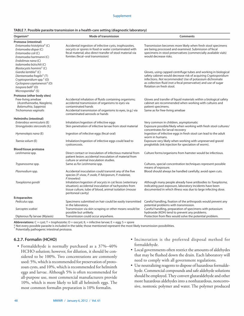
48 MMWR / January 6, 2012 / Vol. 61
Supplement
TABLE 7. Possible parasite transmission in a health-care setting (diagnostic laboratory)
Organism* Mode of transmission Comments
Protozoa (intestinal)Entamoeba histolytica†(C) Accidental ingestion of infective cysts, trophozoites,
oocystsorsporesinfoodorwatercontaminatedwithfecalmaterial;alsodirecttransferofstoolmaterialviafomites(fecal–oraltransmission)
Transmissionbecomesmorelikelywhenfreshstoolspecimensare being processed and examined. Submission of fecal specimensinstoolpreservatives(commerciallyavailablevials)woulddecreaserisks.
Entamoeba dispar (C)Entamoeba coli (C)Entamoeba hartmanni (C)Endolimax nana (C)Iodamoeba bütschlii (C)Blastocystis hominis† (C)Giardia lamblia† (C) Gloves,usingcappedcentrifugetubesandworkinginbiological
safetycabinetwoulddecreaseriskofacquiringCryptosporidium infections. Not recommended:Useofpotassiumdichromateascollectionfluid(notafecalpreservative)anduseofsugarflotation on fresh stool.
Dientamoeba fragilis† (T)Cryptosporidium spp.† (O)Cyclospora cayetanensis† (O)Isospora belli†
(O)
Microsporidia†(S)Protozoa (other body sites)Free-livingamebae(Acanthamoeba, Naegleria, Balamuthia, Sappinia)
Accidentalinhalationoffluidscontainingorganisms;accidental transmission of organisms to eyes via contaminated hands
Glovesandtransferofliquidmaterialswithinabiologicalsafetycabinetarerecommendedwhenworkingwithculturesandpatient specimens.
Trichomonas vaginalis Accidentaltransmissionoforganismstoeyes,(e.g.)viacontaminated aerosols or hands
Sameasforfree-livingamebae
Helminths (intestinal)Enterobius vermicularis (E) Inhalation/ingestion of infective eggs Very common in children, asymptomaticStrongyloides stercoralis (IL) Skin penetration of infective larvae from stool material Exposurepossible/likelywhenworkingwithfreshstoolcultures/
concentrates for larval recoveryHymenolepis nana(E) Ingestionofinfectiveeggs(fecal-oral) Ingestion of infective eggs in fresh stool can lead to the adult
worminhumans.Taenia solium (E) Inhalation/ingestion of infective eggs could lead to
cysticercosis.Exposureverylikelywhenworkingwithunpreservedgravidproglottids(inkinjectionforspeciationofworm).
Blood/tissue protozoaLeishmania spp. Direct contact or inoculation of infectious material from
patientlesion;accidentalinoculationofmaterialfromculture or animal inoculation studies.
Cultureforms/organismsfromhamsterwouldbeinfectious.
Trypanosoma spp. Same as for Leishmania spp. Cultures,specialconcentrationtechniquesrepresentpossiblemeans of exposure.
Plasmodium spp. Accidental inoculation could transmit any of the five species(P. vivax, P. ovale, P. falciparum, P. malariae, P. knowlesi)
Bloodshouldalwaysbehandledcarefully;avoidopencuts.
Toxoplasma gondii Inhalation/ingestionofoocystsincatfeces(veterinarysituation);accidentalinoculationoftachyzoitesfromtissueculture,tubeofblood,animalisolation(mouseperitonealcavity)
Although many people already have antibodies to Toxoplasma, indicating past exposure, laboratory incidents have been documentedinwhichillnesswasduetolargeinfectingdose.
EctoparasitesPediculus spp. Specimens submitted on hair could be easily transmitted
in the laboratory.Carefulhandling,fixationofthearthropodswouldpreventanypotentialproblemswithtransmission.
Sarcoptes scabiei Transmissionviaskinscrapingorothermeanswouldbepossible but unlikely.
Carefulhandling,preparationofspecimenswithpotassiumhydroxide(KOH)tendtopreventanyproblems.
Dipterousflylarvae(Myiasis) Transmissioncouldoccuranywhere. Protectionfromflieswouldsolvethepotentialproblem.
Abbreviations: C=cyst;T=trophozoite;O=oocyst;IL=infectivelarvae;E=egg;S=spore*Noteverypossibleparasiteisincludedinthetable;thosementionedrepresentthemostlikelytransmissionpossibilities.† Potentially pathogenic intestinal protozoa.
6.2.7. Formalin (HCHO)• Formaldehyde is normally purchased as a 37%–40%
HCHO solution; however, for dilution, it should be con-sidered to be 100%. Two concentrations are commonly used: 5%, which is recommended for preservation of proto-zoan cysts, and 10%, which is recommended for helminth eggs and larvae. Although 5% is often recommended for all-purpose use, most commercial manufacturers provide 10%, which is more likely to kill all helminth eggs. The most common formalin preparation is 10% formalin.
• Incineration is the preferred disposal method forformaldehyde.
• Localgovernmentsoftenrestricttheamountsofaldehydesthat may be flushed down the drain. Each laboratory will need to comply with all government regulations.
• Useneutralizingreagentstodisposeofhazardousformalde-hyde. Commercial compounds and safe aldehyde solutions should be employed. They convert glutaraldehyde and other more hazardous aldehydes into a nonhazardous, noncorro-sive, nontoxic polymer and water. The polymer produced
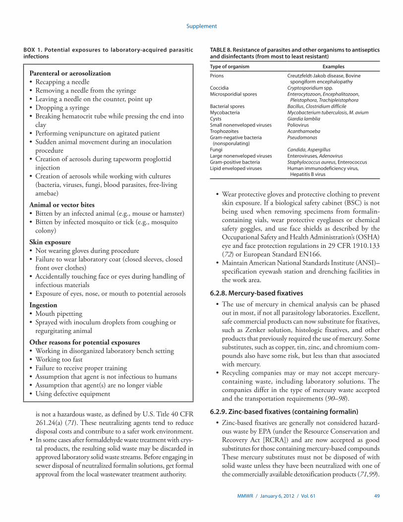
MMWR / January 6, 2012 / Vol. 61 49
Supplement
is not a hazardous waste, as defined by U.S. Title 40 CFR 261.24(a) (71). These neutralizing agents tend to reduce disposal costs and contribute to a safer work environment.
• Insomecasesafterformaldehydewastetreatmentwithcrys-tal products, the resulting solid waste may be discarded in approved laboratory solid waste streams. Before engaging in sewer disposal of neutralized formalin solutions, get formal approval from the local wastewater treatment authority.
•Wearprotectiveglovesandprotectiveclothingtopreventskin exposure. If a biological safety cabinet (BSC) is not being used when removing specimens from formalin-containing vials, wear protective eyeglasses or chemical safety goggles, and use face shields as described by the Occupational Safety and Health Administration’s (OSHA) eye and face protection regulations in 29 CFR 1910.133 (72) or European Standard EN166.
•MaintainAmericanNationalStandardsInstitute(ANSI)–specification eyewash station and drenching facilities in the work area.
6.2.8. Mercury-based fixatives• Theuseofmercuryinchemicalanalysiscanbephased
out in most, if not all parasitology laboratories. Excellent, safe commercial products can now substitute for fixatives, such as Zenker solution, histologic fixatives, and other products that previously required the use of mercury. Some substitutes, such as copper, tin, zinc, and chromium com-pounds also have some risk, but less than that associated with mercury.
• Recycling companiesmayormaynot acceptmercury-containing waste, including laboratory solutions. The companies differ in the type of mercury waste accepted and the transportation requirements (90–98).
6.2.9. Zinc-based fixatives (containing formalin)• Zinc-basedfixativesaregenerallynotconsideredhazard-
ous waste by EPA (under the Resource Conservation and Recovery Act [RCRA]) and are now accepted as good substitutes for those containing mercury-based compounds These mercury substitutes must not be disposed of with solid waste unless they have been neutralized with one of the commercially available detoxification products (71,99).
TABLE 8. Resistance of parasites and other organisms to antiseptics and disinfectants (from most to least resistant)
Type of organism Examples
Prions Creutzfeldt-Jakobdisease,Bovinespongiform encephalopathy
Coccidia Cryptosporidium spp.Microsporidial spores Enterocytozoon, Encephalitozoon,
Pleistophora, TrachipleistophoraBacterial spores Bacillus, Clostridium difficileMycobacteria Mycobacterium tuberculosis, M. aviumCysts Giardia lambliaSmall nonenveloped viruses PoliovirusTrophozoites Acanthamoeba Gram-negativebacteria(nonsporulating)
Pseudomonas
Fungi Candida, AspergillusLarge nonenveloped viruses Enteroviruses, AdenovirusGram-positivebacteria Staphylococcus aureus, EnterococcusLipid enveloped viruses Human immunodeficiency virus,
Hepatitis B virus
BOX 1. Potential exposures to laboratory-acquired parasitic infections
Parenteral or aerosolization• Recappinganeedle• Removinganeedlefromthesyringe• Leavinganeedleonthecounter,pointup• Droppingasyringe• Breakinghematocrittubewhilepressingtheendinto
clay• Performingvenipunctureonagitatedpatient• Suddenanimalmovementduringaninoculation
procedure• Creationofaerosolsduringtapewormproglottid
injection• Creationofaerosolswhileworkingwithcultures
(bacteria, viruses, fungi, blood parasites, free-living amebae)
Animal or vector bites• Bittenbyaninfectedanimal(e.g.,mouseorhamster)• Bittenbyinfectedmosquitoortick(e.g.,mosquito
colony)
Skin exposure• Notwearingglovesduringprocedure• Failuretowearlaboratorycoat(closedsleeves,closed
front over clothes)• Accidentallytouchingfaceoreyesduringhandlingof
infectious materials• Exposureofeyes,nose,ormouthtopotentialaerosols
Ingestion•Mouthpipetting• Sprayedwithinoculumdropletsfromcoughingor
regurgitating animal
Other reasons for potential exposures•Workingindisorganizedlaboratorybenchsetting•Workingtoofast• Failuretoreceivepropertraining• Assumptionthatagentisnotinfectioustohumans• Assumptionthatagent(s)arenolongerviable• Usingdefectiveequipment
50 MMWR / January 6, 2012 / Vol. 61
Supplement
• Disposeofmaterialsinaccordancewithfederal(40CFR261)(71), state, and local requirements.
• Therecommendedcleansingagentiswater.• Zincformalinistoxicbecauseofitsformaldehydecontent.
Dispose by using a licensed waste hauler. Do not mix waste streams unless instructed to do so by your waste hauler. Some wastewater treatment authorities may grant permission for drain disposal of limited amounts if the zinc content is <600 ppm.
• Zincformalinisrecyclableandcanbeneutralizedwithcommercially available detoxification products.
• Zinc-basedfixativesarenowavailablethatdonotcontainformalin. Universal fixatives such as Total-Fix do not con-tain mercury, polyvinyl alcohol, or formalin and can be used for concentration, permanent stained smears, and fecal immunoassays, except for the Entamoeba histolytica and the Entamoeba histolytica/E. dispar group, which require fresh or frozen specimens for testing. Giardia and Cryptosporidium spp. tests can be run on fresh, as well as preserved speci-mens. Dispose of materials in accordance with federal (40 CFR 261) (71), state, and local requirements.
6.2.10. Copper-based fixatives (containing no formalin)•Manylocalitiesrestricttheamountofcoppercompounds
that may be flushed down the drain.• Ensurecompliancewithallgovernmentregulations.
6.2.11. Xylene and alcohols• XyleneiscategorizedasahazardouswasteunderRCRA
and has been assigned EPA hazardous waste no. U239. Use of one of the current commercial substitutes for xylene will avoid the need to address the special handling required for hazardous waste management described here.
• Xylenemaybedisposedofinanorganometallicororganiclaboratory pack that meets the requirements of 40 CFR 264.316 or 265.316 (71). Xylene can be distilled by using a variety of commercially available recycling units to produce reusable xylene.
• Xylenethatcannotbesavedforrecoveryorrecyclingisto be handled as hazardous waste and sent to an RCRA-approved incinerator or disposed in an RCRA-approved waste facility.
• Processing, use, or contamination of this productmaychange the waste management options. State and local disposal regulations may differ from federal disposal regu-lations. Dispose of the container and unused contents in accordance with federal, state and local requirements.
• Thedisposalguidelinesforalcohols(ethylalcohol,isopropylalcohol, methyl alcohol) are identical to those for xylene.
6.3. Working at the Bench• Ingeneral,thesameprecautionsandpracticesthatareused
in microbiology laboratories are applicable to diagnostic parasitology procedures, particularly regarding aerosol transmission (2,33).
• Guidelinesforspecimencollectionandprocessingincludestandard precautions as well as use of recommended equip-ment according to approved methods (e.g., BSCs, fume hoods, centrifuges, sharps, glassware) (2,100,101).
6.4. Personal Precautions
6.4.1. Biological safety cabinet versus fume hood• Althoughuse of a fumehood is notmandatorywhen
processing stool specimens containing formalin, use an OSHA-compliant formalin monitoring program.
• Evenwiththesubstitutionofdehydratingreagentsotherthan xylene, fume hoods are recommended in order to eliminate fecal and solvent odors. A small, table-top model is acceptable.
• ABSCisnotrequiredforprocessingfecalspecimensinthe parasitology laboratory; however, some laboratories use Class I (open-face) or in many cases, a Class II-A2 (lami-nar-flow) BSC for processing all unpreserved specimens.
• ABSC is recommended, especially if the laboratory isprocessing fresh specimens or performing cultures for parasite isolation (Table 7).
6.4.2. Personal protective equipment• Useappropriatehandhygiene(washingandantiseptics)
for laboratory work in a diagnostic parasitology laboratory.•Weardisposableglovesof latex,vinyl,ornitrileduring
accessioning and processing of all specimens for parasito-logic examination, especially when handling blood, body fluids, and stool specimens (100,101); this recommenda-tion applies whether the clinical specimens are fresh or are submitted in fecal preservatives. Always wash hands after glove removal.
6.4.3. Immunization• TheAdvisoryCommittee on ImmunizationPractices
recommends that all persons whose work-related activities involve exposure to blood or other potentially infectious body fluids in a health-care or laboratory setting receive hepatitis B vaccine. Booster doses are not recommended (102).
• Documentation,includingsignedstatementsandrecordsof hepatitis B vaccination or declination, must be kept.
MMWR / January 6, 2012 / Vol. 61 51
Supplement
6.4.4. DisinfectionGeneral recommendations for the microbiology laboratory
are sufficient for use in the diagnostic parasitology section; these would include guidelines for disinfection of countertops, telephones, computers, equipment, and hands-free telephones.
6.5. Dirty versus Clean Areas of the LaboratoryGeneral guidelines for the microbiology laboratory also
apply for the parasitology section of the laboratory. No special recommendations are necessary.
6.6. InstrumentationSafety requirements for the use of instruments are the same
as those used for a general microbiology laboratory and are primarily involved with specimen handling.
6.7. Antibody and Antigen Parasitology TestingSafety requirements for antibody and antigen testing are the
same as those used for a general microbiology or immunology laboratory and are primarily involved with specimen handling.
52 MMWR / January 6, 2012 / Vol. 61
Supplement
7. Mycology Laboratory
Although not a strict requirement, it is recommended that mycology laboratories that culture for filamentous fungi and manipulate those organisms be separate and isolated from the main microbiology laboratory with negative air pressure moving into the room from the main laboratory. Direct access to a Class II biological safety cabinet (BSC) is critical for this activity whether mycology work is conducted in a separate room or in an isolated section of the main laboratory. Most mycology diagnostic work can be conducted in the biosafety level (BSL)-2 laboratory.
7.1. Specimen Receiving and Log-In/SetUp Station
7.1.1. Leaking containersGuidelines for the general microbiology laboratory apply
also for the mycology laboratory. No special recommendations are necessary.
7.1.2. Visible contamination on outside of containerGuidelines for the general microbiology laboratory apply
also for the mycology laboratory. No special recommendations are necessary.
7.1.3. Loose capsGuidelines for the general microbiology laboratory apply
also for the mycology laboratory. No special recommendations are necessary.
7.2. Stains and Disposal
7.2.1. Gram stainThe Gram stain is not the optimum stain for fungus, but if
used particularly for yeast, the same guidelines that apply to bacteriology/clinical microbiology are followed for mycology.
7.2.2. Mycology stains• Calcofluorwhiteisnotconsideredahazardoussubstance;
no special safety measures are required. Use routine labo-ratory procedures: i.e., after skin contact, wash with soap and water; after eye contact, rinse eye well with water; seek medical attention if illness is reported after inhalation or swallowing.
• Followacceptedlaboratoryproceduresappliedtoinfec-tious materials for handling and disposing of slides. Fungal cells stained with calcofluor white can remain viable.
• Ifpotassiumhydroxide(KOH)isusedwiththecalcofluorwhite, more stringent precautions must be taken. Refer to the material safety data sheet for guidance (103).— KOH 10%–15% solution is corrosive. Handle it with
care. KOH may cause burns or irritation to skin, eyes, and respiratory tract. Avoid eye/skin contact and inhalation or ingestion. Use gloves and eye protection if there is a danger of splashing or aerosol formation.
— KOH is not listed as hazardous waste under the Resource Conservation and Recovery Act (RCRA). The small amount used on a slide with calcofluor white can be disposed of with slides containing infectious materi-als. If disposing of larger amounts, do so in accordance with federal, state and local requirements (104–106).
• Lactophenol cotton blue (lactophenol aniline blue orPoirrier’s blue)— In amounts commonly used in a clinical laboratory,
lactophenol cotton blue is acidic; avoid contact with skin, eyes, and clothing.
— Rinse thoroughly with water if spilled.— For fungal slides stained with the fluid, follow accepted
laboratory procedures for handling and disposing of infectious materials.
— If larger amounts are to be produced or disposed of, the product is considered more hazardous, i.e., toxic by inhalation and contact with the skin, and especially if swallowed. Use only with adequate ventilation.
— Dispose as hazardous waste in accordance with federal, state, and local regulations (107–109).
• Indiaink— No special personal protection is required under normal
use conditions; however, the yeast cells can remain viable.— India ink is not a regulated hazardous waste. Dispose
in accordance with applicable federal, state, and local regulations, and dispose of slides safely (110).
• Acid-faststainGuidelines for the tuberculosis laboratory apply also for
the mycology laboratory. No special recommendations are necessary.• Gomorimethenaminesilverstain
— Ingredients are toxic, corrosive, and harmful; avoid contact with skin and eyes.
— Use with adequate ventilation; do not inhale.— Dispose of as a hazardous waste in accordance with
applicable federal, state, and local regulations (111).• Giemsastain
MMWR / January 6, 2012 / Vol. 61 53
Supplement
— Because of its methanol content, Giemsa stain is toxic by inhalation, absorption, or ingestion. Protective gloves and safety goggles are not required but are recommended.
— Dispose of in accordance with federal, state, and local regulations. The preferred method is incineration at an approved facility (112).
7.3. Culture Reading at the Bench (1,113)• Aseparate,closableroomformycologyactivitiesisrecom-
mended but not required.• ConductallculturemanipulationsinaBSCwhetherin
a separate room or within a designated space in the open microbiology laboratory. All mould colonies (filamentous, fuzzy, cottony) must be handled in a Class II BSC. This applies as well to moulds growing on bacteriology plates. See section 7.6 for further information.
• Useshrinksealsortapetosealpetriplates(especiallyifmould begins to grow) in order to prevent accidental open-ing and spread of hyphal segments, conidia, or spores.
• NeverusepetriplatesifCoccidioides immitis is suspected or if a filamentous culture is to be mailed or otherwise trans-ported to another laboratory. Use slants in screw-cap tubes.— Observe all plates and slants for growth before opening.
• Ingeneral,culturesgrowingyeast-likecoloniescanbereadon the open bench in a BSL-2 laboratory; but if the iso-late is suspected of being Cryptococcus neoformans (moist, mucoid colonies) or any dimorphic fungus, move it to a Class IIA2 BSC.
• Neversniffafungalculturetodeterminewhetherithasan odor. Do not open plates containing moulds on the open bench, even if it is a bacteriology work station.
7.4. Personal Precautions
7.4.1. Biosafety cabinet• TheClassIIA2BSCis recommendedforsomemycology
work; i.e., all moulds (fuzzy, wooly, cottony, powdery, or vel-vety) must be handled in the BSC, never on the open bench.
• Formycology,followthesameBSCguidelinesthatapplyto bacteriology.
7.4.2. Personal protective equipment• Guidelinesforthegeneralmicrobiologylaboratoryapply
also for the mycology laboratory.•Wear gloves and removewatches and bracelets when
manipulating a mould culture having the possibility of being a dermatophyte. When the task is completed, wash hands and wrists well.
7.4.3. DisinfectionRecommendations for the general microbiology laboratory
are sufficient for use in the mycology laboratory; these include guidelines for disinfection of countertops and items such as telephones, computers, equipment, and hands-free telephones.
7.4.4. Decontamination and disposal of laboratory waste• Follow the same guidelines that apply to clinical
microbiology.• Ifanautoclaveisunavailableandmedicalwasteishandled
off-site, open plates and tubes containing Coccidioides spp. and completely immerse them in a 1:10 bleach solution overnight before disposing.
7.5. Clean versus Dirty Areas of the LaboratoryGuidelines for the general microbiology laboratory apply
also for the mycology laboratory.
7.6. Select Agents and Pathogenic Moulds• Handleallmould-likecoloniesinaBSC.•Make awet preparation of all culturedmoulds before
setting up a slide culture in order to detect structures that may indicate the possibility of the isolate being a highly pathogenic systemic fungus (113).
• Refrainfromsettingupslideculturesofisolatesthatonwet preparation are suggestive of Histoplasma capsulatum, Blastomyces dermatitidis, Coccidioides immitis, C. posadasii, Paracoccidioides brasiliensis, Penicillium marneffei, or Cladophialophora bantiana. Make every attempt to iden-tify them by well-prepared wet preps and DNA probes if available (113).
• Ifalaboratory-isolatedorganismisidentifiedasH. capsulatum, B. dermatitidis, Coccidioides spp., or Paracoccidioides brasiliensis, BSL-3 practices and facilities are recommended for handling mould-form cultures and environmental samples likely to contain infectious conidia (1).
•Coccidioides spp. are the only fungi currently classified as a select agent requiring registration with CDC/U.S. Department of Agriculture for possession, use, storage and/or transfer (1,114). This may change in the future.
• TodecontaminateCoccidioides isolates when autoclaving is unavailable (i.e., when culture plates are picked up by a contractor for off-site autoclaving), soak the plates and tubes, overnight in a fresh 1:10 bleach solution that completely immerses the opened tubes and plates prior to disposal (see Section 3.5.3).
54 MMWR / January 6, 2012 / Vol. 61
Supplement
7.7. Blood Culture Bench• Guidelinesforthegeneralmicrobiologylaboratoryapply
also for the mycology laboratory.• Platesgrowingmould-likecoloniesmustbeexaminedin
a BSC, not on the open bench.• Ifasmall-celledyeast(~3µm) is detected, consider the
possibility of H. capsulatum, and handle under BSL-2 conditions in a Class 2 BSC (113).
7.8. InstrumentationInstruments used for mycology studies are most commonly
those for continuously monitored blood culture and for yeast identification. Follow the same guidelines that apply to bac-teriology/clinical microbiology.
7.9. Rapid Testing (Kits)• Applytheclinicalmicrobiologyguidelinesformonomor-
phic yeasts in mycology.• Iftheisolateisamold,itmustbehandledintheBSC.
7.10. Molecular TestingFollow the clinical microbiology safety guidelines for
mycology with the additional advisory that mold isolates must be handled in a BSC during extraction of nucleic acids.
MMWR / January 6, 2012 / Vol. 61 55
Supplement
8. Virology Laboratory
8.1. Specimen Processing and Log-In Bench
8.1.1. Biohazards associated with specimen receiving and log-in
The clinical virology laboratory receives a wide variety of clinical specimens for virus detection. Because the infectious nature of this material is largely unknown, special care must be taken to prevent contamination of personnel, the environ-ment, and other clinical specimens.• Handleallclinicalspecimensunderbiosafetylevel(BSL)-2
conditions (1). Some special pathogens and select agents must be handled under more stringent biosafety conditions.
•Wearalaboratorycoat,gloves,andeyeprotectionwhen-ever clinical specimens are handled. This includes the specimen receiving and log-in processes.
• Conductculturesetupandallotherspecimenmanipula-tions in a Class II or higher biological safety cabinet (BSC).
• Compilea“DoNotProcess/DoNotTest”listcontainingthe names of suspect agents that are not to be opened or processed (e.g., smallpox, Ebola virus, vesicular stomatitis virus, foot and mouth virus, swine fever viruses, CDC select agents). Such specimens received for testing need to be promptly and properly packaged and shipped to an appropriate reference laboratory. Contact the appropriate reference laboratory in advance of any forwarding.
• If the laboratoryhas aBSL-3 facility, compile a list ofsuspect agents that must be processed and tested under BSL-3 conditions.
8.1.2. Leaking containers• Donotprocess leaking containersbecause they canbe
a hazard to the technologist, could contaminate the laboratory, or could present an opportunity for specimen comingling and/or contamination that could produce a false result.
• Informtheattendingphysicianorsubmittingveterinar-ian or organization regarding why testing will not be performed, and request a new specimen.
• Placeleakingspecimencontainersintoabiohazardbaganddecontaminate by autoclaving or another approved method.
8.1.3. Visible contamination on outside of containerSpecimens with a small amount of contamination (e.g., a dried
blood spot) on the outside of the container are to be brought to the attention of the laboratory director. The director can examine the specimen and determine if it is suitable for testing and whether it constitutes a hazard to laboratory personnel.
8.1.4. Special precautions for suspicious specimens• Acceptspecimenstransportedtothelaboratorybyenforce-
ment officials in accordance with local and state regulations and following chain-of-custody procedures. However, the purpose of the testing and the suspect agent need to be determined before testing is started. Ensure that appro-priate procedures for chain of custody are in place and followed even if the sample is not processed.
• Thespecimenloginandprocessingpersonnelarerespon-sible for checking the suspect agent “Do Not Process/Do Not Test” list before the specimen is sent on for testing (see 8.1.1). Notify the laboratory director if the suspect agent is on the list.
• IfthelaboratoryhasaBSL-3facility,providethespecimenprocessing and log-in personnel a list of tests and suspect agents that must be handled only in the BSL-3 laboratory.
• Donotprocessortestunapprovedorunusualspecimentypes as described in the laboratory accessioning standard operating procedures.
• Donotacceptany specimen that the laboratory isnotcertified to test.
8.2. Stains, Chemicals, and Disposal• Chemicalhazardsinthevirologyandelectronmicroscopy
laboratory will vary depending on the extent of service pro-vided by the laboratory. The potential hazards presented in this section are intended to be illustrative, not inclusive of all chemicals used, and educational in nature and are not intended to replace material safety data sheet (MSDS) information or state, local, or institutional policies.
• The chemical fumehoodused in the laboratorymustbe certified annually and checked daily for appropriate operation specifications.
8.2.1. AlcoholsEthanol, methanol, isopropyl alcohol, and alcohol blends
are used in the virology laboratory to fix cells, for nucleic acid extraction and precipitation, and as a disinfectant.• Storeconcentratedalcoholsinacabinetratedforflam-
mable storage. Isopropyl and methanol alcohol vapors can be toxic. Use these products only in well-ventilated areas. Isopropyl alcohol can also cause contact dermatitis. Alcohols are effective disinfectants for enveloped viruses, but they have little effect on nonenveloped viruses.
• Donotusealcoholsinclosedspaces.Exposuretosolventfumes can cause eye, nose and throat irritation, drowsiness,
56 MMWR / January 6, 2012 / Vol. 61
Supplement
headaches, and skin dryness. When possible, use these in a fume hood.
• Donotusealcoholsaroundopenflamesorinstrumentsthat cause sparks.
•Wearlatex,vinyl,ornitrilegloveswhenhandlingalcoholsto minimize skin exposure.
• Somealcoholswillcloudplastics;caremustbetakenwhenwiping down plastic instrument faces with alcohols.
8.2.2. AntibioticsAntibiotics in routine use include penicillin, streptomycin,
gentamicin, ciprofloxacin, kanamycin, tetracycline, ampho-tericin B, and neomycin. These antibiotics can be found in culture media and viral transport media.
Concentrated antibiotic mixtures are frequently used to increase the antibiotic concentrations in samples containing large numbers of bacteria or fungi. Concentrated antibiotic solutions can be purchased at 50 times (50×) and 100 times (100×) the working concentration.
Although the risks associated with antibiotic preparation and use are relatively low in the virology laboratory, antibiotic preparation and handling has been associated with hypersensi-tivity reactions and contact dermatitus (115,116) and asthma (116–120) in hospital, pharmaceutical, and animal workers.• Alwaysweargloves,mask,andeyeprotectionwhenhan-
dling antibiotic powders and when preparing or dispensing concentrated antibiotic solutions. Respiratory protection (e.g., fume hood, mask, or positive pressure respirator) may be required in some instances.
• Prevent aerosol generationwhenworkingwith antibi-otic powders and solutions because these aerosols can contaminate the environment, sensitize other laboratory workers (115), and present a hazard to antibiotic-sensitive individuals (115,116).
• Donotallowlaboratorypersonnelwithknownantibioticsensitivities to prepare concentrated antibiotic solutions.
• Itmay also beprudent to excludepregnant employeesfrom preparing concentrated antibiotic solutions because antibiotics might have adverse or unknown effects on the developing fetus.
8.2.3. Bleach solutions (see 3.4.2)
8.2.4. CycloheximideCycloheximide is used as an antibiotic, protein synthesis
inhibitor, and plant growth regulator. In the virology labora-tory, cycloheximide is used in Chlamydia re-feed media.• Cycloheximidepowdersandsolutionsareirritants,causing
redness, itching and burning. Animal studies have shown that
cycloheximide causes adverse reproductive effects, including birth defects, sperm toxicity, and testicular damage. It is not known if cycloheximide can cause similar reproductive effects in humans, so handle it as if it were a reproductive toxin. Consult MSDS documents for more information.
• Thehighest potential for exposure in the laboratory isduring the weighing of cycloheximide powders and during the preparation of cycloheximide solutions. Cycloheximide may enter the body as an aerosol, and orally through dust exposure. Exposure can also occur through hand con-tamination of food, beverages, or cosmetics, or directly by touching the mouth with contaminated hands.
• Handlecycloheximidepowderinachemicalfumehood.•Wear personal protective equipment (PPE), including
laboratory coat and gloves, when handling cycloheximide powders and solutions in order to prevent skin contamina-tion, skin absorption, and/or hand-to-mouth exposure. Wash hands with soap and water after glove removal.
• Cycloheximidedisposal Cycloheximide is inactivated by alkaline solutions
(pH >7.0). Aspirating cycloheximide-containing culture fluids into vacuum traps containing a 1:10 bleach solu-tion will inactivate the chemical. Most soaps and deter-gents are alkaline, and these agents will also inactivate cycloheximide.
8.2.5. Dimethyl sulfoxideDimethyl sulfoxide (DMSO) is used as a cryoprotectant
when freezing cell cultures. DMSO is a powerful solvent and can penetrate skin and latex gloves.•Minimizecontactwithskinandmucousmembranes.•Wearlaboratorycoatandeyeprotectionwhenhandling
DMSO solutions. Double-gloving may be prudent because the chemical will eventually penetrate latex gloves.
• Nitrile gloves,which are commonly used in chemicallaboratories, are rapidly dissolved by DMSO.
• DMSOeasilypenetratestheskin,andsubstancesdissolvedin DMSO may be quickly absorbed. This property has been used as a drug delivery system to allow antifungal medications to penetrate skin, toenails and fingernails. In the laboratory however, DMSO exposure could facilitate the absorption of contaminants. When DMSO comes into contact with the skin, some people report that they can quickly taste an oyster- or garlic-like flavor.
• DMSOismutagenicformammaliansomaticcells,bacte-ria, and yeast. Long-term exposure may cause damage to blood, kidneys, liver, skin, mucous membranes, and eyes. See MSDS documents for more information.
MMWR / January 6, 2012 / Vol. 61 57
Supplement
8.2.6. Electron microscopy stains, fixatives, and buffers• Osmiumtetroxideandglutaraldehydeareusedaselec-
tron microscopy fixatives. Liquid and vapor components are strong fixatives and will quickly fix the skin, mucous membrane and eye tissues of laboratory personnel.— Open vials in the chemical fume hood. Keep vials in
double bottles and seal the tops with parafilm.— Handle vials with disposable gloves.— Use eye protection, gloves, and disposable laboratory
coats when handling the fixative and when fixing tissues.
• Uranyl acetate, phosphotungstic acid, and ammoniummolybdate are used as negative stains in the electron microscopy laboratory. All of these compounds contain heavy metals and are very toxic if inhaled, ingested or introduced through cuts or abrasions. Uranyl acetate is weakly radioactive, and powders need to be kept in a metal container. Phosphotungstic acid is corrosive and causes burns on exposed skin and mucous membranes. Ammonium molybdate is very dangerous in case of eye contact, ingestion, and inhalation.— Laboratory workers must use PPE, including labora-
tory coat, gloves, and eye protection, when handling powders and solutions. See MSDS documents for more information.
— Prepare these stains in a chemical fume hood to prevent inhalation.
— Care must be taken to prevent contamination of work areas with powders.
• Electronmicroscopybuffers suchas sodiumcacodylateand veronal acetate contain arsenic and sodium barbital, respectively. These buffers must be handled with caution.— Prepare buffers in a chemical fume hood to prevent
inhalation of powders.— Use PPE, including laboratory coat, gloves, and eye
protection, when handling powders and solutions. See MSDS documents for more information.
8.2.7. Electron microscopy embedding media(Meth)acrylates and epoxy-based materials are frequently
used to embed biological samples for electron microscopy. Epoxy products include Epon, Araldite, Spurr resin, and Maraglas. Formvar (polyvinyl formal) is used as a support film for electron microscopy grids and for making replicas. Many of these compounds are toxic, carcinogenic or potentially car-cinogenic and are known to cause skin irritation, dermatitis, and skin sensitization. Consult individual MSDS documents for more information.
• Usethesecompoundsinwell-ventilatedareas,preferablyin a chemical fume hood. Many of these chemicals are dissolved in flammable solvents, and they must be kept away from heat and ignition sources.
• Plasticmonomerswillquicklypenetratelatexandvinylgloves; change these gloves frequently when embedding with plastics (121–124).
•Weargloveswhenhandlingortrimmingplastic-embeddedblocks. All the monomers may not be polymerized, and unpolymerized monomers will retain their toxic properties.
• Coverworking areaswith paper towel or plastic-linedabsorbent pads, and clean up spills immediately with alcohol.
• Usesoapandwatertoremoveanyresinsthatcomeintocontact with skin. Do not use alcohol to remove resins from skin because alcohol increases penetration of the resin.
• Disposalofembeddingmediaandfilm-makingsolutions.Embedding materials are generally less hazardous when polymerized or hardened.— Never pour plastic-containing solutions (e.g., propylene
oxide–Epon mixture) down the drain. They will harden in the drain and can plug it.
— Harden all waste before disposal. Store hardened waste in a fume hood and dispose of the container as hazard-ous waste.
— Store discarded containers, beakers, vials, pipettes, and any instruments that have been in contact with resins or support films in puncture-resistant containers in the fume hood until they can be sent off as hazardous waste.
8.2.8. Ethidium bromideEthidium bromide (EtBr) is a DNA intercalating agent that
is commonly used as a nonradioactive marker for visualizing nucleic acid bands in electrophoresis and other gel-based separations. EtBr is a potent mutagen, toxic after acute expo-sure, and is an irritant to the skin, eyes, mouth and the upper respiratory tract.• HandlepureEtBrinachemicalfumehoodbecausethe
powder can easily contaminate the entire laboratory.• DesignateanareawhereEtBrwork isgoingtobeper-
formed, and use EtBr solutions only in that area.• Coversurfaceswithinthedesignatedareawithaplastic-
lined absorbent pad. Replace the pad on a scheduled basis or when it becomes contaminated.
• UsePPE,includinglaboratorycoat,eyeprotectionandgloves when handling EtBr solutions and gels.
Note: Latex gloves provide little protection against EtBr. Nitrile gloves provide an effective short-term barrier. Double-gloving provides increased protection.
58 MMWR / January 6, 2012 / Vol. 61
Supplement
•Washhandsthoroughlyafterremovinggloves.• Application of sodiumhypochlorite solutions to spent
solutions of EtBr will deactivate the ethidium bromide, but the reaction products are mutagenic, according to the Ames test (125). Use an alternative deactivation method, use or a permitted hazardous waste treatment facility to dispose of these spent solutions.
• Useof sodiumhypochlorite solutions inwork areas ofEtBr use is also not recommended.
• EtBrwastemanagement— Collect and manage even small volumes or concentra-
tions of EtBr waste as hazardous waste or follow local regulations.
— Bag materials coming into contact with EtBr, and dispose of as hazardous chemical waste.
— Minimize EtBr solution volumes by adding activated charcoal. The charcoal can be collected by filtration and placed into leak-resistant containers for hazardous waste disposal.
— Place agarose gels containing EtBr into a leak-resistant plastic container and dispose as hazardous waste.
8.2.9. Evans blueEvans blue is used as a counterstain during fluorescence
microscopy. Evans blue powders and solutions are skin irri-tants, but there is no known flammability, carcinogenicity, or teratogenicity warning associated with this compound.• Thehighest potential for exposure in the laboratory is
during the weighing of Evans blue powders and during the preparation of solutions. Breathing powders can cause respiratory irritation. Skin and mucous membrane irrita-tion can also occur.
• HandleEvansbluepowderinachemicalfumehoodtoprevent inhalation.
•WearPPE,includinglaboratorycoat,eyeprotection,andgloves when handling Evans blue powders and solutions to prevent skin contamination.
8.2.10. Guanidinium solutionsGuanidinium chloride, guanidinium thiocyanate, and gua-
nidinium isothiocyanate are chaotropic agents used to disrupt cells and denature proteins (particularly RNases and DNases) during nucleic acid extraction procedures.
These chemicals are strong irritants, and eye exposure can result in redness, irritation and pain. They are toxic if ingested and may cause neurologic disturbances. If inhaled, guanidinium compounds can cause respiratory tract irritation coughing, and shortness of breath.• Handleguanidiniumpowdersinachemicalfumehood
to prevent inhalation.
•Wear PPE, including laboratory coat, gloves, and eyeprotection when handing powders and solutions.
• Donotaddbleachtoanysamplewastecontaininggua-nidinium thiocyanate because of the production of toxic fumes. Guanidinium compounds are reactive with acids and other oxidizers, producing toxic fumes including cya-nide vapors (thiocyanate and isothiocyanate derivatives), hydrochloric acid vapors (guanidinium hydrochloride), and nitrogen oxides (all forms).
8.2.11. Neutral redNeutral red is a pH indicator and a vital stain used in some
plaque assays. It may be harmful if swallowed, inhaled, or absorbed through the skin and can cause irritation to the skin, eyes, and respiratory tract.• Handleneutralredpowderinachemicalfumehoodto
prevent inhalation.•WearPPE, including laboratorycoat andgloves,when
handling neutral red powders and solutions to prevent skin contamination.
• Thehighest potential for exposure in the laboratory isduring the weighing of neutral red powders and during the preparation of solutions.
8.2.12. Merthiolate (thimerosal)Merthiolate, or thimerosal, is a mercury-containing anti-
septic and antifungal agent used as a preservative in some laboratory solutions. Concentrated thimerosal is very toxic when inhaled, ingested, and in contact with skin.•WearPPE, including laboratorycoat andgloves,when
handling merthiolate powders and solutions to prevent skin and mucous membrane exposure.
• Thelowquantitiesusedinsomecommercialreagentsarerelatively safe, but thimerosal exposure can have cumula-tive effects. In the body, merthiolate is metabolized or degraded to ethylmercury (C2H5Hg+) and thiosalicylate. Ethylmercury clears from blood with a half-time of about 18 days, and from the brain in about 14 days.
8.2.13. Organic solvents• Acetoneistheprincipalorganicsolventusedinthevirology
laboratory and is primarily used as a fixative for cell smears.• Acetoneisflammableanditisclassifiedasanirritant,caus-
ing eye damage and skin and respiratory tract irritation. Long-term exposure can result in reproductive, nervous system, kidney, liver and skin damage.
• Storeacetonesinaflammablestoragecabinetandkeepaway from sources of heat, sparks, or flame. Do not store or use acetone in a refrigerator that is not rated as explo-sion proof. Sparks from the refrigeration pump and the
MMWR / January 6, 2012 / Vol. 61 59
Supplement
door-actuated light switch could ignite acetone fumes and cause an explosion.
• Useacetonesinawell-ventilatedarea(orchemicalfumehood) to prevent respiratory irritation.
• Donotusevinylexamglovesforhandlingacetonesandother aggressive organic solvents because vinyl gloves can be dissolved by these agents.
• Latexgloveswilleventuallydissolveinacetone,anddouble-gloving is recommended for short-term acetone usage.
• Acetoneswilldissolveorcloudmanyplastics,andcaremustbe taken to protect plastic devices from acetone exposure.
• Electronmicroscopy— Many volatile solvents, including ethane, propylene
oxide, and ethers, are used in the electron microscopy laboratory. These solvents are extremely flammable and are fire and explosion hazards. Care must be taken to prevent static discharges that could ignite the chemicals.
— Use these solvents in a chemical fume hood to prevent respiratory irritation and minimize the buildup of explosive vapors.
— Wear PPE, including laboratory coat, eye protection, and chemically resistant gloves when handling these chemicals.
— It may be prudent to disconnect flammable gas lines to electron microscopy laboratories to discourage the use of open flames.
— Store small quantities in a well-ventilated flammable storage cabinet, and keep these reagents away from sources of heat, sparks, or flame.
8.2.14. Sodium azideSodium azide is a common preservative in many labora-
tory reagents, including monoclonal antibodies, buffers, and enzyme immunoassay reagents.• Sodiumazideisanacutetoxinandamutagen.Reduceall
contact with this substance to the lowest possible level. • Sodiumazideandhydrazoicacid(HN3, which is formed
from NaN3 in water) are known to produce hypotension (low blood pressure) in laboratory animals and humans and to form strong complexes with hemoglobin, thereby blocking oxygen transport in the blood.
•WearPPE,includinggloves,laboratorycoat,andeyeprotec-tion when handling solutions containing sodium azide.
• Sodiumazideisnotexplosiveexceptwhenheatednearitsdecomposition temperature (300°C) or combined with metals.
• Neverflushsolidorconcentratedsodiumazidesolutionsdown the drain since this practice can cause serious inci-dents when the azide reacts with lead or copper in the drain lines and explodes.
• Dilutesolutions(0.1%)foundinmostlaboratoryreagentsmay be flushed down the sink with copious volumes of water to prevent metal azide buildup. Sodium azide reacts with heavy metals (such as silver, gold, lead, copper, brass, or solder in plumbing systems) and metal salts to form highly explosive compounds such as lead azide and copper azide. These metal azides can explode when the plumbing is repaired or jarred.
• A“skin”designationhasbeenassignedtotheOccupationalSafety and Health Administration (OSHA) Permissible Exposure Limits because of the ability of sodium azide to readily penetrate intact skin. Any dermal exposure can sub-stantially contribute to the overall exposure to sodium azide.
• Sodiumazideisnotcompatibleandmayreactviolentlywithchromyl chloride, hydrazine, bromine, carbon disulfide, dimethyl sulfate, dibromomalonitrile, strong acids (such as hydrochloric, sulfuric and nitric), and acid chlorides.
8.3. Handling Cell Cultures at the Bench• Allcellcultures,whetherinoculatedwithclinicalspeci-
mens or not, are potentially infectious. Unintended or adventitious viral agents have been found in many cell lines, diploid cells, and primary cultures (Table 9). Adventitious agents may be spread during cell culture manipulations and can originate from— latently or persistently infected primary tissue,
secondary cultures, and cell lines; — animal products such as fetal calf serum and trypsin; or— transforming agents (human papilloma virus [HPV],
SV-40, herpesviruses, retroviruses, adenoviruses, and others) used to immortalize cells.
•Manyoftheseagentscangoundetectedbecausetheydonot produce cytopathic effects; therefore, cell passage and archiving can perpetuate these agents for generations.
8.3.1. Cell lines• Primaryculturesofhuman(andpotentiallyanimal)tis-
sue, cells, and blood present the greatest risk for harbor-ing unintended or adventitious infectious agents that can infect humans. As a result, OSHA included human cell lines in its Final Rule on Bloodborne Pathogens (33). Even though OSHA Standards CFR 29, Bloodborne Pathogens, refer to human blood and tissue, adherence to these stan-dards in the veterinary laboratory is advisable. Conduct tissue culture procedures in a Class II BSC.
• Primatecellsandtissuesalsopresentriskstolaboratoryworkers. SV-5 and SV-40 are common contaminants of primary rhesus monkey kidney cells, and cultures from macaques and other Old World monkeys may be latently infected with Herpesvirus simiae (B-virus). B-virus
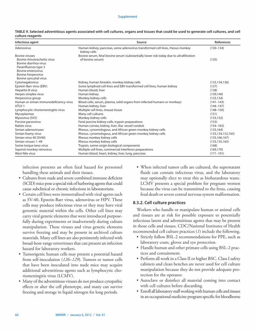
60 MMWR / January 6, 2012 / Vol. 61
Supplement
infection presents an often fatal hazard for personnel handling these animals and their tissues.
• Culturesfromnudeandseverecombinedimmunedeficient(SCID) mice pose a special risk of harboring agents that could cause subclinical or chronic infections in laboratorians.
• Certaincelllineswereimmortalizedwithviralagentssuchas SV-40, Epstein-Barr virus, adenovirus or HPV. These cells may produce infectious virus or they may have viral genomic material within the cells. Other cell lines may carry viral genetic elements that were introduced purpose-fully during experiments or inadvertently during culture manipulation. These viruses and virus genetic elements survive freezing and may be present in archived culture materials. Many cell lines are also persistently infected with broad-host-range retroviruses that can present an infection hazard for laboratory workers.
• Tumorigenichumancellsmaypresentapotentialhazardfrom self-inoculation (126–129). Tumors or tumor cells that have been inoculated into nude mice may acquire additional adventitious agents such as lymphocytic cho-riomeningitis virus (LCMV).
• Manyoftheadventitiousvirusesdonotproducecytopathiceffects or alter the cell phenotype, and many can survive freezing and storage in liquid nitrogen for long periods.
•Wheninfectedtumorcellsarecultured,thesupernatantfluids can contain infectious virus, and the laboratory may optionally elect to treat this as biohazardous waste. LCMV presents a special problem for pregnant women because the virus can be transmitted to the fetus, causing fetal death or severe central nervous system malformation.
8.3.2. Cell culture practicesWorkers who handle or manipulate human or animal cells
and tissues are at risk for possible exposure to potentially infectious latent and adventitious agents that may be present in those cells and tissues. CDC/National Institutes of Health recommended cell culture practices (1) include the following.• StrictlyfollowBSL-2recommendationsforPPE,suchas
laboratory coats, gloves and eye protection.• HandlehumanandotherprimatecellsusingBSL-2prac-
tices and containment.• PerformallworkinaClassIIorhigherBSC.ClassIsafety
cabinets and clean benches are never used for cell culture manipulation because they do not provide adequate pro-tection for the operator.
• Autoclaveordisinfect allmaterial coming into contactwith cell cultures before discarding.
• Enrollalllaboratorystaffworkingwithhumancellsandtissuesin an occupational medicine program specific for bloodborne
TABLE 9. Selected adventitious agents associated with cell cultures, organs and tissues that could be used to generate cell cultures, and cell culture reagents
Infectious agent Source References
Adenovirus Human kidney, pancreas, some adenovirus transformed cell lines, rhesus monkey kidney cells
(130–134)
Bovine viruses Bovine rhinotracheitis virusBovine diarrhea virusParainfluenza type 3Bovine enterovirusBovine herpesvirusBovine syncytial virus
Bovineserum,fetalbovineserum(substantiallylowerrisktodayduetoultrafiltrationofbovineserum) (135)
Cytomegalovirus Kidney,humanforeskin,monkeykidneycells (133,134,136)Epstein-Barrvirus(EBV) SomelymphoidcelllinesandEBV-transformedcelllines,humankidney (137)Hepatitis B virus Human blood, liver (138)Herpes simplex virus Human kidney (139,140)Herpesvirus group Monkey kidney cells (133,134)Human or simian immunodeficiency virus Blood cells, serum, plasma, solid organs from infected humans or monkeys (141–143)HTLV-1 Human kidney, liver (144–147)Lymphocytic choriomeningitis virus Multiple cell lines, mouse tissue (148–150)Mycoplasmas Many cell cultures (151)Myxovirus(SV5) Monkey kidney cells (133,152)Porcine parvovirus Fetal porcine kidney cells, trypsin preparations (153)Rabies virus Human cornea, kidney, liver, iliac vessel conduit (154–163)Simian adenoviruses Rhesus, cynomologous, and African green monkey kidney cells (133,164)Simian foamy virus Rhesus, cynomologous, and African green monkey kidney cells (133,134,152,165)Simianvirus40(SV40) Rhesus monkey kidney cells (135,166,167)Simianviruses1–49 Rhesus monkey kidney cells (133,135,165)Swinetorquetenovirus Trypsin,swine-originbiologicalcomponents (168)Squirrelmonkeyretrovirus Multiple cell lines, commercial interferon preparations (169,170)West Nile virus Human blood, heart, kidney, liver, lung, pancreas (171–191)
MMWR / January 6, 2012 / Vol. 61 61
Supplement
pathogens, and work under the policies and guidelines estab-lished by the institution’s exposure control plan.
• Determineiflaboratorystaffworkingwithhumancellsandtissues need to provide a baseline serum sample (if this is institutional policy based on a risk assessment), be offered hepatitis B immunization and be evaluated by a health-care professional following an exposure incident (1). Similar programs for persons working with nonhuman primate blood, body fluids, and other tissues are recommended.
8.3.3. Biohazards associated with cell culture reading• Driedmediumonthelipofculturetubescouldpresenta
contamination hazard. Handle all culture vessels as if they were contaminated.— Wear gloves and a lab coat when handling viral cultures.— Use eye protection if there is a potential splash hazard.— Perform all culture manipulations in a BSC.— Decontaminate culture tubes and other materials that
come into contact with cell cultures before disposal, using an effective method (usually autoclaving or chemical disinfection) (1).
• Shell vial, tube, andother cultures go throughmultiplemanipulations (e.g., media aspiration, refeeding, inocula-tion) that can generate aerosols. These aerosols and manipu-lations can contaminate the outside of the culture vessel.
• Theoccasionalleakingtubeonanothershelfcouldalsocontaminate the outside of vessels on lower shelves and may not leave visible evidence of the contamination.
8.3.4. Biohazards associated with liquid nitrogen useLiquid nitrogen can become contaminated when ampoules
are broken in the dewar, and contaminants can be preserved in the nitrogen (23). These potentially infectious contami-nants can contaminate other vials in the dewar and generate an infectious aerosol as the liquid nitrogen evaporates. Plastic cryotubes rated for liquid nitrogen temperatures are recom-mended for liquid nitrogen storage because they appear to be sturdier than glass ampoules and are less likely to break in the nitrogen. Glass ampoules are not recommended.
Ampoules and cryotubes can explode when removed from liquid nitrogen creating infectious aerosols and droplets. See Section 3.9 for additional information.
8.4. Personal PrecautionsNo amount of safety engineering can reduce the physical,
chemical, and biological risks in a laboratory environment if personal precautions are not employed consistently and rigor-ously. All laboratory workers and visitors are responsible for fol-lowing established procedures regarding personal precautions.
Directors and supervisors should periodically review their biosafety responsibilities (1).
8.4.1. Biological safety cabinet• Conductallculturemanipulationsinthevirologylabora-
tory in a Class II BSC. These manipulations include, but are not limited to, culture inoculation, feeding, passage, hemadsorption and hemagglutination testing, virus dilu-tions and titrations, cell fixation, immunofluorescent staining, and preparing controls and control slides.
• FollowappropriateBSCsetupandoperationprocedures(1). See Section 3 and Table 5 for guidelines on use of BSCs and descriptions of the characteristics and uses of BSC types.
• CertifyBSCsaccordingtoNSF/ANSIStandard49annu-ally, or after cabinet is moved, HEPA filters are replaced or disinfected, or the unit has undergone any major repairs that could affect the seating or performance of the HEPA filtration system.
• RemovethecontentsoftheBSCanddisinfecttheinteriorof the BSC daily or after a spill or contamination event.— Do not use alcohols as a primary surface disinfectant
in BSCs because alcohols have little or no effect on nonenveloped viruses. Vapors from isopropyl alcohol can be toxic and may cause contact dermatitis. A 1:10 bleach solution provides the best disinfecting activity, but care must be taken to remove the residual chlorine with water because the chlorine will eventually corrode the stainless steel surfaces.
— Remove the floor plate of the cabinet and the front grate monthly or bimonthly for cleaning. Disinfect the floor plate, grate, and the plenum below the floor plate.
— Wipe work surfaces, interior walls and the interior surface of the of the window with a 1:10 solution of household bleach (1) followed by one wiping with water to remove the residual chlorine, and one wiping with 70% ethanol (EtOH). Remove residual chlorine because it will eventually corrode stainless steel surfaces.
— Wipe down any items that will be returned to the BSC.— Let the blower run for at least 4 minutes to remove any
particulates.— Some laboratories leave the sash up and blowers run-
ning at all times, whereas other laboratories turn off the blower and close the sash (if so equipped) at the end of the day.
8.4.2. Personal protective equipment• Mostrecommendationsforthegeneralmicrobiologylabora-
tory are appropriate for the virology laboratory (Section 3).
62 MMWR / January 6, 2012 / Vol. 61
Supplement
• Glovesmustbeworntoprotecthandsfromexposuretohazardous materials and extreme temperatures. In the molecular biology area, gloves are also used to protect the specimen from nucleases that are on the skin. See Sections 8.1–4 for guidelines on use of gloves or other PPE when handling specific stains or chemicals.
8.4.3. DisinfectionDisinfection guidelines for the general microbiology labora-
tory are applicable to the virology laboratory (Section 3).
8.5. Decontamination and Disposal of Laboratory Waste• Disinfectreagentsormaterialscomingintocontactwith
clinical specimens, cell cultures, or virus cultures (includ-ing gloves and PPE) before they are placed into the medical waste stream. Fluids may be disinfected by treating with a 1:10 household bleach (Section 3.4.2 and 3.5) or by autoclaving. Disinfect all other materials by autoclaving before they are placed into the medical waste.
• Decontaminatespecimens,reagents,culturesandequip-ment that come into contact with specimens from patients with suspected Creutzfeldt-Jakob or other prion-associated disease according to local protocols, usually by autoclaving for 1 hour in the presence of 20,000 ppm hypochlorite solutions or 1 N NaOH (1).
8.6. Clean versus Dirty Areas of the Laboratory• Inthevirologylaboratory,thedistinctionbetweendirty
and clean areas is a misnomer; all areas within the labo-ratory present increased opportunities for encountering infectious, chemical, and physical hazards. Laboratory precautions are applicable to all areas of the laboratory.
• Cellculturepreparationandreagentpreparationareasareoften called “clean” areas because no specimens, amplified nucleic acids, or control materials are allowed in these areas. The goal of establishing and policing these “clean” areas is to prevent reagent and cell culture contamination that could produce false results. These are laboratory areas, and laboratory precautions still apply.
8.7. Early Recognition of High-Risk OrganismsRoutine clinical laboratory testing may provide the first evi-
dence of an unexpected bioterrorism event, and routine clinical specimens may also harbor unusual or exotic infectious agents that are dangerous to amplify in culture. Early recognition of these possible high-risk organisms is critical, as is adherence to all fundamentals of laboratory safety. Events that require intervention by a supervisor or laboratory director are listed
(Table 10). Although the majority of events are caused by inadvertent actions and pose no risk, laboratory technologists and directors should be aware that multiple high-risk causes are possible. How the laboratory responds to these trigger events will depend upon whether the laboratory has a BSL-3 facility and the capabilities of the state and local laboratory response network (LRN).
8.7.1. Fluorescent antibody testing bench• Dimstainingofcellswhencontrolsmearsandotherposi-
tive specimens stain strongly can be due to poor antigen expression, sampling crusted lesions, and cellular degra-dation. Dim staining can also occur when the antibody reagent cross-reacts with a similar antigenic epitope arising from antigenic drift, the presence of a viral subspecies, or an unsuspected virus. This type of reaction occurs more frequently when staining with polyclonal antibodies.
• Anunusualpatternofstaining(e.g.,speckledcytoplasmicstaining with a reagent that usually produces nuclear stain-ing) can indicate the presence of an unsuspected virus.
• Staininginanunusualcelltype(e.g.,stainingofsquamouscells rather than ciliated respiratory epithelial cells) could indicate the presence of a different virus or an expected virus with an altered tropism.
• Notifythelaboratorysupervisorordirectorofthesefindings.
8.7.2. Suspicious or unusual results• Knownagentsofbioterrorismorpublichealthemergen-
cies, such as foot and mouth disease, flaviviruses, smallpox, alpha viruses, and hemorrhagic fever viruses, will grow in routine cell cultures used in the clinical laboratory. If a culture produces a cytopathic effect (CPE) pattern that is consistent with one of these agents, do not open for further testing. Contact the attending physician or veterinarian for more information on the patient/animal, and contact the state or local LRN laboratory for further instructions.
• CulturesthatproduceafamiliarCPEpatterninanunusualcelltype and/or an unexpected CPE result from a specific speci-men source (e.g., an eye specimen producing hypertrophic rounding in nearly every cell type) might indicate the presence of an altered or unexpected virus. Contact the physician or veterinarian for more information, and move the specimen into the BSL-3 laboratory for any additional testing.
• Confirmatorytestfailurecanbecausedbyantigenicdriftor operator error during testing. Alternative confirmatory tests such as polymerase chain reaction (PCR) can be used because PCR methods generally present fewer risks than additional fluorescent antibody staining. If PCR testing
MMWR / January 6, 2012 / Vol. 61 63
Supplement
fails to identify the virus, move the culture into the BSL-3 laboratory for further testing.
• IfaBSL-3laboratoryisnotavailable,contactthestateorlocalLRN laboratory for assistance in identifying the viral agent.
8.7.3. Nucleic acid testing• Specimensthatproduceanalteredmeltingcurveshape,too
many peaks, and/or an altered melting temperature (Tm) when controls and other patient curves are normal could indicate poor extraction, the presence of primer dimers, or genetic changes under the primers or probes. These results can also be caused by the presence of an unexpected or unusual (chimeric, recombinant or reassortant) pathogen. Re-extraction and retesting present a modest additional risk. If the results continue to be unusual, the specimen should be referred to the LRN laboratory for additional testing.
• PCR specimenswith altered amplification slopesmaybe the result of genetic variations under the primers and/or probe or the presence of an unusual pathogen. Re-extraction and retesting present a modest additional risk. If the results continue to be unusual, the laboratory should refer the sample to their state or local LRN for additional characterization.
8.8. Hazards Associated with the Electron Microscopy Laboratory
Diagnostic electron microscopy can be a relatively simple and rapid method for morphologic identification of agents in a specimen. Electron microscopy procedures can serve as a general screen to detect novel organisms or organisms that have altered genetic or immunologic properties that render them undetectable by nucleic acid or immunoassay protocols (192). Electron microscopy laboratories share many of the physical, chemical, and biological hazards described for the virology laboratory but also have some unique features.
8.8.1. Flammable and combustible liquidsThe electron microscopy laboratory uses a wide variety of
flammable solvents, and the use of open flames is discouraged (see Section 8.2.13).• Placesolventsrequiringrefrigerationinspecialflammable-
storage refrigerators that minimize exposed electrical con-nections and reduce the opportunity for spark generation.
• Storeflammableliquidsinflammable-storagecabinetsinaccordance with local and state regulations.
TABLE 10. Trigger events requiring supervisor or laboratory director notification or intervention
Event Typical causes High-risk causes
Specimen processing/login benchSpecimenbroughtinbylawenforcementofficialwithachain-of-custodyform
Assaultcaseworkup Potential BT/BC specimen
Suspect agent is on the Do Not Test/Do Not Process list Clerical mistake at order entry Mightbefromapatientwithahigh-riskinfectionUnusual(nonbiological)orunapprovedspecimentype
or containerClericalmistakeatorderentry;
inappropriate order•Powders,environmentalsamples,animalspecimens,clothing,foodsamples,inanimateobjectscouldcontainBT/BC agents.
•MightrepresentanattempttointroduceaBT/BCagentinto the laboratory.
Fluorescent antibody benchDimstainingwhencontrolsandotherpositivespecimens
stain normallyDecreased antigen expression in cells Cross-reactionwithanotherinfectiousagent
Unusualstainingpattern Addedwrongantibodytowell;antibodies ran together during incubation
•Alteredvirusorgenetic/antigenicvariant•Cross-reactionwithanotheragent
Staining unusual cell types Addedwrongantibodytowell;antibodies ran together during incubation
Infectiousagentwithalteredhostrange;cross-reactionwithanotheragent
Cell culture benchCPE pattern and cell tropism are unusual Unusual/unexpectedpathogen;BT/BCagentinspecimenFamiliar CPE pattern but in an unusual cell type Geneticdrift;subspeciespresent Unusual/unexpectedpathogen;BT/BCagentinspecimenUnexpectedCPE/HAdpatternfromtheindicated
specimen sourceClericalerrorwhenenteringsource Unusual/unexpectedpathogen;BT/BCagentinspecimen
Confirmationprotocolsdonotwork Genetic or antigenic drift Unusual/unexpectedpathogen;BT/BCagentinspecimenNucleic acid testing
Altered melting curve shape, too many peaks, altered Tmwhencontrolsandotherpatientcurvesarenormal
Geneticvariationinagent;poorextraction;primerdimers
Unusual/unexpectedpathogen;morethanoneagentpresent;chimeric,recombinantorreassortantpathogen
Change in the slope of the amplification curve Geneticvariationinagent;poorextraction;specimeninhibition
Unusual/unexpectedpathogen;chimeric,recombinantorreassortant pathogen
Abbreviations: BT=bioterrorism;BC=biocrime;CPE=cytopathiceffects;HAd=hemadsorption;Tm = melting temperature.
64 MMWR / January 6, 2012 / Vol. 61
Supplement
• Handleflammableliquidsinfumehoodstominimizevaporbuildup. Ultrasonic cleaning of Wehnelt cap assemblies in an acetone baths must be done in a chemical fume hood.
• Never pour flammable liquids down thedrainbecausethey can cause an explosion.
8.8.2. X-ray hazardsThe electron microscope will generate dangerous levels of
X-rays within the microscope as high-energy electrons strike the metal components. Modern electron microscopes have sufficient shielding and lead-impregnated glass viewing ports that minimize dangers to the operator. However, modifications to the instrument, adding and removing accessories, and some maintenance procedures can compromise the shielding.• Useacalibratedthin-windowGeiger-Müller(GM)survey
meter to verify the shielding effectiveness, and monitor radiation levels— initially at the time of installation;— whenever the microscope is modified; and— periodically as indicated by state, local, or institutional
policies.• Radiationlevelsaretobe<0.5mRat5centimetersfrom
the unit.
8.8.3. Electrical hazardsSee Section 3.7 for information regarding routine electrical
safety in the electron microscopy laboratory.• Specialhigh-voltageandhigh-amperageelectricalsources
are used to power electron microscopes and other equip-ment in the laboratory. Only trained technicians are to service this equipment. Install safety interlocks and power lockouts to prevent activation of electrical circuits while the instrument is being serviced.
• Changingthefilamentinthehigh-voltageelectronguncanpresent an electrical hazard if the grounding rod does not make contact with the Wehnelt cap. A substantial charge can build up on the cap, and the charge must be relieved before touching the cap.
8.8.4. Chemical hazardsSeveral heavy metal stains and aggressive fixatives are used
in the electron microscopy laboratory. See Section 8.2 and the MSDS materials provided by the manufacturers for guidelines for handling stains and fixatives. Embedding and filmmaking materials are chemical hazards, and many of these materials are dissolved in flammable organic solvents.• Limitacuteandlong-termexposuretothesechemicals.• Placeembeddingovensinachemicalfumehoodtomini-
mize exposure to potentially hazardous chemicals.
• Latex gloves are not appropriate for all chemicals, andappropriate glove usage must be emphasized for all labora-tory personnel.
8.8.5. Cryogens and compressed gasesThe most commonly used cryogens used in the electron
microscopy laboratory are liquid nitrogen and liquid helium. Compressed helium, CO2 and nitrogen are also used. Hazards and safety measures associated with these gases are summarized in Sections 3.8 and 3.9.
8.8.6. Specialized equipmentThe electron microscopy laboratory uses a number of special-
ized instruments whose use can be hazardous. For example, evaporators, freeze-driers, freeze-fracture, and sputter coater units use vacuum, and the vessels could implode. Implosion hazards are reviewed in Section 3.13.1.• Topreventeyedamageduringevaporation,usewelder’s
goggles to view the source.• Allowthecomponentstocoolbeforetouchingthem.• Donotbreatheanyoftheevaporatedmetalthatmayflake
off from the surface.• Criticalpointdryerscanbequitedangerousbecauseofthe
high pressures generated within the “bomb” (121,193). Follow the operating and safety procedures described in the operator’s manual for safe operation.— Place a one-half-inch thick lexan shield between the
operator and the bomb.— Do not secure the shield directly over the window of
the bomb where it would receive the full force of an explosion. That force could shatter the shield.
— A polycarbonate face shield is also recommended when observing the contents of the bomb.
— Vent freons or freon substitutes from critical point dryers directly outside or through a fume hood.
8.8.7. Biological hazardsThe biological hazards of the electron microscopy labora-
tory are similar to those of the virology laboratory, and good laboratory practices must be followed.• Conductprimaryspecimenhandlinginabiologicalsafety
cabinet to prevent aerosols and contamination of the laboratory.
• Negativestainingsolutionsmaynotinactivatemicroorgan-isms and viruses. Osmium tetroxide is an effective sterilant, but it cannot be used for all specimens.
8.9. Rapid Testing (Kits)Several FDA-approved, rapid immunodiagnostic tests for
viral antigens and antibodies are available. Originally designed
MMWR / January 6, 2012 / Vol. 61 65
Supplement
for point-of-care or near point-of-care testing, many of these tests are being used for testing in clinical virology laboratories. The following biosafety recommendations are based upon CDC biosafety guidance for handling clinical specimens or isolates containing 2009-H1N1 influenza A virus (194).• Proceduresthatinvolveonlysimplestepssuchasinserting
a swab into medium or pipetting specimens and reagents and are not reasonably expected to generate aerosols may be performed on the bench top using only splash protec-tion. Use the BSC for testing procedures that require cen-trifugation, vortexing, vigorous mixing, or other methods that could generate aerosols.
• Performbenchtoptestinginamannerthatwillpreventsplashes and generation of aerosols. The appropriate PPE for this type of testing consists of a laboratory coat, gloves, eye protection and a face mask, such as a surgical, dental, medical procedure, isolation, or laser mask. A splash shield providing protection of the entire face fulfills the need for separate eye protection and face mask.
• Decontaminateworksurfacesandequipmentwitha1:10bleach solution as soon as possible after specimens are processed.
• Rapidimmunodiagnostictesting,whenperformedinaClass II BSC, does not require use of additional of eye protection and a face mask.
8.10. Molecular LaboratoryMolecular virology laboratories share many of the physical,
chemical and biological hazards described for the virology laboratory, but they also present some unique hazards.
8.10.1. Electrical hazardsSee Section 3.7 for information regarding routine electrical
safety in the molecular virology laboratory. Special high-voltage power sources are used in electrophoresis and nucleic acid sequencing equipment.• Neverusehigh-voltage electrical equipmentnear flam-
mable liquids and gases because an arc could cause an explosion or fire.
• Disconnectthepowertoelectrophoresisequipmentbeforedisassembling the apparatus.
• Only trained technicians should service high-voltageelectrical equipment.
• Nevertamperwithordefeatsafetyinterlocksandpowerlockouts on electrophoresis equipment.
8.10.2. Ultraviolet light hazards• Short-waveultraviolet(UV)viewboxesareoftenusedto
visualize nucleic acid bands in gels. Special care must be taken to protect eyes and completely cover the skin when visualizing and photographing gels on a UV view box.
•Weargloves,long-sleevedlaboratorycoat,andaUV-resistantfull-face shield when working with UV view boxes.
• Useonlyfaceshieldsratedforshort-waveUVlightforthis purpose.
• LocateUVviewboxesoutofthenormaltrafficflowofthelaboratory so that bystanders or persons passing by are not exposed to the ultraviolet light. Other UV light hazards are discussed in Section 3.12.
8.10.3. Chemical hazardsThe chemical hazards unique to the molecular virology labo-
ratory include chloroform, ethidium bromide (Section 8.2.8) and guanidinium-based extraction reagents (Section 8.2.10). Avoid acute and long-term exposure to these.
8.10.4. Biological hazardsThe biological hazards in the molecular virology laboratory
are similar to those of the virology laboratory, and good labora-tory practices must be followed.• Conductprimaryspecimenhandlinginabiologicalsafety
cabinet to prevent aerosols and contamination of the laboratory.
• Extractednucleic acidsmay ormaynot be sterile.Aninadequately performed extraction procedure may not completely remove or inactivate the infectious agents in the specimen. The genome of positive-stranded RNA viruses (poliovirus) is suspected to be infectious, and once introduced into the cell, the viral genome requires no virus-coded proteins or accessory components for virus replication. Although the risks of infection in this man-ner are very small, it is prudent to handle extracted viral nucleic acids as if they were infectious. This practice mir-rors specimen-handling procedures used in other areas of the laboratory and supports a unified specimen-handling policy for the entire laboratory.
66 MMWR / January 6, 2012 / Vol. 61
Supplement
9. Chemistry Laboratory
All specimens of human and animal origin tested by the chemistry, toxicology, or drug-testing laboratory may contain infectious agents. It is imperative to understand and minimize the risk of exposure to patient specimens through surface con-tact, aerosolization, or penetrating injury. Risk mitigation of laboratory-acquired infections is discussed in Sections 2 and 3.
9.1. Automated Analyzers (see also 3.17.3 and 10.6.3)
Automated analyzers frequently have added features to help reduce operator exposures, but they do not totally eliminate the potential for exposure. A common feature in newer systems is closed system sampling.• Chemistry/toxicologyanalyzersoftenhavehigh-velocity
robotic arms and samplers that might cause skin punctures and lacerations. Operate analyzers only with the cover closed.
• Sampleprobesthatmovequicklyordeliverfluidrapidlymay generate aerosols and droplets.
• Always use instruments according tomanufacturerinstructions.
• Ensureinstrumentsafetyshieldsandcontainmentdevicesare in place at time of use.— Limit the amount of hand movement near the sample
probe and liquid-level sensors.— When using instruments for which the operator is required
to wipe sample probes after sampling, wear disposable gloves and use gauze pads with impermeable plastic coat-ing on one side. Newer instruments have automatic probe wash cycles eliminating this source of exposure.
• Topreventspillage,handlesampletraysandsampleswithcaution, and keep them covered when not being manipulated.
• Assumethattheoutsideofbloodtubesiscontaminated,and be prepared to wipe the outside of the tube with the laboratory disinfectant or with a solution of 1:10 house-hold bleach.
• Fillsamplecupsandaliquottubesusingmechanicaldevices(e.g., transfer pipettes), and never decant (pour) them.
• Considereffluentsofclinicalanalyzerstobecontaminatedwith pathogens; some may also be contaminated with haz-ardous chemicals. Their disposal must comply with state and local regulations. Investigate these effluents and consult applicable standards before discharging them in the sewer.
• Followmanufacturer instructions for routine cleaningand trouble-shooting specimen spills on or within an instrument, including the appropriate personal protective equipment (PPE) and type of cleaning solution to be used.— When manufacturer instructions do not include spill
containment and clean-up instructions, collaborate with the manufacturer to develop a standard operating proce-dure (SOP) that will effectively protect the operator and maintain and extend the instrument’s operational life.
— Have fresh 1:10 household bleach solution on hand in case of an emergency spill or breakage after being assured by the manufacturer that chlorine will not damage instrument components.
• Performdailycleaningoffluidicsystemsandsamplerfol-lowing manufacturer instructions.
• Collectwasteintoawastecontainerthatcontainsfreshconcentrated household bleach in sufficient quantity to achieve a final concentration of 10% bleach when the flask is full. Do not collect effluents containing compounds not compatible with bleach (Table 11) into bleach to avoid formation of chlorine gas.
• Donotmixsodiumhypochlorite(bleach)withanyotherchemical unless adequate engineering controls and PPE are in place. Accidental mixing may cause dangerous conditions that could result in injury to personnel and/or damage to property or the environment.
9.2. Tissue Preparation for Chemical/Toxicological Analysis• Considerallunfixedtissuesasbiohazardous,regardlessof
the patient diagnosis or the test(s) ordered.• Theuseofafixativeisnotalwayssufficienttoeliminate
all types of biohazards.• Useabiologicalsafetycabinet(BSC)orPPE,including
gown and gloves. with a fixed containment device for sample aspiration.
• Automatedsample loadingsystemsreducesamplehan-dling and also perform sample vortexing in an enclosure that prevents operator exposure to aerosols and splashes.— Take special care when loading samples onto the instru-
ment tube racks.— For unfixed samples, load instrument tube racks inside a
BSC or wear PPE to protect from splashes and aerosols.
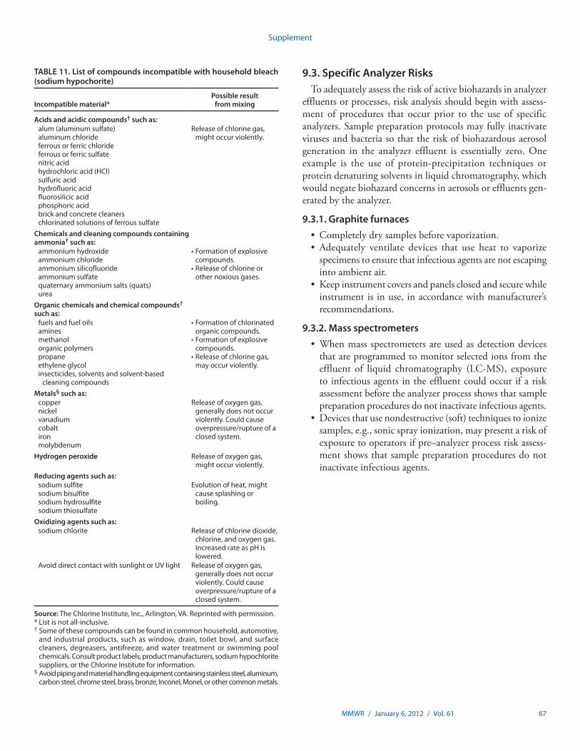
MMWR / January 6, 2012 / Vol. 61 67
Supplement
9.3. Specific Analyzer RisksTo adequately assess the risk of active biohazards in analyzer
effluents or processes, risk analysis should begin with assess-ment of procedures that occur prior to the use of specific analyzers. Sample preparation protocols may fully inactivate viruses and bacteria so that the risk of biohazardous aerosol generation in the analyzer effluent is essentially zero. One example is the use of protein-precipitation techniques or protein denaturing solvents in liquid chromatography, which would negate biohazard concerns in aerosols or effluents gen-erated by the analyzer.
9.3.1. Graphite furnaces• Completelydrysamplesbeforevaporization.• Adequately ventilate devices that use heat to vaporize
specimens to ensure that infectious agents are not escaping into ambient air.
• Keepinstrumentcoversandpanelsclosedandsecurewhileinstrument is in use, in accordance with manufacturer’s recommendations.
9.3.2. Mass spectrometers•Whenmassspectrometersareusedasdetectiondevices
that are programmed to monitor selected ions from the effluent of liquid chromatography (LC-MS), exposure to infectious agents in the effluent could occur if a risk assessment before the analyzer process shows that sample preparation procedures do not inactivate infectious agents.
• Devicesthatusenondestructive(soft)techniquestoionizesamples, e.g., sonic spray ionization, may present a risk of exposure to operators if pre–analyzer process risk assess-ment shows that sample preparation procedures do not inactivate infectious agents.
TABLE 11. List of compounds incompatible with household bleach (sodium hypochorite)
Incompatible material* Possible result
from mixing
Acids and acidic compounds† such as:alum(aluminumsulfate)aluminum chlorideferrous or ferric chlorideferrous or ferric sulfatenitric acidhydrochloricacid(HCl)sulfuric acidhydrofluoric acidfluorosilicic acidphosphoric acidbrick and concrete cleanerschlorinated solutions of ferrous sulfate
Release of chlorine gas, might occur violently.
Chemicals and cleaning compounds containing ammonia† such as:
ammonium hydroxideammonium chlorideammonium silicofluorideammonium sulfatequaternaryammoniumsalts(quats)urea
•Formationofexplosivecompounds.
•Releaseofchlorineorother noxious gases.
Organic chemicals and chemical compounds† such as:
fuels and fuel oilsaminesmethanolorganic polymerspropaneethylene glycolinsecticides,solventsandsolvent-based
cleaning compounds
•Formationofchlorinatedorganic compounds.
•Formationofexplosivecompounds.
•Releaseofchlorinegas,may occur violently.
Metals§ such as:coppernickelvanadiumcobaltironmolybdenum
Release of oxygen gas, generally does not occur violently. Could cause overpressure/rupture of a closed system.
Hydrogen peroxide Release of oxygen gas, might occur violently.
Reducing agents such as:sodium sulfitesodium bisulfitesodium hydrosulfitesodium thiosulfate
Evolution of heat, might cause splashing or boiling.
Oxidizing agents such as:sodium chlorite Release of chlorine dioxide,
chlorine, and oxygen gas. Increased rate as pH is lowered.
AvoiddirectcontactwithsunlightorUVlight Release of oxygen gas, generally does not occur violently. Could cause overpressure/rupture of a closed system.
Source:TheChlorineInstitute,Inc.,Arlington,VA.Reprintedwithpermission.*Listisnotall-inclusive.† Some of these compounds can be found in common household, automotive, and industrialproducts, suchaswindow,drain, toiletbowl, and surfacecleaners,degreasers, antifreeze, andwater treatmentor swimmingpoolchemicals. Consult product labels, product manufacturers, sodium hypochlorite suppliers, or the Chlorine Institute for information.
§ Avoidpipingandmaterialhandlingequipmentcontainingstainlesssteel,aluminum,carbon steel, chrome steel, brass, bronze, Inconel, Monel, or other common metals.
68 MMWR / January 6, 2012 / Vol. 61
Supplement
10. Hematology and Phlebotomy Laboratory
10.1. Specimen Receiving and Log-In/Setup Station
Biosafety guidelines for the hematology laboratory are the same as those for the microbiology laboratory and are described in Section 3.1.
10.2. Work at the Open BenchSee Section 3.2.1.
10.2.1. Standard operating proceduresStandard operating procedures are described in Section 3.1.4.
10.2.2. Manual removal of sealed caps and specimen aliquotting/pipetting
See Section 3.1.5.
10.2.3. Unfixed specimens•Microscopes
— Disinfect the stage, eyepieces, knobs and any other contaminated parts daily.
— Select a disinfectant that will be noncorrosive to the micro-scope and appropriate for potential infectious agents.
• Slidepreparations— Avoid waving slides in the air or using electric fans at
an open bench to air-dry slides.— An electric fan may be used to air-dry slides in a fume
hood and, only if necessary, in a biological safety cabinet (BSC), not on the bench top. Using a fan in the BSC will disrupt airflow and will render the BSC unsuitable for other laboratory work until the airflow has been stabilized.
• Buffycoatsmears Use disposable Wintrobe tubes in place of capillary tubes
to avoid cutting glass tubes and possible exposure to blood and shards of glass.
• Hemacytometer Use extreme caution when using glass hemacytometers
and glass coverslips to avoid punctures from glass shards. Plastic hemacytometers are commercially available and offer repeatable and reliable measurements and analysis. Several hemacytomer designs eliminate the use of cover-slips and allow for exact volume control.
• Bleedingtimes(195)— Wear gloves (see Sections 3.2.2 and 8.4.2).— Use appropriate pediatric/adult template safety devices.
— Ensure devices are reviewed annually in compliance with Occupational Safety and Health Administration (OSHA) Bloodborne Pathogens Standards (33) and CDC guidelines (196).
— Use disposable razors when removal of arm hair from test site is required.
— Dispose of used bleeding time device and razor in sharps container, and filter paper in medical waste container.
• Bonemarrowaspirates— Include appropriate hospital infection control policies
and procedures for patient care settings in the labora-tory policy and procedure for collection of bone mar-row aspirates when collected in these settings.
— Limit what is brought into patient rooms. Bone marrow safety tray kits are marketed by several manufacturers. Use of disposable trephine and aspiration needles is recommended.
— Collect specimens and prepare squash smears and slides in the laboratory.
— If smears must be prepared outside the laboratory, e.g., in patient care settings, use Standard Precautions; use risk and hazards assessment to determine what and when additional personal protective equipment (PPE) might be appropriate; use a flat secure surface to prepare smears and slides; ensure cover/caps on additional specimens are tightly secured before transport; and ensure slides, squash smears, and additional specimens are transported in leakproof and breakage-resistant receptacles.
• Document training and assess competency of labora-tory personnel. Include knowledge of, and adherence to, hospital infection control policies/procedures in patient settings, and the concept of Standard Precautions in all documented training and competency assessments.
10.3. Personal PrecautionsGuidelines for personal precautions, including use of a
BSC (Section 3.3), PPE (Section 3.2.1), and disinfection (Section 3.4) are described in Section 3.
10.4. Decontamination and Disposal of Laboratory Waste (39)
See Section 3.5 for guidelines for decontamination and disposal of laboratory waste.
MMWR / January 6, 2012 / Vol. 61 69
Supplement
10.5. Dirty versus Clean Areas of the LaboratorySee Section 3.16.
10.6. InstrumentationWhether automated or manual, procedures with the poten-
tial for producing specimen aerosols and droplets (e.g., stop-per removal, vortexing, opening or piercing evacuated tubes, automatic sample dispensers) require either PPE or engineering controls designed to prevent exposures to infectious agents.
10.6.1. WaterbathsSee Section 3.17.1.
10.6.2. CentrifugesSee Section 3.17.2.
10.6.3. Automated hematology/hemostasis analyzersAutomated analyzers frequently have added features to help
reduce operator exposures, but these do not totally eliminate potentials for exposure. A common feature in newer systems is closed system sampling. See Sections 3.17.3, 9.1, and 11.6.3 for additional information.• Sampleprobesthatmovequicklyordeliverfluidrapidly
can generate aerosols and droplets.• Alwaysuseinstrumentsaccordingtomanufacturerinstructions.• Ensureinstrumentsafetyshieldsandcontainmentdevices
are in place at time of use.• Limit the amount of handmovementnear the sample
probe and liquid-level sensors.•Wearglovesandusegauzepadswithimpermeableplastic
coating on one side when using instruments for which the operator is required to wipe sample probes after sampling. Newer instruments have automatic probe wash cycles eliminating this source of exposure.
• Handlesampletraysandsampleplateswithcaution,andcover them when not being sampled to prevent spillage.
• Fill sample cups and aliquot tubes usingmechanicaldevices, and never decant (pour) them.
• Assumethateffluentsofclinicalanalyzersarecontami-nated, and dispose in compliance with applicable federal, state, and local environmental regulations.
• Followmanufacturerinstructionsforroutinecleaningandtrouble-shooting specimen spills on or within an instru-ment, including the appropriate PPE and type of cleaning solution to be used.
•Whenmanufacturerinstructionsdonotincludespillcon-tainment and cleanup instructions, collaborate with the manufacturer to develop a standard operating procedure that will effectively protect the operator and maintain and extend the instrument’s operational life.
10.6.4 Flow cytometers (see Section 3.17.3)Occupational exposures in a routine flow cytometry (FCM)
laboratory arise either from sample handling or, more spe-cifically, from aerosols and droplets generated by the flow itself. Flow cytometric applications, e.g., phenotypic analysis, calcium flux evaluations, and apoptosis measurements of unfixed cells, when performed using jet-in-air flow cytometers with extremely high pressure settings can expose operators to potentially hazardous aerosols.• FCMbiosafety procedures should specifically focus on
aerosol containment, waste management and equipment maintenance (197).
• Considerallunfixedmaterials(peripheralleukocytes,bonemarrow, various body fluids, cultured cells, and environ-mental samples) as biohazardous.— Use of a fixative is not always sufficient to eliminate all
types of biohazards.— Use a BSC or PPE with an equivalent fixed contain-
ment device for sample aspiration.— Always wear disposable gloves and protective clothing
when operating a flow cytometer.— Allow only operators with documented training and
experience (professional consensus is 2 years) to per-form potentially biohazardous cell sorting (51).
— Restrict access by allowing only essential personnel dur-ing sorting. Post a notice at the entrance that cell sorting is in process. Personnel wishing to monitor the sort must wear the same PPE as the operator of the cell sorter.
• Cell-sortersareequippedwithanozzletoformajetofmicrodroplets; this step is likely to generate aerosols.
• Instrumentfailuressuchascloggedsortnozzleorairinthefluidic system can drastically increase aerosol formation.
• Newlydesigned safety attachments for cell sorters havebecome commercially available. Some enclosed fluid system flow cytometers perform cell sorting using a fluid switching mechanism. These cytometers, in contrast to jet-in-air cell sorters, do not generate aerosols during cell sorting.
• Efficiencyofaerosolcontrolmeasureson sorter instru-ments is to be tested periodically following manufacturer instructions, particularly when unfixed human cells and known biohazardous samples are acquired or sorted.
• Simplerbead-basedtechnologyformeasuringefficiencyhas been developed.
• Newerflowcytometershaveaddedbiosafetyfeatures—e.g.,enclosed flow cells, droplet containment modules, and auto-mated samplers — for reducing risk of operator exposure to instrument-generated sample droplets and aerosols.
• Ensuretheinstrumentisusedinamannerspecifiedbythe manufacturer.
70 MMWR / January 6, 2012 / Vol. 61
Supplement
• Openandclosetheinstrumentcoverwithcare.• Keepinstrumentcoversandpanelsclosedandsecurewhile
instrument is in use.• Donotdisablesafetyinterlocksand/orsensors.• Placesampletubessecurelyintothesampleintroduction
port. Otherwise it could be blown off once it is pressurized and splash sample onto the operator.— Some sample ports contain a metal sip tube that can
damage gloves when the tube is not inserted carefully.— For better splash protection, wearing safety glasses or chem-
ical splash goggles during acquisition is recommended.— Automated sample loading systems reduce sample
handling and also perform sample vortexing in an enclosure that prevents operator exposure to aerosols and splashes. Take special care when loading samples onto the instrument tube racks with these systems. For unfixed samples, load instrument tube racks inside a BSC or wear PPE to protect from splashes and aerosols.
• Performdailycleaningoffluidicsystemsandsamplerfol-lowing manufacturer instructions.
• Collectwasteintoawastecontainerthatcontainsfreshcon-centrated household bleach in sufficient quantity to achieve a final concentration of 10% when the container is full.
Note: Adding chlorine to a waste container where ammo-nium chloride Tris buffer is used as a lysing agent can release dangerous chlorine gas. Use a broad-spectrum idophor instead.
10.6.5. Automated slide stainers• Ensuretheinstrumentisusedinamannerspecifiedby
the manufacturer.• Keepinstrumentcoversandpanelsclosedandsecurewhile
instrument is in use.
10.6.6. Total or semiautomated hematology test systems• Conducthazardandriskassessmentstoidentifycritical
operations that pose a risk for exposure.• Standardoperatingproceduresshouldinclude
— instructions for troubleshooting tube breakage and specimen spills on conveyor tracts, belts, sorter, aliquot, and cap-piercing probe stations;
— instructions requiring at least daily cleaning and disinfection and cleaning after tube breakage and/or specimen spills; and
— appropriate PPE to be worn when cleaning and trouble-shooting robotics.
Note: HEPA filters or wadded tape held with forceps are helpful for removing fine glass particles.
• If instructions are not provided by themanufacturer,collaborate with the manufacturer to develop trouble-shooting and cleaning procedures that will protect the operator and be compatible with and extend the life of the robotic equipment.
10.7. Rapid Testing (Kits) (Section 3.18)Consider used testing kits to be contaminated, and dispose
of them appropriately in accordance with applicable local and state environmental regulations.
10.8. Molecular Testing (198)• TheClinicalandLaboratoryStandardsInstitutehaspub-
lished standards for nucleic acid amplification assays for hematopathology (199).
• Unidirectionalworkflowandspatialseparationofworkareasmust be strictly adhered to in addition to standard laboratory safety guidelines for open bench and instrument operations.— Reagent preparation is the cleanest area, then specimen
preparation area, and finally product detection area.— Leave transportables, e.g. pens, tape, scissors, glove
boxes, in each designated area (see Section 3.19). Color-coding each area and using color-coded tape and color-coded laboratory coats helps enforce spatial separations of work areas and retain transportables in their designated areas.
— Change laboratory coats and gloves and wash hands before entering each area (see Section 3.19).
10.9. Phlebotomy• Evaluate, select, anduse engineered sharps injury pre-
vention devices that are acceptable for clinical care and provide optimal protection against injuries. Evaluation of engineered sharps injury prevention devices and consid-eration of their prospective use should involve employees who use sharps.
• Establish a process providing annual evaluation andselection of sharps injury prevention devices (196; Section 3.14).
10.9.1. General recommendations (200–203)• Ensuresharpsdisposalcontainersareeasilyaccessiblein
patient rooms and in patient drawing areas; are never more than three-fourths full; and are included in the annual evaluation, selection, and use of sharps injury prevention devices (204).
MMWR / January 6, 2012 / Vol. 61 71
Supplement
• Ensureall applicablepatientcareand infectioncontrolpolices and procedures are strictly adhered to in patient drawing areas.
•Washhandswithsimplesoapandwaterorantimicrobialsolution to protect against external and internal exposure to bloodborne pathogens. Ensure hands are washed before gloving; after gloves are removed; after contact with each patient or patient sample; before leaving the laboratory, drawing station, or patient room; before eating; and after hands have touched a possibly contaminated surface.
• Establishaprocessandprocedureforspecimentransportwithin and, if applicable, outside the facility (93).— If patient specimens are transported from the drawing
station to another area of the facility, ensure they are transported in a secondary container that has a tight-fitting latchable cover and is constructed of material to contain blood spills.
— The phlebotomy service procedure manual must include spill response and spill cleanup instructions for all areas of the hospital where there is potential for specimen spills (including such areas as elevators and stairwells).
— Ensure courier services employed by the laboratory enforce the laboratory policy for transportation of specimens, and ensure documentation of personnel training and competency assessment regarding speci-men transport (including procedures for spill response, cleanup, and incident reporting).
10.9.2. Dirty versus clean areas in the laboratorySee Section 3.16.
10.9.3. Pneumatic tube systemsSee Section 3.1.6.
10.9.4. Personal precautionsSee Section 3.2.
10.9.5 Disinfection of work spaceSee Section 3.4.1.Regardless of the method, the purpose of decontamination is
to protect the phlebotomist, the patient and the environment,
and anyone who enters a patient room/drawing station or who handles materials that have been carried into or out of the patient room/drawing station.• Limitthephlebotomymaterialsbroughtintoapatientroom.• Routinelycleanenvironmentalsurfacesbeforesettingup
the patient room/drawing station work area, and again before leaving the patient room or after each patient in the drawing station work area.
• Disinfectanyitem(e.g.,pen,telephone)touchedwithusedgloves.
• Donotusealcoholsoralcohol-basedsolutionstodisinfectsurface areas because they evaporate readily, which signifi-cantly decreases efficacy. Instead use aqueous disinfectants such as 1:10 dilution of household bleach or the hospital-recommended disinfectant.
• Usedisinfectants recommended for environmental sur-faces, such as 1:10 dilution of household bleach or other EPA- registered disinfectants effective against hepatitis B virus, human immunodeficiency virus, and other blood-borne pathogens (205).
10.9.6. Disinfecting patient room work areas and drawing stations
See Section 3.4.1.• Includeinstructionsintheproceduremanualregarding
what PPE to use, how to clean, what disinfectant to use, and how to dispose of the materials. Post the instructions in the phlebotomy office for reference.
• Allowdriedbloodorbodyfluidatleast20minutes’contactwith the tuberculocidal disinfectant to allow permeation and easy removal.
• Neveruseaknifeorotherinstrumenttoscrapedriedbloodor body fluid from surface areas because this can generate aerosols.
10.9.7. Documentation of training and competency assessment in phlebotomy
Assessment includes knowledge of, and adherence to, any applicable hospital infection control policies/procedures in patient settings and the concept of Standard Precautions.
72 MMWR / January 6, 2012 / Vol. 61
Supplement
11. Blood Bank
11.1. Transfusion-Transmitted DiseasesMany infectious agents are transmitted through transfusion
of infected blood; these include hepatitis B virus, hepatitis C virus, human immunodeficiency viruses 1 and 2, human T-cell lymphotropic viruses (HTLV-I and II), cytomegalovirus, parvovirus B19, West Nile virus , dengue virus, trypanosomia-sis, malaria, and variant Creutzfeldt-Jakob disease. The AABB provides information on transfusion-transmitted diseases as well, available at http://www.aabb.org/Pages/Homepage.aspx.
11.2. Bloodborne Pathogen Standard The Occupational Safety and Health Administration’s
(OSHA) Bloodborne Pathogen Standard, 29 CFR 1910.1030 must be adhered to in the blood bank laboratory (33).
11.3. Specimen Receiving and Log-In/Setup Station
Guidelines for receiving and logging specimens and handling specimen containers are described in Section 3.1.
11.4. Work at the Open BenchWritten procedures for blood bank include specific work
practices and work practice controls to mitigate potential expo-sures. Standard operational procedures (SOPs) and procedure manuals are described in Section 3.1.4.
11.4.1. Unfixed specimens•Microscopes
— Disinfect the stage, eyepieces, knobs and any other contaminated parts after use or according to a specified schedule determined by the laboratory.
— Select an appropriate disinfectant that will be noncor-rosive to the microscope (see Section 3.4.1).
• Slidepreparations— Replace glass with plastic where possible.— Avoid waving slides in the air or using electric fans at
an open bench to air-dry slides.— If other instruments are used to dry slides, have a risk
assessment performed to measure aerosol risks.•Manualcellwashing Perform a risk/hazard assessment.
— For saline washing of cell suspensions, use of automated cell washers reduces some, but not all, of the hazards associated with this procedure.
— Dumping saline washes into pour-off containers can generate splashes and aerosols. Splashing can be mini-mized by using semi-automated pipettes and keeping
liquid levels in pour-off containers below one-fourth full. Wear fixed shields or appropriate water-resistant personal protective equipment (PPE) to protect from splashes.
— When shaking small test tubes for resuspending red cell pellets and reading end-point agglutination, use fixed bench-top shields or wear appropriate PPE to protect from splashes and sprays.
• Performproceduresusingaplasmaextractororexpressorwith appropriate face and eye protection to protect from sprays and possible explosion of the blood/component bags.
• Useappropriatefaceandeyeprotectionwhencuttingdonorsegments during either confirmatory testing or other testing.
•Wipeoutersurfacesofbloodbagsandcomponentswitha towel moistened with appropriate disinfectant before release for infusion, ensuring that the disinfectant will not compromise the plastic bag.
• Ifengineeringcontrolsareinplacetopreventsplashesorsprays at blood bank workbenches, the requirement for PPE may be modified on the basis of an assessment and evidence of the effectiveness of the engineering control to prevent exposure to splashes or sprays.
11.4.2. Biological safety cabinetA Class II biological safety cabinet (BSC; see Section 3.3) is
required for all aerosol-generating processes.
11.4.3. Personal protective equipmentSee Section 3.2.2.
11.4.4. DisinfectionSee Section 3.4.
11.4.5. Decontamination and disposal of laboratory waste
See Section 3.5 for discussion, including a waste manage-ment plan.
11.5. Clean versus Dirty Areas of the LaboratorySee Section 3.16.
11.6. InstrumentationSee Section 3.17.
11.6.1. Refrigerators and freezersFor all refrigerators and freezers in the blood bank, establish a
cleaning and maintenance protocol that will minimize contamina-tion and extend the life of the equipment and also maintain the sophisticated cooling systems blood bank refrigerators require to
MMWR / January 6, 2012 / Vol. 61 73
Supplement
provide uniform and quick temperature recovery when needed. (also see Section 3) Most newer blood bank laboratory refrigerators and freezers are stainless steel and have painted finishes and remov-able trays, which make cleaning and sanitizing an easier process.•Manufacturerinstructionsforuseandcareofbloodbank
refrigerators and freezers usually include recommended PPE and type of disinfectant.
• CollaboratewiththemanufacturertoestablishSOPsforcleaning and maintenance if instructions are not provided with the equipment.
• Cleanupbloodspillsimmediately.• Clean refrigerator handles and outside doors around
handles at the end of each shift.• Labeltherefrigeratorwiththeuniversalbiohazardsymbol
and the word “Biohazard.”
11.6.2. Automated blood bank analyzersAutomated or semi-automated instruments are now available
that are adapted either to donor collection settings or patient transfusion settings. Although these instruments have the potential to replace much of the open bench testing in blood banks and donor collection settings, manual testing is still being used for some antibody detection and verification procedures and in smaller laboratories. All blood bank automated analyz-ers currently approved for use in the United States have added features to help reduce operator exposures, but they have not totally eliminated potential for exposure.• Sampleprobesthatmovequicklyordeliverfluidrapidly
may generate aerosols and droplets. • Ensureinstrumentsareusedaccordingtomanufacturer
instructions.• Ensureinstrumentsafetyshieldsandcontainmentdevices
are in place at time of use.• Limittheamountofhandmovementnearsampleprobes
and liquid-level sensors.•Wearglovesandusegauzepadswithimpermeableplastic
coating on one side when using instruments for which the operator is required to wipe sample probes.
• Handlesampletrays,samplecardsandsampleplateswithcau-tion, and cover when not being sampled to prevent spillage.
• Fill aliquot tubes usingmechanical devices, andneverdecant (pour).
• Considereffluentsofclinicalanalyzerstobecontaminated,and dispose in compliance with state and local regulations.
• Followmanufacturer instructions for routine cleaningand trouble-shooting of specimen spills on or within an instrument, including the appropriate PPE and type of cleaning solution to be used.
•Whenmanufacturer instructions do not include spillcontainment and cleanup instructions, collaborate with
the instrument manufacturer to develop an SOP that will effectively protect the operator and maintain and extend the instrument’s operational life.
11.6.3. Total or semiautomated test systemsSee Section 10.6.6.
11.7. Test Kits and Reagent TraysSee Section 10.7.
11.8. Donor Blood Collection, Apheresis, and Disposition
Donor collection and apheresis areas are considered patient care settings, and all applicable hospital patient care and infec-tion control polices/procedures must be strictly adhered to.• Establishandmaintainprocessesandprocedurestocontrol
the quality of infectious disease testing and safe disposition and transport of all collected blood and blood products.
• UseStandardPrecautions.• Use risk andhazard assessments to determinewhat or
when additional PPE might be appropriate.• Ensurecover/capsonanyadditionalspecimensaretightly
secured before transport.• Placebloodorbloodproductsthatarebeingtransported
from the collection site to another location in a secondary container which, in addition to maintaining a specified temperature range, also has a tight-fitting cover and is constructed of material to contain blood spills.
• Placesingle-donorunitsorcomponentsissuedfortrans-fusion within a secondary container to contain spills, especially when environmental conditions might cause rupture (e.g., pneumatic tube systems).
• Selectanduseengineeredsharpsinjurypreventiondevicesthat are acceptable for clinical care and provide optimal protection against injuries. See Section 3.14.1 for discus-sion of preventing punctures and cuts.
• Establishaprocesstoprovideannualevaluationofuseandselection of sharps injury prevention devices (196).
• Provideeasyaccesstosharpsdisposalcontainers;neverfillthem more than three-fourths full; and include them in the annual evaluation, selection, and use of sharps injury prevention devices.
• Donotstoredonorbloodandcomponentswithpatientspecimens and reagent trays.
• Documenttrainingandassesscompetencyoflaboratorystaff.Include knowledge of, and adherence to, hospital infection control policies/procedures in patient settings and the con-cept of Standard Precautions in all documented training.
• Discardoutdatedblood,bloodcomponents,andtissueincompliance with federal, state and local regulations.
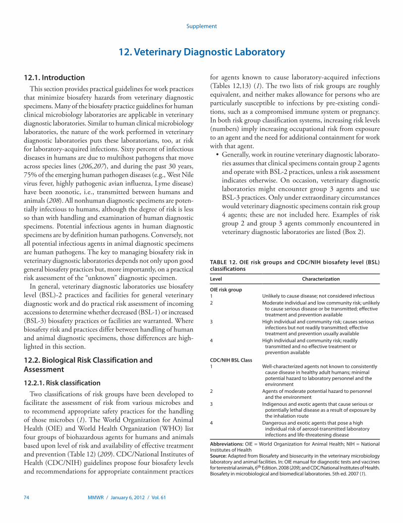
74 MMWR / January 6, 2012 / Vol. 61
Supplement
12. Veterinary Diagnostic Laboratory
12.1. IntroductionThis section provides practical guidelines for work practices
that minimize biosafety hazards from veterinary diagnostic specimens. Many of the biosafety practice guidelines for human clinical microbiology laboratories are applicable in veterinary diagnostic laboratories. Similar to human clinical microbiology laboratories, the nature of the work performed in veterinary diagnostic laboratories puts these laboratorians, too, at risk for laboratory-acquired infections. Sixty percent of infectious diseases in humans are due to multihost pathogens that move across species lines (206,207), and during the past 30 years, 75% of the emerging human pathogen diseases (e.g., West Nile virus fever, highly pathogenic avian influenza, Lyme disease) have been zoonotic, i.e., transmitted between humans and animals (208). All nonhuman diagnostic specimens are poten-tially infectious to humans, although the degree of risk is less so than with handling and examination of human diagnostic specimens. Potential infectious agents in human diagnostic specimens are by definition human pathogens. Conversely, not all potential infectious agents in animal diagnostic specimens are human pathogens. The key to managing biosafety risk in veterinary diagnostic laboratories depends not only upon good general biosafety practices but, more importantly, on a practical risk assessment of the “unknown” diagnostic specimen.
In general, veterinary diagnostic laboratories use biosafety level (BSL)-2 practices and facilities for general veterinary diagnostic work and do practical risk assessment of incoming accessions to determine whether decreased (BSL-1) or increased (BSL-3) biosafety practices or facilities are warranted. Where biosafety risk and practices differ between handling of human and animal diagnostic specimens, those differences are high-lighted in this section.
12.2. Biological Risk Classification and Assessment
12.2.1. Risk classificationTwo classifications of risk groups have been developed to
facilitate the assessment of risk from various microbes and to recommend appropriate safety practices for the handling of those microbes (1). The World Organization for Animal Health (OIE) and World Health Organization (WHO) list four groups of biohazardous agents for humans and animals based upon level of risk and availability of effective treatment and prevention (Table 12) (209). CDC/National Institutes of Health (CDC/NIH) guidelines propose four biosafety levels and recommendations for appropriate containment practices
for agents known to cause laboratory-acquired infections (Tables 12,13) (1). The two lists of risk groups are roughly equivalent, and neither makes allowance for persons who are particularly susceptible to infections by pre-existing condi-tions, such as a compromised immune system or pregnancy. In both risk group classification systems, increasing risk levels (numbers) imply increasing occupational risk from exposure to an agent and the need for additional containment for work with that agent.• Generally,workinroutineveterinarydiagnosticlaborato-
ries assumes that clinical specimens contain group 2 agents and operate with BSL-2 practices, unless a risk assessment indicates otherwise. On occasion, veterinary diagnostic laboratories might encounter group 3 agents and use BSL-3 practices. Only under extraordinary circumstances would veterinary diagnostic specimens contain risk group 4 agents; these are not included here. Examples of risk group 2 and group 3 agents commonly encountered in veterinary diagnostic laboratories are listed (Box 2).
TABLE 12. OIE risk groups and CDC/NIH biosafety level (BSL) classifications
Level Characterization
OIE risk group1 Unlikelytocausedisease;notconsideredinfectious2 Moderateindividualandlowcommunityrisk;unlikely
tocauseseriousdiseaseorbetransmitted;effectivetreatment and prevention available
3 Highindividualandcommunityrisk;causesseriousinfectionsbutnotreadilytransmitted;effectivetreatment and prevention usually available
4 Highindividualandcommunityrisk;readilytransmitted and no effective treatment or prevention available
CDC/NIH BSL Class1 Well-characterizedagentsnotknowntoconsistently
causediseaseinhealthyadulthumans;minimalpotential hazard to laboratory personnel and the environment
2 Agents of moderate potential hazard to personnel and the environment
3 Indigenous and exotic agents that cause serious or potentially lethal disease as a result of exposure by the inhalation route
4 Dangerous and exotic agents that pose a high individualriskofaerosol-transmittedlaboratoryinfectionsandlife-threateningdisease
Abbreviations: OIE=WorldOrganization forAnimalHealth;NIH=NationalInstitutes of HealthSource: Adapted from Biosafety and biosecurity in the veterinary microbiology laboratoryandanimalfacilities.In:OIEmanualfordiagnostictestsandvaccinesfor terrestrial animals, 6thEdition.2008(209);andCDC/NationalInstitutesofHealth.Biosafetyinmicrobiologicalandbiomedicallaboratories.5thed.2007(1).
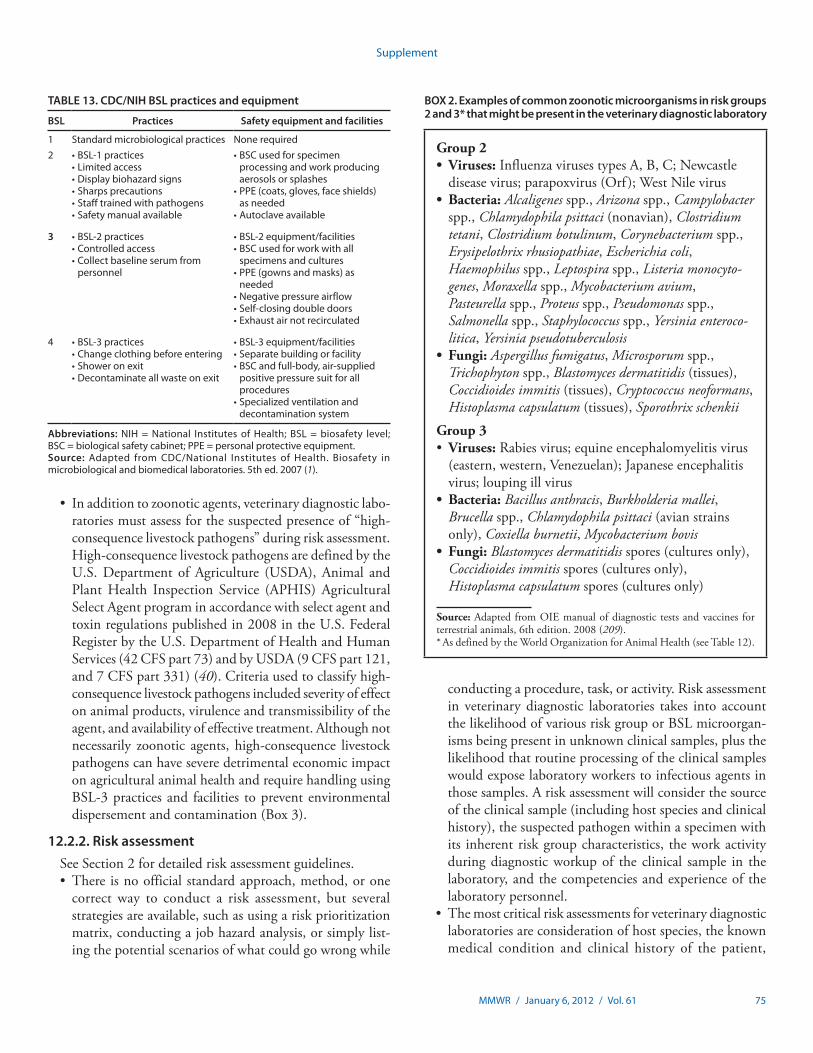
MMWR / January 6, 2012 / Vol. 61 75
Supplement
• Inadditiontozoonoticagents,veterinarydiagnosticlabo-ratories must assess for the suspected presence of “high-consequence livestock pathogens” during risk assessment. High-consequence livestock pathogens are defined by the U.S. Department of Agriculture (USDA), Animal and Plant Health Inspection Service (APHIS) Agricultural Select Agent program in accordance with select agent and toxin regulations published in 2008 in the U.S. Federal Register by the U.S. Department of Health and Human Services (42 CFS part 73) and by USDA (9 CFS part 121, and 7 CFS part 331) (40). Criteria used to classify high-consequence livestock pathogens included severity of effect on animal products, virulence and transmissibility of the agent, and availability of effective treatment. Although not necessarily zoonotic agents, high-consequence livestock pathogens can have severe detrimental economic impact on agricultural animal health and require handling using BSL-3 practices and facilities to prevent environmental dispersement and contamination (Box 3).
12.2.2. Risk assessmentSee Section 2 for detailed risk assessment guidelines.• Thereisnoofficialstandardapproach,method,orone
correct way to conduct a risk assessment, but several strategies are available, such as using a risk prioritization matrix, conducting a job hazard analysis, or simply list-ing the potential scenarios of what could go wrong while
conducting a procedure, task, or activity. Risk assessment in veterinary diagnostic laboratories takes into account the likelihood of various risk group or BSL microorgan-isms being present in unknown clinical samples, plus the likelihood that routine processing of the clinical samples would expose laboratory workers to infectious agents in those samples. A risk assessment will consider the source of the clinical sample (including host species and clinical history), the suspected pathogen within a specimen with its inherent risk group characteristics, the work activity during diagnostic workup of the clinical sample in the laboratory, and the competencies and experience of the laboratory personnel.
• Themostcriticalriskassessmentsforveterinarydiagnosticlaboratories are consideration of host species, the known medical condition and clinical history of the patient,
TABLE 13. CDC/NIH BSL practices and equipment
BSL Practices Safety equipment and facilities
1 Standard microbiological practices Nonerequired2 •BSL-1practices
•Limitedaccess•Displaybiohazardsigns•Sharpsprecautions•Stafftrainedwithpathogens•Safetymanualavailable
•BSCusedforspecimenprocessingandworkproducingaerosols or splashes
•PPE(coats,gloves,faceshields)as needed
•Autoclaveavailable
3 •BSL-2practices•Controlledaccess•Collectbaselineserumfrom
personnel
•BSL-2equipment/facilities•BSCusedforworkwithall
specimens and cultures•PPE(gownsandmasks)as
needed•Negativepressureairflow•Self-closingdoubledoors•Exhaustairnotrecirculated
4 •BSL-3practices•Changeclothingbeforeentering•Showeronexit•Decontaminateallwasteonexit
•BSL-3equipment/facilities•Separatebuildingorfacility•BSCandfull-body,air-supplied
positive pressure suit for all procedures
•Specializedventilationanddecontamination system
Abbreviations: NIH=National InstitutesofHealth; BSL=biosafety level;BSC=biologicalsafetycabinet;PPE=personalprotectiveequipment.Source: Adapted from CDC/National Institutes of Health. Biosafety in microbiologicalandbiomedicallaboratories.5thed.2007(1).
BOX 2. Examples of common zoonotic microorganisms in risk groups 2 and 3* that might be present in the veterinary diagnostic laboratory
Group 2• Viruses: Influenza viruses types A, B, C; Newcastle
disease virus; parapoxvirus (Orf ); West Nile virus• Bacteria:Alcaligenes spp., Arizona spp., Campylobacter
spp., Chlamydophila psittaci (nonavian), Clostridium tetani, Clostridium botulinum, Corynebacterium spp., Erysipelothrix rhusiopathiae, Escherichia coli, Haemophilus spp., Leptospira spp., Listeria monocyto-genes, Moraxella spp., Mycobacterium avium, Pasteurella spp., Proteus spp., Pseudomonas spp., Salmonella spp., Staphylococcus spp., Yersinia enteroco-litica, Yersinia pseudotuberculosis
• Fungi:Aspergillus fumigatus, Microsporum spp., Trichophyton spp., Blastomyces dermatitidis (tissues), Coccidioides immitis (tissues), Cryptococcus neoformans, Histoplasma capsulatum (tissues), Sporothrix schenkii
Group 3• Viruses: Rabies virus; equine encephalomyelitis virus
(eastern, western, Venezuelan); Japanese encephalitis virus; louping ill virus
• Bacteria:Bacillus anthracis, Burkholderia mallei, Brucella spp., Chlamydophila psittaci (avian strains only), Coxiella burnetii, Mycobacterium bovis
• Fungi:Blastomyces dermatitidis spores (cultures only), Coccidioides immitis spores (cultures only), Histoplasma capsulatum spores (cultures only)
Source: Adapted from OIE manual of diagnostic tests and vaccines for terrestrial animals, 6th edition. 2008 (209).* As defined by the World Organization for Animal Health (see Table 12).
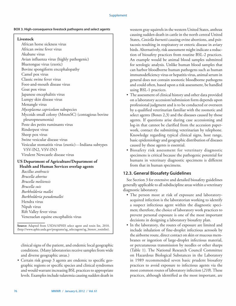
76 MMWR / January 6, 2012 / Vol. 61
Supplement
clinical signs of the patient, and endemic local geographic conditions. (Many laboratories receive samples from wide and diverse geographic areas.)
• Certainriskgroup3agentsareendemictospecificgeo-graphic regions or specific species and clinical syndromes and would warrant increasing BSL practices to appropriate levels. Examples include tularemia causing sudden death in
western gray squirrels in the western United States, anthrax causing sudden death in cattle in the north central United States, Coxiella burnetii causing ovine abortions, and psit-tacosis resulting in respiratory or enteric disease in aviary birds. Alternatively, risk assessment might indicate a reduc-tion of biosafety practices from routine BSL-2 practices. An example would be animal blood samples submitted for serologic analysis. Unlike human blood samples that can harbor bloodborne human pathogens such as human immunodeficiency virus or hepatitis virus, animal serum in general does not contain zoonotic bloodborne pathogens and could often, based upon a risk assessment, be handled using BSL-1 practices.
• Theassessmentofclinicalhistoryandotherdataprovidedon a laboratory accession/submission form depends upon professional judgment and is to be conducted or overseen by a qualified veterinarian familiar with the zoonotic and select agents (Boxes 2,3) and the diseases caused by those agents. If questions arise during case accessioning and log-in that cannot be clarified from the accession paper-work, contact the submitting veterinarian by telephone. Knowledge regarding typical clinical signs, host range, basic epidemiology and geographic distribution of diseases caused by these agents is essential.
• Biosafety risk assessment for veterinary diagnosticspecimens is critical because the pathogenic potential for humans in veterinary diagnostic specimens is different from that in human specimens.
12.3. General Biosafety GuidelinesSee Section 3 for extensive and detailed biosafety guidelines
generally applicable to all subdiscipline areas within a veterinary diagnostic laboratory.• The personmost at risk of exposure and laboratory-
acquired infection is the laboratorian working to identify a suspect infectious agent within the diagnostic speci-men; therefore, the choice of laboratory work practices to prevent personal exposure is one of the most important decisions in designing a laboratory biosafety plan.
• Inthelaboratory,theroutesofexposurearelimitedandinclude inhalation of fine-droplet infectious aerosols by the airborne route, direct contact on skin or mucous mem-branes or ingestion of large-droplet infectious material, or percutaneous transmission by needles or other sharps (Table 1). The National Research Council Committee on Hazardous Biological Substances in the Laboratory in 1989 recommended seven basic prudent biosafety practices to avoid exposure to infectious agents via the most common routes of laboratory infection (210). These practices, although identified as the most important, are
BOX 3. High-consequence livestock pathogens and select agents
LivestockAfrican horse sickness virusAfrican swine fever virusAkabane virusAvian influenza virus (highly pathogenic)Bluetongue virus (exotic)Bovine spongiform encephalopathyCamel pox virusClassic swine fever virusFoot-and-mouth disease virusGoat pox virusJapanese encephalitis virusLumpy skin disease virusMenangle virusMycoplasma capricolum subspeciesMycoids small colony (MmmSC) (contagious bovine pleuropneumonia)
Peste des petits ruminants virusRinderpest virusSheep pox virusSwine vesicular disease virusVesicular stomatitis virus (exotic)—Indiana subtypes VSV-IN2, VSV-IN3
Virulent Newcastle disease virus
US Department of Agriculture/Department of Health and Human Services overlap agentsBacillus anthracisBrucella abortusBrucella melitensisBrucella suisBurkholderia malleiBurkholderia pseudomalleiHendra virusNipah virusRift Valley fever virusVenezuelan equine encephalitis virus
Source: Adapted from USDA/APHIS select agent and toxin list, 2010. (http://www.aphis.usda.gov/programs/ag_selectagent/ag_bioterr_toxinlist).
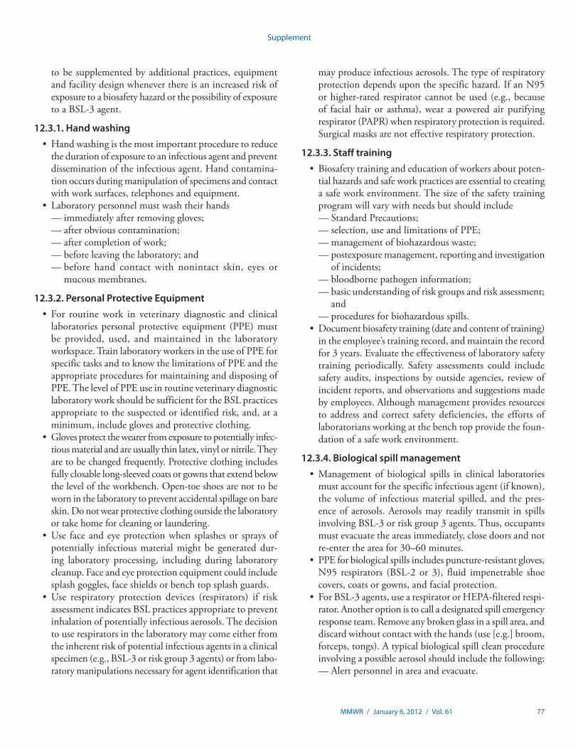
MMWR / January 6, 2012 / Vol. 61 77
Supplement
to be supplemented by additional practices, equipment and facility design whenever there is an increased risk of exposure to a biosafety hazard or the possibility of exposure to a BSL-3 agent.
12.3.1. Hand washing• Handwashingisthemostimportantproceduretoreduce
the duration of exposure to an infectious agent and prevent dissemination of the infectious agent. Hand contamina-tion occurs during manipulation of specimens and contact with work surfaces, telephones and equipment.
• Laboratorypersonnelmustwashtheirhands— immediately after removing gloves;— after obvious contamination;— after completion of work;— before leaving the laboratory; and— before hand contact with nonintact skin, eyes or
mucous membranes.
12.3.2. Personal Protective Equipment• For routinework in veterinary diagnostic and clinical
laboratories personal protective equipment (PPE) must be provided, used, and maintained in the laboratory workspace. Train laboratory workers in the use of PPE for specific tasks and to know the limitations of PPE and the appropriate procedures for maintaining and disposing of PPE. The level of PPE use in routine veterinary diagnostic laboratory work should be sufficient for the BSL practices appropriate to the suspected or identified risk, and, at a minimum, include gloves and protective clothing.
• Glovesprotectthewearerfromexposuretopotentiallyinfec-tious material and are usually thin latex, vinyl or nitrile. They are to be changed frequently. Protective clothing includes fully closable long-sleeved coats or gowns that extend below the level of the workbench. Open-toe shoes are not to be worn in the laboratory to prevent accidental spillage on bare skin. Do not wear protective clothing outside the laboratory or take home for cleaning or laundering.
• Use faceandeyeprotectionwhensplashesor spraysofpotentially infectious material might be generated dur-ing laboratory processing, including during laboratory cleanup. Face and eye protection equipment could include splash goggles, face shields or bench top splash guards.
• Use respiratory protection devices (respirators) if riskassessment indicates BSL practices appropriate to prevent inhalation of potentially infectious aerosols. The decision to use respirators in the laboratory may come either from the inherent risk of potential infectious agents in a clinical specimen (e.g., BSL-3 or risk group 3 agents) or from labo-ratory manipulations necessary for agent identification that
may produce infectious aerosols. The type of respiratory protection depends upon the specific hazard. If an N95 or higher-rated respirator cannot be used (e.g., because of facial hair or asthma), wear a powered air purifying respirator (PAPR) when respiratory protection is required. Surgical masks are not effective respiratory protection.
12.3.3. Staff training• Biosafetytrainingandeducationofworkersaboutpoten-
tial hazards and safe work practices are essential to creating a safe work environment. The size of the safety training program will vary with needs but should include— Standard Precautions;— selection, use and limitations of PPE;— management of biohazardous waste;— postexposure management, reporting and investigation
of incidents;— bloodborne pathogen information;— basic understanding of risk groups and risk assessment;
and— procedures for biohazardous spills.
• Documentbiosafetytraining(dateandcontentoftraining)in the employee’s training record, and maintain the record for 3 years. Evaluate the effectiveness of laboratory safety training periodically. Safety assessments could include safety audits, inspections by outside agencies, review of incident reports, and observations and suggestions made by employees. Although management provides resources to address and correct safety deficiencies, the efforts of laboratorians working at the bench top provide the foun-dation of a safe work environment.
12.3.4. Biological spill management•Management of biological spills in clinical laboratories
must account for the specific infectious agent (if known), the volume of infectious material spilled, and the pres-ence of aerosols. Aerosols may readily transmit in spills involving BSL-3 or risk group 3 agents. Thus, occupants must evacuate the areas immediately, close doors and not re-enter the area for 30–60 minutes.
• PPEforbiologicalspillsincludespuncture-resistantgloves,N95 respirators (BSL-2 or 3), fluid impenetrable shoe covers, coats or gowns, and facial protection.
• ForBSL-3agents,usearespiratororHEPA-filteredrespi-rator. Another option is to call a designated spill emergency response team. Remove any broken glass in a spill area, and discard without contact with the hands (use [e.g.] broom, forceps, tongs). A typical biological spill clean procedure involving a possible aerosol should include the following:— Alert personnel in area and evacuate.
78 MMWR / January 6, 2012 / Vol. 61
Supplement
— Close doors and do not re-enter area for 30–60 minutes (post sign forbidding entry to the area), based on the number of air exchanges.
— Alert laboratory supervisor.— Don PPE appropriate for type of spill.— Remove and discard broken glass or other objects
(without contact with hands).— Absorb the spill with absorbent material.— Discard contaminated material in a biohazardous waste
container.— Clean spill site with aqueous detergent.— Decontaminate area with appropriate disinfectant.— Rinse spill site with water and allow site to dry.— Copy contaminated laboratory forms and discard into
the biohazard waste container.— Place all disposable contaminated cleanup material in
the biohazard bag and treat as infectious waste.— Wash hands.— Prepare a spill/incident report, identify cause of spill,
and determine remedial action.• Ifaspilloccursinabiologicalsafetycabinet(BSC),do
not turn off the cabinet fan. Minor spills in a BSC can be absorbed with absorbent paper. If infectious material flows into the grille, wipe all items in the cabinet with disinfectant and remove them. Close the drain valve and pour disinfectant onto the surface and through the grille into the drain pan. Allow appropriate contact time, then drain, rinse and dry.
•Whenbreakageoccursinacentrifuge(whichinherentlywould produce aerosols), keep the centrifuge tightly closed for 30 minutes before decontamination commences.
• Ifaspecimentubebreakswithintheplasticscrew-cappedcanister in a centrifuge:— Turn the motor off.— Remove the canister immediately and place in a BSC.— Notify senior person in charge and other colleagues
working in the area.— While wearing protective clothing, open the canister
under the safety cabinet.— Pour a 1:10 dilution of bleach or a noncorrosive disin-
fectant into the canister to decontaminate all surfaces. Let the canister soak in bleach or disinfectant solution for 10 minutes. Clean canister thoroughly.
— Do not pick up broken glass with gloved hands. Use forceps or cotton held in forceps, or tongs or hemostats, and dispose into a biosafety sharps container.
— Discard all nonsharp contaminated materials from canis-ter into a red biohazard bag for biohazard waste disposal.
— Swab unbroken capped tubes with the same disinfec-tant; then swab again, wash with water and dry.
— Treat all materials used during the cleanup as infectious waste.
Note: If the specimen tube breaks in a centrifuge that does not have individual canisters but does have a bio-hazard cover and sealed rotor, follow the manufacturer’s instructions for cleaning and decontamination.
12.3.5. Immunization• TheAdvisoryCommitteeonImmunizationPractices,in
addition to recommending immunization of health-care personnel with vaccines recommended for all adults (influ-enza, measles/mumps/rubella, varicella, and tetanus/diph-theria/pertussis), recommends meningococcal or hepatitis B vaccination for those at risk for occupational exposure (211). These agents are not present in animal diagnostic specimens. In veterinary diagnostic laboratories, it is not recommended that laboratorians be immunized routinely against potential risk group 3 or BSL-3 pathogens. The only situation in which immunization is to be considered is against rabies for laboratorians processing a large number of specimens potentially containing rabies virus (e.g., routinely processing central nervous tissues from animals with neurologic disease compatible with rabies) (Table 14) (212).
12.4. Pathology (Necropsy and Surgical Pathology)
See Section 5 for detailed biosafety guidelines applicable to necropsy, surgical pathology, and histology working areas in a veterinary diagnostic laboratory.
12.5. ParasitologySee Section 6.
12.6. MycologySee Section 7.
12.7. VirologySee Section 8.
12.8. ToxicologySee Section 9.
12.9. Hematology/SerologySee Section 10.
12.10. Molecular Diagnostics and Rapid TestsBiosafety guidelines to be followed when conducting molecu-
lar diagnostic testing (i.e., polymerase chain reaction [PCR]) or using rapid tests such as enzyme-linked immunosorbent assay (ELISA) can be specific to the particular testing being
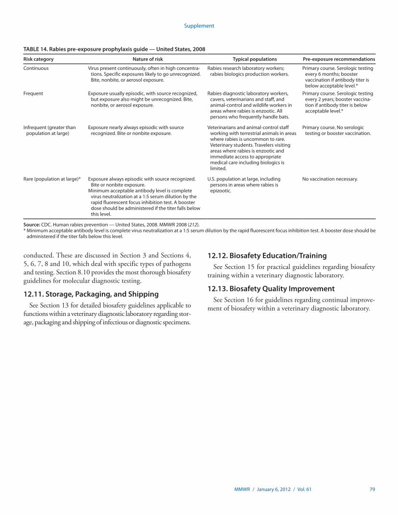
MMWR / January 6, 2012 / Vol. 61 79
Supplement
conducted. These are discussed in Section 3 and Sections 4, 5, 6, 7, 8 and 10, which deal with specific types of pathogens and testing. Section 8.10 provides the most thorough biosafety guidelines for molecular diagnostic testing.
12.11. Storage, Packaging, and ShippingSee Section 13 for detailed biosafety guidelines applicable to
functions within a veterinary diagnostic laboratory regarding stor-age, packaging and shipping of infectious or diagnostic specimens.
TABLE 14. Rabies pre-exposure prophylaxis guide — United States, 2008
Risk category Nature of risk Typical populations Pre-exposure recommendations
Continuous Virus present continuously, often in high concentra-tions. Specific exposures likely to go unrecognized. Bite, nonbite, or aerosol exposure.
Rabiesresearchlaboratoryworkers;rabiesbiologicsproductionworkers.
Primary course. Serologic testing every6months;boostervaccination if antibody titer is belowacceptablelevel.*
Frequent Exposureusuallyepisodic,withsourcerecognized,but exposure also might be unrecognized. Bite, nonbite, or aerosol exposure.
Rabiesdiagnosticlaboratoryworkers,cavers, veterinarians and staff, and animal-controlandwildlifeworkersinareaswhererabiesisenzootic.Allpersonswhofrequentlyhandlebats.
Primary course. Serologic testing every2years;boostervaccina-tionifantibodytiterisbelowacceptablelevel.*
Infrequent(greaterthanpopulationatlarge)
Exposurenearlyalwaysepisodicwithsourcerecognized. Bite or nonbite exposure.
Veterinariansandanimal-controlstaffworkingwithterrestrialanimalsinareaswhererabiesisuncommontorare.Veterinary students. Travelers visiting areaswhererabiesisenzooticandimmediate access to appropriate medical care including biologics is limited.
Primary course. No serologic testing or booster vaccination.
Rare(populationatlarge)* Exposurealwaysepisodicwithsourcerecognized.Bite or nonbite exposure.
Minimum acceptable antibody level is complete virus neutralization at a 1:5 serum dilution by the rapid fluorescent focus inhibition test. A booster doseshouldbeadministeredifthetiterfallsbelowthis level.
U.S.populationatlarge,includingpersonsinareaswhererabiesisepizootic.
No vaccination necessary.
Source:CDC.Humanrabiesprevention—UnitedStates,2008.MMWR2008(212).*Minimumacceptableantibodyleveliscompletevirusneutralizationata1:5serumdilutionbytherapidfluorescentfocusinhibitiontest.Aboosterdoseshouldbeadministeredifthetiterfallsbelowthislevel.
12.12. Biosafety Education/TrainingSee Section 15 for practical guidelines regarding biosafety
training within a veterinary diagnostic laboratory.
12.13. Biosafety Quality ImprovementSee Section 16 for guidelines regarding continual improve-
ment of biosafety within a veterinary diagnostic laboratory.
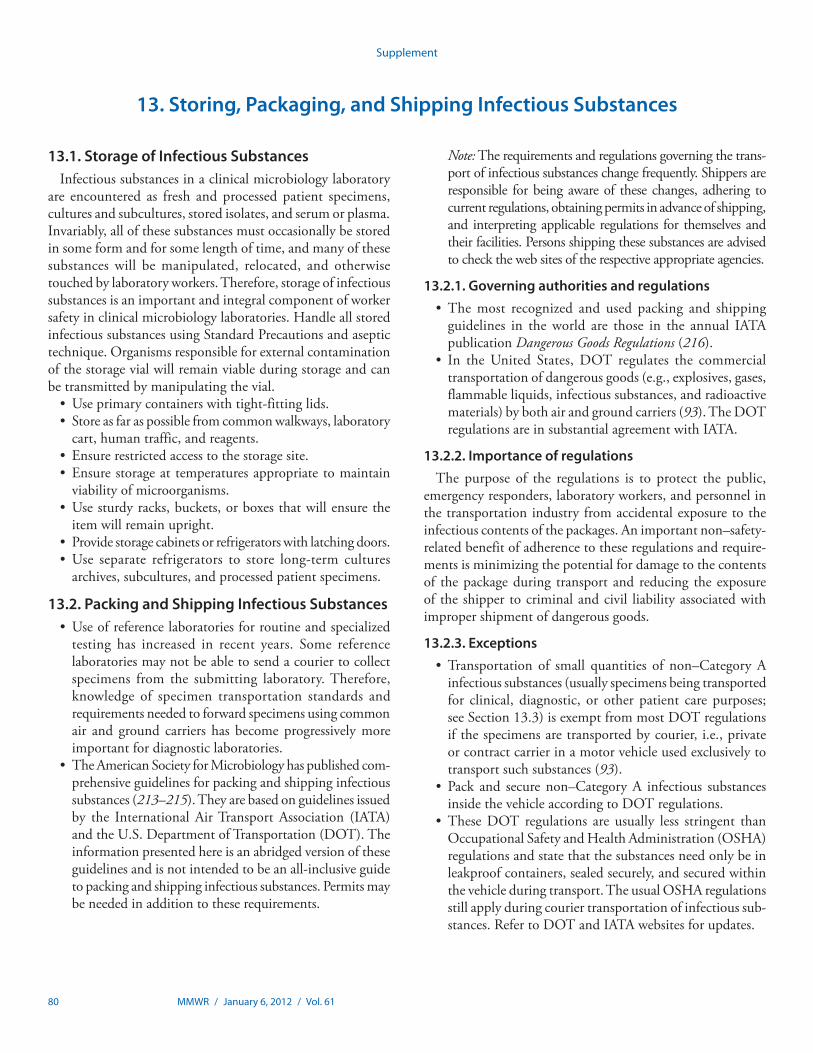
80 MMWR / January 6, 2012 / Vol. 61
Supplement
13. Storing, Packaging, and Shipping Infectious Substances
13.1. Storage of Infectious SubstancesInfectious substances in a clinical microbiology laboratory
are encountered as fresh and processed patient specimens, cultures and subcultures, stored isolates, and serum or plasma. Invariably, all of these substances must occasionally be stored in some form and for some length of time, and many of these substances will be manipulated, relocated, and otherwise touched by laboratory workers. Therefore, storage of infectious substances is an important and integral component of worker safety in clinical microbiology laboratories. Handle all stored infectious substances using Standard Precautions and aseptic technique. Organisms responsible for external contamination of the storage vial will remain viable during storage and can be transmitted by manipulating the vial.• Useprimarycontainerswithtight-fittinglids.• Storeasfaraspossiblefromcommonwalkways,laboratory
cart, human traffic, and reagents.• Ensurerestrictedaccesstothestoragesite.• Ensurestorageattemperaturesappropriatetomaintain
viability of microorganisms.• Usesturdyracks,buckets,orboxesthatwillensurethe
item will remain upright.• Providestoragecabinetsorrefrigeratorswithlatchingdoors.• Use separate refrigerators to store long-term cultures
archives, subcultures, and processed patient specimens.
13.2. Packing and Shipping Infectious Substances• Useofreferencelaboratoriesforroutineandspecialized
testing has increased in recent years. Some reference laboratories may not be able to send a courier to collect specimens from the submitting laboratory. Therefore, knowledge of specimen transportation standards and requirements needed to forward specimens using common air and ground carriers has become progressively more important for diagnostic laboratories.
• TheAmericanSocietyforMicrobiologyhaspublishedcom-prehensive guidelines for packing and shipping infectious substances (213–215). They are based on guidelines issued by the International Air Transport Association (IATA) and the U.S. Department of Transportation (DOT). The information presented here is an abridged version of these guidelines and is not intended to be an all-inclusive guide to packing and shipping infectious substances. Permits may be needed in addition to these requirements.
Note: The requirements and regulations governing the trans-port of infectious substances change frequently. Shippers are responsible for being aware of these changes, adhering to current regulations, obtaining permits in advance of shipping, and interpreting applicable regulations for themselves and their facilities. Persons shipping these substances are advised to check the web sites of the respective appropriate agencies.
13.2.1. Governing authorities and regulations• Themost recognized and used packing and shipping
guidelines in the world are those in the annual IATA publication Dangerous Goods Regulations (216).
• In theUnited States,DOT regulates the commercialtransportation of dangerous goods (e.g., explosives, gases, flammable liquids, infectious substances, and radioactive materials) by both air and ground carriers (93). The DOT regulations are in substantial agreement with IATA.
13.2.2. Importance of regulationsThe purpose of the regulations is to protect the public,
emergency responders, laboratory workers, and personnel in the transportation industry from accidental exposure to the infectious contents of the packages. An important non–safety-related benefit of adherence to these regulations and require-ments is minimizing the potential for damage to the contents of the package during transport and reducing the exposure of the shipper to criminal and civil liability associated with improper shipment of dangerous goods.
13.2.3. Exceptions• Transportation of small quantities of non–CategoryA
infectious substances (usually specimens being transported for clinical, diagnostic, or other patient care purposes; see Section 13.3) is exempt from most DOT regulations if the specimens are transported by courier, i.e., private or contract carrier in a motor vehicle used exclusively to transport such substances (93).
• Pack and securenon–CategoryA infectious substancesinside the vehicle according to DOT regulations.
• TheseDOT regulations are usually less stringent thanOccupational Safety and Health Administration (OSHA) regulations and state that the substances need only be in leakproof containers, sealed securely, and secured within the vehicle during transport. The usual OSHA regulations still apply during courier transportation of infectious sub-stances. Refer to DOT and IATA websites for updates.
MMWR / January 6, 2012 / Vol. 61 81
Supplement
13.2.4. Specific regulations• The safe and legal transport of infectious substances is
based on the following mandated activities:— training every 2 (IATA) or 3 (DOT) years for persons
handling infectious substances for shipment;— classification and naming of infectious substances;— selection of correct packaging materials;— packing shipments correctly;— placing appropriate markings and labels onto the outer
package; and— documenting relevant aspects of each package and its
contents.
13.2.5. U.S. Postal ServiceThe U.S. Postal Service publishes its own regulations in the
USPS Domestic Mail Manual (96). The USPS regulations for mailing hazardous materials generally adhere to DOT regula-tions; however, consult the USPS Domestic Mail Manual for specific needs and requirements.
13.3. Classification of Infectious Substances
13.3.1. ClassificationAll shipped goods must be classified using a three-step pro-
cess to define dangerous goods that are shipped by commercial carriers. Classification allows the shipper to select the proper IATA packing instructions and directions to use, and provides information necessary to complete required documentation (a Shipper’s Declaration for Dangerous Goods) if the substance is a Category A infectious substance.
13.3.2. Steps of classification• Thematerial is classified into one of the nine IATA-
specified classes (Class 1 through Class 9) of dangerous goods. Infectious and toxic substances are Class 6 danger-ous goods; dry ice is a Class 9 dangerous good. Class 6 and Class 9 substances usually are the only dangerous goods shipped by laboratorians.
• Class6substancesmustbecategorizedintoeitherDivision6.1 (toxic substances) or Division 6.2 (infectious substances).
• Division6.2infectioussubstancesmustbeclassifiedintoone of nine IATA-specified types of infectious substances: Category A infectious substance, Category B infectious substance, patient specimens, exempt human or animal specimens, genetically modified organisms, exempt sub-stances, biological products, infected animals, or medical waste (Table 15).
• Ifthesubstanceisdeterminedtobeeithera“patientspeci-men” or an “organism” and is not obviously a Category A or Category B substance but meets the criteria of or has
characteristics of a Category A or Category B substance, the shipper must classify it as a Category A or Category B substance. Otherwise, the substance must be classified as an “exempt human or animal specimen” or a “genetically modified organism” (Class 9), respectively.
13.3.3. Category A infectious substancesA Category A substance is “an infectious substance which
is transported in a form that, when exposure to it occurs, is capable of causing permanent disability, or life-threatening or fatal disease to otherwise healthy humans or animals” (93).• CategoryAsubstancesarespecificallydesignatedandlisted
by IATA and DOT. The list of Category A substances is not all-inclusive; the shipper is allowed by IATA to perform a thorough risk assessment and to use discretion and professional judgment when deciding if a substance meets Category A criteria. Category A substances must be assigned UN number UN2814 or UN2900 (Figure 2).
Note: Certain Category A infectious substances are consid-ered Category A only if the substance is in culture form, i.e., concentrated; this distinction is clearly indicated in the specific IATA list.
• CertainCategoryApathogenshavebeendesignatedasagentsof bioterrorism and are known as select agents (40). Federal regulations require shippers to have special registration and permits to possess, use, transfer, and receive select agents.
13.3.4. Category B infectious substancesA Category B substance is “an infectious substance that
does not meet the criteria for inclusion in Category A” (93). Category B substances are not in a form generally capable of causing disability, life-threatening illness, or fatal disease.
TABLE 15. Types and classifications of IATA division 6.2 infectious substances
Type of infectious substance IATA classification
Category A substance Category ACategory B substance CategoryB*Patient specimen Meets Category A criteria Category A Meets Category B criteria Category B Does not meet Category A or B criteria Exempt human or animal specimenExempt human or animal specimen Exempt human or animal specimenGenetically modified microorganism Meets Category A criteria Category A Meets Category B criteria Category B Does not meet Category A or B criteria Genetically modified organismExempt substance NoneBiological product† NoneInfected animal† NoneMedicalwaste† None
Abbreviation: IATA: International Air Transport Association*ThepropershippingnameforCategoryBsubstancesisBiologicalSubstance,
Category B. †Substance is not addressed in detail in these guidelines.
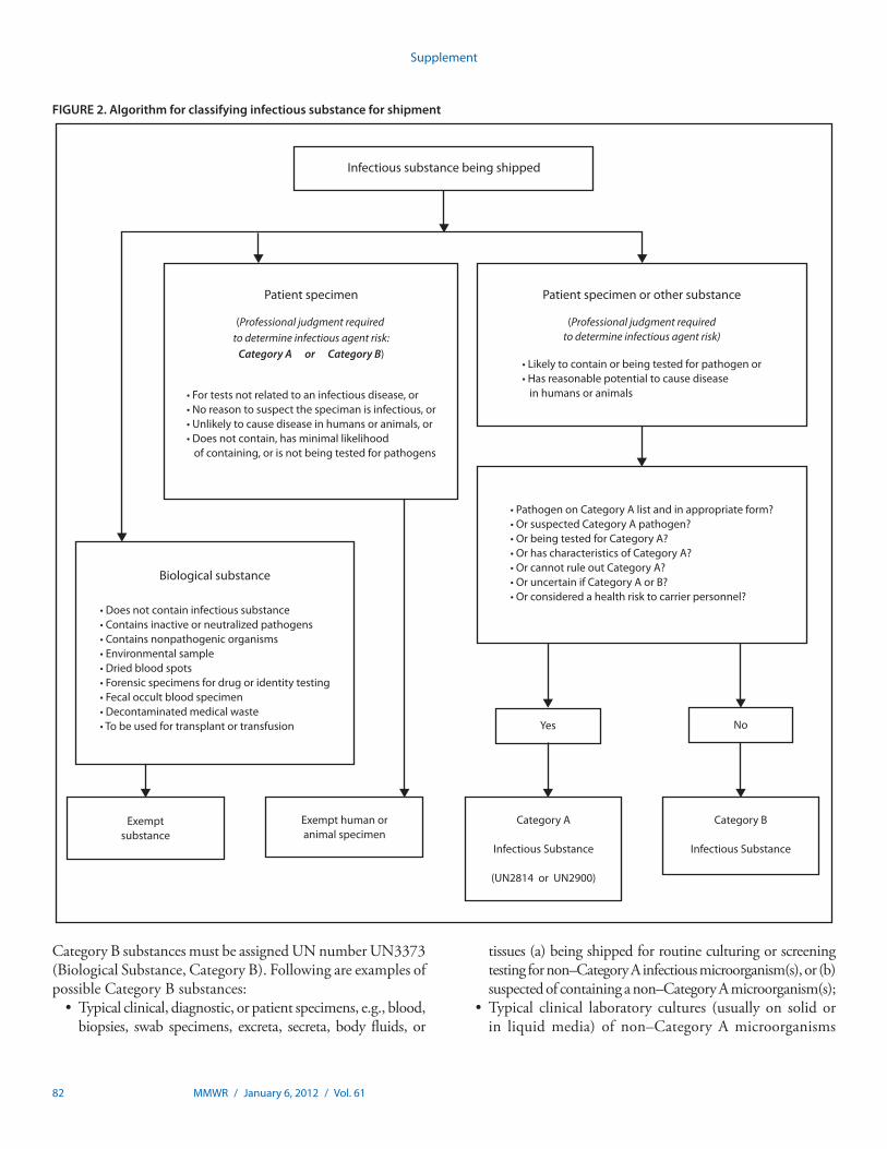
82 MMWR / January 6, 2012 / Vol. 61
Supplement
Infectious substance being shipped
Patient specimen or other substance
(Professional judgment requiredto determine infectious agent risk)
• Likely to contain or being tested for pathogen or• Has reasonable potential to cause disease in humans or animals
• Pathogen on Category A list and in appropriate form?• Or suspected Category A pathogen?• Or being tested for Category A?• Or has characteristics of Category A?• Or cannot rule out Category A?• Or uncertain if Category A or B?• Or considered a health risk to carrier personnel?
Yes No
Category A
Infectious Substance
(UN2814 or UN2900)
Category B
Infectious Substance
Exemptsubstance
Exempt human oranimal specimen
(Professional judgment required to determine infectious agent risk:
Category A or Category B)
Biological substance
• Does not contain infectious substance• Contains inactive or neutralized pathogens• Contains nonpathogenic organisms• Environmental sample• Dried blood spots• Forensic specimens for drug or identity testing• Fecal occult blood specimen• Decontaminated medical waste• To be used for transplant or transfusion
• For tests not related to an infectious disease, or• No reason to suspect the speciman is infectious, or• Unlikely to cause disease in humans or animals, or• Does not contain, has minimal likelihood of containing, or is not being tested for pathogens
Patient specimen
FIGURE 2. Algorithm for classifying infectious substance for shipment
Category B substances must be assigned UN number UN3373 (Biological Substance, Category B). Following are examples of possible Category B substances:• Typicalclinical,diagnostic,orpatientspecimens,e.g.,blood,
biopsies, swab specimens, excreta, secreta, body fluids, or
tissues (a) being shipped for routine culturing or screening testing for non–Category A infectious microorganism(s), or (b) suspected of containing a non–Category A microorganism(s);
• Typical clinical laboratory cultures (usually on solidorin liquid media) of non–Category A microorganisms
MMWR / January 6, 2012 / Vol. 61 83
Supplement
routinely encountered and manipulated in clinical micro-biology laboratories (Figure 2).
13.3.5. Exempt human (or animal) specimensExempt human or animal body site specimens are those
for which there is “minimal likelihood there are pathogens present” (93). Examples of such specimens include urine or serum to be tested for glucose, cholesterol, hormone levels, prostate-specific antigen, and analytes used to evaluate heart and kidney function.• Professionaljudgmentandknowledgeofpatientmedical
history may used to determine if the specimen is an infec-tious risk or contains pathogens.
• Exempthumanoranimalspecimenshavelessstringentpackaging requirements than do Category A and Category B substances. IATA requires outer packages containing exempt human or animal specimens to be clearly labeled as “Exempt Human Specimen” or “Exempt Animal Specimen.” DOT does not require this label on outer packages (Figure 2).
13.3.6. Exempt substancesMany substances commonly encountered in clinical labo-
ratories are exempt from strict infectious substance shipping requirements (Figure 2). Examples of such substances are• Substancesthatdonotcontaininfectioussubstancesor
are unlikely to cause disease in humans and animals;•Mostenvironmentalsamples(e.g.,food,soil);• Substances that contain neutralized or inactivated
microorganisms;• Substancestobetestedforalcoholordrugs,pregnancy
indicators, cancer, and antibodies;• Samplessubmittedforforensicanalysis;• Driedbloodspotsandfecaloccultbloodscreenspecimens;• Bloodandbloodcomponentscollectedforthepurpose
of transfusion or transplantation;• Food andDrugAdministration (FDA)-approved and
FDA-licensed biological products; and• <30mLof10%formalinperprimarycontainerwhenthe
formalin is used as a preservative.
13.3.7. Patient specimens• A“patientspecimen”ismaterialcollecteddirectlyfrom
humans or animals for diagnostic, treatment, prevention, investigational, or research purposes.
• PatientspecimensthatmeetCategoryAorCategoryBcriteriamust be classified as Category A or Category B substances.
• Patient specimens thatmeet neitherCategory A norCategory B criteria must be treated as exempt human or animal specimens (Figure 2).
13.3.8. Genetically modified organismsGenetically modified organisms usually meet either
Category A or Category B criteria. If this is not the case, the organism must be classified as a “genetically modified micro-organism” (Class 9, Miscellaneous Dangerous Goods) and packed and shipped as such.
13.3.9. Biological productsVirtually all commercially available biological products are
exempt from regulations for packing and shipping infectious substances. Examples of biological products include bacterial typing sera, vaccines, bacterial antigens, antimicrobial agents, reagents for identifying bacteria, and reagents used in antimi-crobial susceptibility testing.
13.3.10. Infected animal• A live, intentionally infected animal that is knownor
reasonably expected to contain an infectious substance cannot be transported by air unless the substance cannot be transported by any other means. An exemption from DOT will be required.
• Consultationwithindividualcommercialcarriersisadvisedif either live or dead infected animals need to be shipped.
13.3.11. Medical waste•MedicalwastethatcontainsCategoryAorCategoryB
infectious substances must be packed and shipped as such and assigned number UN2814, UN2900, or UN3373.
•Medicalwastethatisreasonablybelievedtohavealowprobability of containing infectious substances must be packed and shipped as “medical waste not otherwise speci-fied” (n.o.s) (UN3291).
13.4. Naming Category A and Category B Substances• Afterclassifyingthesubstance,theshippermustidentify
(officially name) the Category A and Category B infectious substances by assigning the substance one of the >3,000 IATA-specified and internationally recognized UN num-bers and proper shipping names listed in the blue pages section of the IATA Dangerous Goods Regulations (216).
• Thislistprovides14informationalitemsforeachoftheproper shipping names; the items correspond to the infor-mation needed to complete the Shipper’s Declaration for Dangerous Goods. Fortunately, only seven of the 3,000 proper shipping names are used by most clinical microbiol-ogy laboratories:— Two for Category A substances that affect humans;— Two for Category A infectious substances that affect
animals;
84 MMWR / January 6, 2012 / Vol. 61
Supplement
— One for a Category B infectious substance;— One for genetically modified organisms; — One for dry ice.
• The blue pages provide proper shipping names,UNnumbers, packing instructions, quantity limits, and other information related to packing and shipping substances.
13.5 Packing Instructions and Packing Substances
13.5.1. Packing instructions and directions• IATApacking instructions (PI) describe theminimum
standards for safe transport of infectious substances.• The instructionsusedby clinical laboratories are those
that relate to shipping Category A infectious substances (PI 620), Category B infectious substances (PI 650), and dry ice (PI 954).
• There are no specifically PI-numbered instructions forspecimens classified as “exempt human or animal speci-mens”; however, IATA provides directions that must be followed (Table 16).
13.5.2. Marking and labeling outer packages• Shippers are responsible for the propermarking and
labeling of the outer shipping container (commonly, a cardboard box).
• Themarkingsandlabelscommunicateessentialinforma-tion regarding the shipper and consignee of the package, nature and weight of the contents of the package, the potential hazard of the substance, how the substance is packed, and information to be used in case of an emer-gency (Figures 3,4).
13.5.3. Specific markings and labelsThe following list cites the situations requiring a marker or
label, and the specific markings or labels for that situation.• Shipperandconsignee—shipper’sandconsignee’sname
and address;• Responsibleperson—nameandtelephonenumberofsome-
one who can answer general questions about the shipment;• CategoryA Substance— aClass 6 diamond-shaped
“Infectious Substance” label, and a proper shipping name (not the technical name), UN number, and quantity label;
• Category B Substance— “Biological Substance,Category B” and “UN3373” labels;
• Dryice—Class9“MiscellaneousDangerousGoods”andweight of dry ice label;
• Packageorientation—arrowsonoppositesidesofpack-ages containing >50 mL of a liquid or frozen liquid;
• Exemptpatientspecimens—“ExemptHumanSpecimen”or “Exempt Animal Specimen” label;
• OuterpackagecontainingCategoryAinfectioussubstance— a “UN” label; a UN inside of a circle, and a series of letters and numbers that indicate the type of package, class of goods the package is designed to carry, manufacturing date, authorizing agency, and manufacturer.
13.6. Documentation
13.6.1. Shipper’s Declaration for Dangerous Goods• AShipper’sDeclarationisalegalcontractbetweentheshipper
and carrier. It is required to document the shipment of Category A infectious substances and must be accurate and legible.
• EssentiallyalloftheIATA-specifiedtechnical informationrequired to complete the “Nature and Quantity of Dangerous Goods” section of the Declaration can be found in the blue pages of IATA Dangerous Goods Regulations (216).
13.6.2. Emergency response telephone number• DOT(butnotIATA)regulationsstatethatanemergency
response telephone number must be provided on Shipper’s Declarations that accompany shipments of Category A infectious substances.
• Thenumbermustbemonitoredatalltimesbyaperson(notan answering machine, message service, or pager) who has knowledge of the hazards of the material being shipped and emergency response and incident mitigation information in case a handler comes in contact with the released contents of the package. This number also can be that of a commercial service which can provide the appropriate mitigation infor-mation. If the telephone number of a commercial service is used, the contract number or service agreement number assigned by the commercial service must be entered on the document, as well as the name of the service provider.
13.6.3. AirbillsIATA carriers are required to prepare airbills to describe air
cargo and accompany shipments in transit. Some dangerous goods shipments, such as Biological Substances Category B ship-ments, require preparation of this document but not a Shipper’s Declaration. Specific preparation instructions are detailed in each IATA package instruction and in the “Documentation” section of the Dangerous Goods Regulations.
13.7. RefrigerantsPackaging must be leakproof when wet ice is used. Dry
ice is a Class 9 dangerous good; it must be packaged accord-ing to PI 954, and its use requires completion of a Shipper’s Declaration if it is used to ship a Category A substance.
Note: Dry ice is an explosion hazard and must never be placed into a tightly sealed container. Dry ice must be placed outside the secondary container, and the outer packaging must permit the release of CO2.
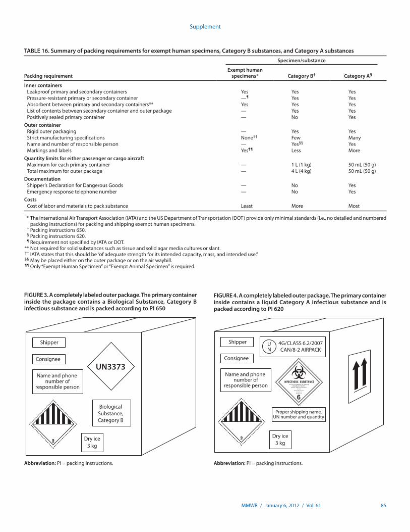
MMWR / January 6, 2012 / Vol. 61 85
Supplement
TABLE 16. Summary of packing requirements for exempt human specimens, Category B substances, and Category A substances
Packing requirement
Specimen/substance
Exempt human specimens* Category B† Category A§
Inner containersLeakproof primary and secondary containers Yes Yes YesPressure-resistantprimaryorsecondarycontainer —¶ Yes YesAbsorbentbetweenprimaryandsecondarycontainers** Yes Yes YesListofcontentsbetweensecondarycontainerandouterpackage — Yes YesPositively sealed primary container — No Yes
Outer containerRigid outer packaging — Yes YesStrict manufacturing specifications None†† Few ManyName and number of responsible person — Yes§§ YesMarkings and labels Yes¶¶ Less More
Quantity limits for either passenger or cargo aircraftMaximum for each primary container — 1L(1kg) 50mL(50g)Total maximum for outer package — 4L(4kg) 50mL(50g)
DocumentationShipper’s Declaration for Dangerous Goods — No YesEmergency response telephone number — No Yes
CostsCost of labor and materials to pack substance Least More Most
* The InternationalAirTransportAssociation(IATA)andtheUSDepartmentofTransportation(DOT)provideonlyminimalstandards(i.e.,nodetailedandnumberedpackinginstructions)forpackingandshippingexempthumanspecimens.
† Packing instructions 650. § Packing instructions 620. ¶ RequirementnotspecifiedbyIATAorDOT. ** Notrequiredforsolidsubstancessuchastissueandsolidagarmediaculturesorslant. †† IATAstatesthatthisshouldbe“ofadequatestrengthforitsintendedcapacity,mass,andintendeduse.” §§ Maybeplacedeitherontheouterpackageorontheairwaybill. ¶¶ Only“ExemptHumanSpecimen”or“ExemptAnimalSpecimen”isrequired.
FIGURE 4. A completely labeled outer package. The primary container inside contains a liquid Category A infectious substance and is packed according to PI 620
Shipper
Consignee
Name and phone number of
responsible person
Dry ice3 kg
Proper shipping name,UN number and quantity
UN
4G/CLASS 6.2/2007CAN/8-2 AIRPACK
INFECTIOUS SUBSTANCEIN CASE OF DAMAGE OR LEAKAGE
IMMEDIATLEY NOTIFYPUBLIC HEALTH AUTHORITY
IN U.S.A.
NOTIFY DIRECTOR-CDC
ATLANTA, GA
404/633-5313
Abbreviation: PI = packing instructions.
FIGURE 3. A completely labeled outer package. The primary container inside the package contains a Biological Substance, Category B infectious substance and is packed according to PI 650
Shipper
Consignee
Name and phone number of
responsible person
Dry ice3 kg
BiologicalSubstance,Category B
UN3373
Abbreviation: PI = packing instructions.
86 MMWR / January 6, 2012 / Vol. 61
Supplement
13.8. Training and Certification• Anyoneinvolvedinpackingandshippinginfectioussub-
stances must receive formal training in this activity; every 2 years by IATA, and every 3 years by DOT. The essential components of a training program must include— general awareness and familiarity with packing and
shipping infectious substances;— importance, nature, and contents of IATA and DOT
regulations;— hands-on and/or demonstrations of packaging and
packing techniques;— marking and labeling;— documentation of shipments of dangerous goods;— safety training;— pre- and posttraining testing; and— issuance of a certificate after successful completion of
the training.• Acceptabletrainingmaterialsandmethodsincludemanu-
als, training courses, and workshops, all of which are com-mercially available from professional organizations and commercial suppliers of packaging materials for dangerous goods. A training program or workshop that includes didactic, hands-on training and demonstrations can be developed by any hospital, laboratory, school, institution, or other facility.
• IATAandDOTrequireallaspectsoftrainingtobedocu-mented. The most important document used to prove appropriate and timely training is a certificate issued after training is complete.— Employers are to keep a record for each employee who
is trained.— The record should include employee’s name, loca-
tion and date of training, name of the trainer, course content, documentation of testing, and a copy of the certificate of training. IATA and DOT certification is valid for 2 and 3 years, respectively.
• DOT, through its Pipeline andHazardousMaterialsSafety Administration (PHMSA), and the Federal Aviation Administration have authority to perform unannounced inspections of clinical laboratories whose employees pack and ship infectious substances, to inspect these facilities for compliance with the training regulations, and to inspect training records at these facilities. Facilities that do not comply with prescribed regulations are subject to substantial fines.

MMWR / January 6, 2012 / Vol. 61 87
Supplement
14. Emergency Procedures and Responsibilities
The risk of acquiring a laboratory-associated infection (LAI) after physically contacting a microorganism (an “exposure”) in the workplace is real, always present, and an integral part of working in a diagnostic laboratory, and in particular the clinical microbiology laboratory. The potential for an exposure exists whenever a laboratorian manipulates and transports microor-ganisms, processes and stores patient specimens, and operates instruments used in the process. Diagnostic laboratories can be safe places to work if standard and appropriate safe work practices and procedures are easily accessible, understood by employees, enforced, and followed. These procedures are to be properly outlined in an exposure control plan and labora-tory manuals. These plans are composed of essential elements related to preventing an exposure, and, equally important, they describe employer and employee involvement and responsibili-ties before and after an exposure. Appropriate actions taken after an exposure can greatly reduce or even eliminate the chance that an exposure will result in an LAI. Well-designed plans with the full support of the director and higher management can reduce workers’ chances of exposures to microorganisms and can help ensure a culture of safety in diagnostic laboratories.
14.1. Responsibilities of Employers Before an Exposure
14.1.1. Exposure control plan• Employersneedtoestablishaformalplanoutliningwork
practices and procedures to minimize the potential for an exposure.
• Employersare tohavea formalplantoaddressactionsto be taken after an employee is exposed. The plan is to incorporate at least the elements presented here (or the equivalent) and be easily accessible to employees.
• Boththeemployerandtheemployeemustembracetheplan and rigorously participate in protecting themselves, their colleagues, the staff, and patients.
• Developtheplanbeforeafirstexposurewithinputfromthe director of the laboratory, laboratory workers, and representatives of employee health and infection control or a local site medical services provider to ensure the inclusion of appropriate immunization, postexposure prophylaxis, and infection control measures.
• Document annual reviewof safermedical devices andother advances in research technology that can reduce or eliminate potential risks of exposure.
• Developa“safetyculture”withappropriatebuy-infromall levels of employees, including upper management
and front-line workers. Develop a nonpunitive reporting structure for potential occupational exposures, and have provisions in place for employees who have self-reported changes in health status to be reassigned, if required, without negatively influencing their job performance or their performance review.— Ensure that all potential incidents are reported
regardless of whether the employee thinks an expo-sure occurred. The incident is to be evaluated by the employer and occupational physician to determine if postexposure prophylaxis is needed.
— Medical practices designated to perform these medical evaluations should be provided with the current U.S. Public Health Service recommendations for medical evaluations and postexposure prophylaxis for the infec-tious agents in use.
— Work collaboratively with occupational medical pro-viders to ensure they are equipped to evaluate clinical laboratory workers who may have had occupational exposure to a variety of infectious agents.
14.1.2. Documentation of potential exposures• Develop a record or form to include the following
information:— Date, time, and location of the exposure;— Employee’s name and employee number;— Names of other employees related to the incident (e.g.,
witnesses);— Details of the incident or exposure; brand names and
manufacturer of any devices or instruments involved, including lot numbers of medical devices and personal protective equipment (PPE) used at the time of exposure;
— Relevant health information/status of the exposed employee at the time of the exposure;
— Immediate or remedial actions taken, including first aid;— Actions recommended that the exposed employee take
immediately or the same day, (e.g., visits to employee health or the emergency department, administration of chemoprophylaxis, consultations with physicians);
— Results of discussions with employee health clinicians;— Monitoring and follow-up plans;— Space to record results of monitoring and follow-up
plans; — Appropriate signatures (minimum: those of the
employee and the employee’s immediate supervisor).
88 MMWR / January 6, 2012 / Vol. 61
Supplement
14.1.3. Emergency response equipment and facilities• Employeesmustreceivetrainingontheproperuseofall
emergency equipment.• Firstaidkitsmustbevisibleandeasilyaccessiblewithin
the laboratory. Eye wash stations and safety showers meeting American National Standards Institute (ANSI) specifications must be readily accessible and comply with ANSI standards and any Occupational Safety and Health Administration (OSHA) standards mandating their place-ment. Cleansing skin abrasions, flushing eyes, or shower-ing after skin contact can greatly reduce or even eliminate the chance that an exposure will result in an LAI.
• Employeesaretobemadeawareandfrequentlyremindedof the importance and locations of emergency equip-ment and facilities. Ensure that procedures are in place to document that all equipment is functioning properly (e.g., ANSI standards for eyewashes and showers) and within acceptable expiration dates (e.g., first aid kit).
14.1.4. ImmunizationsThe Advisory Committee on Immunization Practices, in
addition to recommending immunization of health-care per-sonnel with vaccines recommended for all adults (influenza, measles/mumps/rubella, varicella, and tetanus/diphtheria/pertussis), recommends meningococcal or hepatitis B vaccina-tion for those at risk for occupational exposure (211,217,218).•Microbiologistswho are routinely exposed to isolates
of Neisseria meningitidis should be vaccinated (217). Laboratorians manipulating N. meningitidis isolates are among the groups at increased risk for meningococcal disease, and fatal laboratory-acquired meningococcal disease has been documented (218).
• Immunizationofemployeesingeneral,andthesespecialsituations in particular, is most effectively addressed in institutional employee health or infection control poli-cies. Consult CDC’s Vaccines website for information and updated immunization recommendations (http://www.cdc.gov/vaccines). Employers are to make laboratorians aware of the existence and availability of all vaccines.
14.1.5. Education of employees• Employeesmustreceiveinitialtrainingandrefreshertrain-
ing at least annually, and this training is to be documented. Training will encompass all the necessary laboratory procedures employed in the facility to reduce the risk of exposure. Training includes the following information:— Engineering controls in place (including safety
equipment);
— Proper procedures used in the manipulation of samples; — PPE and procedures to be followed in the event of an
exposure.• Employeesaretobemadeawareandfrequentlyreminded
that— Many reasonable and helpful actions taken after an
exposure can greatly reduce or even eliminate the chance that an exposure will result in an LAI.
— These actions are detailed in an exposure management plan.
— Employee health clinicians and infection control practi-tioners are available for consultation after an exposure.
— Immunizations appropriate for workers in clinical microbiology laboratories are available.
• Theyaretoalsobemadeawareofsymptomsofinfectionsand follow-up procedures.
• Document that theemployeehas readandunderstoodall procedures that have been adopted in support of the laboratory’s formal exposure management plan.
14.2. Responsibilities of Employees Before an Exposure
It is the responsibility of laboratory employees to do the following:• FollowStandardPrecautionsandotherestablishedinsti-
tutional laboratory safety practices at all times.• Neverperformlaboratoryprocedures,manipulatemicro-
organisms, process patient specimens, or operate microbi-ology identification instruments if doing so will increase the risk for an LAI.
• Knowwhattodoimmediatelyifanexposureorsuspectedexposure has occurred.— Be familiar with laboratory infection control manuals
and procedures and where they are located.— Know the location of first aid kits, eye wash stations,
and showers, and how to use them.• Keepthesupervisorinformedoftheirhealthstatus(e.g.,
immunocompetency, cuts, abrasions, pregnancy, breathing problems) to determine the possibility of an increased LAI risk while performing a particular job.
Note: Employees are advised to report changes in health status, but they cannot be compelled to do so. A provision should be developed and in place regarding how to handle this situation.
• Knowanyunique signs and symptomsof any aerosol-transmitted infectious agent that is routinely isolated and manipulated in the laboratory. Report such symptoms to supervisors if experienced.
MMWR / January 6, 2012 / Vol. 61 89
Supplement
14.3. Responsibilities of Employers After an Exposure
14.3.1. Determination of the extent of exposure• ThelikelihoodthatanexposurewillresultinanLAIis
directly related to several variables, including— concentration of the microorganism to which the
employee was exposed;— physical form of the microorganism (e.g., broth, colony,
lyophilized, aerosol);— innate virulence of the microorganism;— length of time the worker was exposed to the
microorganism;— proper use of PPE;— immunocompetency status of the exposed worker; and— portal of entry (intact, inflamed, abraded, or cut skin;
needle stick; mucous membrane; respiratory route; oral route).
• Immediatelyafteranyexposure:— Examine (together with the exposed employee, if pos-
sible) all details of the exposure incident.— Determine the degree to which these variables (and
others if applicable) were involved in the exposure.— Make a decision regarding the degree of exposure. — Agree to an acceptable course of action to prevent or
reduce the chances of the employee’s developing an LAI.• Communication or consultationwith the employee’s
health-care provider may be necessary, depending upon the severity of exposure.
Note: Most exposures in laboratories are mitigated by thorough hand washing, and although some are significant enough to document and take additional actions, all are potentially risky.
• Initiatearootcauseanalysistodetermineallfactsregardingthe exposure, the root causes, and an action plan to correct identified causes. The action plan needs to be developed, implemented, and monitored.
• After an incident, retrainingofpersonnelmaybewar-ranted, and procedure manuals may require review to ensure appropriate safety measures are listed.
14.3.2. Documentation of exposures• Documentonanexposureincidentformalldetailsofthe
exposure and all decisions related to the exposure. The form is to be signed by the employee and the employee’s immediate supervisor.
• Gather information from personnelwhomight havewitnessed the incident or assisted in the clean-up.
• Allowemployeesaccesstomedicalservicesforconsultation.
14.3.3. Consultation with employee health cliniciansThe employee and the supervisor of an employee who has expe-
rienced a potential exposure are to contact the employee health physician or nurse and discuss the exposure. These clinicians are the persons most likely to provide advice regarding timely chemopro-phylaxis and to able to administer appropriate antimicrobial agents.
14.3.4. Counseling exposed employees• Keeptheexposedemployeewellinformedduringthepost-
exposure period as information continues to be gathered and documented.
• Telltheemployeeallthefindingsoftheexposureinves-tigation, the supervisor’s opinion regarding the extent of exposure, what and when actions are expected to happen, whom he/she should see for medical consultation, where additional information can be found, and which postex-posure prophylaxis is advised and available.
• Counseltheemployeetoseekmedicaladviceandtreat-ment for any acute illnesses that occurs after the exposure and during follow-up. The employee should receive a copy of the initial and final exposure report forms.
14.3.5. Exposure to Mycobacterium tuberculosis• Approachestosituationsinwhichanemployeehaslikely
been exposed to Mycobacterium tuberculosis are best addressed in institutional employee health or infection control policies, many of which are based on CDC guidelines (219).
• IfanemployeeisexposedtoM. tuberculosis, e.g., by pos-sible inhalation of aerosolized M. tuberculosis, the employer should arrange to have the employee skin- or blood-tested to determine if an exposure has occurred. The results of this test can be compared with the employee’s baseline or previous annual test to detect a conversion.
14.3.6. Exposure to Neisseria meningitidis• Employees who are exposed percutaneously to a
N. meningitidis isolate from a sterile site should receive treatment with an appropriate antibiotic.
• EmployeeswhohaveamucosalexposuretoaN. meningitidis isolate from a sterile site should also receive antimicrobial chemoprophylaxis.
14.3.7. Exposure to bloodborne pathogens• Approachestoemployeepercutaneousormembraneexposures
to bloodborne pathogens, e.g., human immunodeficiency virus and hepatitis viruses B and C, are most effectively addressed in institutional employee health or infection control policies, many of which are based on CDC guidelines (220,221). These policies address the availability of postexposure prophylaxis for exposure to hepatitis B and human immunodeficiency viruses.
90 MMWR / January 6, 2012 / Vol. 61
Supplement
14.4. Responsibilities of Employees After an Exposure• Neverassumealaboratoryinjuryorexposureisinsignifi-
cant or unimportant.• Employeesmustbe empowered to report all incidents,
with the goal of protecting themselves, their colleagues, and their families without fear of reprisal. Report all exposures to the supervisor immediately, and discuss the
exposure to determine what, if any, actions need to be taken. Actively participate in the documentation of the exposure, and provide pertinent information that will be used in the development of the corrective-action plan
• Cooperatefullywiththelaboratory’sapprovedpostexpo-sure processes, and follow prudent medical advice.
• Followthedirectionsofthesupervisortothedegreetheyare judged to be reasonable.
MMWR / January 6, 2012 / Vol. 61 91
Supplement
15. Biosafety Education
Biosafety education efforts begin even before an employee begins working in the laboratory. The employer must develop an accurate job description so that the employee understands the job responsibilities. Knowledge, skills, and abilities needed for the job are to be defined. Evaluate incoming employees to see if they meet these criteria. Develop a mentoring plan and fill any training gaps before employees are placed in a position that would put them at risk for exposure. Evaluate and docu-ment the employees’ competency before they are allowed to work independently.
Educational opportunities to reinforce safe behaviors must be ongoing and supported by all levels of management and staff. In accordance with Occupational Safety and Health Administration (OSHA) requirements, education about the risks of exposure to infectious agents begins with a new employee’s first orientation to the laboratory or assignment to technical work and is to be specific to the tasks the employee performs. Training must include an explanation of the use and limitations of methods that will reduce or prevent exposure to infectious materials. These include engineering controls, work practices, and personal protective equipment. Annual retraining for these employees must be provided within 1 year of their original training and should emphasize information on new engineering controls and practices. Annual safety training offers a chance to review key biosafety measures that may be forgotten during everyday work pressures.
The responsibility for overseeing the safety education of labora-tory personnel must be clearly assigned. This responsibility may be delegated to the biosafety officer or other staff member who has been given additional training through specialized courses or work experience and whose competency to perform the training has been verified. Because laboratory tests might be performed outside a traditional laboratory setting (e.g., doctor’s office, out-patient clinic, community setting), these recommendations for training and education must be adapted to suit the employees performing the tests and the person who is overseeing them.
15.1. Biosafety Training/ExercisesEmployee training can be accomplished by any of several
methods, and nearly all of these can be adapted or combined to fit the needs of employees in a particular laboratory.• Livedemonstrationsareeffectivebecausetheyallowinter-
action between the laboratory worker and an expert who can ask and answer questions.
• Demonstrationsofcommonproblemsorexposuresthatlaboratory scientists might encounter in their normal work duties can be presented for individuals or groups. This
might be done by rigging (or “pre-positioning”) a labora-tory area or work station with deliberate problems or safety errors and asking, “What’s wrong here and how would you address it?” “What would you do if you encountered this problem?” For example:— Set up a biological safety cabinet (BSC) with excess
clutter inside, or demonstrate someone going in and out of the hood excessively.
— Practice what to do if you drop a liquid culture, possibly of Mycobacterium tuberculosis, in the mycobacteriology laboratory.
— Discuss what to do if a syringe is sent to the laboratory with a needle attached.
— How would you handle a broken tube with a specimen in the centrifuge?
— Demonstrate what you would do if you got a splash of a bacterial suspension in your face/eyes/mouth.
— Demonstrate ejecting pipette tips where they could bounce up and strike the laboratory scientist in the face, and demonstrate the correct procedure.
— Discuss how to deal with a leaking specimen in the pneumatic tube.
• Allproperbehavior for avoidingordealingwith safetybreaches such as those highlighted in these demonstration events should be described in a standard safety practices and procedures section in the laboratory safety manual.
• Monthlysafetymini-presentationscanbepresentedduringregularly scheduled laboratory meetings. Employees who have had a “near-miss” or actual incident could share their experience with their coworkers, in accordance with insti-tutional privacy considerations. Having this information coming from their coworkers would increase its impact. Elicit and discuss creative solutions to prevent biosafety problems.
• Alternatively,eachlaboratoryemployeecouldtakeaturnatpresenting at a staff meeting what they perceive as a biosafety hazard. This would also help to keep everyone aware of daily safety issues so easily forgotten in a busy workplace.
15.2. Educational Reinforcement• Educationalreinforcementthroughcommunicationsand
quizzes is to be ongoing and random. Unpredictable quiz-zes or challenges might be most effective because they force quick, deliberate thought about how to respond. A specific scenario could be handed to a randomly selected technolo-gist, and some laboratories may have the technological capacity to send safety challenges to individual workers when they sign on for the day. Alternatively, educational
92 MMWR / January 6, 2012 / Vol. 61
Supplement
reinforcement might be as simple as having a question of the week posted in the laboratory. These approaches would enable safety exercises to be carried out for all shifts.
• Ifmeetingpresentationsarenotpossible,shortone-pagewritten “safety puzzles” can be shared in the break room over coffee, or posted on the lockers or on the laboratory bulletin board. An example is a “what’s wrong with this picture?” format.— Show a photo of a person working with moulds outside
the biosafety cabinet.— Show someone working in the biosafety cabinet with
objects blocking the vent of the biosafety hood.— Show a person vortexing bacterial suspensions for
antimicrobial susceptibility testing without a lid on the tube.
— Post the correct answers with the next safety quiz.• Signs remindingpersonnel to complywith safetypoli-
cies might increase awareness and compliance. OSHA, CDC, and other organizations provide job aids that can be downloaded and printed. However, because signs that remain unchanged quickly lose their effectiveness, rotate these signs with new messages that are designed to keep staff alert to the hazards at the work stations.
• ConsulttheAmericanBiologicalSafetyAssociationwebsite(http://www.absa.org/resbslinks.html) for links to numerous government agencies and other professional organizations, rules/regulations/guidelines, information on biosecurity and bioterrorism, technical links and papers, and information on their week-long biosafety review course.
15.3. Annual Checklist of Critical Safety Items and Procedures
There is no one “official” set of questions for an annual safety checklist. Although many common activities might be performed by all personnel, customize the list to reflect the actual job duties. Analyze each work station for the type of biosafety risks associated with it, and target the checklist to each of these risks. If practical, ask individual laboratorians to draft their own checklists for the duties they perform, and have their list reviewed by their supervisor and safety officer.
15.4. Assessment and DocumentationEmployee training and competency assessment should be
documented for the following:• Constructingandproperlylabelingcontainersformedical
waste;
• Followingallfederal,state,andlocalregulationsregard-ing waste management, including handling, disposal, and storage of medical waste;
• Transportationofmedicalwaste,includinganyrequiredDepartment of Transportation (DOT) labeling of transport containers (e.g., with a “known infectious substance” label);
• Transportationofspecimens;• Useofappropriatesupplies—e.g.,containers,plasticbags,
labeling — for transport of all laboratory specimens both within and outside of the hospital facility;
• Followingallfederal,state,andlocalregulationsregardingtransport of laboratory specimens; and
• Transportationofspecimensoutsidethefacility(93).
15.5. Monitoring Compliance with Safety Procedures• Toreinforcetheimportanceofsafetytrainingandeduca-
tion, make it a part of the annual performance review. During this review, ensure that the employee participates in annual training, follows policies, and actively participates in laboratory-wide safety quizzes. Those who have experienced a series of incidents during the evaluation period are to be counseled and receive more intense retraining in those areas.
• Ensure that employees understand the importance ofreporting incidents to Employee Health and their super-visor or manager. Explain that this is important for the employee’s protection and that linking an injury or infec-tion to an incident that may have occurred in the work-place will help to compensate the employee appropriately and improve practices to protect others.
• Develop an expedited exposure control planwherebya laboratory employee is “streamlined” through the employee health process and is, therefore, more likely to report an incident.
• The supervisor ormanagermust follow up on everyemployee who has incurred a problem to protect the employee and to make sure these incidents are monitored and systematic risks are eliminated.
• Conductanannualreviewofengineeringcontrolsbecausechanges in practices and technology may put the labora-tory staff at new risks. New engineering controls should be reviewed by appropriate persons, as warranted, e.g., the safety officer, risk management, purchasing, or employ-ees who use needles and other sharps in the course of their duties.

MMWR / January 6, 2012 / Vol. 61 93
Supplement
• Laboratorysectionscanappointa“safetycaptainofthemonth,” rotating the position among employees. The safety captain is to reinforce good safety behaviors and encourage coworkers to comply with safety policies.
• Reservepunitivemeasures for egregiousor repeat viola-tions, and create a positive attitude about safety rather than a negative one with administrative consequences. In this regard, the laboratory staff could become a cadre of safety
“shoppers,” who could be encouraged to report on any new safety ideas implemented by the staff as well as examples of unsafe practices or situations they see occurring in the laboratory. The purpose is not to report people, but to pro-mote a safe work environment and describe situations they see where safe practices are not being followed so that safety can be reinforced and recognized at the next staff meeting.
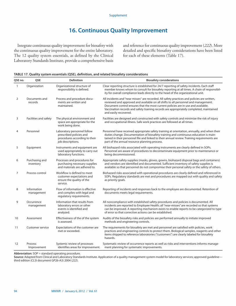
94 MMWR / January 6, 2012 / Vol. 61
Supplement
16. Continuous Quality Improvement
Integrate continuous quality improvement for biosafety with the continuous quality improvement for the entire laboratory. The 12 quality system essentials, as defined by the Clinical Laboratory Standards Institute, provide a comprehensive basis
TABLE 17. Quality system essentials (QSE), definition, and related biosafety considerations
QSE no. QSE Definition Biosafety considerations
1 Organization Organizationalstructureofresponsibility is defined.
Clear reporting structure is established for 24/7 reporting of safety incidents. Each staff memberknowswhomtoconsultforbiosafetyreportingatalltimes.Achainofresponsibil-ity for overall compliance leads directly to the head of the organizational unit.
2 Documents and records
Process and procedure docu-mentsarewrittenandmaintained.
Allincidentsand“nearmisses“arerecorded.Allsafetypracticesandpoliciesarewritten,reviewedandapprovedandavailableonallshiftstoallpersonnelandmanagement.Document control ensures that the most current policies are in use and available. Vaccination records and safety training records are appropriately completed, maintained and easily recovered.
3 Facilities and safety The physical environment and space are appropriate for the workbeingdone.
Facilitiesaredesignedandconstructedwithsafetycontrolsandminimizetheriskofinjuryandoccupationalillness.Safeworkpracticesarefollowedatalltimes.
4 Personnel Laboratorypersonnelfollowprescribed policies and procedures according to their jobdescriptions.
Personnelhavereceivedappropriatesafetytrainingatorientation,annually,andwhentheirduties change. Documentation of biosafety training and continuous education is main-tainedintheirpersonnelfileandlinkedtotheirannualreview.Trainingrequirementsarepart of the annual resource planning process.
5 Equipment Instrumentsandequipmentareused appropriately to carry out laboratory functions.
AllbiohazardrisksassociatedwithoperatinginstrumentsareclearlydefinedinSOPs.Personnelareawareofprocedurestodecontaminateequipmentpriortomaintenanceorbeing decommissioned.
6 Purchasing and inventory
Processes and procedures for purchasing necessary supplies and materials are adhered to.
Appropriatesafetysupplies(masks,gloves,gowns,biohazarddisposalbagsandcontainers)and vendors are identified and documented. Sufficient inventory of safety supplies is available so that personnel do not compromise their personal safety or the safety of others.
7 Process control Workflowisdefinedtomeetcustomer expectations and ensurethequalityoftheservice.
BiohazardrisksassociatedwithoperationalproceduresareclearlydefinedandreferencedinSOPs.Regulatorystandardsaremetandproceduresaremappedoutwithqualityandsafetyas priority goals.
8 Information management
Flowofinformationiseffectiveandcomplieswithlegalandregulatoryrequirements.
Reporting of incidents and responses back to the employee are documented. Retention of documentsmeetslegalrequirements.
9 Occurrencemanagement
Information that results from laboratory errors or other events is identified and analyzed.
Allnoncompliancewithestablishedsafetyproceduresandpoliciesisdocumented.AllincidentsarereportedtoEmployeeHealth;all“near-misses”arerecordedsothatsystemscan be improved. A reporting mechanism exists to enable reports to be categorized to type of error so that corrective actions can be established.
10 Assessment Effectiveness of the of the system is assessed.
Audits of the biosafety risks and policies are performed annually to initiate improved methods and engineering controls.
11 Customer service Expectations of the customer are met or exceeded.
Therequirementsforbiosafetyaremetandpersonnelaresatisfiedwithpolicies,workpractices and engineering controls to protect them. Biological samples, reagents and other itemsshippedtoreferencelaboratories(“customers”)areclearlylabeledforbiosafetyhazards.
12 Process Improvement
Systemicreviewofprocessesidentifies areas for improvement.
Systematicreviewofoccurrencereportsaswellasrisksandinterventionsinformsmanage-ment planning for systematic improvements.
Abbreviation:SOP=standardoperatingprocedure.Source: AdaptedfromClinicalandLaboratoryStandardsInstitute.Applicationofaqualitymanagementsystemmodelforlaboratoryservices;approvedguideline—thirdedition(CLSIdocumentGP26-A3)2004(222).
and reference for continuous quality improvement (222). More detailed and specific biosafety considerations have been listed for each of these elements (Table 17).
MMWR / January 6, 2012 / Vol. 61 95
Supplement
Acknowledgments
We acknowledge the assistance of Tanya Graham, DVM, South Dakota State University, Brookings, SD; Larry Thompson, DVM, PhD, Nestle Purina Pet Care, St. Louis, MO; R. Ross Graham, DVM, PhD, Merrick and Company; Corrine Fantz, PhD, Emory University, Atlanta, GA; Thomas Burgess, PhD, and Quest Diagnostics, Tucker, GA.
We appreciate the review and input into the document provided by the Office of Health and Safety, CDC; American Association of Veterinary Laboratory Diagnosticians; American Biological Safety Association; College of American Pathologists; American Society for Microbiology; Association of Public Health Laboratories; and subject matter experts at CDC.
References
1. CDC/National Institutes of Health. Biosafety in microbiological and biomedical laboratories. 5th ed. Available at http://www.cdc.gov/biosafety/publications/bmbl5/BMBL.pdf.
2. Clinical and Laboratory Standards Institute. Protection of laboratory workers from occupationally acquired infections; approved guideline—fourth edition (CLSI document M29-A4). Wayne, PA: Clinical and Laboratory Standards Institute; 2011. In press.
3. CDC. Laboratory-acquired vaccinia exposures and infections—United States, 2005–2007. MMWR 2008;57:401–4.
4. CDC. Laboratory-acquired brucellosis—Indiana and Minnesota, 2006. MMWR 2008;57:39–42.
5. CDC. Update: potential exposures to attenuated vaccine strain Brucella abortus RB51 during a laboratory proficiency test—United States and Canada, 2007. MMWR 2008;57:36–9.
6. CDC. Laboratory exposure to Burkholderia pseudomallei—Los Angeles, California, 2003. MMWR 2004;53:988–90.
7. CDC. Laboratory-acquired meningococcal disease—United States, 2000. MMWR 2002;51:141–4.
8. CDC. Suspected cutaneous anthrax in a laboratory worker—Texas, 2002. MMWR 2002;51:279–81.
9. CDC. Laboratory-acquired West Nile virus infections—United States, 2002. MMWR 2002;51:1133–5.
10. CDC. Laboratory-acquired human glanders—Maryland, May 2000. MMWR 2000;49:532–5.
11. CDC. Epidemiologic notes and reports. Laboratory-acquired meningococcemia—California and Massachusetts. MMWR 1991;40: 46–47,55.
12. Jacobson JT, Orlob RB, Clayton JL. Infections acquired in clinical laboratories in Utah. J Clin Microbiol 1985;21:486–9.
13. Harrington JM, Shannon HS. Incidence of tuberculosis, hepatitis, brucellosis, and shigellosis in British medical laboratory workers. Br Med J 1976;1:759–62.
14. Reid DD. Incidence of tuberculosis among workers in laboratories. Br Med J 1957;2:10–14.
15. Vesley D, Hartmann HM. Laboratory-acquired infections and injuries in clinical laboratories: a 1986 survey. Am J Public Health 1988;78: 1213–15.
16. Walker D, Campbell D. A survey of infections in United Kingdom laboratories, 1994–1995. J Clin Pathol 1999;52:415–8.
17. Sejvar JJ, Johnson D, Popovic T, et al. Assessing the risk of laboratory-acquired meningococcal disease. J Clin Microbiol 2005;43:4811–14.
18. Sewell DL. Laboratory-associated infections and biosafety. Clin Microbiol Rev 1995;8:389-405.
19. Pike RM. Laboratory-associated infections: incidence, fatalities, causes, and prevention. Annu Rev Microbiol 1979;33:41–66.
20. Herwaldt, BL. Laboratory-acquired parasitic infections from accidental exposures. Clin Microbiol Rev 2001;14:659–88.
21. Pragay DA, Howard SF, Gill ES. Clinical laboratory accidents, and some recommended remedies. Clin Chem 1980;26:1107–8.
22. Kyes K. Blood safety. Clinical Laboratory Products. June 2007. Available at http://www.clpmag.com/issues/articles/2007-06_02.asp.
23. Collins CH. Laboratory-acquired infections: history, incidence, causes, and prevention. 3rd ed. London: Butterworth-Heinemann, Ltd; 1993.
24. Pike RM. Laboratory-associated infections: incidence, fatalities, causes, and prevention. Annu Rev Microbiol 1979;33:41–66.
25. Pike RM. Laboratory-associated infections: summary and analysis of 3921 cases. Health Lab Sci 1976;13:105–14.
26. Pike RM. Past and present hazards of working with infectious agents. Arch Pathol Lab Med 1978;102:333–6.
27. Harding AL, Byers KB. Epidemiology of laboratory-associated infections. In: Fleming DO, Hunt DL, eds. Biological safety: principles and practices. 3rd ed. Washington, DC: ASM Press; 2000:35–54.
28. Vesley D, Hartmann HM. Laboratory-acquired infections and injuries in clinical laboratories: a 1986 survey. Am J Public Health. 1988;78:1213–15.
29. National Fire Protection Association. Section 5.1.3.3.2 (7). In: NFPA 99 Standard for health care facilities, 2005 Edition., Quincy, MA: National Fire Protection Association; 2005:37.
30. Zohar D. The effects of leadership dimensions, safety climate, and assigned priorities on minor injuries in work groups. Journal of Organizational Behavior 2002;23:75–92.
31. Zohar D, Luria G. A multilevel model of safety climate: Cross-level relationships between organization and group-level climates. J. Appl Psychol 2005;90:616–28.
32. Clinical and Laboratory Standards Institute. Laboratory design: approved guideline—second edition (CLSI document GP18-A2). Wayne, PA: Clinical and Laboratory Standards Institute; 2007.
33. Occupational Safety and Health Administration. Occupational safety and health standards. Z. Toxic and hazardous substances. Bloodborne pathogens. Standard no.1910.1030. Available at http://www.osha.gov/pls/oshaweb/owadisp.show_document?p_table=standards&p_ id=10051.
34. International Federation of Biosafety Associations. Biosafety compen-dium on regulations and information sources. Available at http://www.internationalbiosafety.org/english/internlCompendium.asp.
35. American Biological Safety Organization. Available at http://www. absa.org. 36. World Health Organization. Laboratory biosafety manual, 3rd edition.
Geneva: World Health Organization, 2004. Available at http://www.who.int/csr/resources/publications/biosafety/Biosafety7.pdf.
37. Occupational Safety and Health Administration. Occupational safety and health standards. I. Personal protective equipment. Standard no. 1910.132. Available at http://www.osha.gov/pls/oshaweb/owadisp.show_document?p_table=STANDARDS&p_id=9777.
38. Rutala W, Weber D. Use of inorganic hypochlorite (bleach) in health-care facilities. Clin Microbiol Rev 1997;597–610.
39. Clinical and Laboratory Standards Institute. Clinical laboratory waste management; approved guideline—third edition (CLSI document GP05-A3). Wayne, PA: Clinical and Laboratory Standards Institute; 2011.
40. CDC, US Department of Agriculture, Animal and Plant Health Inspection Service. National select agent registry. Available at http://www.selectagents.gov.
41. Miller JM. The Select Agent Rule and its impact on clinical laboratories. Clin Microbiol Newsletter 2006;28(8):57–63.
96 MMWR / January 6, 2012 / Vol. 61
Supplement
42. Johansen BV. Hazards related to EM-laboratory instrumentation. Ultrastruct Pathol 1984;7:219–25.
43. Bance GN, Barber VC, Sholdice JA. Safety in the SEM laboratory—1981 update. Scan Electron Microsc 1981;II:87–94.
44. Fleming DO, Richardson JH, Tulis JJ, Vesley D. Laboratory safety: principles and practices. 2nd ed. Washington DC: American Society for Microbiology; 1995.
45. Clinical and Laboratory Standards Institute. Implementing a needlestick and sharps injury prevention program in the clinical laboratory (CLSI document CLSI X03RE). Wayne, PA: Clinical and Laboratory Standards Institute; 2002.
46. CDC. Guidelines for preventing the transmission of Mycobacterium tuberculosis in health-care settings, 2005. MMWR 2005;54 (No. RR-17).
47. Collins CH. Laboratory acquired infections. Med Lab Sci 1980;37: 291–8.
48. Chatigny MA, Hatch MT, Wolochow H, et al. Studies on release and survival of biological substances used in recombinant DNA laboratory procedures. Recombinant DNA technical bulletin. Bethesda, MD: US Department of Health and Human Services, National Institutes of Health. 1979.
49. Wiedbrauk DL, Stoerker J. Quality assurance in the molecular virology laboratory. In: Wiedbrauk DL, Farkas DH, eds. Molecular methods for virus detection. San Diego, CA: Academic Press; 1995: 25–37.
50. Darlow HM. Safety in the microbiology laboratory: an introduction. In: Shapton DA, Board RG, eds. Safety in microbiology. London: Academic Press; 1972:1–19.
51. Schmid I, Lambert C, Ambrozak D, Marti GE, Moss DM, Perfetto SP. International Society for Analytical Cytology biosafety standard for sorting of unfixed cells. Cytometry Part A. 2007;6:414–37.
52. Kubica GP. Your tuberculosis laboratory: are you really safe from infection? Clinical Microbiology Newsletter 1990;12:85–7.
53. Murray PR, Baron EJ, Horgensen JH, Pfaller MA, Yolken RH. Manual of clinical microbiology, Eighth edition. Washington, DC: ASM Press; 2003.
54. Fleming DO, Hunt DL. Biological safety: principles and practices. Washington, DC: ASM Press; 2006.
55. Occupational Safety and Health Administration. Occupational safety and health standards. I. Personal protective equipment: Respiratory protection. Standard no. 1910.134. Section 4. Available at http://www.osha.gov/pls/oshaweb/owadisp.show_document?p_table=STANDARDS &p_id=12716.
56. Nolte K, Taylor D, Richmond J. Biosafety considerations for autopsy. Am J Forensic Med Pathol 2002;23(2):107–22.
57. Collins C, Grange J. Tuberculosis acquired in laboratories and necropsy rooms. Communicable Disease and Public Health 1999;2:161–7.
58. Burnett L, Lunn G, Coico R. Biosafety: guidelines for working with pathogenic and infectious microorganisms. In: Curr Protoc Microbiol.[serial online] May 2009; DOI: 10.1002/9780471729259.mc01a01s13.
59. Nolte KB, Foucar K, Richmond JY. Hanta viral biosafety issues in the autopsy room and laboratory: concerns and recommendations. Hum Pathol 1996;27:1253–4.
60. De Carli G, Puro V, Scognamiglio P, Ippolito G. Infection with hepatitis C virus transmitted by accidental needlesticks. Clin Infect Dis 2003;37:1718–9.
61. Nyberg M, Suni J, Haltia M. Isolation of human immunodeficiency virus (HIV) at autopsy one to six days postmortem. Am J Clin Pathol 1990;94:422–5.
62. Petrosillo N, Puro V, De Carli G, Ippolito G, SIROH Group. Risks faced by laboratory workers in the AIDs era. J Biol Regul Homeost Agents 2001;15:243–8.
63. CDC. Updated US Public Health Service guidelines for the management of occupational exposures to HBV, HCV, and HIV and recommendations for postexposure prophylaxis. MMWR 2001;50 (No. RR-11).
64. Brown P, Gibbs CJ, Gajdusek D, Cathala F, LaBauge R. Transmission of Creutzfeldt-Jakob disease from formalin-fixed, paraffin-embedded human brain tissue. N Engl J Med 1986;315:1614–5.
65. Sitwell L LB, Attack E, Izukawa, D. Creutzfeldt-Jakob disease in histopathology technicians N Engl J Med 1988;318:854.
66. Wilkins D WA, Cossar YE, Miller DC. Creutzfeldt-Jakob disease in histopathology technicians. N Engl J Med 1988;318:853–4.
67. Miller D. Creutzfeldt-Jakob disease in histopathology technicians. N Engl J Med 1988;318:853–4.
68. Canadian Food Inspection Agency. Biosafety guidelines developed for National TSE Veterinary Diagnostic Laboratory Network. Version 1.0, March 8, 2002.
69. Newsom S, Rowlands C, Mathews J, Elliot CJ. Aerosols in the mortuary. J Clin Pathol 1983;36:127–32.
70. Occupational Safety and Health Administration. Occupational safety and health standards. Z. Toxic and hazardous substances. 1910.1048, Formaldehyde. Available at http://www.osha.gov/pls/oshaweb/owadisp.show_document?p_table=STANDARDS&p_id=10075.
71. Code of Federal Regulations. Title 40, Protection of Environment. Available at http://ecfr.gpoaccess.gov/cgi/t/text/text-idx?c=ecfr; sid=4990e762d7b81851bef18f82dc851826;rgn0=div5;view=text; node=40%3A25.0.1.1.2;idno=40;cc=ecfr#40:25.0.1.1.2.3.1.5.
72. Occupational Safety and Health Administration. Occupational safety and health standards. I. Personal protective equipment. 1910.133, Eye and face protection. Available at http://www.osha.gov/pls/oshaweb/owadisp.show_document?p_table=STANDARDS&p_id=9778.
73. National Tuberculosis Advisory Committee. Guidelines for Australian Mycobacteriology Laboratories. Communicable Diseases Intelligence 2006;30:116–28.
74. Hedwell E. Incidence of tuberculosis among medical students at Lund University. American Review of Tuberculosis 1940;41:770–80.
75. Weston J, Locker G. Frequency of glove puncture in the postmortem room. J Clin Pathol 1992;45:177–8.
76. Templeton GL, Illing LA, Young L, Cave D, Stead WW, Bates JH. The risk for transmission of Mycobacterium tuberculosis at the bedside and during autopsy. Ann Intern Med 1995;122:922–5.
77. Meyer J. TB Plagues Office of the LA Coroner. Los Angeles Times April 25, 1997;A1–27. Available at http://articles.latimes.com/1997-04-25/news/mn-52228_1_coroner-s-office.
78. Kappel T, Reinarts J, Schmid J, Holter J, Azar M. The viability of Mycobacterium tuberculosis in formalin-fixed pulmonary autopsy tissue: review of the literature and brief report. Hum Pathol 1996; 27:1361–4.
79. Ford A. Taking stock of biosafety perils in surg path. CAP Today 2009; 23:1.
80. Gärtner H-V, Seidl C, Luckenbach C, et al. Genetic analysis of a sarcoma accidentally transplanted from a patient to a surgeon. N Engl J Med 1996;335:1494–7.
81. Goette D, Jacobson, KW, Doty RD. Primary inoculation tuberculosis of the skin; prosector’s paronychia. Arch Dermatol 1978;114:567–9.
82. Vetter J. Microbial aerosols from a freezing microtome. American Society for Clinical Microbiology Summary Report; 1977.
83. CDC. Tuberculosis infection associated with tissue processing—California. MMWR 1981;30(6):73–4.
MMWR / January 6, 2012 / Vol. 61 97
Supplement
84. Smith J. The hazard to pathologists from tuberculosis specimens. Hum Pathol 1996;27:1251–2.
85. Gerston K, Blumberg L, Tshabalala V, Murray J. Viability of mycobacteria in formalin-fixed lungs. Hum Pathol 2004;35:571–5.
86. Richard M, Biacabe A, Perret-Liaudet A, McCardle L, Ironside J, Kopp N. Protection of personnel and environment against Creutzfeldt-Jakob disease in pathology laboratories. Clin Expl Pathol 1999;47: 192–200.
87. Clinical and Laboratory Standards Institute. Procedures for the recovery and identification of parasites from the intestinal tract; approved guideline—second edition (CLSI document M28-A2). Wayne, PA: Clinical and Laboratory Standards Institute; 2005.
88. Wheatley W. A rapid staining procedure for intestinal amoebae and flagellates. Am J Clin Pathol 1951;21:990–1.
89. Garcia LS. Diagnostic medical parasitology, 5th ed. Washington, DC: ASM Press; 2007.
90. International Air Transport Association. 2005[a]. Dangerous Goods Regulations, 46th edition. Montreal, Canada: International Air Transport Association; 2005.
91. International Air Transport Association. 2005[b]. Dangerous Goods Regulations, 46th ed. Addendum II. March 22. Montreal, Canada: International Air Transport Association; 2005.
92. International Air Transport Association. 2005[c]. Dangerous Goods Regulations, 46th ed. Addendum III. July 5. Montreal, Canada: International Air Transport Association; 2005.
93. US Department of Transportation, Pipeline and Hazardous Materials Safety Administration. Hazardous materials: infectious substances; harmonization with the United Nations recommendations; final rule (49 CFR Parts 171 et al.). Federal Register 2006;71:32244–63. Available at https://hazmatonline.phmsa.dot.gov/services/publication_documents/Transporting%20Infectious%20Substances%20Safely.pdf.
94. US Department of Transportation, Research and Special Programs Administration. Harmonization with the United Nations Recommendations, International Maritime Dangerous Goods Code, and International Civil Aviation Organization’s Technical Instructions; final rule (CFR 42, Parts 171, 172, et al.). Federal Register 2004;69:76044–76187 (http://www.labsafety.com/refinfo/fedreg/FRPDF/122004.pdf).
95. US Department of Transportation, Research and Special Programs Administration. Hazardous materials: revision to standards for infectious substances and genetically modified organisms; final rule (CFR 42, Parts 171 et al.). Federal Register 2002;67:53118–53144.
96. US Postal Service. Domestic mail manual. Hazardous materials (601.10.1). 2011. Available at http://pe.usps.com/cpim/ftp/manuals/dmm300/full/mailingStandards.pdf.
97. World Health Organization. Transport of infectious substances. Background to the 17 amendments adopted in the 13th revision of the United Nations Model Regulations guiding the transport of infectious substances. Geneva: World Health Organization; 2004. Available at http://www.who.int/csr/resources/publications/WHO_CDS_CSR_LYO_2004_9/en.
98. World Health Organization. 2005. Guidance on Regulations for the Transport of Infectious Substances 2009–2010. Geneva: World Health Organization; 2009. Available at http://www.who.int/csr/resources/publications/biosafety/WHO_HSE_EPR_2008_10/en/index.html.
99. Garcia LS, Shimizu RY, Shum A, Bruckner DA. Evaluation of intestinal protozoan morphology in polyvinyl alcohol preservative: comparison of zinc sulfate and mercuric chloride based compounds for use in Schaudinn’s fixative. J Clin Microbiol 1993;31:307–10.
100. Jungbauer FH, van der Harst JJ, Groothoff JW, Coenraads PJ. Skin protection in nursing work: promoting the use of gloves and hand alcohol. Contact Dermatitis 2004;51:135–40.
101. Kampf G, Kramer A. Epidemiologic background of hand hygiene and evaluation of the most important agents for scrubs and rubs. Clin Microbiol Rev 2004;17:863–93.
102. CDC. A comprehensive immunization strategy to eliminate transmission of hepatitis B virus infection in the United States. Recommendations of the Advisory Committee on Immunization Practices. Part II: Immunization of adults. MMWR 2006;55 (No. RR-16).
103. Calcofluor white reagent dropper (material safety data sheet). Franklin Lakes, NJ: Becton Dickinson; 2011. Available at http://www.bdregdocs.com/msds/view/msds-usa-261195.pdf.
104. Potassium hydroxide 10% w/v (material safety data sheet). Austin, TX: Science Stuff, Inc.; 2006. Available at http://www.sciencestuff.com/msds/C2360.html.
105. Calcobrite fungi staining kit (material safety data sheet). Guildford, UK: Clin-Tech, LTD; 2009. Available at http://www.clin-tech.co.uk/msds/63115X.pdf.
106. Potassium hydroxide (10–45%) solutions and concentrates (material safety data sheet). Phillipsburg, NJ: Mallinckrodt Baker, Inc.; 2008. Available at http://www.jtbaker.com/msds/englishhtml/P5887.htm.
107. Lactophenol cotton blue stain droppers (material safety data sheet). Franklin Lakes, NJ: Becton Dickinson; 2003. Available at http://www.bd.com/ds/productCenter/261188.asp.
108. BactiDrop lactophenol aniline blue (material safety data sheet). Hants, UK: Oxoid; 2007. Available at http://www.oxoid.com/pdf/msds/EN/R21526.pdf.
109. Lactophenol–cotton blue mounting fluid (material safety data sheet). Gibbstown, NJ: EMD Chemicals; 2010. Available at http://www.emdchemicals.com/lactophenol/EMD_CHEM-R03465/p_uuid? attachments=MSDS.
110. Higgins waterproof black drawing India ink (material safety data sheet). Bellwood, IL: Sanford Corporation; 2001. Available at http://web.grcc.cc.mi.us/Pr/msds/visualarts-photography/HIGGINS%20Waterproof %20Black%20Drawing%20Indian%20Ink.pdf.
111. Methenamine silver stain (package insert). Carpinteria, CA: Dako North America; 2008. Available at http://www.dako.com/us/ searchresultlist?search=methenamine+silver+stain.
112. Giemsa stain (material safety data sheet). Torrance, CA: Medical Chemical Corp.; 2006. Available at http://www.med-chem.com/msds/ 591A.pdf.
113. Larone DH. Medically important fungi, a guide to identification, 4th ed., Washington, DC: ASM Press; 2002.
114. Stevens DA, Clemons KV, Levine HB, et al. Expert opinion: what to do when there is Coccidioides exposure in a laboratory. Clin Infect Dis 2009; 49:919–23.
115. Indoor allergens: assessing and controlling adverse health effects. In: Pope AM, Patterson R, eds. Washington, DC: National Academy Press; 1993.
116, Rudzki E, Rebandel P, Rebandel B. Occupational allergy to antibiotics. Med Pr 1986;37:383–7.
117. Coutts II, Dally MB, Taylor AJ, Pickering CA, Horsfield N. Asthma in workers manufacturing cephalosporins. Br Med J (Clin Res Ed) 1981;283:950.
118. Davies RJ, Hendrick DJ, Pepys J. Asthma due to inhaled chemical agents: ampicillin, benzyl penicillin, 6 amino penicillanic acid and related substances. Clin Allergy 1974;4:227-47.
119. Malo J-L, Cartier A. Occupational asthma in workers of a pharmaceutical company processing spiramycin. Thorax 1988;45:371–7.
98 MMWR / January 6, 2012 / Vol. 61
Supplement
120. Paggiaro PL, Loi AM, Toma A. Bronchial asthma and dermatitis due to spiramycin in a chick breeder. Clin Allergy 1979;9:571–4.
121. Bance GN, Barber VC, Sholdice JA. Safety in the SEM laboratory—1981 update. Scan Electron Microsc 1981;II:87–94.
122. Drury P. Safety in electron microscopy and protective glove hazards. Can J Med Technol 1980;42:80–2.
123. Pegum JS, Medhurst FA. Dermatitis from penetration of rubber gloves by acrylic monomer. Br Med J 1971;276:141–3.
124.ToblerM,WüthrichB,FreiburghausAU.Contactdermatitis fromacrylate and methacrylate compounds in Lowicryl embedding media for electron microscopy. Contact Dermatitis 1990;23:96–102.
125. Lunn G, Sansone EB. Ethidium bromide: destruction and decontami-nation of solutions. Anal Biochem 1987;162:453–8. PMID 3605608.
126. Gärtner HV, Seidl C, Luckenbach C, et al. Genetic analysis of a sarcoma accidentally transplanted from a patient to a surgeon. N Engl J Med 1996;335:1494–6.
127. Nadler SH, Moore GE. Immunotherapy of malignant disease. Arch Surg 1969;99:376–81.
128. Scanlon EF, Hawkins RA, Fox WW, Smith WS. Fatal homotransplanted melanoma: a case report. Cancer 1965;18:782–89.
129. Southam CM. Homotransplantation of human cell lines. Bull NY Acad Med 1958;34:416–23.
130. Blohme I, Nyberg G, Jeansson S, Svalander C. Adenovirus infection in a renal transplant patient. Transplant Proc 1992;24:295.
131. Myerowitz RL, Stalder H, Oxman MN, et al. Fatal disseminated adenovirus infection in a renal transplant recipient. Am J Med 1975;59:591–8.
132. Asim M, Chong-Lopez A, Nickeleit V. Adenovirus infection of a renal allograft. Am J Kidney Dis 2003;41:696–701.
133. Hsuing GD. Latent virus infections in primate tissues with special reference to simian viruses. Bacteriol Rev 1968;32:185–205.
134. Swack NS, Hsuing GD. Endogenous agents in primary cell cultures with special reference to latent viruses. In Vitro 1974;10:260–7.
135. Hopps HE. Origin of endogenous and exogenous agents in cell cultures. In Vitro 1974;10:243–6.
136. Patel R, Paya CV. Infections in solid-organ transplant recipients. Microbiol Rev 1997;10:86–124.
137. van Gelder T, Kroes LC, Mulder A, Gratama JW, Weimar W. A living-related kidney donor as the source of a nearly fatal primary Epstein-Barr virus infection following transplantation. Transplantation 1994;58:852–4.
138. Davis C, Gretch DR, Carithers RL. Hepatitis B and transplantation. Infect Dis Clin North Am 1995;9:925–41.
139. Dummer JS, Armstrong J, Somers J, et al. Transmission of infection with herpes simplex virus by renal transplantation. J Infect Dis 1987;155:202–6.
140. Goodman JL. Possible transmission of herpes simplex virus by organ transplantation. Transplantation 1989;47:609–13.
141. Schwarz A, Hoffmann F, L’age-Stehr, J, Tegzess AM, Offermann G. Human immunodeficiency virus transmission by organ donation. Outcome in cornea and kidney recipients. Transplantation 1987; 1:21–4.
142. Sotir M, Switzer W, Schable C, Schmitt J, Vitek C, Khabbaz RF. Risk of occupational exposure to potentially infectious nonhuman primate materials and to simian immunodeficiency virus. J Med Primatol 1997;26:233–40.
143. Khabbaz RF, Rowe T, Murphey-Corb M, et al. Simian immunodeficiency virus needlestick accident in a laboratory worker. Lancet 1992; 340:271–3.
144. Gonzalez-Perez MP, Munoz-Juarez L, Cardenas FC, Zarranz Imirizaldu JJ, Carranceja JC, Garcia-Saiz A. Human T-cell leukemia virus type I infection in various recipients of transplants from the same donor. Transplantation 2009;75:1006–11.
145. Nakatsuji Y, Sugai YF, Watanabe S, et al. HTLV-I-associated myelopathy manifested after renal transplantation. J Neurol Sci 2000;177: 154–6.
146. Remesar MC, del Pozo AE, Pittis MG, Mangano AM, Sen L, Briones L. Transmission of HTLV-I by kidney transplant. Transfusion 2000;40:1421–2.
147. Toro C, Rodes B, Poveda E, Soriano V. Rapid development of subacute myelopathy in three organ transplant recipients after transmission of human T-cell lymphotropic virus type I from a single donor. Transplantation 2003;75:102–4.
148. CDC. Lymphocytic choriomeningitis virus infection in organ transplant recipients—Massachusetts, Rhode Island, 2005. MMWR 2005;54:537–9.
149. Dykewicz CA, Dato VM, Fisher-Hoch SP, Ostroff SM, Gary H Jr, McCormick JB. Lymphocytic choriomeningitis outbreak associated with nude mice in a research institute. JAMA 2010;267:1349–53.
150. Mahy BW, Dykewicz C, Fisher-Hoch S, Ostroff S, Sanchez A. Virus zoonoses and their potential for contamination of cell cultures. Dev Biol Stand 1991;75:183–9.
151. Schmitt M, Pawlita M. High-throughput detection and multiplex identification of cell contaminations. Nucleic Acids Res Advance Access 2009;37(18):e119. Available at http://nar.oxfordjournals.org/content /37/18/e119.full.
152. Emery JB, York CJ. Occurrence of a hemadsorption virus in normal monkey tissue culture. Virology 1960;11:313–5.
153. Mengling WL. Porcine parvovirus: frequency of naturally occurring transplacental infection and viral contamination of fetal porcine kidney cell cultures. Am J Vet Res 1975;36:41–4.
154. Srinivasan A, Burton EC, Kuehnert MJ, et al. Transmission of rabies virus from an organ donor to four transplant recipients. N Engl J Med 2005;352:1103–11.
155. Robertson I. Corneal transplants and rabies. Med J Aust 1979;2:697. 156. Martín-Dávila P, Fortún J, López-Vélez R, et al. Transmission of tropical
and geographically restricted infections during solid-organ transplantation. Clin Microbiol Rev 2008;21:60–96.
157. Javadi MA, Fayaz A, Mirdehghan SA, Ainollahi B. Transmission of rabies by corneal graft. Cornea 2009;15:431–3.
158. Houff SA, Burton RC, Wilson RW, et al. Human-to-human transmission of rabies virus by corneal transplant. N Engl J Med 1979; 300: 603–4.
159. Hellenbrand W, Meyer C, Rasch G, Steffens T, Amon A. Cases of rabies in Germany following organ transplantation. Eur Surveill 2005; 10:E050224.
160. Gode GR, Bhide NK. Two rabies deaths after corneal grafts from one donor. Lancet 1988;2:791.
161. CDC. Human-to-human transmission of rabies via corneal transplant —Thailand. MMWR. 1981;30:473–4.
162. Baer GM, Shaddock JH, Houff SA, Harrison AK, Gardner JJ. Human rabies transmitted by corneal transplant. Arch Neurol 2009;39: 103–7.
163. Patient received cornea: rabies case linked to transplant. American Medical News 1978;21:3.
164. Hull RN, Minner JR, Smith JW. New viral agents recovered from tissue cultures of monkey kidney cells. I. Origin and properties of cytopathic agents SV1, SV2, SV4, SV5, SV6, SV11, SV12, and SV15. Am J Hyg 1956;63:204–15.
MMWR / January 6, 2012 / Vol. 61 99
Supplement
165. Rustigian R, Johnston T, Reihart H. Infection of monkey kidney tissue cultures with virus-like agents. Proc Soc Exp Biol Med 1955; 88:8–16.
166. Peden K, Sheng L, Yacobucci M, et al. Recovery of strains of the polyomavirus SV40 from rhesus monkey kidney cells dating from the 1950s to the early 1960s. Virology 2008;370:63–76.
167. Sweet BH, Hilleman MR. The vacuolating virus, SV40. Proc Soc Exp Biol Med 1960;105:420–7.
168. Kekarainen T, Martínez-Guinó L, Segalés J. Swine torque teno virus detection in pig commercial vaccines, enzymes for laboratory use and human drugs containing components of porcine origin. J Gen Virol 2000;90:648–53.
169. Middleton PG, Miller S, Ross JA, Steel CM, Guy K. Insertion of SMRV-H viral DNA at the c-myc gene locus of a BL cell line and presence in established cell lines. Int J Cancer 1992;54:451–4.
170. Sun R., Grogan E, Shedd D, et al. Transmissible retrovirus in Epstein-Barr virus-producer B95-8 cells. Virology 1995;209:374–83.
171. Antony S. Severe meningo-encephalitis and death in a renal transplant recipient resulting from West Nile virus infection. J Natl Med Assoc 2004;96:1646–7.
172. Armali Z, Ramadan R, Chlebowski A, Azzam ZS. West Nile meningo-encephalitis infection in a kidney transplant recipient. Transplant Proc 2003;35:2935–6.
173. Bragin-Sanchez D, Chang PP. West Nile virus encephalitis infection in a heart transplant recipient: a case report. J Heart Lung Transplant 2005;24:621–3.
174. CDC. West Nile virus activity–United States, September 26–October 2, 2002, and investigations of West Nile virus infections in recipients of blood transfusion and organ transplantation. JAMA 2002;288: 1975–6.
175. CDC. West Nile virus infection in organ donor and transplant recipients–Georgia and Florida, 2002. JAMA 2002;288:1465–6.
176. CDC. Update: West Nile virus screening of blood donations and transfusion-associated transmission—United States. MMWR 2004; 53:281–4.
177. CDC. Transfusion-associated transmission of West Nile virus—Arizona, 2004. MMWR. 2004;53:842–4.
178. CDC. West Nile virus infections in organ transplant recipients—New York and Pennsylvania, August–September, 2005. MMWR 2005;54: 1021–3.
179. DeSalvo D, Roy-Chaudhury P, Peddi R, et al. West Nile virus encephalitis in organ transplant recipients: another high-risk group for meningoencephalitis and death. Transplantation 2004;77.
180. Hardinger KL, Miller B, Storch GA, Desai NM, Brennan DC. West Nile virus-associated meningoencephalitis in two chronically immunosuppressed renal transplant recipients. Am J Transplant 2003;3:1312–5.
181. Harrington T, Kuehnert MJ, Kamel H, et al. West Nile virus infection transmitted by blood transfusion. Transfusion 2003;43:1018–22.
182. Iwamoto M, Jernigan DB, Guasch A, et al. Transmission of West Nile virus from an organ donor to four transplant recipients. N Engl J Med 2003;348:2196–203.
183. Kleinschmidt-DeMasters BK, Marder BA, Levi ME, et al. Naturally acquired West Nile virus encephalomyelitis in transplant recipients: clinical, laboratory, diagnostic, and neuropathological features. Arch Neurol 2004;61:1210–20.
184. Kumar D, Prasad GVR, Zaltzman J, Levy GA, Humar A. Community-acquired West Nile virus infection in solid-organ transplant recipients. Transplantation 2004;77.
185. Kusne S, Smilack J. Transmission of West Nile virus by organ transplantation. Liver Transplant 2005;11:239–41.
186. Montgomery SP, Brown JA, Kuehnert MJ, et al. Transfusion-associated transmission of West Nile virus. United States 2003 through 2005. Transfusion 2006;46:2038–46.
187. Murtagh B, Wadia Y, Messner G, Allison P, Harati Y, Delgado R. West Nile virus infection after cardiac transplantation. J Heart Lung Transplant 2005;24:774–6.
188. Pealer LN, Marfin AA, Petersen LR, et al. Transmission of West Nile virus through blood transfusion in the United States in 2002. N Engl J Med 2003;349:1236–45.
189. Ravindra K, Freifeld A, Kalil A, et al. West Nile virus associated encephalitis in recipients of renal and pancreas transplants: case series and literature review. Clin Infect Dis 2004;38:1257–60.
190. Shephert JC, Subramanian A, Montgomery RA, et al. West Nile virus encephalitis in a kidney transplant recipient. Am J Transplant 2004; 4:830–3.
191. Trijzelaar B. Regulatory affairs and biotechnology in Europe: III. Introduction into good regulatory practice—validation of virus removal and inactivation. Biotherapy 1993;62:93–102.
192. Hazelton PR, Gelderblom HR. Electron microscopy for rapid diagnosis of infectious agents in emergent situations. Emerg Infect Dis 2003;9:294–303.
193. Johansen BV. Hazards related to EM-laboratory instrumentation. Ultrastruct Pathol 1984;7:219–25.
194. CDC. Interim biosafety guidance for all individuals handling clinical specimens or isolates containing 2009-H1N1 influenza A virus (Novel H1N1), including vaccine strains. August 15, 2009. Available at: http://cdc.gov/h1n1flu/guidelines_labworkers.htm. 2-16-2010.
195. Clinical and Laboratory Standards Institute. Performance of the bleeding time test; approved guideline—fourth edition (CLSI document H45-A2).Wayne, PA: Clinical and Laboratory Standards Institute; 2005.
196. CDC. Workbook for designing, implementing, and evaluating a sharps injury prevention program. Atlanta, GA: US Department of Health and Human Services, CDC. Available at http://www.cdc.gov/sharps safety/pdf/sharpsworkbook_2008.pdf.
197. Clinical and Laboratory Standards Institute. Clinical flow cytometric analysis of neoplastic hematolymphoid cells; approved guideline—second edition (CLSI document H43-A2). Wayne, PA: Clinical and Laboratory Standards Institute; 2007.
198. Clinical and Laboratory Standards Institute. Collection, transport, and processing of blood Specimens for testing plasma-based coagulation assays and molecular hemostasis assays; approved guideline—fifth edition (CLSI document H21-A5). Wayne, PA: Clinical and Laboratory Standards Institute; 2008.
199. Clinical and Laboratory Standards Institute. Nucleic acid amplification assays for molecular hematopathology; approved guideline (CLSI document MM05-A). Wayne, PA: Clinical and Laboratory Standards Institute; 2003.
200. Clinical and Laboratory Standards Institute. Collection, Transport, Preparation, and storage of specimens for molecular methods; approved guideline (CLSI document MM13-A). Wayne, PA: Clinical and Laboratory Standards Institute; 2005.
201. Clinical and Laboratory Standards Institute. Procedures for the collection of diagnostic blood specimens by venipuncture; approved standard—sixth edition (CLSI document H03-A6). Wayne, PA: Clinical and Laboratory Standards Institute; 2007.
100 MMWR / January 6, 2012 / Vol. 61
Supplement
202. Clinical and Laboratory Standards Institute. Procedures and devices for the collection of diagnostic capillary blood specimens; approved standard—sixth edition (CLSI document H04-A6). Wayne, PA: Clinical and Laboratory Standards Institute; 2008.
203. Clinical and Laboratory Standards Institute. Procedures for the handling and processing of blood specimens for common laboratory tests; approved guideline—fourth edition (CLSI document H18-A4). Wayne, PA: Clinical and Laboratory Standards Institute; 2010.
204 CDC. Selecting, evaluating, and using sharps disposal containers. Cincinnati, OH: US Department of Health and Human Services, CDC, National Institute for Occupational Safety and Health; 1998. Available at http://www.cdc.gov/niosh/pdfs/97-111.pdf.
205. US Environmental Protection Agency. Selected EPA-registered disinfectants. Washington DC: US Environmental Protection Agency; 2009. Available at http://www.epa.gov/oppad001/chemregindex.htm.
206. American Veterinary Medical Association. One health: a new professional imperative. Schaumburg, IL: American Veterinary Medical Association; 2008. Available at http://www.avma.org/onehealth.
207. Torrey EF, Yolken RH. Beasts of the earth. New Brunswick, NJ: Rutgers University Press; 2005.
208. Taylor LH, Latham SM, Woolhouse ME. Risk factors for human disease emergence. Philos Trans R Soc Lond B Biol Sci 2001;356:983–9.
209. Biosafety and biosecurity in the veterinary microbiology laboratory and animal facilities. In: OIE manual of diagnostic tests and vaccines for terrestrial animals. Paris, France: World Organization of Animal Health (OIE), Office International Des Epizooties; 2008.
210. The National Research Council, Committee on Hazardous Biological Substances in the Laboratory. Biosafety in the laboratory. Prudent practices for the handling and disposal of infectious materials. Washington, DC: National Academy Press; 1989. Available at http://www.nap.edu/openbook.php?isbn=0309039754.
211. Immunization Action Coalition. Healthcare personnel vaccination recommendations. St. Paul, MN: Immunization Action Coalition; 2011. Available at http://www.immunize.org/catg.d/p2017.pdf.
212. CDC. Human rabies prevention—United States, 2008: Recommen-dations of the Advisory Committee on Immunization Practices. MMWR 2008;57(No. RR-3).
213. American Society for Microbiology. Sentinel level clinical microbiology laboratory guidelines. Washington, DC: American Society for Microbiology; 2010. Available at http://www.asm.org/?option=com_content&view=article&id=6342&Itemid=639.
214. Gray LD, Snyder JW. Sentinel laboratory guidelines for suspected agents of bioterrorism and emerging infectious diseases. Packing and shipping infectious substances. Washington, DC: American Society for Microbiology; 2010. Available at http://www.asm.org/images/pdf/Clinical/ps11-15-10final.pdf.
215. Gray LD, Snyder JW. Packing and shipping biological materials. In: Fleming DO, Hunt DL, eds. Biological safety: principles and practices, 4th edition. Washington, DC: ASM Press; 2006.
216. International Air Transport Association. Dangerous goods regulations, 52nd. edition. Montreal, Canada: International Air Transport Association; 2011.
217. CDC. Prevention and control of meningococcal disease. Recommendations of the Advisory Committee on Immunization Practices. MMWR 2005;54(No. RR-7).
218. CDC. Laboratory-acquired meningococcal disease—United States, 2000. MMWR 2000;51:141–4.
219. CDC. Guidelines for preventing the transmission of Mycobacterium tuberculosis in health-care settings, 2005. MMWR 2005;54 (No. RR-17).
220. CDC. Updated US Public Health Service guidelines for the management of occupational exposure to HIV and recommendations for postexposure prophylaxis. MMWR 2005;54(No, RR-9).
221. CDC. Updated US Public Health Service guidelines for the management of occupational exposure to HBV, HCV, and HIV and recommendations for postexposure prophylaxis. MMWR 2001;50(No. RR-11).
222. Clinical and Laboratory Standards Institute. Application of a quality management system model for laboratory services; approved guideline—third edition (CLSI document GP26-A3). Wayne, PA: Clinical and Laboratory Standards Institute; 2004.
MMWR / January 6, 2012 / Vol. 61 101
Supplement
Blue Ribbon Panel for Issues of Clinical Laboratory SafetyKathleen G. Beavis, MD, College of American Pathologists, Chicago, Illinois; Ellen Jo Baron, PhD, Stanford, California; William R. Dunn, MS, Greater New York Hospital Association Regional Laboratory Task Force, New York, New York; Larry Gray, PhD, American Society for Microbiology, Cincinnati, Ohio; Bill Homovec, MPH, American Clinical Laboratory Association, Burlington, North Carolina; Michael Pentella, PhD, Association of Public Health Laboratories, Iowa City, Iowa; Bruce Ribner, MD, Atlanta, Georgia; William A. Rutala, PhD, Chapel Hill, North Carolina; Daniel S. Shapiro, MD, Burlington, Massachusetts; Lisa A. Skodack-Jones, MT, Salt Lake City, Utah; Christine Snyder, American Society for Clinical Laboratory Science, Helena, Montana; Robert L. Sunheimer, MS, American Society for Clinical Pathology, Syracuse, New York; Christina Z. Thompson, MS, American Biological Safety Association, Greenfield, Indiana.CDC Staff: Nancy L. Anderson, MMSc; Rex Astles, PhD; D. Joe Boone, PhD; David S. Bressler, MS; Roberta Carey, PhD; Casey Chosewood, MD; Mitchell L. Cohen, MD; Judy Delaney, MS; Thomas L. Hearn, DrPH; Kathleen F. Keyes, MS; Davis Lupo, PhD; Robert Martin, DrPH; Alison C. Mawle, PhD; Terra McConnel; J. Michael Miller, PhD; Shana Nesby, DVM; Janet K. Nicholson, PhD; John P. O’Connor, MS; Anne Pollock; John C. Ridderhof, DrPH; Pamela Robinson; Elizabeth G. Weirich, MS; Ae S. Youngpairoj.
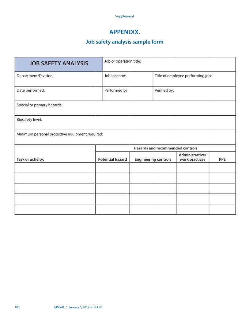
102 MMWR / January 6, 2012 / Vol. 61
Supplement
APPENDIX. Job safety analysis sample form
JOB SAFETY ANALYSIS Job or operation title:
Department/Division: Job location: Titleofemployeeperformingjob:
Date performed: Performed by Verified by:
Special or primary hazards:
Biosafety level:
Minimumpersonalprotectiveequipmentrequired:
Task or activity:
Hazards and recommended controls
Potential hazard Engineering controlsAdministrative/work practices PPE
U.S. Government Printing Office: 2012-523-043 Region IV ISSN: 1546-0738
The Morbidity and Mortality Weekly Report (MMWR) Series is prepared by the Centers for Disease Control and Prevention (CDC) and is available free of charge in electronic format. To receive an electronic copy each week, visit MMWR’s free subscription page at http://www.cdc.gov/mmwr/mmwrsubscribe.html. Paper copy subscriptions are available through the Superintendent of Documents, U.S. Government Printing Office, Washington, DC 20402; telephone 202-512-1800.
Address all inquiries about the MMWR Series, including material to be considered for publication, to Editor, MMWR Series, Mailstop E-90, CDC, 1600 Clifton Rd., N.E., Atlanta, GA 30333 or to [email protected].
All material in the MMWR Series is in the public domain and may be used and reprinted without permission; citation as to source, however, is appreciated.
Use of trade names and commercial sources is for identification only and does not imply endorsement by the U.S. Department of Health and Human Services.
References to non-CDC sites on the Internet are provided as a service to MMWR readers and do not constitute or imply endorsement of these organiza-tions or their programs by CDC or the U.S. Department of Health and Human Services. CDC is not responsible for the content of these sites. URL addresses listed in MMWR were current as of the date of publication.
Supplement