gram staining - syracuse high school · •fix the dried film by passing it briefly through the...
TRANSCRIPT

Gram Staining

Gram Staining
• Gram +
• Gram -
Cell Wall

Gram-Positive Bacteria• Take up the crystal violet
stain used in the test, and
then appear to be purple-
colored when seen
through a microscope.
• This is because the thick
peptidoglycan layer in the
bacterial cell wall retains
the stain after it is washed
away from the rest of the
sample, in the
decolorization stage of
the test.

Gram-Negative Bacteria• Does not retain violet stain
after decolorization.
– Their peptidoglycan layer
is located under a layer of
fat.
– The alcohol used in
decolorization degrades
the outer lipid layer making
the cell wall incapable of
retaining the crystal violet.
• This causes bacteria to take
up the counterstain (safranin)
and appear red or pink.

Gram Staining

Gram Staining

Process:
Can be done with either:• A Liquid Medium or• A Solid Medium

Removing Inoculum from
a Liquid Medium
• Flame the lip of the culture tube. This creates a convection current which forces air out of the tube and prevents airborne contaminants from entering the tube.
• Keeping the culture tube at an angle, insert the inoculating loop and remove a loopful of inoculum.
• Again flame the lip of the culture tube.
• Replace the cap.

Transfer a sample of the
medium to the slide.
• Allow the medium to air dry.

Removing Inoculum from a Solid
medium• Place several loopfuls of tap water on a
slide.
• Sterilize the inoculating loop.
• Allow it to cool before moving on.
• Scrape off a small amount of the organisms
and close the lid.
• Transfer a small sample of the colony to the drop, and emulsify (mix with inoculating loop).
1
3
2
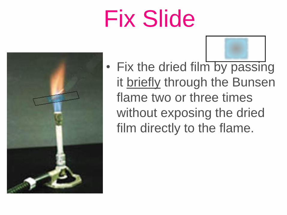
Fix Slide
• Fix the dried film by passing
it briefly through the Bunsen
flame two or three times
without exposing the dried
film directly to the flame.

Gram Staining
Flood for 1 minute with Crystal Violet
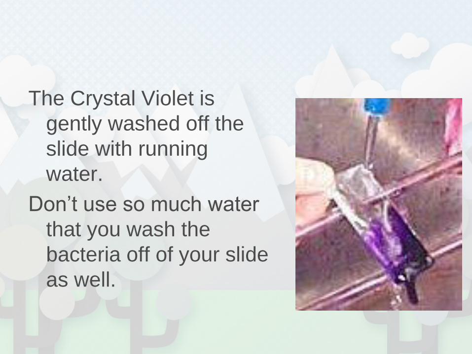
The Crystal Violet is
gently washed off the
slide with running
water.
Don’t use so much water
that you wash the
bacteria off of your slide
as well.

Gram Staining
Flood with Grams Iodine for 1 minute

Gram Staining
Decolorize with 95% ethyl alcohol.Caution: Do not over-decolorize. Add reagent drop by drop until alcohol runs almost clear, showing only a blue tinge.

Gram Staining
Counterstain with safranin for 45 seconds.


Gram
Staining

Gram staining is one type of
DIFFERENTIAL Stain.
•Crystal (Violet)•Iodine•Alcohol {Ethanol}•Safranin

Shape
• Cocci
– round
• Rod
–Bacillus
• spiral

Shape
• Cocci
– round
• Rod
–Bacillus
• spiral1
2
3

Cocci Morphology

Gram-Positive Bacteria• Gram-Positive cocci
• Clusters: usually characteristic of
Staphylococcus spp., such as S. aureus.

Gram-Positive Bacteria• Gram-Positive cocci
• Chain: usually characteristic of Streptococcus
spp., such as S. pneumoniae, B group
streptococci

Gram-Positive Bacteria• Gram-Positive cocci
• Tetrad: usually characteristic of Micrococcus
spp.

Gram-Positive Bacteria• Gram positive bacilli
• Thick : usually characteristic of Clostridium
spp., such as C. perfringens, C. septicum,
• C. tetanomorphum
•
• Thin: usually characteristic of Listeria spp.
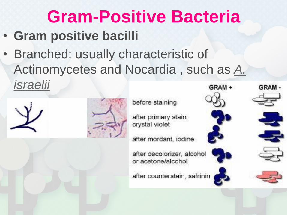
Gram-Positive Bacteria• Gram positive bacilli
• Branched: usually characteristic of
Actinomycetes and Nocardia , such as A.
israelii

Gram-negative bacteria• Gram-negative cocci
• Diplococci: usually characteristic of
Neiseria spp., such as N. meningitidis

Gram-negative bacteria• Gram-negative cocci
• Coccobacilli: usually characteristic of
Acinetobacter spp., which can be either Gram-
positive or Gram-negative, and is often Gram-
variable.
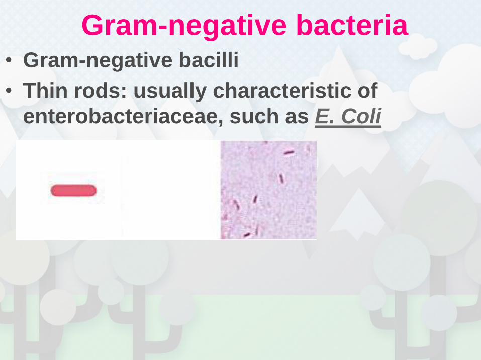
Gram-negative bacteria• Gram-negative bacilli
• Thin rods: usually characteristic of
enterobacteriaceae, such as E. Coli

Gram-negative bacteria• Gram-negative bacilli
• Coccobacilli: usually characteristic of
Haemophilus spp., such as H. influenzae

Gram-negative bacteria• Gram-negative bacilli
• Curved: usually characteristic of Vibrio
spp.; Campylobactor spp., such as V.
cholerae

Gram-negative bacteria• Gram-negative bacilli
• Thin needle shape: usually characteristic of
Fusobacterium spp.

Gram Negative
1 2

Gram-Negative Diplococcus
12

Gram-Positive Coccus Chains
1
2

Gram Positive TetraCoccus
12

Gram Negative Sarcinae

Gram Positive Cocci Clusters
1
2

Gram-Negative Rod
12

Gram Positive Club Shaped Rod
12

Branching Rods
12

Gram negative Comma form rods
12

Gram Positive Spore-forming Bacilli
Bacillus megaterium

Gram Positive Spore-forming Bacilli
Bacillus megaterium

Spirillum 1
2
3
4

Spirillum volutans

What shape are these bacteria?
Are they Gram + or
Gram - ?

Bacillus megaterium

Enterococcus faecalis
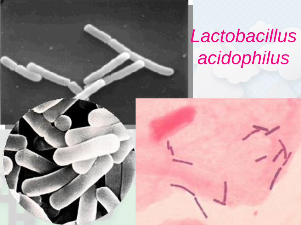
Lactobacillus
acidophilus

Micrococcus luteus

Rhodospirillum rubrum

Sarcina
aurantiaca

Staphylococcus epidermidis

Gram + Bacilli

Actinomyces

Lactobacillus

Listeria monocytogenes

Gram - Bacilli

Bacteroides fragilis

Campylobacter

Fusobacterium nucleatum

Pseudomonas aeruginosa -
encapsulated

Gram + Cocci

viridans streptococci

Pneumococci

Staphylococcus

Streptococcus