ghrelin inhibits the development of mouse preimplantation embryos in vitro
TRANSCRIPT

Ghrelin Inhibits the Development of MousePreimplantation Embryos in Vitro
KAZUHIRO KAWAMURA, NAOKI SATO, JUN FUKUDA, HIDEYA KODAMA, JIN KUMAGAI,HIDEO TANIKAWA, AKIRA NAKAMURA, YOKO HONDA, TOSHIHARU SATO, AND
TOSHINOBU TANAKA
Department of Obstetrics and Gynecology (K.K., N.S., J.F., J.K., H.T., Y.H., T.S., T.T.), Department of Medical InformationScience (A.N.), and Faculty of Health Science (H.K.), Akita University School of Medicine, Akita 010-8543, Japan
Although ghrelin acts as a modulator of feeding behavior andenergy metabolism in the central nervous system, recent stud-ies have implicated the peripheral actions of ghrelin in re-productive tissues. Here, we investigated the expression ofghrelin and its receptor (GHS-R) in mouse oocyte and preim-plantation embryos, and we examined the role of ghrelin inthe regulation of early embryo development. Both ghrelin andGHS-R mRNAs were detected in morula or more advancedembryo stages. As for the origin of ghrelin, both ghrelin mRNAand protein were identified in the uterine endometrium. Thelevels of ghrelin in uterine fluid as well as plasma were sig-nificantly increased in fasting mice compared with animals
with free access to foods. Addition of ghrelin to culture mediainhibited the development of two-cell embryos to the hatchedblastocysts, and the inhibitory effects of ghrelin were abol-ished by an antagonist for the GHS-R. In addition, ghrelinsignificantly decreased the number of total cells, inner cellmass, and trophectoderm cells in blastocysts. These observa-tions suggest that ghrelin could inhibit the development ofpreimplantation embryos during fasting. Thus, ghrelin mayact as a peripheral factor to avoid the excess metabolicdemands imposed by pregnancy during malnutritionalstates. (Endocrinology 144: 2623–2633, 2003)
GH SECRETAGOGUES (GHS) are a group of artificiallysynthesized peptidyl and nonpeptidyl molecules
known to release GH in vivo by acting through a seven-transmembrane, G protein-coupled receptor, GHS-receptor(GHS-R; Refs. 1–6). Recently, ghrelin was identified as anendogenous ligand for the GHS-R (6). Ghrelin is a 28-aminoacid peptide with an essential n-octanoyl modification at theSer3 residue and is primarily expressed in neuroendocrineX/A-like cells of the gastric mucosa, pituitary gland, andhypothalamus (7, 8). The structure of ghrelin is highly con-served between rodents and human with changes in onlytwo residues (6). In addition to its potent GH-releasing ac-tivity, ghrelin stimulates food intake through the modulationof the expression of hypothalamic neuropeptide Y (NPY)and/or the agouti-related protein (9–11). Ghrelin inducesadiposity in rodent by increasing food intake and reducingfat utilization (12). Thus, ghrelin is considered to play animportant role in the regulation of feeding behavior andenergy metabolism by mainly acting at the central nervoussystem (9–12).
Although ghrelin is expressed in the central nervous sys-tem to regulate diverse functions, ghrelin transcripts havealso been detected in several peripheral tissues, such as kid-ney, hematopoietic immune cells, placenta, lung, pancreas,testis, and stomach (13–19). In addition, the GHS-R is ex-pressed in diverse peripheral tissues (14, 20). Although theexact functional significance of peripheral ghrelin is un-
known, ghrelin inhibits the secretion of testosterone by testisLeydig cells under the stimulation of human chorionic go-nadotropin (hCG) and cAMP (19), thus suggesting novelroles of ghrelin in the reproductive system.
In animals with insufficient nutrient intake, the fertilitypotential is suppressed, probably due to adaptive responsesto evade the excess metabolic demands imposed by preg-nancy (21, 22). The level of plasma ghrelin was known toelevate under malnutritional states (12, 23–26). Some GHS,including ghrelin, were shown to negatively regulate cellviability and proliferation (27–29). Ghrelin was demon-strated to inhibit the proliferation of breast cancer cells (29).These findings have provided the basis to hypothesize thatghrelin may inhibit development of embryos when maternalnutrient intake is insufficient.
The aim of this study was to investigate whether ghrelincould inhibit development of preimplantation embryos inmice. We sought to determine 1) the temporal expression ofghrelin and GHS-R mRNAs in oocytes and preimplantationembryos up to the hatched blastocyst stage; 2) whetherghrelin is secreted by the reproductive tracts and binds topreimplantation embryos; and 3) the effects of ghrelin treat-ment on preimplantation embryo development. Our resultsdemonstrate that mouse preimplantation embryos expressboth ghrelin and GHS-R, and ghrelin secreted from repro-ductive tracts could inhibit the development of early em-bryos through the GHS-R.
Materials and MethodsCollection of mouse oocytes and preimplantation embryos
Female IVCS mice, aged 9 wk, (Institute for Animal Reproduction,Ibaragi, Japan) were superovulated with a single ip injection of 10 IU of
Abbreviations: EIA, Enzyme immunoassay; FAM, Tri-5 (and Tri-6)carboxyfluorescein; GHRP-6, GH-releasing peptide-6; GHS, GH secre-tagogue(s); GHS-R, GHS receptor; hCG, human chorionic gonadotropin;HTF, human tubal fluid; ICM, inner cell mass; NPY, neuropeptide Y; TE,trophectoderm.
0013-7227/03/$15.00/0 Endocrinology 144(6):2623–2633Printed in U.S.A. Copyright © 2003 by The Endocrine Society
doi: 10.1210/en.2003-0033
2623
The Endocrine Society. Downloaded from press.endocrine.org by [${individualUser.displayName}] on 26 August 2014. at 11:55 For personal use only. No other uses without permission. . All rights reserved.

pregnant mare serum gonadotropin (Sigma, St. Louis, MO), followed48 h later by 10 IU of hCG (Sigma). Two-cell stage embryos wereobtained by flushing the oviducts of the mated mice at 46–47 h after hCGinjection. The embryos were washed three times with M2 medium(Sigma). Subsequently, groups of 10–15 embryos were placed in 30-�ldrops of the human tubal fluid (HTF) medium (30), covered by mineraloil, and cultured at 37 C in 5% CO2 in air. For RT-PCR analysis, four-cell,eight-cell, morula, blastocyst, and hatched blastocyst-stage embryoswere collected from cultures in individual micro-drop at 50–52, 68–70,90–92, 118–120, and 142–144 h after hCG injection, respectively. Un-fertilized oocytes were also obtained from oviducts of unplugged miceat 18–20 h after hCG injection.
All procedures involving the care and use of animals were approvedby the Animal Research Committee, Akita University School of Medi-cine (Akita, Japan).
RT-PCR and nested PCR
The methods of RT-PCR for oocytes and preimplantation embryoswere described previously (31, 32). Briefly, poly (A)� mRNA was iso-lated from 15 mouse oocytes or preimplantation embryos of severalstages (two-cell, four-cell, eight-cell, morula, blastocyst, and hatchedblastocyst), and each mRNA sample was reverse transcribed into cDNA.Exogenous rabbit �-globin mRNA (Life Technologies, Inc., Rockville,MD) was added to each sample before mRNA extraction to evaluate theefficiency of mRNA extraction and the RT procedure. The amount ofcDNA subjected to each PCR was equivalent to the number of genomes(e.g. one two-cell stage embryo or one quarter of an eight-cell stageembryo), so that each PCR product was derived from the same numberof transcribing genomes.
The primers for ghrelin and GHS-R were based on GenBank accessionno. AB035701 and AF332997, respectively, as shown in Table 1. The PCRwas performed according to the programs described in the legend ofTable 1. For positive controls for ghrelin and GHS-R, mouse placenta andbrain cDNAs were also amplified. For negative controls, the specimenin which water was substituted for mRNA was amplified. Because of thelow number of oocytes and preimplantation embryos under study,heminested PCR was needed to obtain optimal results. Furthermore,total RNA was extracted from uterus of pregnant mice at d 4.5 aftermating, and RT-PCR for ghrelin was performed.
The PCR products were separated by 2% agarose gel electrophoresis(Agarose-LE, Nacalai Tesque, Inc., Kyoto, Japan) in the presence ofethidium bromide (Sigma), and visualized with a UV transilluminator(Funakoshi, Tokyo, Japan). To confirm identity, bands of each PCRproduct were eluted from the agarose gel using the QIAquick gel ex-traction kit (QIAGEN KK, Tokyo, Japan), ligated into the pDrive Cloningvector (QIAGEN KK), and cloned in accordance with standard proto-cols. Plasmid DNA was recovered using Quantum Prep Plasmid Mini-prep kit (Bio-Rad Laboratories, Inc., Hercules, CA), cycle sequenced, andanalyzed in a ABI 100 DNA sequencer (PE Applied Biosystems, Tokyo,Japan) using T7 or SP6 site-specific primers.
Binding of fluorescent ghrelin to mousepreimplantation embryos
For binding studies, two-cell stage embryos were cultured in the HTFmedium at 37 C in 5% CO2 in air. When the embryos reached the four-cellor blastocyst stage, the medium was replaced with 1, 10, and 100 nm offluorescent ghrelin conjugated with Tri-5 (and Tri-6) carboxyfluorescein(FAM; Phoenix Pharmaceuticals, Inc., Belmont, CA) in the HTF mediumand incubated for 30 min at 37 C. After three washes, these embryos werefixed with 4% paraformaldehyde in PBS for 15 min at room temperatureand washed three times. To determine the background fluorescence,embryos were incubated only with the HTF medium. Nonspecific bind-ing of FAM-ghrelin (100 nm) was estimated by coincubating with a100-fold excess of unlabeled ghrelin (Phoenix Pharmaceuticals, Inc.). Thefluorescence signals in embryos were visualized using a confocal laserscanning microscope (LSM 410, Carl Zeiss, Oberkochen, Germany). Op-tical sections with intervals of 1 �m were taken with a 63�/1.4 PlanApochromat objective, and the fluorescent images were obtained as 8-bitimages of TIF format files. After the calculation of total numbers of pixelsfor the whole embryo, the numbers of fluorescent pixels, which corre-spond to the binding site of FAM-ghrelin to GHS-R, were measured bySigmaScan Pro 5.0 (SPSS Japan Inc., Tokyo, Japan).
Immunohistochemistry
Uteri were obtained from 9-wk-old pregnant mice at d 4.5 after matingand fixed with 4% paraformaldehyde in PBS for 6 h at 4 C. Fixed frozentissue sections were blocked with 10% normal goat serum (DAKO Corp.,Kyoto, Japan) for 30 min at room temperature. Samples were incubatedwith rabbit antighrelin serum (Phoenix Pharmaceuticals, Inc.) with a di-lution of 1:150 in 1% PBS-BSA/0.1% Triton X-100 (Sigma), overnight at 4C. After three washes in cold PBS, samples were incubated with 1.0 �g/mlof goat antirabbit Cy3 fluorescein antibody (Chemicon, Temecula, CA) in1% PBS-BSA/0.1% Triton X-100, for 1 h at room temperature in the dark.After three washes in cold PBS, slides were covered in a drop of antifademounting medium (DAKO Corp.) and analyzed under an epifluorescencemicroscope (Olympus Corp., Tokyo, Japan). For negative controls, sectionswere subjected to the same method, except that the primary antiserum wasreplaced by the same dilutions of normal rabbit serum (DAKO Corp.) orby the primary antiserum preabsorbed with the ghrelin peptide (PhoenixPharmaceuticals, Inc.) at 50 �g/ml.
Enzyme immunoassay (EIA)
Ghrelin concentrations in plasma and uterine fluid were measuredusing a ghrelin EIA kit (Phoenix Pharmaceuticals, Inc.) with a sensitivityof 0.9 ng/ml, and the intra- and interassay coefficients of variation wereless than 5% and 14%, respectively. For measurement of ghrelin in theuterine fluid, uteri were collected from 10 nonpregnant 9-wk-old miceat the day of estrus and 9-wk-old pregnant mice at d 4.5 after mating,all with free access to a standard rodent diet. This pregnant stage cor-responded to the embryonic stage of blastocyst. After 48 h of fasting(only access to water was allowed), samples were also obtained from 10
TABLE 1. Primers used for RT-PCR and nested PCR, PCR cycles and temperatures for amplification of the different cDNA
Transcript PCRround Primer sequence (5�-3�) Size of product (bp) Annealing (C)
Ghrelin 1st Sense CCATCTGCAGTTTGCTGCTA 394 60Antisense CGGATGTGAGTTCTTGCTCA
2nd Sense CCATCTGCAGTTTGCTGCTA 350 60Antisense GCCTGTCCGTGGTTACTTGT
GHSR1a 1st Sense CTGCTCTGCAAACTCTTCCA 375 64Antisense CTTCCTCCCGATGAGACTGT
2nd Sense CTGCTCTGCAAACTCTTCCA 354 64Antisense GAGCACAGTGAGGCAGAAGA
�-actin Sense GGACCTCACTGACTACCTCATGAA 524 55Antisense GGTGGAAGGTGGTCAACACCTAG
�-globin Sense GCAGCCACGGTGGCGAGTAT 257 55Antisense GTGGGACAGGAGCTTGAAAT
PCR cycles: denaturation at 94 C for 30 sec and extension at 72 C for 30 sec, total 35 cycles were performed.
2624 Endocrinology, June 2003, 144(6):2623–2633 Kawamura et al. • Ghrelin Inhibits Embryo Development
The Endocrine Society. Downloaded from press.endocrine.org by [${individualUser.displayName}] on 26 August 2014. at 11:55 For personal use only. No other uses without permission. . All rights reserved.
nonpregnant mice at the day of estrus and at d 4.5 after mating. Uterinefluid samples were obtained by flushing the uterine cavities with 10 �lof 0.1% 3-[(3-cholamidopropyl) dimethylammonio]-1-propanesulfonatebuffer (Sigma) containing 0.6 TIU/ml of aprotinin (Sigma) and centri-fuged at 2000 � g for 5 min. Each supernatant was stored at �70 C untilassay. The plasma samples were obtained from the same mice describedabove and stored at �70 C until assay. Ghrelin content in uterine fluidand plasma samples was measured by EIA in duplicate.
Embryo cultures
Two-cell stage embryos were collected as described above. The em-bryos were washed three times with the M2 medium (Sigma). Groupsof 10–15 embryos randomly selected were placed in 30-�l drops of HTFmedium covered by mineral oil with or without synthetic rat ghrelin(Phoenix Pharmaceuticals, Inc.). The mature peptide of rat ghrelin isidentical to the mouse ghrelin (Ref. 6; GenBank accession no. AB035701).Embryos were cultured over 72 h up to the hatched blastocyst stage at37 C in 5% CO2 in air.
To examine whether the effects of ghrelin on preimplantation em-bryos were mediated through GHS-R, embryos were cultured in HTFmedium containing 10 mm of an antagonist for GHS-R, [d-Lys-3]GH-releasing peptide-6 (GHRP-6) (Peninsula Laboratories, Inc., San Carlos,CA; Ref. 10) with or without 100 nm of ghrelin. Furthermore, embryoswere cultured in HTF medium containing 100 nm of des-octanoyl 3ghrelin (Phoenix Pharmaceuticals, Inc.), an analog lacking biologicalactivity (6). For controls, embryos were cultured in HTF medium aloneor containing 100 nm of ghrelin.
Embryonic development was monitored daily by phase-contrast mi-croscopy (Olympus Corp.), and the rate of embryo development wasassessed.
Differential labeling of inner cell mass (ICM) andtrophectoderm (TE) nuclei
Embryos at the two-cell stage were cultured with 10 nm of ghrelin for56 h, and the numbers of ICM and TE cells of each blastocyst werecounted by the differential labeling technique using two polynucleotide-specific fluorochromes (propidium iodide and bisbenzimide; Hoechst33342, Sigma) as described previously (31). After staining, the blasto-cysts were mounted on a glass slide, and the number of total, TE, andICM cells in each blastocyst was counted under an epifluorescencemicroscope (Olympus Corp.).
Statistical analysis
To analyze the effect of ghrelin on embryo development, ordinal un-paired comparison t test and the analysis of sources of variation, F test, aswell as the trend analysis (33–35) were performed on all of the possiblepairs. The logarithms of observed four ghrelin concentrations are orderedfrom �1 (log [0.1 nm]) to 2 (log [100 nm]) with equal interval, 1. Thus, multiplecomparisons of a series of observations at four different concentrations canbe considered as those of ordered groups. In other words, further infor-mation about the nature of dependency induced by logarithmic concen-tration of ghrelin can be elucidated by using trend analysis. Consideringlinear (df1 � 1, df2 � n � k � 1), quadratic (df1 � 2, df2 � n � k � 2), cubic(df1 � 3, df2 � n � k � 3), and quartic (df1 � 4, df2 � n � k � 4), wheren is a number of embryos used in each experiment and k is the number ofexperiments, trend analyses were performed. The t tests were performedwith SPSS 10.1 (SPSS, Inc., Japan Inc.), and the calculations for the F testsand the trend analysis were performed with Excel 2001 (Microsoft Corp.,Redmond, WA), according to published procedures (33).
The one-way ANOVA followed Fisher’s protected least significantdifference test was used to evaluate differences in ghrelin protein con-centrations in plasma and uterine fluid, and the Mann-Whitney U testwas performed for the comparison of the number of total blastomeres,ICM, and TE cells.
ResultsTemporal expression of ghrelin and GHS-R mRNAs inmouse oocytes and preimplantation embryos
RT-PCR and nested PCR were performed to detectmRNAs for ghrelin and GHS-R in the mouse oocytes and
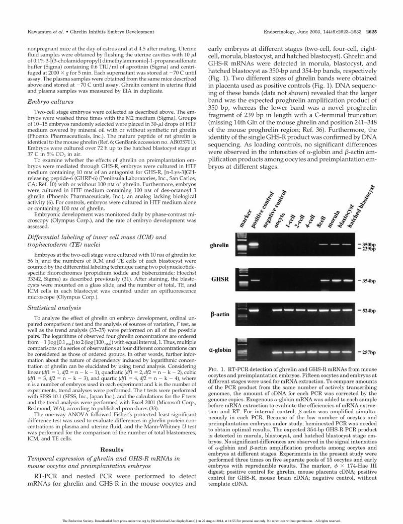
early embryos at different stages (two-cell, four-cell, eight-cell, morula, blastocyst, and hatched blastocyst). Ghrelin andGHS-R mRNAs were detected in morula, blastocyst, andhatched blastocyst as 350-bp and 354-bp bands, respectively(Fig. 1). Two different sizes of ghrelin bands were obtainedin placenta used as positive controls (Fig. 1). DNA sequenc-ing of these bands (data not shown) revealed that the largerband was the expected proghrelin amplification product of350 bp, whereas the lower band was a novel proghrelinfragment of 239 bp in length with a C-terminal truncation(missing 14th Gln of the mouse ghrelin and position 241–348of the mouse proghrelin region; Ref. 36). Furthermore, theidentity of the single GHS-R product was confirmed by DNAsequencing. As loading controls, no significant differenceswere observed in the intensities of �-globin and �-actin am-plification products among oocytes and preimplantation em-bryos at different stages.
FIG. 1. RT-PCR detection of ghrelin and GHS-R mRNAs from mouseoocytes and preimplantation embryos. Fifteen oocytes and embryos atdifferent stages were used for mRNA extraction. To compare amountsof the PCR product from the same number of actively transcribinggenomes, the amount of cDNA for each PCR was corrected by thegenome copies. Exogenous �-globin mRNA was added to each samplebefore mRNA extraction to evaluate the efficiencies of mRNA extrac-tion and RT. For internal control, �-actin was amplified simulta-neously in each PCR. Because of the low number of oocytes andpreimplantation embryos under study, heminested PCR was neededto obtain optimal results. The expected 354-bp GHS-R PCR productis detected in morula, blastocyst, and hatched blastocyst stage em-bryos. No significant differences are observed in the signal intensitiesof �-globin and �-actin amplification products among oocytes andembryos at different stages. Experiments in the present study wereperformed three times on five separate pools of 15 oocytes and earlyembryos with reproducible results. The marker, � � 174-Hae IIIdigest; positive control for ghrelin, mouse placenta cDNA; positivecontrol for GHS-R, mouse brain cDNA; negative control, withouttemplate cDNA.
Kawamura et al. • Ghrelin Inhibits Embryo Development Endocrinology, June 2003, 144(6):2623–2633 2625
The Endocrine Society. Downloaded from press.endocrine.org by [${individualUser.displayName}] on 26 August 2014. at 11:55 For personal use only. No other uses without permission. . All rights reserved.

Binding of fluorescent ghrelin to mousepreimplantation embryos
To confirm the expression of functional GHS-R in mousepreimplantation embryos, binding studies were performedusing FAM-ghrelin. The clustered fluorescent signals weredetected homogeneously in both ICM and TE cells at theblastocyst stage (Fig. 2, A–E), and the ratios of fluorescentpixels per embryo were saturated at 10 nm of FAM-ghrelin
(Fig. 2B and Table 2). Embryos treated with 100 nm of FAM-ghrelin (Fig. 2C and Table 2) showed a similar level of flu-orescent signals as compared with those treated with 10 nmof FAM-ghrelin (Fig. 2B and Table 2). In contrast, in four-cellstage embryos, no obvious signals were observed even inembryos treated with 100 nm of FAM-ghrelin (Fig. 2F). Forcontrols, embryos incubated only with HTF medium alsoshowed no signal (Fig. 2, E and H). Furthermore, no signals
FIG. 2. Binding of fluorescent ghrelin to mouse preimplantation embryos. Shown are confocal images of optical sections of the following. A–E,Blastocyst stage embryos; F–H, four-cell stage embryos. Embryos were incubated with 1, 10, and 100 nM of FAM-ghrelin in HTF medium for30 min at 37 C in 5% CO2 in air and fixed in 4% paraformaldehyde. The clustered fluorescent signals of FAM-ghrelin are found homogeneouslyin both ICM and TE cells at 1.0 nM (A). The clustered fluorescent signals increase in 10 nM (B), but are saturated in 100 nM (C). Four-cell stageembryos lack fluorescent signals even at 100 nM (F). For controls of background fluorescence and nonspecific bindings, embryos were incubatedonly with HTF medium (E and H) and with a 100-fold excess of unlabeled ghrelin (D and G). The fluorescent signals in these controls are muchweaker than specific signals in A–C. Confocal images were taken at magnification of �63. Consistent signals were observed in at least threeexperiments in which a total of five four-cell and blastocyst stage embryos were surveyed.
2626 Endocrinology, June 2003, 144(6):2623–2633 Kawamura et al. • Ghrelin Inhibits Embryo Development
The Endocrine Society. Downloaded from press.endocrine.org by [${individualUser.displayName}] on 26 August 2014. at 11:55 For personal use only. No other uses without permission. . All rights reserved.

could be detected when embryos were incubated with thefluorescent ligand in the presence of a 100-fold excess ofunlabeled ghrelin (Fig. 2, D and G).
Detection of ghrelin mRNA and protein inmouse endometrium
We hypothesize that embryos bearing GHS-R could beactivated by ghrelin secreted by the uterus. RT-PCR wasperformed to detect ghrelin mRNA in the mouse uterus.Ghrelin mRNA was detected in uterus as a 394-bp band (Fig.3a). As described above, the lower band corresponded to thetruncated proghrelin fragment of 286 bp. Immunohisto-chemical staining was performed to detect ghrelin protein inthe mouse endometrium. According to the sequence analy-sis, the lower band was a novel mouse ghrelin fragment, whichcorresponded to the rat des-Gln14-ghrelin (36). The rat des-Gln14-ghrelin was identified as a splice variant of ghrelin alsowith GH-releasing activity. However, the role of this novelform of mouse ghrelin in reproductive tract is unknown. Atthe protein level, the luminal and glandular epithelia of theendometrium were stained with the ghrelin antibody (Fig.3b-A). The specificity of this immunoreactivity was demon-strated by the absence of staining in specimens incubatedwith nonimmunized serum (data not shown) and preab-sorbed primary antiserum for ghrelin (Fig. 3b-B).
Determination of ghrelin level in the mouse uterine fluid
To further examine whether ghrelin is secreted by theendometrium, the levels of ghrelin in plasma and uterinefluid were measured using the ghrelin EIA. Plasma ghrelinconcentrations were significantly increased in both nonpreg-nant and pregnant mice at 48 h after fasting as compared withnonfasted mice (both P � 0.05; Fig. 4A). There were nosignificant differences in plasma ghrelin concentrations be-tween pregnant and nonpregnant mice (Fig. 4A). Similarresults were obtained from uterine fluid samples (Fig. 4B).The ghrelin concentrations of uterine fluid were significantlyincreased in both nonpregnant and pregnant mice at 48 hafter fasting as compared with nonfasted mice (both P �0.0001; Fig. 4B). The level of ghrelin in uterine fluid of preg-nant mice was slightly higher than that of nonpregnant mice,but the difference did not reach a significant level (Fig. 4B).
The effect of ghrelin on the development of preimplantationembryos in vitro
We hypothesized that increases of ghrelin in the uterinefluid of fasting animals could regulate early embryo de-velopment and determined the effects of ghrelin treatmenton the in vitro development of mouse preimplantation
FIG. 3. a, RT-PCR detection of ghrelin mRNA from uterus of preg-nant mice at d 4.5. Total RNA was extracted from uterus, andRT-PCR was performed. For internal control, �-actin was amplifiedsimultaneously. Based on the DNA sequencing of ghrelin PCRproducts, the larger molecular weight band corresponded to anexpected ghrelin amplification product of 394 bp, and a smaller286-bp product corresponded to a transcript with a C-terminaltruncation (missing 14th Gln of the mouse ghrelin and position241–348 of the mouse proghrelin) is detected in uterus. Experi-ments in the present study were performed three times with re-producible results. The marker, � � 174-Hae III digest; positivecontrol, mouse placenta cDNA; negative control, distilled water. b,Immunofluorescence staining of ghrelin in mouse endometrium.Samples were fixed in 4% paraformaldehyde and stained usingrabbit antighrelin serum with a dilution of 1:150 as primary an-tibodies and 1.0 �g/ml of goat antirabbit Cy3 fluorescein antibodyas secondary antibodies. Immunoreactivity is detected in the lu-minal and glandular epithelia of endometrium (bA). Absorptionwith 50 �g/ml of ghrelin peptide before immunostaining abolishedall positive staining (bB). Original magnification, �200. Consistentstaining was observed in at least 3 experiments in which a total of10 mouse endometrium were surveyed.
TABLE 2. Ratio of fluorescent pixels obtained from confocalmicroscopic eight-bit images of mouse blastocyst
GhrelinNumber offluorescentpixels (A)
Number of pixels inwhole embryo’s
area (B)
Ratio of pixels perembryo (A/B)
1 nM 7,959 � 973 52,065 � 357 0.1530 � 0.004210 nM 27,147 � 3,767 52,866 � 2,710 0.5048 � 0.1025100 nM 38,221 � 3,008 57,903 � 2,260 0.6594 � 0.0996
A total of five embryos were used in each concentration.
Kawamura et al. • Ghrelin Inhibits Embryo Development Endocrinology, June 2003, 144(6):2623–2633 2627
The Endocrine Society. Downloaded from press.endocrine.org by [${individualUser.displayName}] on 26 August 2014. at 11:55 For personal use only. No other uses without permission. . All rights reserved.

embryos. Two-cell stage embryos were cultured in thepresence of 0.1, 1, 10, and 100 nm of rat ghrelin. In eachexperiment, 20 –28 embryos were used in each group,consisting of six observations, and the experiment was
repeated five times. Results of examination of a total of123–159 embryos in each group were summarized in Fig.5. Up to 36 h of culture, ghrelin treatment showed no effecton the development of preimplantation embryos to themorula stage. After 48 and 56 h of culture, 100 nm ofghrelin significantly inhibited the development of em-bryos from morula to blastocyst and from blastocyst toexpanded blastocyst stage (P � 0.02 and 0.003 for none vs.100 nm, respectively) with embryo development retardedat morula and blastocyst stages, respectively. After 72 h ofculture, the rates of formation of hatched blastocyst fromexpanded blastocyst were significantly inhibited by 10 and100 nm of ghrelin (P � 0.007 for none vs. 10 nm, and P �0.002 for none vs. 100 nm). The slopes of observed ratiobetween the consecutive two points on the logarithmicconcentrations of ghrelin were compared in each devel-opmental stage. The absolute value of the slope between1.0 and 10 nm of ghrelin was the highest for all the stagesof embryos as well as for the culture periods, 48 h, 56 h,and 72 h (data not shown). Thus, a threshold value in theinhibitory effect of ghrelin existed between 1.0 and 10 nm.In addition, the observed P values of F test revealed thatany developmental stages were not significant in eitherlinearity, quadratic, cube, or quartic (0.997 � P � 0.526 forall developmental stages and at all culture periods). Thus,the nature of the inhibitory effect of ghrelin in the devel-opment of preimplantation embryos may be explained asa more complicated trend.
To confirm the specificity of the inhibitory effect of ghrelinon the preimplantation embryos, the effects of an antagonistfor GHS-R, [d-Lys-3]GHRP-6, and a nonbioactive form ofghrelin, des-octanoyl 3 ghrelin, were examined by an addi-tional five sets of experiments. Two-cell stage embryos werecultured with 1) HTF medium alone; 2) 100 nm of ghrelin; 3)10 mm of [d-Lys-3]GHRP-6; 4) 100 nm of ghrelin and 10 mmof [d-Lys-3]GHRP-6; and 5) 100 nm of des-octanoyl 3 ghrelin.In each experiment, 22–28 embryos were used in each group,consisting of 6 observations, and the experiment was re-peated 5 times. A total of 145–168 embryos were tested ineach group, and the results were summarized in Fig. 6. Theinhibitory effects of ghrelin on the development of embryosfrom morula to the blastocyst, blastocyst to expanded blas-tocyst, and expanded blastocyst to hatched blastocyst stagewere significantly blocked by treatment of [d-Lys-3]GHRP-6(P � 0.008, P � 0.001, and P � 0.000, vs. 100 nm of ghrelin,respectively). [d-Lys-3]GHRP-6 alone and des-octanoyl 3 gh-relin showed little effect in the suppression of embryodevelopment.
The effect of ghrelin on the regulation of cell numbers incultured mouse blastocyst
The numbers of total, TE, and ICM cells of blastocyst after56 h of culture with or without ghrelin are summarized inFig. 7. Blastocysts cultured with 10 nm of ghrelin had asignificantly lower total cell number, as compared with blas-tocysts cultured in HTF medium alone. The decrease in thetotal cell number of ghrelin-treated blastocysts resulted frominhibition of the proliferation of both ICM and TE cells, and
FIG. 4. The levels of ghrelin in mouse plasma (A) and uterine fluid (B)during early pregnancy. Bars represent mean � SEM (n � 10). Non-pregnant, Nonpregnant mouse 9 wk of age at estrus; Pregnant on D �4.5, pregnant mouse 9 wk of age at d 4.5; Fasting, 48 h of fasting (onlyaccess to water was allowed.) Each uterine fluid was obtained byflushing the uterine cavities with 10 �l of 0.1% 3-[(3-cholamidopropyl)dimethylammonio]-1-propanesulfonate buffer containing 0.6 TIU/mlof aprotinin and centrifuged at 2000 � g for 5 min. The plasmasamples were obtained simultaneously. Ghrelin concentration wasdetermined by EIA. Data were analyzed one-way ANOVA followedFisher’s protected least significant difference test. *, P � 0.0001; †,P � 0.05.
2628 Endocrinology, June 2003, 144(6):2623–2633 Kawamura et al. • Ghrelin Inhibits Embryo Development
The Endocrine Society. Downloaded from press.endocrine.org by [${individualUser.displayName}] on 26 August 2014. at 11:55 For personal use only. No other uses without permission. . All rights reserved.

FIG. 5. The dose-dependent effects of ghrelin on the in vitro development of mouse preimplantation embryos. In each experiment, 20–28embryos were used in each group, consisting of 6 observations, and the experiment was repeated 5 times. A total of 123–159 embryos wereexamined in each group; 0 nM � 159, 0.1 nM � 120, 1.0 nM � 123, 10 nM � 124, 100 nM � 130. Values are mean � SEM. The data were analyzedby unpaired comparison t test and the analysis of sources of variation, F test, as well as the trend analysis. a, P � 0.02 for none vs. 100 nM;b, P � 0.003 for none vs. 100 nM; c, P � 0.007 for none vs. 10 nM; d, P � 0.002 for none vs. 100 nM.
Kawamura et al. • Ghrelin Inhibits Embryo Development Endocrinology, June 2003, 144(6):2623–2633 2629
The Endocrine Society. Downloaded from press.endocrine.org by [${individualUser.displayName}] on 26 August 2014. at 11:55 For personal use only. No other uses without permission. . All rights reserved.

FIG. 6. The effects of an antagonist for GHS-R, [D-Lys-3]GHRP-6 and a nonbioactive form of ghrelin, des-octanoyl 3 ghrelin. In the additionalfive sets of experiments, embryos were cultured in: 1) HTF medium alone; 2) 100 nM of ghrelin; 3) 10 mM of [D-Lys-3]GHRP-6; 4) 100 nM of ghrelinand 10 mM of [D-Lys-3]GHRP-6; and 5) 100 nM of des-octanoyl 3 ghrelin. In each experiment, 22–28 embryos were used in each group, consistingof exactly 6 observations, and the experiment was repeated 5 times. Between 145 and 168 embryos were tested in each group; 1) 168, 2) 145,3) 145, 4) 159, and 5) 159. None, HTF medium only. Values are mean � SEM. The data were analyzed by unpaired comparison t test and theanalysis of sources of variation, F test. *, P � 0.01, vs. 100 nM of ghrelin.
2630 Endocrinology, June 2003, 144(6):2623–2633 Kawamura et al. • Ghrelin Inhibits Embryo Development
The Endocrine Society. Downloaded from press.endocrine.org by [${individualUser.displayName}] on 26 August 2014. at 11:55 For personal use only. No other uses without permission. . All rights reserved.

the inhibitive effect was equally observed in TE cells and ICMcells (both P � 0.05).
Discussion
In the present study, we demonstrate the temporal ex-pression of ghrelin and GHS-R mRNAs in mouse oocytes andpreimplantation embryos. Both ghrelin and GHS-R mRNAswere expressed in mouse morula, blastocyst, and hatchedblastocyst stage embryos. Using binding assays, fluorescent-labeled ghrelin could bind to both ICM and TE cells at blas-tocyst stage as clustering patterns, suggesting the existenceof receptor-ligand complexes (37, 38). In contrast, four-cellstage embryos lacked specific fluorescent signals. The resultsof both RT-PCR and binding assay also suggest that four-cellstage embryos do not express functional receptors. Thus,through the receptor-mediated process, exogenously sup-plemented ghrelin could be taken into embryos expressingthe GHS-R. Ghrelin protein was expressed in the epithelia ofendometrium as shown by immunohistochemistry. Further-more, ghrelin mRNA was expressed in uterus of early preg-nant mice. Thus, these data strongly suggested that ghrelinis produced by endometrial epithelium.
We further confirmed whether ghrelin was secreted dur-ing early embryogenesis by the reproductive tract of mouse.Ghrelin could be detected in uterine fluid, and the level wassignificantly increased in fasting mice as compared withthose with free access to foods. These findings suggest thatghrelin is produced and secreted from endometrial epithe-lium and may regulate the function(s) of preimplantationembryo during its development in a paracrine/autocrinemanner.
Accumulated evidence indicates that a number of growthfactors and cytokines contribute in a paracrine and/or au-
tocrine fashion to the rate of embryo development, the pro-portion of embryos developing to the blastocyst stage, thecell number in the blastocyst, energy metabolism, and apo-ptosis (reviewed in Ref. 39). Supplementation of culture me-dium with exogenous growth factors and cytokines affectsthe development of preimplantation embryos via paracrinepathways (reviewed in Ref. 39). Although much of the workon the stimulating effects of growth factors and cytokines onpreimplantation embryos has been carried out using cultureconditions, there have been only a few reports concerninginhibitory factors. TNF-� was reported to inhibit cell prolif-eration in blastocyst and induce apoptosis in the ICM (40–44). Furthermore, TNF-� decreases the ability of embryos todifferentiate into fetuses after implantation (44). Interferon-�inhibited blastocyst formation and trophoblast outgrowthafter attachment in vitro (45, 46). The data obtained from thepresent study have shown that the addition of ghrelin tomouse embryo culture media can inhibit preimplantationembryo development from two-cell stage embryo to the blas-tocyst, fully expanded blastocyst, and hatched blastocyst invitro in a dose-dependent manner. This effect was blocked byan antagonist for GHS-R, [d-Lys-3]GHRP-6. Therefore,ghrelin is one of the inhibitory factors for the developmentof preimplantation embryos. Although ghrelin could inhibitthe development of embryos as a paracrine factor, treatmentof antagonist for GHS-R alone showed little effect on thedevelopment of embryos. Because the levels of ghrelin in theembryo culture medium were under the sensitivity of assaydetection (data not shown), the level of ghrelin secreted fromembryo itself may be insufficient for inducing inhibitoryeffects. Thus, further studies will be required to elucidate theautocrine mechanism within mouse preimplantation embryos.
It is well known that disorders in nutritional status candisrupt the complex interplay of gonadotropins and gonadalhormones, which are essential for fertility. Suppression ofpulsatile LH secretion has been reported after fasting or foodrestriction in mammals, including rodents and humans (47–55). Fasting-induced suppression of LH is considered to bea result of reduced secretion of GnRH from the hypothala-mus, because fasted animals show LH pulses similar in quan-tity and magnitude to fed ones when administrated exoge-nous GnRH (53, 56–60). Previous reports have demonstratedthat plasma ghrelin levels rose in response to food restrictionor fasting as well as aging (12, 21–24, 61). Thus, ghrelin mayact as a peripheral factor to avoid the excess metabolic de-mands imposed by reproduction during insufficient nutrientintake.
In the present study, fasting for 48 h led to increasedsecretion of ghrelin into uterine fluid as observed in plasmasamples. The inhibiting effect of ghrelin on embryo devel-opment was observed when the concentration of ghrelinexceeded to threshold between 1.0 nm and 10 nm. The bind-ing assay also showed that the levels of fluorescent signalsin embryos were saturated at 10 nm of FAM-ghrelin. Theconcentration of ghrelin in uterine fluid of fasting mice at d4.5 of pregnancy was determined to be 7.02 � 0.70 ng/ml(2.12 � 0.21 nm), a level found to regulate embryodevelopment.
At the blastocyst stage, the embryo consists of two typesof cell lineage, TE and ICM cells. The TE cells are necessary
FIG. 7. The effect of ghrelin on the number of total cells (Total), ICMcells, and TE cells in 56-h cultured blastocysts. Values are mean � SEMof blastocysts cultured in 10 nM of ghrelin (G; n � 30) and blastocystscultured in HTF medium alone (C; n � 30). Data were analyzed byMann-Whitney U test. *, P � 0.05, significantly different from cor-responding control.
Kawamura et al. • Ghrelin Inhibits Embryo Development Endocrinology, June 2003, 144(6):2623–2633 2631
The Endocrine Society. Downloaded from press.endocrine.org by [${individualUser.displayName}] on 26 August 2014. at 11:55 For personal use only. No other uses without permission. . All rights reserved.

for implantation and subsequent formation of the placentaand extraembryonic membranes. The ICM cells form all threegerm layers and all tissues of the embryo, as well as ex-traembryonic membranes. Thus, cell numbers in the TE, inthe ICM, or in both cell populations of blastocyst are theindicators of embryo growth and viability (62). AlthoughTNF-� treatment predominantly suppresses the ICM lineage(40–44), ghrelin decreases the total cell number of blastocystsas a result of reduction of the numbers of both ICM and TEcells. These differences may be caused by the differentialexpression pattern of specific receptors for TNF-� andghrelin in blastocyst. The TNF-� receptors were shown to belocalized mainly in ICM (41), whereas fluorescent ghrelinwas detected in both ICM and TE cells.
Recently, some GHS were reported to inhibit proliferationof thyroid, breast, and lung cancer cell lines as assessed bythymidine incorporation and cell proliferation (27–29).Among the GHS, ghrelin was shown to inhibit thymidineincorporation and proliferation of a breast cancer cell line atconcentrations close to its binding affinity (29); however,several conflicting data have been reported in other cell lines.Ghrelin simulated proliferation of prostate cancer cell line(63) and cardiomyocyte cell line (64).
In conclusion, we demonstrate the temporal expression ofghrelin and GHS-R mRNAs in mouse preimplantation em-bryos. Both ghrelin and GHS-R mRNAs were detected aftermorula stage embryos. Ghrelin was produced and secretedfrom reproductive tracts, and the level of ghrelin was ele-vated with fasting. Furthermore, high levels of ghrelin couldinhibit the development of mouse preimplantation embryosthrough its specific receptor, GHS-R. These observationsstrongly suggest that ghrelin could inhibit the developmentof preimplantation embryos under malnutritional status.
Acknowledgments
We thank Dr. Aaron J. Hsueh (Stanford University School of Medi-cine, Stanford, CA) for reading this manuscript.
Received January 10, 2003. Accepted February 10, 2003.Address all correspondence and requests for reprints to: Kazuhiro
Kawamura, Department of Obstetrics and Gynecology, Akita UniversitySchool of Medicine, Hondo 1-1-1, Akita 010-8543, Japan. E-mail:[email protected].
This work was supported by a Grant-in Aid for Scientific Research (C:14571535) from the Japanese Ministry of Education, Science, Sports andCulture.
References
1. Smith RG, Van Der Ploeg LHT, Howard AD, Feighner SD, Cheng K, HickeyGJ, Wyvratt Jr MJ, Fisher MH, Nargund RP, Patchett AA 1997 Peptidomi-metic regulation of growth hormone secretion. Endocr Rev 18:621–645
2. Bowers CY 1998 Growth hormone-releasing peptide (GHRP). Cell Mol Life Sci54:1316–1329
3. Casanueva FF, Dieguez C 1999 Growth hormone secretagogues: physiologicalrole and clinical utilities. Trends Endocrinol Metab 10:30–38
4. Howerd AD, Feighner SC, Cully DF, Arena JP, Liberator PA, Rosenblum CI,Hamelin M, Hreniuk DL, Palyha OC, Anderson J, Paress PS, Diaz C, ChouM, Liu KK, McKee KK, Pong SS, Chaung LY, Elbrecht A, Dashkevicz M,Heavens R, Rigby M, Sirinathsinghji DJ, Dean DC, Melillo DG, Patchett AA,Nargund RP, Griffin PR, Demartino JA, Gupta SK, Schaeffer JM, Smith RG,Van Der Ploeg LHT 1996 A receptor on pituitary and hypothalamus thatfunctions in growth hormone release. Science 273:974–977
5. McKee KK, Palyha OC, Feighner SC, Hreniuk DL, Tan CP, Phillips MS,Smith RG, Van Der Ploeg LHT, Howard AD 1997 Molecular analysis of ratpituitary and hypothalamic growth hormone secretagogue receptors. MolEndocrinol 11:415–423
6. Kojima M, Hosoda H, Date Y, Nakazato M, Matsuo H, Kangawa K 1999Ghrelin is a growth-hormone acylated peptide from stomach. Nature 402:656–660
7. Kojima M, Hosoda H, Matsuo H, Kangawa K 2001 Ghrelin: discovery of thenatural endogenous ligand for the growth hormone secretagogue receptor.Trends Endocrinol Metab 12:118–122
8. Date Y, Kojima M, Hosoda H, Sawaguchi A, Mondal MS, Suganuma T,Matukura S, Kangawa K, Nakazato M 2000 Ghrelin, a novel growth hormone-releasing acylated peptide, is synthesized in a distinct endocrine cell type inthe gastrointestinal tracts of rats and humans. Endocrinology 141:4255–4261
9. Wren AM, Small CJ, Ward HL, Murphy KG, Dakin CL, Taheri S, KennedyAR, Roberts GH, Morgan DG, Ghatei MA, Bloom SR 2000 The novel hy-pothalamic peptide ghrelin stimulates food intake and growth hormone se-cretion. Endocrinology 141:4235–4328
10. Nakazato M, Murakami N, Date Y, Kojima M, Matsuo H, Kangawa K,Matsukura S 2001 A role for ghrelin in the central regulation of feeding. Nature409:194–198
11. Shintani M, Ogawa Y, Ebihara K, Aizawa-Abe M, Miyanaga F, Takaya K,Hayashi T, Inoue G, Hosoda K, Kojima M, Kangawa K, Nakao K 2001Ghrelin, an endogenous growth hormone secretagogue, is a novel orexigenicpeptide that antagonizes leptin action through the activation of hypothalamicneuropeptide Y/Y1 receptor pathway. Diabetes 50:227–232
12. Tschop M, Smiley DL, Heiman ML 2000 Ghrelin induces adiposity in rodents.Nature 407:908–913
13. Gnanapavan S, Kola B, Bustin SA, Morris DG, McGee P, Fairclough P,Bhattacharya S, Carpenter R, Grossman AB, Korbonits M 2002 The tissuedistribution of the mRNA of ghrelin and subtypes of its receptor, GHS-R, inhumans. J Clin Endocrinol Metab 87:2988–2991
14. Mori K, Yoshimoto A, Takaya K, Hosoda K, Ariyasu H, Yahata K, MukoyamaM, Sugawara A, Hosoda H, Kojima M, Kanagawa K, Nakao K 2000 Kidneyproduces a novel acylated peptide, ghrelin. FEBS Lett 486:213–216
15. Hattori N, Saito T, Yagyu T, Jiang B-H, Kitagawa K, Inagaki C 2001 GH, GHreceptor, GH secretagogue receptor, and ghrelin expression in human T cells,B cells, and neutrophils. J Clin Endocrinol Metab 86:4284–4291
16. Gualillo O, Caminos JE, Blanco M, Garcia-Caballero T, Kojima M, KangawaK, Dieguez C, Casanueva F 2001 Ghrelin, a novel placental-derived hormone.Endocrinology 142:788–794
17. Volante M, Fulcheri E, Allia E, Cerrato M, Pucci A, Papotti M 2002 Ghrelinexpression in fetal, infant, and adult lung. J Histochem Cytochem 50:1013–1021
18. Date Y, Nakazato M, Hashiguchi S, Dezaki K, Mondal MS, Hosoda H,Kojima M, Kangawa K, Arima T, Matsuo H, Yada T, Matsukura S 2002Ghrelin is present in pancreatic �-cells of humans and rats and stimulatesinsulin secretion. Diabetes 51:124–129
19. Tena-Sempere M, Barreiro ML, Gonzalez LC, Gaytan F, Zhang FP, CaminosJE, Pinilla L, Casanueva FF, Dieguez C, Aguilar E 2002 Novel expression andfunctional role of ghrelin in rat testis. Endocrinology 143:711–725
20. Papotti M, Ghe C, Cassoni P, Catapano F, Deghenghi R, Ghigo E, MuccioliG 2000 Growth hormone secretagogue binding sites in peripheral humantissues. J Clin Endocrinol Metab 85:3803–3807
21. I’Anson H, Foster DL, Foxcroft GR, Booth PJ 1991 Nutrition and reproduction.Oxf Rev Reprod Biol 13:239–311
22. Wade GN, Schneider JE, Li HY 1996 Control of fertility by metabolic cues.Am J Physiol 270:E1–E19
23. Horvath TL, Diano S, Sotonyi P, Heiman M, Tschop M 2001 Minireview:ghrelin and the regulation of energy balance—a hypothalamic perspective.Endocrinology 142:4163–4169
24. Toshinai K, Mondal MS, Nakazato M, Date Y, Murakami N, Kojima M,Kangawa K, Matsukura S 2001 Upregulation of ghrelin expression in thestomach upon fasting, insulin-induced hypoglycemia, and leptin administra-tion. Biochem Biophys Res Comm 281:1220–1225
25. Bagnasco M, Kalra PS, Kalra SP 2002 Ghrelin and leptin pulse discharge infed and fasted rats. Endocrinology 143:726–729
26. Gualillo O, Caminos JE, Nogueiras R, Seoane LM, Arvat E, Ghigo E, Casan-ueva FF, Dieguez C 2002 Effect of food restriction on ghrelin in normal-cyclingfemale rats and in pregnancy. Obes Res 10:682–687
27. Cassoni P, Papotti M, Catapano F, Ghe C, Deghenghi R, Ghigo E, MuccioliG 2000 Specific binding sites for synthetic growth hormone secretagogues innon-tumoral and neoplastic human thyroid tissue. J Endocrinol 165:139–146
28. Ghe C, Cassoni P, Catapano F, Marrocco T, Deghenghi R, Ghigo E, MuccioliG, Papotti M 2002 The antiproliferative effect of synthetic peptidyl GH secre-tagogue in human CALU-1 lung carcinoma cells. Endocrinology 143:484–491
29. Cassoni P, Papotti M, Corrado G, Catapano F, Sapino A, Graziani A, Degh-enghi R, Reissmann T, Ghigo E, Muccioli G 2002 Identification, character-ization, and biological activity of specific receptors for natural (ghrelin) andsynthetic growth hormone secretagogues and analogs in human breast car-cinoma and cell lines. Endocrinology 86:1738–1745
30. Quinn P, Kerin JF, Waners GM 1985 Improved pregnancy rate in human invitro fertilization with the use of a medium on the composition of human tubalfluid. Fertil Steril 44:493–498
31. Kawamura K, Sato N, Fukuda J, Kodama H, Kumagai J, Tanikawa H, Na-kamura A, Tanaka T 2002 Leptin promotes the development of mouse pre-implantation embryos in vitro. Endocrinology 143:1922–1931
2632 Endocrinology, June 2003, 144(6):2623–2633 Kawamura et al. • Ghrelin Inhibits Embryo Development
The Endocrine Society. Downloaded from press.endocrine.org by [${individualUser.displayName}] on 26 August 2014. at 11:55 For personal use only. No other uses without permission. . All rights reserved.

32. Kawamura K, Fukuda J, Kodama H, Kumagai J, Kumagai A, Tanaka T 2001Expression of Fas and Fas ligand mRNA in rat and human preimplantationembryos. Mol Hum Reprod 7:431–436
33. Winer BJ, Brown DR, Michels K 1991 Statistical principles in experimentaldesign. 3rd ed. New York: McGraw-Hill
34. Altman DG 1991 Practical statistics for medical research. London: Chapman,Hall
35. Maxwell SE, Delaney HD 2000 Designing experiments and analyzing data.London: Lawrence Erlbaum Associates
36. Hosoda H, Kojima M, Matsuo H, Kangawa K 2000 Purification and charac-terization of rat des-Glin14-ghrelin, a second endogenous ligand for the growthhormone secretagogue receptor. J Biol Chem 275:21995–22000
37. Beaudet A, Nouel D, Stroh T, Vandenbulcke F, Dal-Farra C, Vincent JP 1998Fluorescent ligands for studying neuropeptide receptors by confocal micros-copy. Braz J Med Biol Res 31:1479–1489
38. Fabry M, Langer M, Rothen-Rutishauser B, Wunderli-Allenspach H, HockerH, Beck-Sickinger AG 2000 Monitoring of the internalization of neuropeptideY on neuroblastoma cell line SK-N-MC. Eur J Biochem 267:5631–5637
39. Hardy K, Spanos S 2002 Growth factor expression and function in the humanand mouse preimplantation embryo. J Endocrinol 172:221–236
40. Pampfer S, Moulaert B, Vanderheyden I, Wuu YD, De Hertogh R 1994 Effectof tumor necrosis factor � on rat blastocyst and glucose metabolism. J ReprodFertil 101:199–206
41. Pampfer S, Wuu YD, Vanderheyden I, De Hertogh R 1994 Expression oftumor necrosis factor-� (TNF�) receptors and selective effect of TNF� on theinner cell mass in mouse blastocysts. Endocrinology 134:206–212
42. Pampfer S, Vanderheyden I, Vesela J, De Hertogh R 1995 Neutralization oftumor necrosis factor � (TNF�) action on cell proliferation in rat blastocysts byantisense oligodeoxyribonucleotides directed against TNF� p60 receptor. BiolReprod 52:1316–1326
43. Pampfer S, Vanderheyden I, McCracken JE, Vesela J, De Hertogh R 1997Increased cell death in rat blastocysts exposed to maternal diabetes in utero andto high glucose or tumor necrosis factor-� in vitro. Development 124:4827–4836
44. Wuu YD, Pampfer S, Becquet P, Vanderheyden I, Lee KH, De Hertogh R 1999Tumor necrosis factor � decreases the viability of mouse blastocysts in vitro andin vivo. Biol Reprod 60:479–483
45. Hill DJ, Strain AJ, Milner RDG 1987 Growth factors in embryogenesis. In:Clarke JR, ed. Oxford reviews of reproductive biology. Oxford, UK: OxfordUniversity Press; 398–455
46. Haimovici F, Hill JA, Anderson DJ 1991 The effects of soluble products ofactivated lymphocytes and macrophages on blastocyst implantation events invitro. Biol Reprod 44:69–75
47. Badger TM, Lynch EA, Fox PH 1985 Effects of fasting on luteinizing hormonedynamics in the male rat. J Nutr 115:788–797
48. Cagampang FRA, Maeda K-I, Yokoyama A, Ota K 1990 Effect of fooddeprivation on the pulsatile LH release in the cycling and ovariectomizedfemale rat. Horm Metab Res 22:269–272
49. Blank JL, Desjardins C 1985 Differential effects of food restriction on pituitary-testicular function in mice. Am J Physiol 248:R181–R189
50. Morin LP 1986 Environment and hamster reproduction: responses to phase-specific starvation during estrus cycle. Am J Physiol 251:R663–R669
51. Foster DL, Olster DH 1985 Effect of restricted nutrition on puberty in the lamb:patterns of tonic luteinizing hormone (LH) secretion and competency of the LHsurge system. Endocrinology 116:375–381
52. Thomas GB, Mercer JE, Karalis T, Rao A, Cummins JT, Clarke IJ 1990 Effectof restricted feeding on the concentrations of growth hormone (GH), gonad-otropins, and prolactin (PRL) in plasma, and on the amounts of messengerribonucleic acid for GH, gonadotropin subunits, and PRL in the pituitaryglands of adult ovariectomized ewes. Endocrinology 126:1361–1367
53. Cameron JL, Nosbisch C 1991 Suppression of pulsatile luteinizing hormoneand testosterone secretion during short term food restriction in the adult malerhesus monkey (Macaca mulatta). Endocrinology 128:1532–1540
54. Rojdmark S 1987 Influence of short-term fasting on the pituitary-testicular axisin normal men. Horm Res 25:140–146
55. Cameron JL, Weltzin TE, McConaha C, Helmreich DL, Kaye WH Slowing ofpulsatile luteinizing hormone secretion in men after forty-eight hours of fast-ing. J Clin Endocrinol Metab 73:35–41
56. Bronson FH 1986 Food-restricted, prepubertal, female rats: rapid recovery ofluteinizing hormone pulsing with excess food, and full recovery of pubertaldevelopment with gonadotropin-releasing hormone. Endocrinology 118:2483–2487
57. Foster DL, Ebling FJP, Micka AF, Vannerson LA, Bucholtz DC, Wood RI,Suttie JM, Fenner DE 1989 Metabolic interfaces between growth and repro-duction. I. Nutritional modulation of gonadotropin, prolactin, and growthhormone secretion in the growth-limited female lamb. Endocrinology 125:342–350
58. Bergendahl M, Perheentupa A, Huhtaniemi I 1991 Starvation-induced sup-pression of pituitary-testicular function in rats is reversed by pulsatile go-nadotropin-releasing hormone substitution. Biol Reprod 44:413–419
59. Kile JP, Alexander BM, Moss GE, Hallford DM, Nett TM 1991 Gonadotropin-releasing hormone overrides the negative effect of reduced dietary energy ongonadotropin synthesis and secretion in ewes. Endocrinology 128:843–849
60. Aloi JA, Bergendahl M, Iranmanesh A, Veldhuis JD 1997 Pulsatile intrave-nous gonadotropin-releasing hormone administration averts fasting-inducedhypogonadotropism and hypoandrogenemia in healthy, normal weight men.J Clin Endocrinol Metab 82:1543–1548
61. Liu YL, Yakar S, Otero-Corchon V, Low MJ, Liu JL 2002 Ghrelin gene ex-pression is age-dependent and influenced by gender and the level of circu-lating IGF-I. Mol Cell Endocrinol 189:97–103
62. Van Soom A, Ysebaert MT, de Kruif A 1997 Relationship between timing ofdevelopment, morula morphology and cell allocation to inner cell mass andtrophectoderm in in-vitro produced bovine embryos. Mol Reprod Dev 47:47–56
63. Jeffery PL, Herington AC, Chopin LK 2002 Expression and action of thegrowth hormone releasing peptide ghrelin and its receptor in prostate cancercell lines. J Endocrinol 172:R7–R11
64. Pettersson I, Muccioli G, Granata R, Deghenghi R, Ghigo E, Ohlsson C,Isgaard J 2002 Natural (ghrelin) and synthetic (hexarelin) GH secretagoguesstimulate H9c2 cardiomyocyte cell proliferation. J Endocrinol 175:201–209
Kawamura et al. • Ghrelin Inhibits Embryo Development Endocrinology, June 2003, 144(6):2623–2633 2633
The Endocrine Society. Downloaded from press.endocrine.org by [${individualUser.displayName}] on 26 August 2014. at 11:55 For personal use only. No other uses without permission. . All rights reserved.