genotoxicity of quercetin in cultured mammalian cells
TRANSCRIPT
Mutation Research, 136 (1984) 9-21 9 Elsevier
MTR 00853
Genotoxicity of quercetin in cultured mammalian cells
J.C.M. van der Hoeven, I.M. Bruggeman and F.M.H. Debets Department of Toxicology, Agricultural University, De Dreijen 12, Wageningen, and I Notox Pathobiology Research B. V., Haringvliet 100,
3011 TH Rotterdam (The Netherlands)
(Received 21 April 1983) (Revision received 18 November 1983)
(Accepted 21 November 1983)
Summary
The naturally occurring flavonol, quercetin, was investigated concerning its ability to induce SCEs and HGPRT-deficient mutants in V79 Chinese hamster cells, and HGPRT- and TK-deficient mutants in mouse lymphoma L5178Y cells. V79 cells were exposed to quercetin in monolayer, under exponential growing condition, in suspension in the presence of liver homogenate, and in co-cultivation with primary chick embryo hepatocytes. No induction of HGPRT-deficient mutants was observed. Furthermore, under standard conditions, no relevant increase in the number of SCEs could be detected. If, however, the cells were exposed simultaneously to quercetin and BrdUrd, a greater than 3-fold increase in the number of SCEs was observed. This induction was dose-related for both quercetin and BrdUrd. Treatment of L5178Y cells with quercetin did not result in an increase in HGPRT-deficient mutants. At the TK locus a weak increase in the number of TK-deficient mutants was found. Addition of liver homogenate abolished this effect. The inability of quercetin to induce SCEs and point mutations in mammalian cells, and the fact that the clastogenic effect of quercetin, whereby it induces TK-deficient mutants in mouse lymphoma L5178Y cells, is abolished by the addition of liver homogenate, may explain the negative outcome of the majority of carcinogenicity studies on quercetin in mammals.
Quercetin, the most commonly occurring been reported. Quercetin was found to be the most flavonoid in food plants, has been found to be potent of this group of mutagens in the mutagenic in the Salmonella/microsome assay Salmonella/microsome test. This polyphenolic (Bjeldanes and Chang, 1977; Sugimuraet al., 1977; compound is directly mutagenic towards tester Brown and Dietrich, 1979; Hardigree and Epler, strains TA98, TA100, TA1537 and TA1538. It is 1978; MacGregor and Jurd, 1978). At this moment an inducer of frameshift mutations. Addition of the mutagenic activity of at least 28 flavonoids has liver homogenate increases the mutagenic effect.
The activation of quercetin to a more mutagenic compound, of unknown identity, is carried out by
Abbrevations: BrdUrd, 5-bromo-2'-deoxyuridine; BU, 5-bro- non-inducible soluble enzymes (Hardigree and mouracil; CP, cyclophosphamide; DMN, dimethylnitrosamine; Epler, 1978; MacGregor and Jurd, 1978). EMS, ethyl methanesulfonate; HBSS, Hanks' balanced salt solution; 3MC, 3-methylcholanthrene; SCD, sister°chromatid Although quercetin is a potent mutagen in the differentiation; SCE, sister-chromatid exchange; 6TG, 6-thio- Salmonel la/microsome assay, it was found, to be guanine, only weakly mutagenic in E. coli (Hardigree and
0165-1218/84/$03.00 © 1984 Elsevier Science Publishers B.V.

10
Epler, 1978) and negative in B. subtilis (MacGre- 1982). In in vitro cell transformation assays, gor and Sacks, 1979). The response was positive in quercetin was found to be a weak inducer of Saccharomyces cerevisiae (Hardigree and Epler, transformed colonies of Ba lb /c 3T3 cells (Meltz 1978) and in Drosophila melanogaster (Watson, and MacGregor, 1981) and hamster embryo cells 1982). In the latter test system, very high con- (Umezawa et al., 1977). centrations of quercetin (13-55 g / l ) were used. In Data on carcinogenicity of quercetin are con- in vitro mammalian cell systems, quercetin was flicting. In the fifties, quercetin was reported to be found to be genotoxic. It induced chromosomal non-carcinogenic in a study on a small number of aberrations in Chinese hamster ovary (CHO) cells rats (Ambrose et al., 1952). Recently, Pamukcu et (MacGregor et al., 1980; Carver et al., 1983) and al. (1980) reported that quercetin induced a high DNA single-strand breaks in mouse lymphoma incidence of intestinal and bladder tumors in rats. L5178Y cells (Meltz and MacGregor, 1981). Quercetin was, however, found to be non-carcino-
No induction of SCEs in CHO cells was ob- genic in four studies on mice, rats and golden served (MacGregor et al., 1980; Carver et al., hamsters, although very high doses of quercetin 1983), whereas a dose-dependent increase in SCEs were applied - - levels as high as 10% in the diet was found in cultured human lymphoblastoid NL3 (Saito et al., 1980; Hirono et al., 1981; Hosaka and cells (Sugimura, 1979), HE2144 human fibroblasts, Hirono, 1981; Morino et al., 1982). Don-6 and B-131 Chinese hamster fibroblasts and The present study was carried out to investigate human lymphocytes (Yoshida et al., 1980). Maruta the reasons for the contradictory results reported et al. (1979) reported that quercetin induced in the literature on the genotoxic effects of HGPRT-deficient mutants in V79 Chinese ham- quercetin in mammalian cells in vitro. Induction ster cells both in the presence and absence of $9 of SCEs and H G P R T mutants by quercetin in V79 mix. In CHO cells no activity was observed at the Chinese hamster cells was therefore investigated HGPRT, APRT and Na+/K+-ATPase loc i (Carve r under different conditions in the presence and et al., 1983). In the same study, quercetin was absence of exogenous metabolic activation sys- weakly mutagenic towards the TK +/ locus terns. Experiments reported on the induction of (Carver et al., 1983). In the L5178Y TK +/ assay, TK-deficient mutants in L5178Y mouse lymphoma quercetin induced TFT-resistant mutants in the cells were repeated and extended such that in the absence of $9 mix (Amacher et al., 1979, 1980; same population of exposed cells, HGPRT-defi- Meltz and MacGregor, 1981). In contrast to the cient mutants could be determined. results obtained in the Salmonella/microsome as- say, addition of S9 mix resulted in a decrease in Malerials and methods the mutagenic activity (Meltz and MacGregor, 1981). HPLC analysis
Sahu and coworkers (1981)reported induction The purity of the quercetin (Fluka AG, of micronuclei after i.p. administration of querce- Germany) was confirmed by HPLC analysis. tin to mice. These results do not correspond with Quercetin analysis was carried out using a Lichro- those of MacGregor 's group (MacGregor, 1979; sorb ® RP18 column (L 25 cm, I.D. 4.6 mm; MacGregor and Wehr, 1981), who found quercetin Chrompack, Middelburg, The Netherlands) using to be negative in the micronucleus test after i.p. methanol /wate r /ace t ic acid ( 5 0 : 4 5 : 5 ) a s solvent. and p.o. exposure. Oral administration of querce- The absorbance was measured at 254 nm. Querce- tin did not produce an effect in the host-mediated tin concentrations in aqueous solutions were mea- assay, whereas i.p. administration gave a positive sured after extraction with 3 equal volumes of response (Aeschbacher, 1980). Urine of rats proved ethyl acetate. The combined ethyl acetate fractions to be mutagenic in the Salmonella/microsome as- were evaporated to dryness and the obtained re- say after i.p. or p.o. administration of 1 g / k g sidue dissolved in DMSO. 20 /~1 of this solution quercetin, indicating that quercetin a n d / o r muta- was used for HPLC analysis. The recovery of this genic metabolites can reach the body fluids, method was > 95%. Quercetin was not teratogenic in the rat (Willhite,
11
Chemicals nitrosamine and heliotrine are found to be geno- Dimethylnitrosamine (DMN) and ethyl me- toxic for V79 cells co-cultivated with primary chick
thanesulfonate (EMS) were purchased from embryo hepatocytes (Van der Hoeven et al., in Merck-Schuchart; 6-thioguanine (6TG) and quer- preparation). In the present study, primary chick cetin were o b t a i n e d f rom F luka A.G. ; hepatocytes were also used for metabolic activa- cyclophosphamide (CP) was from Asta-Werke tion in addition to experiments with rat liver ho- A.G.; and 5-bromo-2'-deoxyuridine (BrdUrd)and mogenate. In short: 5-106 V79 cells were co- 3-methylcholanthrene (3MC) were from Sigma cultivated in 6-cm petri dishes together with Chemicals. 3 .5-5.0.106 hepatocytes, from 15-day-old chick
embryos. In this way,-N79 cells were exposed for Cell lines and culturing 40 h to the test compound. After exposure the cells
Chinese hamster cells (V79) were grown in were trypsinized and treated as described for Ham's F10 (Flow) supplemented with 10% new- monolayer exposure in the absence of a metabolic born calf serum (Gibco), penicillin (Gist-Brocades, activation system. Because no good discrimination 200 uni ts /ml) and streptomycin (Gist-Brocades, is possible between hepatocytes and V79 cells no 100 #g /ml) . Cells were routinely grown at 37°C in direct survival was determined. Costar 75-cm 2 flasks in an atmosphere of 5% CO 2. Semi-confluent cultures were trypsinized (0,25% Sister-chromatid exchange test with V79 Chinese
hamster cells trypsin (Difco) and 0.05% EDTA) and subcultured at ratios ranging from I : 10 to 1 : 20. SCEs were prepared by a slight modification of
the method described by Perry and Wolff (1974).
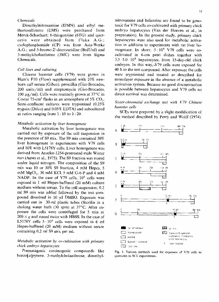
Metabolic activation by liver homogenate 02h 20h 4~h 68h 72h Metabolic activation by liver homogenate was ' ~ . ~ { , ~
carried out by exposure of the cell suspension in . . . . . the presence of $9 mix. The $9 mix contained 30% 02 ~h ~8, 32h liver homogenate in experiments with V79 cells and 10% with L5178Y cells. Liver homogenate was u L_J
derived from Aroclor-1254-pretreated male Wistar 02h 20h ~4h ~bh 1~ t 5 rats (Ames et al., 1975). The $9 fraction was stored c I _ I .............. :i
under liquid nitrogen. The composition of the $9 mix was 10 or 30% $9 fraction, 4 mM Hepes, 5 02h ~0h 32h ~h ~h
? 1. i l ..... I ] mM MgCI2, 30 mM KCI, 5 mM G-6-P and 4 mM D . . . . . . [ NADP. In the case of V79 cells, 10 6 cells were exposed in 1 ml Hepes-buffered (20 mM) culture o2, 20, 32h ~ ~bh medium without serum. To the cell suspension, 0.2 E ;~_~ ml $9 mix was added followed by the test com-
0 50 52h 76h 80h pound dissolved in 10 #l DMSO. Exposure was F [ .:~ y carried out in 30-ml plastic tubes (Sterilin in a x shaking water bath (30 rpm) at 37°C. After ex-
0 18n 58 60h 80h 8/+h posure the cells were centrifuged for 5 min at t . ' 4 200 × g and rinsed twice with HBSS. In the case of x L5178Y cells 5 -10 6 cells were exposed in 6 ml Hepes-buffered (20 mM) medium without serum I Cell affochmenf ~ ] ~ $9 . . . .
containing 0.2 ml $9 mix per ml. EZ3 N . . . . I growth ~ Exp . . . . . foq ..... f,n
~ ] BrdUrd ~n presence of ernbryomc chick hepotocytes
Metabofic activation by co-cultivation with primary E~ Quercetin exp . . . . . X Trypsinizahon
chick embryo hepatocytes EZ3 [okch . . . . .
Premutagenic carcinogenic compounds like Fig. 1. Various methods used for exposure of V79 cells to benzo[a]pyrene, 3-methylcholanthrene, dimethyl- quercetin in SCE experiments.
12
Various protocols of exposure of the cells were EMS (120 /~g/ml) was used as a positive con- used (see Fig. 1). In the case of exposure in trol in all experiments without exogenous meta- monolayer, V79 Chinese hamster cells were seeded bolic activation systems. In all cases, > 1 on flame-sterilized slides (20 cm 2) placed in 10-cm SCE/chromosome was observed. In experiments plastic petri dishes (Greiner). After 2 h the cells with metabolic activation systems, various pre- were attached and complete culture medium was mutagens were used as positive controls as men- added to the dishes to a total volume of 10 ml. The tioned in the respective tables. compound dissolved in DMSO (final concentra- No statistical treatment of the SCE results was tions 0.2%) was added 18 h later. At that time, performed. In the case of SCEs, a statistically 0 .5-1.0.105 cells were present on the slides. After significant result is not always evidence of geno- 24 h of exposure the medium was removed, the toxic activity. For example, a compound that is cells were rinsed twice with Hanks ' balanced salt not genotoxic but that elongates the S phase of the solution (HBSS) and fresh medium containing cell cycle can increase the SCE rate by 30% (see BrdUrd (10/ IM) was added for 28 h. During the also Jongen et al., 1981). This increase may be very last 4 h colchicine (0.1 ~ g / m l ) was present (method significant in a statistical way, but is not the result A, Fig. 1). In the case of exposure in the presence of genotoxic activity. We consider, therefore, a of liver homogenate, cells were treated with result to be positive in the SCE assay when a quercetin in suspension (method B), in contrast to positive dose-response curve is obtained at non- the other experiments on the induction of SCEs toxic concentrations in the absence of BrdUrd (see which were performed with cells in monolayer, also Results). For metabolic activation, co-cultivation of V79 cells with primary chick embryo hepatocytes was Forward mutation test with V79 Chinese hamster used as well (method G). After exposure the cells cells
were seeded on the slides. After cell attachment The forward mutation test on 6-thioguanine the cells were treated with BrdUrd (methods B and resistance in V79 Chinese hamster cells was per- G). In other experiments cells were simultaneously formed according to the method of Van Zeeland exposed to quercetin and BrdUrd (methods C, D and Simons (1975, 1976a,b). Cells were exposed to and E) or the cells were pretreated with BrdUrd quercetin following 3 different procedures. The (method F). After colchicine treatment the medium first procedure consisted of a 24-h treatment with was removed, the cells were rinsed with HBSS, quercetin in monolayer. The cells were exposed to swollen with 0.56% KC1 for 15 rain at room tern- different concentration of quercetin during 24 h. perature and fixed in a 3 : 1 mixture of methanol After exposure the cells were collected by trypsini- and acetic acid. The slides were dried overnight, zation. To determine survival about 200 cells were washed with distilled water, treated for 10 min seeded in 9-cm petri dishes (Costar) and incubated with Hoechst (fluorochrome 33258, 5 /~g/ml) in for 6-7 days. The colonies were then fixed in phosphate buffer (pH 7.0) in the dark, rinsed in methanol, stained with Giemsa (10%)and counted. distilled water and exposed to UV radiation At least 2.5.105 cells were subcultured to allow (HBO-Hg, 100 W) for 4 h in a phosphate citrate expression of mutations. Cell densities were kept buffer (pH 7.0). Subsequently the cells were stained between 2.5 • 105 and 4 .106 cells per 75 c m 2. After in 5% Giemsa in SOrensens' buffer (pH 6.8) for 10 7 days of expression cells were seeded in 9-cm min. The preparations were dried overnight and petri dishes. Each dish contained 105 cells in 10 ml SCEs were scored at a magnification of 1000 × . complete medium supplemented with 6TG (10 The number of sister-chromatid exchanges was #g /ml ) . Colonies were fixed, stained and scored expressed as the number of SCEs/chromosome after 10 days. For estimation of survival again 200 because the V79 cell line is pseudodiploid. At each cells were plated. For determination of both muta- dose level at least 25 metaphases were scored. In tion frequency and survival 5 plates per dose level some cases (Tables 1, 3, 4, 5 and 6) experiments were used. Mutation frequencies are expressed as were carried out in duplicate (indicated with a en mutants per 105 survivors. In the two other proce- b). dures, cells were exposed in the presence of exoge-
13
nous metabol ic ac t ivat ion systems. Metabo l i c in c loning med ium supp lemented with 5 ~ g / m l ac t iva t ion was achieved by apply ing co-cul t ivat ion 6 T G and 3 . 1 0 5 cells in c loning med ium with 50 with ei ther p r imary chick embryo hepatocytes or ~ g / m l BrdUrd . Fo r each locus, 10 plates were liver homogena te as descr ibed for the SCE test. A n used. At the same t ime cloning efficiencies were effect was considered to be mutagenic when the de te rmined as descr ibed for direct survival mea- induced muta t ion frequency was at least 3 t imes surements . Colonies were counted by eye after higher than the spon taneous muta t ion frequency 10-13 days of incubat ion. Muta t ion frequencies for that specific exper iment (Bradley et al., 1981). are expressed as the number of mutan t s per 10 5
survivors. A doubl in~ of the spontaneous muta t ion Forward mutation test with L5178Y mouse rate is considered to be a mutagenic effect (Clive lymphoma cells et al., 1979).
The muta t ion test with mouse l y m p h o m a L5178Y cells was carr ied out as descr ibed by Clive
Results et al. (1979), with minor modif icat ions . Pr ior to the start of the tests, s tock cells were grown for 24
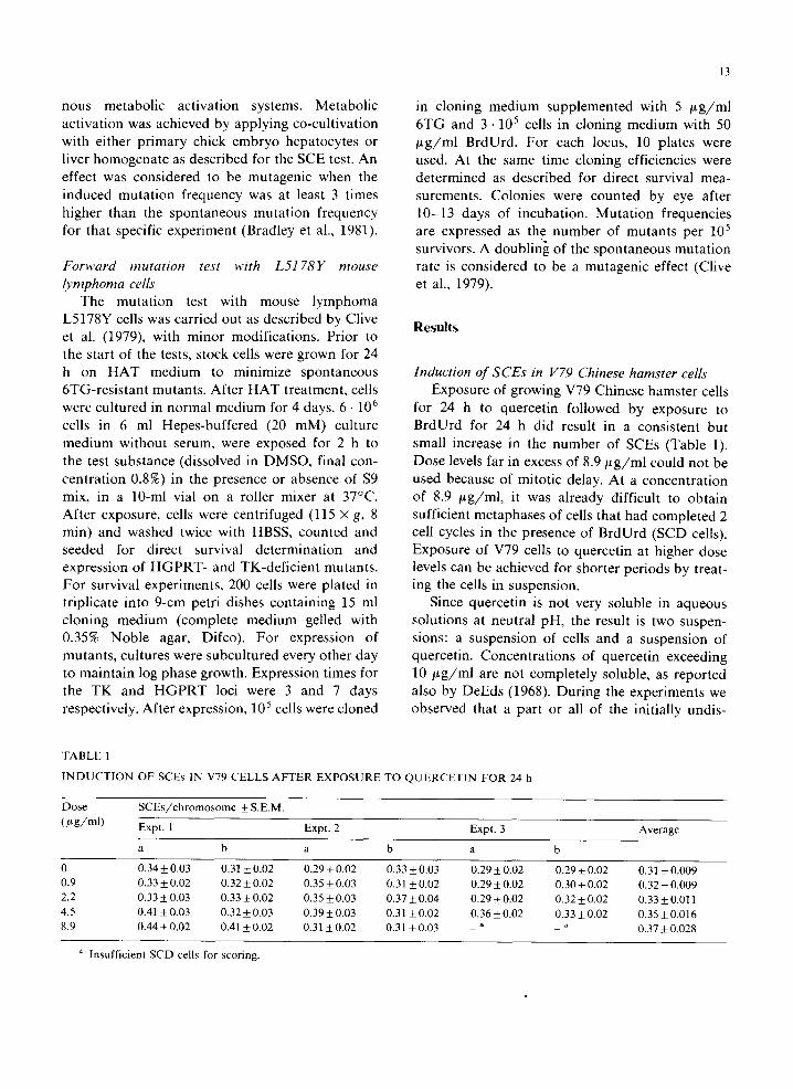
h on H A T med ium to minimize spontaneous Induction of SCEs in V79 Chinese hamster cells 6TG-res i s tan t mutants . Af ter H A T t reatment , cells Exposure of growing V79 Chinese hamste r cells were cul tured in normal med ium for 4 days. 6 - 106 for 24 h to quercet in fol lowed by exposure to cells in 6 ml Hepes-buf fe red (20 mM) cul ture BrdUrd for 24 h did result in a consis tent but med ium without serum, were exposed for 2 h to small increase in the number of SCEs (Table 1). the test subs tance (dissolved in D M S O , final con- Dose levels far in excess of 8 . 9 / ~ g / m l could not be cen t ra t ion 0.8%) in the presence or absence of $9 used because of mi tot ic delay. At a concent ra t ion mix, in a 10-ml vial on a roller mixer at 37°C. of 8.9 /~g/ml, it was a l ready difficult to ob ta in Af te r exposure, cells were centr i fuged (115 x g, 8 sufficient metaphases of cells that had comple ted 2 rain) and washed twice with HBSS, counted and cell cycles in the presence of B rdUrd (SCD cells). seeded for direct survival de te rmina t ion and Exposure of V79 cells to quercet in at higher dose express ion of H G P R T - and TK-def ic ien t mutants , levels can be achieved for shorter per iods by treat- F o r survival exper iments , 200 cells were p la ted in ing the ceils in suspension. t r ipl icate into 9-cm petr i dishes conta in ing 15 ml Since quercet in is not very soluble in aqueous c loning med ium (comple te med ium gelled with solut ions at neutra l pH, the result is two suspen- 0.35% N o b l e agar, Difco). F o r expression of sions: a suspension of cells and a suspension of mutants , cul tures were subcul tured every other day quercet in. Concen t ra t ions of quercet in exceeding to ma in ta in log phase growth. Express ion t imes for 1 0 / ~ g / m l are not comple te ly soluble, as repor ted the T K and H G P R T loci were 3 and 7 days also by DeEds (1968). Dur ing the exper iments we respectively. Af te r expression, 10 5 cells were cloned observed that a par t or all of the ini t ial ly undis-
TABLE 1
INDUCTION OF SCEs IN V79 CELLS AFTER EXPOSURE TO QUERCETIN FOR 24 h
Dose SCEs/chromosome _+ S.E.M.
(/~g/ml) Expt. 1 Expt. 2 Expt. 3 Average
a b a b a b
0 0.34_+0.03 0.31_+0.02 0.29_+0.02 0.33_+0.03 0.29_+0.02 0.29_+0.02 0.31+_0.009 0.9 0.33 _+ 0.02 0.32 _+ 0.02 0.35 _+ 0.03 0.31 4- 0.02 0.29 -+ 0.02 0.30 4-_ 0.02 0.32 _+ 0.009 2.2 0.33_+0.03 0.33_+0.02 0.35-+0.03 0 .37-+0.04 0.29_+0.02 0.32_+0.02 0.33_+0.011 4.5 0.41-+0.03 0.32+0.03 0.39_+0.03 0.31_+0.02 0.36_+0.02 0 .33-+0.02 0.35-+0.016 8.9 0.44_+0.02 0.41 +0.02 0.31 -+0.02 0.31 _+0.03 _ a ~ 0.37_+0.028
a Insufficient SCD cells for scoring.
14
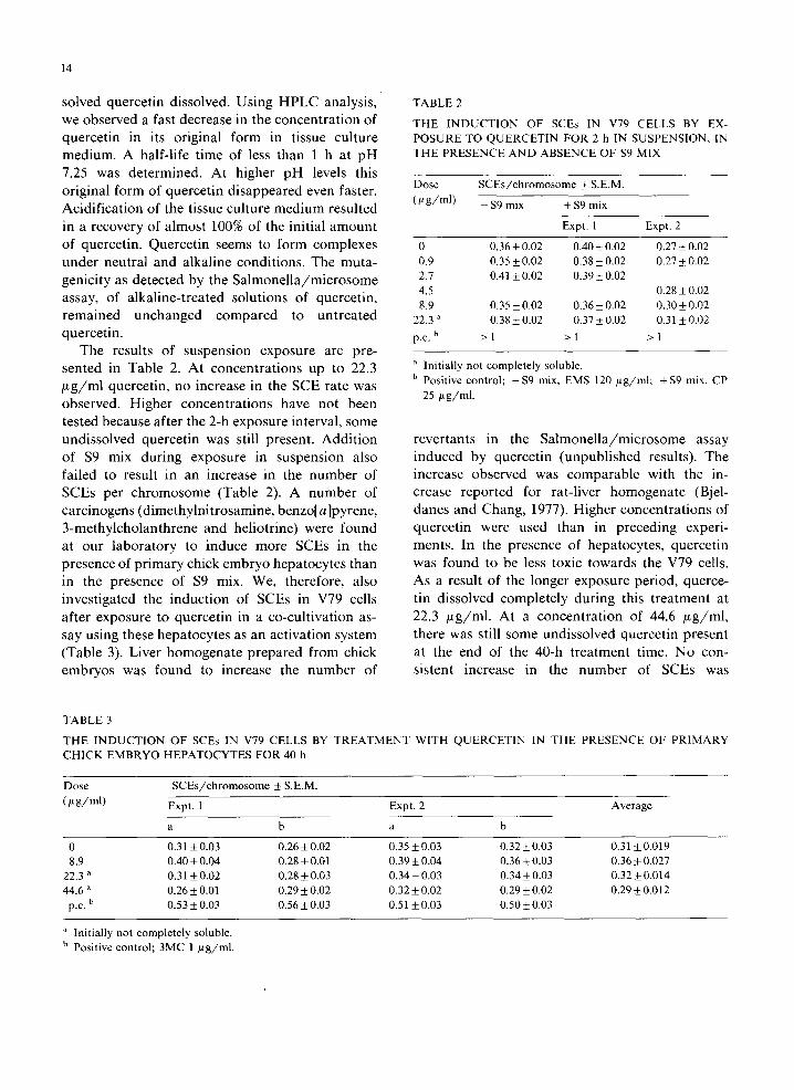
solved quercetin dissolved. Using HPLC analysis, TABLE 2
we observed a fast decrease in the concentra t ion of THE INDUCTION OF SCEs IN V79 CELLS BY EX- quercetin in its original form in tissue culture POSURE TO QUERCETIN FOR 2 h IN SUSPENSION, IN medium. A half-life time of less than 1 h at pH THE PRESENCE AND ABSENCE OF $9 MIX
7.25 was determined. At higher pH levels this original form of quercet in disappeared even faster. D o s e SCEs/chromosome + S.E.M.
Acidif icat ion of the tissue culture medium resulted (t2g/ml) - s9 mix + $9 mix
in a recovery of almost 100% of the initial amoun t Expt. 1 Expt. 2
of quercetin. Quercetin seems to form complexes 0 0.36_+0.02 0.40_+0.02 0.27_+0.02 under neutral and alkaline conditions. The muta- 0.9 0.35 + 0.02 0.38 + 0.02 0.27_+ 0.02 genicity as detected by the Sa lmone l l a /mic rosome 2.7 0.41 + 0.02 0.39_+ 0.02 assay, of alkaline-treated solutions of quercetin, 4.5 0.28+0.02
8.9 0.35+0.02 0.36+0.02 0.30+_0.02 remained unchanged compared to unt rea ted 22.3 a 0.38+0.02 0.37_+0.02 0.31+0.02 quercetin, p.e. b > 1 > 1 > 1
The results of suspension exposure are pre- sented in Table 2. At concentra t ions up to 22.3 a Initially not completely soluble.
b Positive control; -$9 mix, EMS 120 /~g/ml; + $9 mix, CP /2g/ml quercetin, no increase in the SCE rate was 25 ~t g/ml.
observed. Higher concentra t ions have not been
tested because after the 2-h exposure interval, some undissolved quercetin was still present. Addi t ion revertants in the Sa lmone l l a /mic rosome assay of $9 mix during exposure in suspension also induced by quercetin (unpubl ished results). The failed to result in an increase in the number of increase observed was comparable with the in- SCEs per chromosome (Table 2). A number of crease reported for rat-liver homogenate (Bjel-
carcinogens (dimethylni t rosamine, benzo[a]pyrene, danes and Chang, 1977). Higher concentrat ions of 3-methylcholanthrene and he l io t r ine )were found quercetin were used than in preceding experi- at our laboratory to induce more SCEs in the ments. In the presence of hepatocytes, quercetin
presence of pr imary chick embryo hepatocytes than was found to be less toxic towards the V79 cells. in the presence of $9 mix. We, therefore, also As a result of the longer exposure period, querce- investigated the induct ion of SCEs in V79 cells tin dissolved completely during this t reatment at
after exposure to quercet in in a co-cult ivation as- 22.3 # g / m l . At a concentra t ion of 44.6 /~g/ml, say using these hepatocytes as an activation system there was still some undissolved quercetin present
(Table 3). Liver homogenate prepared from chick at the end of the 40-h t reatment time. No con- embryos was found to increase the n u m b e r of sistent increase in the number of SCEs was
TABLE 3
THE INDUCTION OF SCEs IN V79 CELLS BY TREATMENT WITH QUERCETIN IN THE PRESENCE OF PRIMARY CHICK EMBRYO HEPATOCYTES FOR 40 h
D o s e SCEs/chromosome ! S.E.M.
(/~ g/ml) Expt. 1 Expt. 2 Average
a b a b
0 0.31 +_0.03 0.26_+0.02 0.35 _+0.03 0.32+0.03 0.31 +0.019 8.9 0.40 _+ 0.04 0.28 + 0.01 0.39 -+ 0.04 0.36 _+ 0.03 0.36 -+ 0.027
22.3 a 0.31 _+ 0.02 0.28 + 0.03 0.34 _+ 0.03 0.34 _+ 0.03 0.32 _+ 0.014 44.6 a 0.26 _+ 0.01 0.29 _+ 0.02 0.32 _+ 0.02 0.29 _+ 0.02 0.29 _+ 0.012
p.c. b 0.53 _+ 0.03 0.56 + 0.03 0.51 _+ 0.03 0.50 _+ 0.03
a Initially not completely soluble. ~' Positive control; 3MC 1 /~g/ml.
15
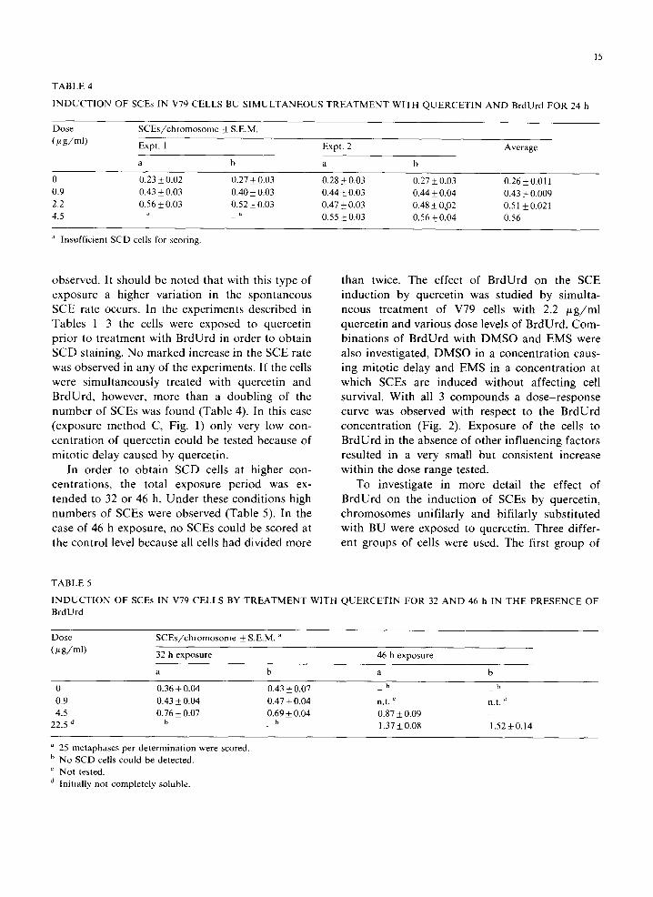
TABLE 4
INDUCTION OF SCEs IN V79 CELLS BU SIMULTANEOUS TREATMENT WITH QUERCETIN AND BrdUrd FOR 24 h
Dose SCEs/chromosome ± S.E.M.
(#g/ml) Expt. 1 Expt. 2 Average
a b a b
0 0,23 ± 0.02 0.27 + 0,03 0.28 ± 0.03 0.27 _+ 0.03 0.26 ± 0.011 0.9 0.43 ± 0.03 0.40 ± 0,03 0.44 ± 0.03 0.44 + 0.04 0.43 ± 0.009 2.2 056 ± 0.03 0.52 ± 0.03 0.47 ± 0.03 0.48 ± 002 0.51 _+ 0.021 4.5 - ~' - " 0.55 ± 0.03 0.56 ± 0.04 0.56
Insufficient SCD cells for scoring.
obse rved . It shou ld be n o t e d that wi th this type of t han twice. T h e ef fec t of B r d U r d on the S C E
e x p o s u r e a h igher va r i a t ion in the s p o n t a n e o u s i n d u c t i o n by q u e r c e t i n was s tud ied by s imul ta -
S C E rate occurs . In the e x p e r i m e n t s desc r ibed in neous t r e a t m e n t o f V79 cells wi th 2.2 ~ g / m l
T a b l e s 1 3 the cells were exposed to que rce t i n q u e r c e t i n and va r ious dose levels of B r d U r d . Corn-
p r i o r to t r e a t m e n t wi th B r d U r d in o rde r to o b t a i n b i n a t i o n s of B r d U r d wi th D M S O and E M S were
S C D sta ining. N o m a r k e d increase in the S C E rate a lso inves t iga ted , D M S O in a c o n c e n t r a t i o n caus-
was o b s e r v e d in any of the expe r imen t s . I f the cells ing mi to t i c de lay and E M S in a c o n c e n t r a t i o n at
were s i m u l t a n e o u s l y t rea ted wi th que rce t i n and wh ich S C E s are i n d u c e d w i thou t a f fec t ing cell
B r d U r d , however , m o r e than a d o u b l i n g of the survival . W i t h all 3 c o m p o u n d s a d o s e - r e s p o n s e
n u m b e r o f S C E s was f o u n d (Tab le 4). In this case cu rve was obse rved wi th respec t to the B r d U r d
( e x p o s u r e m e t h o d C, Fig. 1) on ly very low con- c o n c e n t r a t i o n (Fig. 2). E x p o s u r e of the cells to
c e n t r a t i o n of q u e r c e t i n cou ld be tes ted because of B r d U r d in the absence o f o the r i n f luenc ing fac tors
m i t o t i c de lay caused by querce t in , resu l ted in a ve ry smal l bu t cons i s t en t increase
In o rde r to o b t a i n S C D cells at h ighe r con- w i th in the dose r ange tested.
cen t r a t i ons , the to ta l e x p o s u r e pe r iod was ex- T o inves t iga te in m o r e deta i l the ef fec t of
t e n d e d to 32 or 46 h. U n d e r these c o n d i t i o n s high B r d U r d on the i n d u c t i o n of S C E s by querce t in ,
n u m b e r s o f S C E s were obse rved (Tab le 5). In the c h r o m o s o m e s un i f i l a r ly and bi f i la r ly subs t i tu ted
case of 46 h exposure , no SCEs cou ld be scored at w i th B U were exposed to querce t in . Th ree d i f fer -
the con t ro l level because all cells had d iv ided m o r e en t g roups o f cells were used. T h e first g roup of
TABLE 5
INDUCTION OF SCEs IN V79 CELLS BY TREATMENT WITH QUERCETIN FOR 32 AND 46 h IN THE PRESENCE OF BrdUrd
Dose SCEs/chromosome ± S.E.M. a
(~g/ml) 32 h exposure 46 h exposure
a b a b
0 0.36_+0.04 0.43±0.07 _ b _ h 0.9 0.43 ± 0.04 0.47 ± 0.04 n.t. c n.t. ~ 4.5 0.76 ± 0.07 0.69 ± 0.04 0.87 ± 0.09 -
22.5 d _ b _ b 1.37_+0.08 1.52_+0.14
25 metaphases per determination were scored. b No SCD cells could be detected. c Not tested. ,t Initially not completely soluble.
16
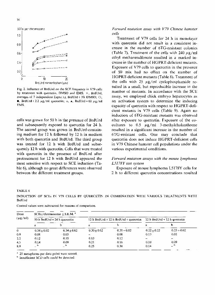
SEEs per chromosome Forward mutation assay with V79 Chinese hamster
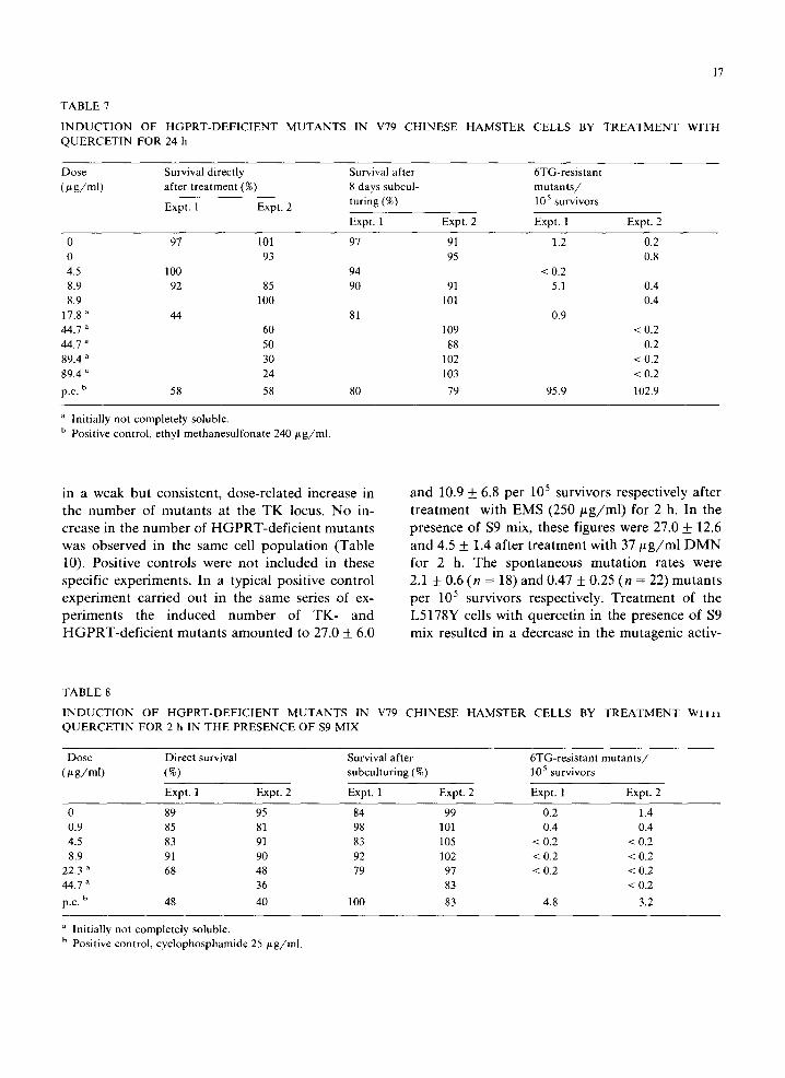
1.0 ~ ~ • cells i ~ J T r e a t m e n t of V79 cells for 24 h in m o n o l a y e r
- J " wi th q u e r c e t i n d id no t resul t in a cons i s t en t in- 0.8 ,• • - ~ - • " , - ~ " . . . . f c rease in the n u m b e r of 6 T G - r e s i s t a n t co lon ies
o--,~" ( T a b l e 7). T r e a t m e n t of the cells wi th 2 4 0 / x g / m l 0.6- __--o
o-.~- ethyl m e t h a n e s u l f o n a t e resu l ted in a m a r k e d in- / / .... D
// D -_D . . . . . . 04 o/, - c rease in the n u m b e r of H O P R T - d e f i c i e n t mu tan t s .
. . . . . . . . . . . . . . . × × x x E x p o s u r e of V79 cells to que rce t i n in the p resence
0.2- o f $9 mix had no ef fec t on the n u m b e r of
0 1'0 20 H G P R T - d e f i c i e n t m u t a n t s (Tab le 8). T r e a t m e n t of
BrdUrdconcentrafion(l~rnI the cells wi th 25 ~ g / m l c y c i o p h o s p h a m i d e re- su l ted in a small , bu t r e p r o d u c i b l e increase in the
Fig. 2. Influence of BrdUrd on the SCE frequency in V79 cells n u m b e r of mu tan t s . In a c c o r d a n c e wi th the S C E by treatment with quercetin, DMSO and EMS. ×, BrdUrd,
averages of 7 independent Expts; D, BrdUrd+ 3% DMSO; O, assay, we e m p l o y e d chick e m b r y o h e p a t o c y t e s as e, BrdUrd+2.2 /.tg/ml quercetin; A A, BrdUrd+60 /~g/ml an ac t i va t i on sys tem to d e t e r m i n e the i nduc ing
EMS. c a p a c i t y o f q u e r c e t i n wi th respect to H G P R T - d e f i -
c ien t m u t a n t s in V79 cells ( T a b l e 9). A g a i n no
i n d u c t i o n o f 6 T G - r e s i s t a n t m u t a n t s was obse rved
cells was g r o w n for 50 h in the p re sence of B r d U r d a f te r exposu re to querce t in . E x p o s u r e of the co-
and subsequen t l y exposed to q u e r c e t i n for 24 h. cu l tu re s to 0.5 / ~ g / m l 3 - m e t h y l c h o l a n t h r e n e
T h e second g roup was g r o w n in B r d U r d - c o n t a i n - resu l ted in a s ign i f i can t increase in the n u m b e r of
ing m e d i u m for 12 h fo l lowed by 12 h in m e d i u m 6 T G - r e s i s t a n t cells. O n e m a y c o n c l u d e that
wi th b o t h q u e r c e t i n and B r d U r d . T h e th i rd g roup q u e r c e t i n does n o t i nduce H G P R T - d e f i c i e n t cells
was t rea ted for 12 h wi th B r d U r d and subse- in V79 Ch inese h a m s t e r cell p o p u l a t i o n s u n d e r the
q u e n t l y 12 h wi th querce t in , Cel ls that were t rea ted va r i ous e x p e r i m e n t a l cond i t ions .
w i th que rce t i n in the p re sence of B r d U r d af te r
p r e t r e a t m e n t for 12 h wi th B r d U r d a p p e a r e d the Forward mutation assays with the mouse lymphoma m o s t sens i t ive wi th respect to S C E i n d u c t i o n (Ta- L5178Y test system ble 6), a l t hough no great d i f fe rences were obse rved E x p o s u r e o f m o u s e l y m p h o m a L5178Y cells for
b e t w e e n the d i f f e r en t t r e a t m e n t groups . 2 h to d i f fe ren t q u e r c e t i n c o n c e n t r a t i o n s resul ted
TABLE 6
INDUCTION OF SCEs IN V79 CELLS BY QUERCETIN IN COMBINATION WITH VARIOUS TREATMENTS WITH BrdUrd
Control values were subtracted for reasons of comparison.
Dose SCEs/chromosome ___ S.E.M. "
(/~g/ml) 50 h BrdUrd + 24 h quercetin 12 h BrdUrd + 12 h BrdUrd + quercetin 12 h BrdUrd + 12 h quercetin
a b a b a b
0 0.34 +_ 0.02 0.34 +_ 0.02 0.30 _+ 0.02 0.31 _+ 0.02 0.22 _+ 0.22 0.25 _+ 0.02 0.9 0.08 - 0.03 - 0.00 0.13 0.02 2.2 0.12 0.10 0.13 0.12 - - 4.5 0.14 0.09 0.21 0.16 0.10 0.09 8.9 _ b _ b 0.25 0.30 0.14 _ b
a 25 metaphases per data point were scored. b Insufficient SCD cells could be detected.
17
TABLE 7
INDUCTION OF HGPRT-DEFICIENT MUTANTS IN V79 CHINESE HAMSTER CELLS BY TREATMENT WITH QUERCETIN FOR 24 h
Dose Survival directly Survival after 6TG-resistant ( ~ g/ml) after treatment (%) 8 days subcul- mutants/
Expt. 1 Expt. 2 turing (%) 105 survivors
Expt. 1 Expt. 2 Expt. 1 Expt. 2
0 97 101 97 91 1.2 0.2 0 93 95 0.8 4.5 100 94 < 0.2 8.9 92 85 90 91 5.1 0.4 8.9 100 101 0.4
17.8 a 44 81 0.9 44.7 a 60 109 < 0.2 44.7 ~ 50 88 0.2 89.4 " 30 102 < 0.2 89.4 ~ 24 103 < 0.2
p.c. b 58 58 80 79 95.9 102.9
Initially not completely soluble. b Positive control, ethyl methanesulfonate 240 ~g/ml.
in a w e a k b u t c o n s i s t e n t , d o s e - r e l a t e d i n c r e a s e in a n d 10.9 + 6.8 p e r 105 s u r v i v o r s r e s p e c t i v e l y a f t e r
t h e n u m b e r of m u t a n t s a t t he T K locus. N o in- t r e a t m e n t w i t h E M S (250 ~ g / m l ) for 2 h. I n t he
c r e a s e in t he n u m b e r of H G P R T - d e f i c i e n t m u t a n t s p r e s e n c e o f $9 mix , t he se f igures were 27.0 __+ 12.6
w a s o b s e r v e d in t he s a m e cell p o p u l a t i o n ( T a b l e a n d 4.5 + 1.4 a f t e r t r e a t m e n t w i t h 37 ~ g / m l D M N
10), Pos i t i ve c o n t r o l s we re n o t i n c l u d e d in t he se fo r 2 h. T h e s p o n t a n e o u s m u t a t i o n r a t e s we re
spec i f i c e x p e r i m e n t s . In a t yp i ca l p o s i t i v e c o n t r o l 2.1 __+ 0.6 ( n = 18) a n d 0 .47 + 0.25 ( n = 22) m u t a n t s
e x p e r i m e n t c a r r i e d o u t in the s a m e ser ies of ex- p e r 105 s u r v i v o r s respec t ive ly . T r e a t m e n t o f t he
p e r i m e n t s the i n d u c e d n u m b e r of T K - a n d L 5 1 7 8 Y cel ls w i t h q u e r c e t i n in t he p r e s e n c e o f $9
H G P R T - d e f i c i e n t m u t a n t s a m o u n t e d to 27.0 + 6.0 m i x r e s u l t e d in a d e c r e a s e in t he m u t a g e n i c ac t iv -
TABLE 8
INDUCTION OF HGPRT-DEFICIENT MUTANTS IN V79 CHINESE HAMSTER CELLS BY T R E A T M E N T Wltra QUERCETIN FOR 2 h IN THE PRESENCE OF $9 MIX
Dose Direct survival Survival after 6TG-resistant mutants/ (/~g/ml) (%) subculturing (%) 105 survivors
Expt. 1 Expt. 2 Expt. 1 Expt. 2 Expt. 1 Expt. 2
0 89 95 84 99 0.2 1.4 0.9 85 81 98 101 0.4 0.4 4.5 83 91 83 105 < 0.2 < 0.2 8.9 91 90 92 102 < 0.2 < 0.2
22.3 a 68 48 79 97 < 0.2 < 0.2 44.7 a 36 83 < 0.2
p.c. b 48 40 100 83 4.8 3.2
Initially not completely soluble. b Positive control, cyclophosphamide 25/~g/ml.
18
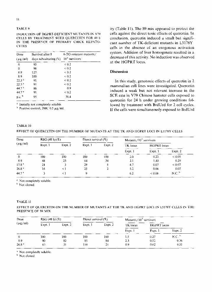
TABLE 9 ity (Tab le 11). T h e $9 mix a p p e a r e d to p r o t e c t the
INDUCTION OF HGPRT-DEFICIENT MUTANTS IN V79 cells aga ins t the d i rec t toxic e f fec ts of que rce t in . In CELLS BY TREATMENT WITH QUERCETIN FOR 40 h conclusion, quercetin induced a small but signifi- IN THE PRESENCE OF PRIMARY CHICK HEPATO- c a n t number of TK-deficient mutants in L5178Y CYTES cel ls in the a b s e n c e of an e x o g e n o u s ac t iva t ion
Dose Survival after 8 6-TG-resistant mutants/ sys t em. A d d i t i o n of l iver h o m o g e n a t e r e su l t ed in a
(/~g/ml) days subculturing (%) 105 survivors d e c r e a s e of this act ivi ty . N o i n d u c t i o n was o b s e r v e d at the H G P R T locus.
0 95 < 0.2 0 96 < 0.2 8.9 127 < 0.2 Discussion 8.9 100 < 0.2
22.3 a 91 < O.2 In this s tudy, g e n o t o x i c e f fec t s of q u e r c e t i n in 2
22.3 a 91 < O.2 m a m m a l i a n cell l ines were • ~,e - ~;nves ' ;"a 'e ' t . "~ - n u e r c e ' ; n 44.7 a 86 0.9 44.7 ~ 91 < O.2 i n d u c e d a weak b u t no t r e l evan t inc rease in the
b 95 38.4 S C E ra te in V79 C h i n e s e h a m s t e r ceils e x p o s e d to p.c. q u e r c e t i n for 24 h u n d e r g r o w i n g c o n d i t i o n s fol-
a Initially not completely soluble, l o w e d by t r e a t m e n t wi th B r d U r d for 2 cell cycles. b Positive control, 3MC 0.5 ~t g/ml. If the cells were s i m u l t a n e o u s l y e x p o s e d to B r d U r d
TABLE 10
EFFECT OF QUERCETIN ON THE NUMBER OF MUTANTS AT THE TK AND HGPRT LOCI IN L5178Y CELLS
Dose RSG (48 h) (%) Direct survival (%) Mutants/10 s survivors
(/~g/ml) Expt. 1 Expt. 2 Expt. 1 Expt. 2 TK locus HGPRT locus
Expt. 1 Expt. 1 Expt. 2
0 100 100 100 100 2.0 0.22 < 0.05 8.9 48 23 64 34 2.1 1.40 0.29
17.8 ~ 21 3 29 5 4.7 0.07 < 0.07 26.8 a 16 < 1 25 2 5.2 0.06 0.07
44.7 ~ 3 < 1 9 6.2 < 0.06 N.C. b
a Not completely soluble. b Not cloned.
TABLE 11
EFFECT OF QUERCETIN ON THE NUMBER OF MUTANTS AT THE TK AND HGPRT LOCI IN L5178Y CELLS IN THE PRESENCE OF $9 MIX
Dose RSG (48 h) (%) Direct survival (%) Mutants/105 survivors
(/~g/ml) Expt. 1 Expt. 2 Expt. 1 Expt. 2 TK locus HGPRT locus
Expt. 1 Expt. 1 Expt. 2
0 100 100 100 100 1.5 0.27 N.C. b 8.9 90 82 85 84 2.3 0.52 0.36
26.8 a 83 31 110 21 0.9 0.62 0.51
Not completely soluble. b Not cloned.
19
and quercetin, however, a marked, more than 3- mutations at the H G P R T locus of V79 cells was fold increase in the SCE rate was observed. Ex- found either in the presence or absence of exoge- posure of V79 cells to BrdUrd in the presence of nous activation systems. Our results conflict with 2.25 /~g/ml quercetin resulted in a dose-response the data reported by Maruta et al. (1979), who curve with respect to the BrdUrd concentration, reported induction of HGPRT-deficient mutants Similar results were observed with DMSO and in V79 cell populations after exposure to quercetin EMS. V79 cells exposed to quercetin in the pres- for 48 h in the absence of an exogenous activation ence of Aroclor-1254-induced liver homogenate or system or exposure for 1 h in the presence of primary hepatocytes, did not show any effect on activated liver homogenate. It should be noted the number of SCEs. Apparently neither quercetin that the experimental protocol used by Maruta nor its metabolites produce lesions which result in and coworkers may be (partly) responsible for an increase in SCEs in V79 cells. The small effect their positive results. They used cell populations on the SCE rate following exposure to quercetin with high numbers of spontaneous mutants (3-18 for 24 h is probably a result of a prolongation of per 105 survivors). Increased numbers of mutants the cell cycle. Similar weak effects were found if were only observed at toxic concentrations of V79 cells were exposed to high DMSO concentra- quercetin. At these conditions, selection of pre-ex- tions (Jongen et al., 1981). Since simultaneous isting mutant cells may have occurred. exposure to quercetin and BrdUrd resulted in an Conflicting results obtained in various toxico- increase in the SCE rate, perhaps quercetin did logical studies may bear some relationship to the inhibit the enzyme poly(ADP-ribose)-polymerase, effects observed concerning the solubility of Inhibitors of this enzyme are potent inducers of quercetin at different pH values. Apparently, SCEs when simultaneous exposure is applied quercetin can be present in aqueous solutions in at (Natarajan et al., 1981). In this study, chro- least two distinct forms. These forms may have mosomes bifilarly substituted with BU were found different capacities to penetrate into cells and to be as sensitive for quercetin as unifilarly sub- therefore, have different effects. stituted chromosomes. This is in contrast with the The results of our study are in accordance with polymerase inhibitors which affect bifilarly sub- those reported by Carver et al. (1983), who found stituted chromosomes more than unifilarly sub- no induction of forward mutations in CHO cells at stituted chromosomes with respect to SCE induc- the HGPRT, APRT and Na+ /K+-ATPase loci, tion. This discrepancy between quercetin and real However, as has also been reported by others polymerase inhibitors cannot be explained. SCE (Carver et al., 1983; Amacher et al., 1979, 1980; induction may, therefore, be influenced by more Meltz and MacGregor, 1981), quercetin induced factors than reported to date. This (these) same forward mutations at the TK locus in mammalian factor(s) is (are) probably also involved in the cells in vitro. The fact that quercetin merely in- induction of SCEs when V79 cells are simulta- duced mutations at the TK locus and not at the neously exposed to DMSO or EMS and BrdUrd. HGPRT, APRT and Na+/K+-ATPase loci in Among these factors may be prolongation of the S mammalian cells indicates that quercetin exhibits phase and more incorporation of BU in the DNA a mutagenic response in mammalian cells by caus- resulting in an increase in the ' spontaneous ' SCE ing gross chromosomal lesions (Clive et al., 1979; rate. The results reported by Sugimura (1979) and Hozier et al., 1981). Indeed various authors have Yoshida et al. (1980) may also be the result of reported the clastogenic activity of quercetin in cooperative action of quercetin and BrdUrd, since Chinese hamster fibroblasts, Chinese hamster in these studies an increase in the number of SCEs ovary cells, human fibroblasts and PHA-stimu- was found in various mammalian cells after lated human lymphocytes (Yoshida et al., 1980; simultaneous exposure to BrdUrd and quercetin. Carver et al., 1983). Our results are in agreement with those of Carver The decrease in mutagenic activity at the TK et al. (1983) who observed no induction of SCEs locus of L5178Y cells in the presence of liver by quercetin in CHO cells, homogenate is an unexpected finding since the
In the present study no induction of forward addition of $9 mix markedly enhanced the muta-
2O
genicity of quercetin in the Salmonella/microsome Activation of flavonol glycosides by mixed glycosidases a s s a y (e.g. Bjeldanes and Chang, 1977). This phe- from rat cecal bacteria and other sources, Mutation Res.,
n o m e n o n a n d the fac t t h a t q u e r c e t i n i n d u c e d o n l y 66, 223-240. gross chromosomal aberrations in mammalian Carver, J.H., A.V. Carrano and J.F. MacGregor (1983) Genetic
effects of the flavonols quercetin, kaempferol, and galangin cells, i n d i c a t e t h a t t h e m e c h a n i s m s of t he e f fec t s of on Chinese hamster ovary cells in vitro, Mutation Res., 113, q u e r c e t i n in b a c t e r i a a n d m a m m a l i a n cells a re no t 45-60. comparable. Inactivation of the clastogenic prop- Clive, D, K.O. Johnson, J.F.S. Spector, A.G. Batson and
M.M.M. Brown (1979) Validation and characterization of e r t i e s of q u e r c e t i n b y l iver h o m o g e n a t e in a s says the L5178Y/TK +/ mouse lymphoma mutagen assay sys-
w i t h m a m m a l i a n cells a n d the fac t t h a t q u e r c e t i n tem, Mutation Res., 59, 61 108.
d o e s n o t i n d u c e S C E s a n d p o i n t m u t a t i o n s in Hardigree, A.A., and J.L. Epler(1978)Comparativemutagene- m a m m a l i a n cells m a y e x p l a i n w h y q u e r c e t i n d i d sis of plant flavonoids in microbial systems, Mutation Res.,
n o t p r o v e to b e c a r c i n o g e n i c in t he m a j o r i t y o f the 58, 231-239. a n i m a l t es t s ( S a i t o et al., 1980; H i r o n o et al., 1981; Hirono, I., I. Heno, S. Hosaka, H. Takanashi, T. Matsushima, H o s a k a a n d H i r o n o , 1981; M o r i n o et al., 1982). T. Sugimura and S. Natori (1981) Carcinogenicity examina-
tion of quercetin and rutin in ACI rats, Cancer Lett., 13, 15 21.
Acknowledgements Hosaka, S., and I. Hirono (1981) Carcinogenicity test of quercetin by pulmonary adenoma bioassay in strain A mice,
T h e s e i n v e s t i g a t i o n s we re f i n a n c i a l l y s u p p o r t e d Gann, 72, 327 328. Hozier, J., J. Sawyer, M. Moore, B. Howard and D. Clive
by a grant from the Dutch Cancer Society (1981) Cytogenetic analysisof the L5178Y TK+/- -~ TK / (Koningin Wilhelmina Fonds, LHW 79-1). mouse lymphoma mutagenesis assay system, Mutation Res.,
W e t h a n k Mr . W . M . F . J o n g e n for p r e p a r a t i o n 84, 169-181. o f the l iver h o m o g e n a t e a n d Mrs . J. H a g e n s - Jongen, W.M.F., P.H.M. Lohman, M.J. Kottenhagen, G.M. K l e i n h o o n t e v a n Os a n d Mrs . M.E . V e r k o r e n for Alink, F. Berends and J.H. Koeman (1981) Mntagencity typing the manuscript, testing of dichloromethane in short-term mammalian test
systems, Mutation Res., 81,203 213. MacGregor, J.T. (1979) Mutagenicity studies of flavonoids in
References vivo and in vitro, Toxicol Appl. Pharmacol., 48, A47. MacGregor, J.T., and L. Jurd (1978) Mutagenicity of plant
Aeschbacher, H.U. (1980) Risk Assessment of muta- flavonoids; Structural requirements for mutagenic activity gens/carcinogens in food, Paper presented at the 10th in Salmonella typhimurium, Mutation Res., 54, 297-309. Annual Meeting of the European Environmental Mutagen MacGregor, J.T., and L.E. Sacks (1979) The Bacillus subtilis Society. multigene sporulation test: Sensitivity to known mutagens
Amacher, D.E., S.C. Paillet and V.A. Ray (1979) Point muta- and carcinogens, Environ. Mutagen., 1, 121. tions at the thymidine kinase locus in L5178Y mouse MacGregor, J.T., and C.M. Wehr (1981) Bone marrow and lymphoma cells, I. Application to genetic toxicological test- peripheral blood erythrocyte micronucleus frequencies in ing, Mutation Res., 64, 391-406. mice exposed to plant flavonols, Paper presented at the
Amacher, D.E., S.C. Paillet, G.N. Turner, V.A. Ray and D.S. 13th Annual Meeting of the American Environmental Salsburg (1980) Point mutations at the thymidine kinase Mutagen Society. locus in L5178Y mouse lymphoma cells, II, Test validation MacGregor, J.T., A.V. Carrano and J.H. Carver (1980) Genetic and interpretation, Mutation Res., 72, 447-474. effects of flavonols in Chinese hamster ovary (cito) cells in
Ambrose, A.M., D.J. Robbins and F. DeEds (1952) Compara- vitro and in rabbit lymphocytes and mouse erythroblasts in tive toxicities of quercetin and quercitrin, J. Am. Pharma- vivo, Environ. Mutagen., 2, 231. col. Assoc. Sci. Ed., 41, 119-122. Maruta, A., K. Enaka and M. Umeda (1979) Mutagenicity of
Ames, B.N., J. McCann and E. Yamasaki (1975) Methods for quercetin and kaempferol on cultured mammalian cells, detecting carcinogens and mutagens with the Sai- Gann, 70, 273-276. monella/mammalian microsome mutagenicity test, Muta- Meltz, M.L., and J.T. MacGregor (1981) Activity of the plant tion Res., 31, 347-364. flavonol quercetin in the mouse lymphoma L5178Y TK +/
Bjeldanes, L.F., and G.W. Chang (1977) Mutagenic activity of mutation, DNA single-strand break and Balb/c 3T3 chemi- quercetin and related compounds, Science, 197, 577-578. cal transformation assays, Mutation Res., 88, 317 324.
Bradley, M.O., B. Bhuyan, M.C. Francis, R. Langenbach, A. Morino, K., N. Matsukura, T. Kawachi, H. Ohgaki, T. Sugimura Peterson and E. Huberman (1981) Mutagenesis by chemical and I. Hirono (1982) Carcinogenicity test of quercetin and agents in V79 Chinese hamster cells: A review and analysis rutin in golden hamsters by oral administration, Carcino- of the literature, Mutation Res., 87, 81-142. genesis, 3, 93-97.
Brown, J.P., and P.S. Dietrich (1979) Mutagenicity of plant Natarajan, A.T., I. Csuk/~s and A.A. van Zeeland (1981) Con- flavonols in the Salmonella/mammalian microsome test; tribution of incorporated 5-bromodeoxypuridine in DNA to
21
the frequencies of sister-chromatid exchanges induced by formation of hamster embryo cells by quercetin, Toxicol. inhibitors of poly-(ADP-ribose)-polymerase, Mutation Res., Lett., 1, 175-178. 84, 125-132. Van der Hoeven, J.C.M., I.M. Bruggeman, I. van Dis and Y.
Pamukcu, A.M., S. Jalciner, J.F. Hatcher and G.F. Bryan van Erp (1984) Embryonic chick hepatocytes as a metabolic (1980) Quercetin, a rat intestinal and bladder carcinogen activation system in in vitro genotoxicity assays, I. Induc- present in bracken fern ( Pteridium aquilinum ), Cancer Res., tion of SCEs in V79 Chinese hamster cells by premutagenic 40, 3468-3472. compounds, in preparation.
Perry, P., and S. Wolff (1974) New Giemsa method for the Van Zeeland, A.A., and J.W.I.M. Simons (1975) Ploidy level differential staining of sister chromatids, Nature (London), and mutation to hypoxanthine-guanine-phosphoribosyl- 251,156 158. transferase (HGPRT) deficiency in Chinese hamster cells,
Sahu, R.K., R. Basu and A. Sharma (1981) Genetic toxicologi- Mutation Res., 28, 239-250. cal testing of some plant flavonoids by the micronucleus Van Zeeland, A.A., and J.W.I.M. Simons (1976a) The use of test, Mutation Res., 89, 69-74. correction factors in the determination of mutant frequen-
Saito~ D.~ A. Shirai, T. Matsushima, T. Sugimura and 1. Hirono cies in populations of human diploid skin fibroblasts, Muta- (1980) Test of carcinogenicity of quercetin, a widely distrib- tion Res., 34, 149-158. uted mutagen in food, Teratogen. Carcinogen. Mutagen.~ 1, Van Zeeland, A.A., and J.W.I.M. Simons (1976b) Linear 213-221. dose-response relationships after prolonged expression
Sugimura, T. (1979)Naturally occurring genotoxic carcinogens, times in V79 Chinese hamster cells, Mutation Res., 35, in: E.C. Miller, J.A. Miller, I. Hirono, T. Sugimura and S. 129-138. Takayama (Eds.), Naturally Occurring Carci- Watson, W.A.F. (1982) The mutagenic activity of quercetin and nogens-Mutagens and Modulators of Carcinogenesis, Jpn. kaempferol in Drosophila melanogaster, Mutation Res., 103, Sci. Soc. Press, Tokyo, pp. 241-261. 145 147.
Sugimura, T., M. Nagao, T. Matsushima, T. Yahagi, Y. Seino, Willhite, C.C. (1982) Teratogenic potential of quercetin in the A. Shirai, M. Sawamura, S. Natori, K. Yoshihira, M. rat, Food Chem. Toxicol., 20, 75-79. Fukuoka and M. Kuroyanagi (1977) Mutagenicity of flavone Yoshida, M.A., M. Sasaki, K. Sugimura and T. Kawachi (1980) derivatives, Proc. Jpn. Acad., Ser. B, 53, 194-197. Cytogenetic effects of quercetin on cultured mammalian
Umezawa, K., T. Matsushima, T. Sugimura, T. Hirakawa, M. cells, Proc. Jpn. Acad., Ser. B, 56, 443-447. Tanaka, Y. Katoh and S. Takayama (1977) In vitro trans-