genetic instability induced by low doses of x-rays in hamster cells
TRANSCRIPT

Genetic instability induced by low doses of x-rays in hamster cells
A. SEOANE, A. GUERCI & F. DULOUT{
Centro de Investigaciones en Genetica Basica y Aplicada (CIGEBA), Facultad de Ciencias Veterinarias, Universidad
Nacional de La Plata, La Plata, Argentina
(Received 28 October 2006; revised 17 November 2006; accepted 20 November 2006)
AbstractPurpose: Genomic instability involves time delayed events and can be manifested as elevated rates of heritable changes inthe progeny of irradiated cells. To study the induction of chromosomal instability by very low doses of radiation ChineseHamster Ovary (CHO) cells were exposed to 10 – 50 milisieverts (mSv) (�10 – 50 miligrays (mGy)) of x-rays.Materials and methods: Control and irradiated cell populations were assayed for chromosomal aberrations and assessedusing a micronucleus test and anaphase-telophase analysis at the first cell division post-irradiation and at every fourpopulation doublings thereafter up to 16 population doublings post-irradiation.Results: Frequencies of micronuclei, anaphase-telophase alterations and chromosomal aberrations were increased when thecells were analysed immediately after x-ray exposure. Micronuclei and anaphase-telophase alterations showed significantlyincreased frequencies when they were analysed at 12 and 16 population doublings after exposure to 50 mSv. Chromosomalaberrations increased significantly at 12 and 16 population doublings after exposure to 10 mSv and 50 mSv.Conclusions: Our results are consistent with the presence of a phenomenon by which the initial DNA damage in thesurviving cells is memorized. Micronuclei and achromatic lessions were the main cytogenetic damage observed in cellsexposed to very low doses of x-rays, indicating that these low doses are able to induce genetic instability.
Keywords: Genetic instability, ionizing radiation, low doses
Introduction
It is widely accepted that ionizing radiation is a
mutagenic agent capable of inducing deleterious
effects in human beings. The biological effects of
exposure to radiation were considered for many years
to be a consequence of direct DNA damage that was
not correctly restored by metabolic repair processes.
Although it is assumed that the risk of genotoxic
damage increases in proportion to the dose of
radiation, the situation is much less clear at low
doses (Brenner et al. 2003). Recently, several lines of
evidence have suggested new pathways for radiation-
induced genetic damage, mainly after low dose
exposure (Little 2003, Morgan 2003). Two non-
targeted phenomena, genomic instability and the
bystander effect, have been described over the last
two decades suggesting that ionizing radiation
exposure may induce damage in cells that are not
being directly irradiated. Genomic instability
involves time delayed events whereas the bystander
effect includes the occurrence of damage in cells that
are not themselves directly transversed by radiation.
Genomic instability can be manifested as elevated
rates of heritable changes in the progeny of irradiated
cells. It can be measured as chromosomal aberra-
tions, micronucleus formation, gene mutations and
microsatellite instabilities as well as other end points,
but chromosomal changes are the best described
(Morgan 2003, Limoli et al. 2000). Stable aberrations
can be transmitted through many generations of cell
replication (Little 2003). They can appear de novo in
the progeny of cells that survive the original radiation
exposure (Smith et al. 2003). A great variety of papers
have reported the induction of chromosomal instabil-
ity by low dose radiation exposure. Different cellular
types such as lymphocytes, HPV-G transfected
keratinocytes, CHO and other cells showed evidence
of delayed chromosomal damage after exposure to
g- or w-rays (0.1 – 12 Grays) (Gy) or a-particles
Correspondence: A. Seoane, Centro de Investigaciones en Genetica Basica y Aplicada (CIGEBA), Facultad de Ciencias Veterinarias, Universidad Nacional de
La Plata, Calle 60 y 118, CC 296 B1900AVW La Plata, Argentina. E-mail: [email protected]{Deceased.
Int. J. Radiat. Biol., Vol. 83, No. 2, February 2007, pp. 81 – 87
ISSN 0955-3002 print/ISSN 1362-3095 online � 2007 Informa UK Ltd.
DOI: 10.1080/09553000601129085
Int J
Rad
iat B
iol D
ownl
oade
d fr
om in
form
ahea
lthca
re.c
om b
y U
nive
rsita
ets-
und
Lan
desb
iblio
thek
Due
ssel
dorf
on
12/0
5/13
For
pers
onal
use
onl
y.

(Holmberg et al. 1998, Bartolleto et al. 2001,
Mothersill et al. 2000, Little et al. 1997, Ponnaiya
et al. 2004). Elevated rates of micronucleated cells
were observed in the progeny of primary human
fibroblasts and V-79 cells irradiated with w rays or
a-particles (Belyakov et al. 1999, Trott et al. 1998,
Jamali & Trott 1996). On the other hand, no evidence
of persistent transmissible genomic instability was
found in a study of blood lymphocytes in the bone
marrow of radiation workers with internal deposits of
plutonium (Whitehouse & Tawn 2001) or in normal
diploid human fibroblasts (AG1521A) surviving after
exposure in G(0) to low- and high-LET radiation
(Dugan & Bedford 2003).
In order to establish the induction of chromosomal
instability by very low doses of radiation CHO cells
were exposed to 10 – 50 milisievert (mSv) (�10 –
50 mGy) of w-rays. Chromosomal aberrations and
micronuclei were analysed at various times after the
initial radiation exposure.
Material and methods
Cells
CHO cells obtained from the American Type
Culture Collection (Manassas, VA, USA) were
cultured in Ham’s F10 medium (Gibco BRL, Grand
Island, NY, USA) supplemented with 10% foetal
bovine serum (Notocor Laboratories, Province of
Cordoba, Argentina) and antibiotics (50 IU penicil-
lin and 50 mg/ml streptomycin) (Bago Laboratories,
Buenos Aires, Argentina) in a humidified atmo-
sphere with 5% CO2. Cells were cultured in Falcon
T-25 flasks (Nunc, Nalge Nunc International,
Denmark) with 10 ml culture medium.
Experimental design
Irradiation treatments were performed when the cells
were 90 – 95% confluent. Two radiation doses were
employed: 10 and 50 mSv, based on previous work
in our laboratory (Guerci et al. 2003, 2004) and the
dosimetry reported in epidemiological studies of
human exposures to ionizing radiation (Barquinero
et al. 1993, Paz y Mino et al. 1995, Balakrishnan &
Rao 1999, Cardoso et al. 2001, Cavallo et al. 2002).
The x-ray apparatus used was from Dental San Justo
company (Buenos Aires, Argentina) operated at
65 kV and 5 mA. Doses were determined by using
a Keithley Digital 35617 EBS microchamber
PTW N 2336/414 dosimeter (C-Com Industries,
Robertville, MO, USA). The dose rate employed was
50 mSv/min. Radiation was given from above
through the medium and exposure times were 12
and 60 sec for 10 and 50 mSv respectively. For the
irradiation treatment 10 ml of fresh medium was
placed on the attached cells to avoid the presence of
detached cells.
After treatment, cells were trypsinized, resus-
pended and divided into two fractions. One fraction
was cultured for cytogenetic analysis and the other
fraction continued in culture for 16 passages. A
control group remained unirradiated.
The doubling time of CHO cells under these
culture conditions was periodically checked in the
laboratory using a bromodeoxyuridine technique
(BrdU) and it varie between 12 and 15 h (Grillo &
Dulout, 1995) for both control and irradiated cells.
Control and irradiated populations were assayed
for chromosomal aberrations, frequency of micro-
nuclei and anaphase-telophase analysis at the first
cell division post-irradiation, and at every four
population doublings thereafter up to 16 population
doublings post-irradiation. Each experiment was
repeated twice and mean values are shown in
Tables I – V.
Cytogenetic analysis
Cells were cultured as monolayers for 15 – 16 hr.
Two hours before fixation colchicine (Merk,
Darmstadt, Germany), 1 mg/ml final concentration,
was added to the cultures. Cells were then removed
from each flask by trypsinization and agitation. The
cell suspension was centrifuged and the pellet
resuspended in 5 ml of hypotonic solution (KCl
0.075 M) for 20 – 22 min at 378C. One ml of fixative
(methanol:acetic acid 3:1) was added to the suspen-
sion before the cells were pelleted and resuspended
in fixative. Cells were dropped onto clean slides and
stained with 4% Giemsa for 10 min.
Table I. Mean frequencies (average+ standard error) of micronuclei, structural chromosome aberrations and anaphase-telophase alterations
in irradiated and non-irradiated CHO cells. Analysis performed immediately after irradiation.
Chromosomal aberrations (%)
Treatment Micronuclei (%)
Achromatic
lesions
Chromatid
breaks
Isochromatid
breaks Abnormal cells
Anaphase-telophase
alterations (%)
– 4.0+0.06 – – – – 0.5+ 0.07
10 mSv 11.0+0.10 2.0+ 0.14 1.0+0.09 – 3.0+ 0.17 2.0+ 0.14
50 mSv 9.0+0.09 7.0+ 0.25 2.0+0.14 – 9.0+ 0.28 4.0+ 0.19
82 A. Seoane et al.
Int J
Rad
iat B
iol D
ownl
oade
d fr
om in
form
ahea
lthca
re.c
om b
y U
nive
rsita
ets-
und
Lan
desb
iblio
thek
Due
ssel
dorf
on
12/0
5/13
For
pers
onal
use
onl
y.

At least 200 metaphases per experimental point
were scored for cytogenetic aberrations under light
microscopy and classified following the criteria
recommended by Archer and co-workers (Archer
et al. 1981) and WHO (World Health Organization
1985). Non-staining or very lightly stained chromo-
some regions in one or both chromatids were
considered as achromatic lesions (gaps) when there
was no displacement of the chromatid fragment(s)
distal to the lesion. If there was a displacement of the
distal chromatid fragment or the non-staining region
was wider than the chromatid width, the aberration
was scored as a break.
Data for aberration frequencies in irradiated
and control cells were statistically analyzed using
the w2-test.
Micronucleus test
The cytokinesis-blocked micronucleus assay was
modified from Fenech and Morley (1985). Cells
were cultured as monolayers for 30 h. About 16 h
Table III. Mean frequencies (average+ standard error) of micronuclei, structural chromosome aberrations and anaphase-telophase
alterations in irradiated and non-irradiated CHO cells. Analysis performed 8 cycles after irradiation.
Chromosomal aberrations (%)
Treatment
Micronuclei
(%)
Achromatic
lesions
Chromatid
breaks
Isochromatid
breaks Abnormal cells
Anaphase-telophase
alterations (%)
– 4+ 0.06 0.5+0.07 0.5+0.07 – 1.0+0.09 0.5+ 0.07
10 mSv 5+ 0.07 1.5+0.12 0.5+0.07 – 2.0+0.14 2.0+ 0.14
50 mSv 10+ 0.09 2.5+0.15 1.0+0.09 – 3.5+0.18 3.5+ 0.15
Table IV. Mean frequencies (average+ standard error) of micronuclei, structural chromosome aberrations and anaphase-telophase
alterations in irradiated and non-irradiated CHO cells. Analysis performed 12 cycles after irradiation.
Chromosomal aberrations (%)
Treatment
Micronuclei
(%)
Achromatic
lesions
Chromatid
breaks
Isochromatid
breaks Abnormal cells
Anaphase-telophase
alterations (%)
– 4+ 0.06 2.0+ 0.14 – – 2.0+ 0.14 1.0+ 0.09
10 mSv 11+ 0.10 9.0+ 0.28 – – 9.0+ 0.28 3.5+ 0.18
50 mSv 24+ 0.15 14.0+ 0.34 1.0+0.09 1.0+ 0.09 16.0+ 0.36 5.0+ 0.21
Table V. Mean frequencies (average+ standard error) of micronuclei, structural chromosome aberrations and anaphase-telophase
alterations in irradiated and non-irradiated CHO cells. Analysis performed 16 cycles after irradiation.
Chromosomal aberrations (%)
Treatment
Micronuclei
(%) Achromatic lesions Chromatid breaks
Isochromatid
breaks Abnormal cells
Anaphase-telophase
alterations (%)
– 5+0.07 0.5+0.07 – – 0.5+0.07 1.5+ 0.12
10 mSv 8+0.08 3.0+0.17 1.0+0.09 – 4.0+0.19 2.5+ 0.15
50 mSv 18+0.13 7.0+0.25 2.0+0.14 1.0+ 0.09 10.0+0.30 5.0+ 0.21
Table II. Mean frequencies (average+ standard error) of micronuclei, structural chromosome aberrations and anaphase-telophase
alterations in irradiated and non-irradiated CHO cells. Analysis performed 4 cycles after irradiation.
Chromosomal aberrations (%)
Treatment
Micronuclei
(%)
Achromatic
lesions
Chromatid
breaks
Isochromatid
breaks Abnormal cells
Anaphase-telophase
alterations (%)
– 5.0+ 0.07 1.0+0.09 1.0+0.09 – 2.0+0.14 1.0+ 0.09
10 mSv 5.0+ 0.07 0.5+0.14 0.5+0.14 – 1.0+0.09 1.0+ 0.09
50 mSv 5.0+ 0.07 2.5+0.15 1.0+0.09 – 3.5+0.18 1.5+ 0.12
Genetic instability induced by x-rays in CHO cells 83
Int J
Rad
iat B
iol D
ownl
oade
d fr
om in
form
ahea
lthca
re.c
om b
y U
nive
rsita
ets-
und
Lan
desb
iblio
thek
Due
ssel
dorf
on
12/0
5/13
For
pers
onal
use
onl
y.
before harvesting b-cytochalasin (3 mg/ml final con-
centration) (Sigma, St. Louis, MO, USA) was
added. Cells were then removed by trypsinization
and agitation. The cell suspension was centrifuged
and the pellet resuspended in 5 ml of fixative
(methanol:acetic acid 3:1), the cells were washed
with fresh fixative for three times, resuspended,
dropped onto clean slides and stained with 4%
Giemsa for 10 min. One-thousand binucleated cells
were analysed per experimental point. Fenech and
coworkers (Fenech et al. 2003) scoring criteria for
micronuclei determinations were used. The w2-test
was used for statistical analysis.
Anaphase-telophase analysis
Cells were cultured as monolayers in 246 36 mm
cover-glasses attached with a small drop of silico-
nized grease (Merk, Darmstadt, Germany) to the
bottom of 90 mm Petri dishes, as was described in a
previous work (Seoane & Dulout 1994). Cell
harvesting was accomplished by adding an equal
volume of fixative (methanol-acetic acid 3:1) to the
culture medium. After 10 min two changes of
fixative were made. Cover-glasses were stained with
carbol fuchsin and attached with DPX mounting
medium (Sigma, St. Louis, MO, USA) to coded
slides. The cytogenetic alterations analysed were
chromatin bridges, chromosomal fragments and
lagging chromosomes. At least 200 anaphase-telo-
phase images were observed per experimental point.
The Sokal and Rohlf ‘G’ method (Sokal & Rohlf
1979) was used in order to compare the treatment
conditions with the negative control.
Results
To assay for the appearance of delayed cytogenetic
alterations, irradiated populations of CHO cells were
examined at every four population doublings up to
16 population doublings post irradiation (Figures 1 –
3). As expected, frequencies of micronuclei,
anaphase-telophase alterations and chromosomal
aberrations were increased compared to control
cells, when the cells were analysed immediately after
x-ray exposure (Table I). Chromosomal aberration
frequencies were significantly different to that of
controls at 10 and 50 mSv exposure (p50.01 and
p50.001 respectively). Anaphase-telophase altera-
tion frequencies only showed statistically significant
differences when cells were exposed to 50 mSv
(p50.01). No significant increases were observed
for other frequencies.
Tables II – V show micronuclei, structural chro-
mosomal aberrations and anaphase-telophase altera-
tions percentages analysed after 4, 8, 12 and
16 population doublings respectively. Very small
statistically insignificant increases were observed at
the three end points when the cells were analysed
after four and eight population doublings for
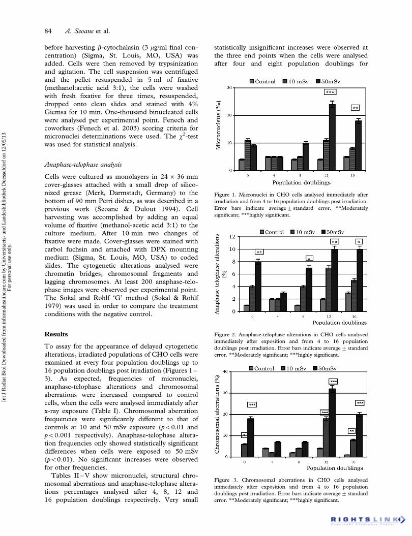
Figure 1. Micronuclei in CHO cells analysed immediately after
irradiation and from 4 to 16 population doublings post irradiation.
Error bars indicate average+ standard error. **Moderately
significant; ***highly significant.
Figure 2. Anaphase-telophase alterations in CHO cells analysed
immediately after exposition and from 4 to 16 population
doublings post irradiation. Error bars indicate average+ standard
error. **Moderately significant; ***highly significant.
Figure 3. Chromosomal aberrations in CHO cells analysed
immediately after exposition and from 4 to 16 population
doublings post irradiation. Error bars indicate average+ standard
error. **Moderately significant; ***highly significant.
84 A. Seoane et al.
Int J
Rad
iat B
iol D
ownl
oade
d fr
om in
form
ahea
lthca
re.c
om b
y U
nive
rsita
ets-
und
Lan
desb
iblio
thek
Due
ssel
dorf
on
12/0
5/13
For
pers
onal
use
onl
y.

exposure to 10 and 50 mSv. Only anaphase-
telophase alterations showed significant differences
after eight population doublings (p50.05).
Micronuclei showed significantly increased fre-
quencies when they were analysed at 12 and 16
population doublings after exposure to 50 mSv
(p50.001 and p50.01 respectively). A similar
situation was observed for anaphase-telophase ana-
lysis (p50.01 and p50.05 respectively). Chromoso-
mal aberrations increased significantly at 12 and 16
population doublings after exposure to 10 mSv
(p50.001 and p50.01 respectively) and 50 mSv
(p50.001).
No correlation analysis could be performed
because only two x-rays doses were used. However,
alteration frequencies were higher in cells exposed to
50 mSv than those exposed to 10 mSv for all end
points analysed (Figures 1 – 3).
Likewise, frequencies of micronucleus, anaphase-
telophase alterations and chromosomal aberrations
were higher at 12 than at 16 population doublings.
Discussion
Ionizing radiation is a physical agent that induces
single and double strand breaks, base damage and
DNA-protein crosslinks. Generally it is agreed that
the most important lesion for the genetic risk of
ionizing radiation exposure is the double strand
breaks (DSB) which when not repaired or misre-
paired can lead to genetic changes. The conse-
quences of the induction of DSB can be observed
cytogenetically at the first mitosis immediately
following exposure to ionizing radiation as transloca-
tions, ring chromosomes, dicentrics, gaps and
double minutes (Smith et al. 2003). In the present
work only gaps or breaks were observed and no
increase in chromosome type aberrations was de-
tected at the first mitosis after exposure. A similar
situation was observed at delayed times. Clastogenic
events expressed as micronuclei, anaphase chromo-
some fragments and subchromatid or chromatid type
aberrations could be scored.
Achromatic lesions have been the subject of
controversy for years since their significance is not
clear. Brecher (1977) has suggested that, rather than
being completely separate phenomena, gaps and
breaks are different manifestations of the same events
and that gaps may be incomplete breaks. In this
sense Dulout and coworkers (1983, 1985) have
reported that gaps are transformed into breaks by
blockage of the G2 check point by caffeine. Recent
studies showed significant increases of gaps after low
dose x-ray exposure (Guerci et al. 2003). In addition,
similar results were found in air crew members
(Cavallo et al. 2002) and hospital workers
(Hagelstrom et al. 1995, Paz y Mino et al. 1995,
Guerci 2004). These studies all agree in attributing
the increase of gaps or chromatid type aberrations to
chronic exposure to low level ionizing radiation.
Micronuclei originate from chromosome breaks or
lagging chromosomes. Results described in the
present work show that immediately after irradiation
as well as after 12 and 16 population doublings an
increase of micronuclei, anaphase-telophase altera-
tions (lagging fragments) and achromatic lesions
(gaps) could be observed. This fact is a clear
indication that a number of the gaps are true
chromatid or chromosome breaks and that the
biological signification of gaps should be revaluated.
On the other hand, genomic instability can be
manifested as stable aberrations transmitted through
many generations of cell replication such as chromo-
somal reduplication, translocations or small deletions
(Little 2003). In addition, it can be characterized by
the de novo appearance of chromosomal damage in
the progeny of cells that survived the original
radiation exposure (Smith et al. 2003, Belyakov
et al. 1999). Our results are consistent with these last
observations because cytogenetic aberrations and
micronuclei were observed immediately after expo-
sure and after 12 replication cycles, but not (or not
significantly) at replication cycles four to eight.
Although we are unable to establish a mechanism
for these effects, our results agree with the presence
of a phenomenon by which the initial DNA damage
in the surviving cells is made permanent or ‘memor-
ized’. In this way Little (2003) hypothesized that
radiation may induce changes affecting the genes
directly and/or affecting either chromosomes or the
cytoplasm in such a way as to increase the instability
of the gene system. Susuki and coworkers (2003)
have suggested that radiation exposure causes non-
lethal, potentially unstable chromosome regions that
are transmissible for many generations after irradia-
tion. These unstable regions could cause delayed
DNA breakage.
Numerous studies have established the occurrence
of radiation-induced chromosomal instability in
various cells types (Holmberg et al. 1998, Mothersill
et al. 2000, Little et al. 1997, Limoli et al. 1999,
Trott et al. 1998, Lorimore et al. 1998). In most of
these studies, the cells were exposed to higher doses
than those used in our experiments. Micronucleus
induction at delayed times was reported for doses
higher than those used in this work (Jamali & Trott
1996, Trott et al. 1998, Belyakov et al. 1999).
Results described could reflect a complex response
where very low doses of x-rays induced mutationally
unstable cell survival instead of cell death. In this
sense, Mothersill and Seymour (2003) support the
idea that affected cells can die or live with damage
and recover badly or well. If the cell does not recover
well, it can die or perpetuate the damage.
Genetic instability induced by x-rays in CHO cells 85
Int J
Rad
iat B
iol D
ownl
oade
d fr
om in
form
ahea
lthca
re.c
om b
y U
nive
rsita
ets-
und
Lan
desb
iblio
thek
Due
ssel
dorf
on
12/0
5/13
For
pers
onal
use
onl
y.

In conclusion, cytogenetic damage could be ob-
served in the progeny of cells exposed to very low
doses of x-rays. Our results are consistent with the
idea that low doses and low dose rates of x-rays may
induce genomic instability. However, further studies
are necessary to confirm these ideas.
Acknowledgements
This work is part of the ‘Proyecto integrado de
mutagenesis, carcinogenesis y teratogenesis ambien-
tal’ of the ‘Programa de Incentivos para Docentes-
Investigadores de Universidades Nacionales’. A. B.
Guerci is a fellowship of the National University of La
Plata. The authors are grateful to Prof. Juan Andrieu
for the calibration of the irradiation equipment.
We wish to dedicate this work to the memory of
Fernando N. Dulout who has recently passed away.
References
Archer PG, Bender M, Bloom A, Brewen J, Carano A, Preston R.
1981. Guidelines for cytogenetic studies in mutagen-exposed
human populations. In: Bloom AD, editor. Guidelines for
cytogenetic studies on human populations exposed to muta-
genic and reproductive hazards. March of Dimes Birth Defects
Foundation. New York. pp 1 – 35.
Balakrishnan S, Rao S. 1999. Cytogenetic analysis of peri-
pheral blood lymphocytes of occupational workers exposed
to low levels of ionizing radiaton. Mutation Research 442:
37 – 42.
Barquinero JF, Barrios L, Caballin M, Miro R, Ribas M, Subıas A,
Egozcue J. 1993. Cytogenetic analysis of lymphocytes from
hospital workers occupationally exposed to low levels of
ionizing radiaton. Mutation Research 286:275 – 279.
Bartolleto E, Mognato M, Ferraro P, Canova S, Cherubini R,
Celotti L, Russo A. 2001. Chromosome instability induced in
the cell progeny of human T lymphocytes irradiated in G(o)
with gamma-rays. Mutagenesis 16:529 – 537.
Belyakov OV, Prise K, Trott K, Michael B. 1999. Delayed
lethality, apoptosis and micronucleus formation in human
fibroblasts irradiated with x-rays or alpha-particles. Interna-
tional Journal of Radiation Biology 75:985 – 993.
Brecher S. 1977. Ultrastructural observations of x-ray induced
chromoatid gaps. Mutation Research 42:249 – 267.
Brenner DJ, Doll R, Goodhead D, Hall E, Land C, Little JB,
Lubin J, Preston D, Preston R, Puskin J, Ron E, Sachs R,
Samet J, Setlow R, Zaider M. 2003. Cancer risk attributable to
low doses of ionizing radiation: assessing what we really know.
Proceedings of the National Academy of Science USA
25:13761 – 13766.
Cardoso R, Takahashi-Hyodo, Peitl S, Ghilardi-Neto Jr. T,
Sakamoto-Hojo E. 2001. Evaluation of chromosomal aberra-
tions, micronuclei and sister chromatid exchangesin hospital
workers chronically exposed to ionizing radiation. Teratogen-
esis, Carcinogenesis and Mutagenesis 21:431 – 439.
Cavallo D, Marinaccio A, Perniconi B, Tomao P, Pecoriello V,
Moccaldi R, Iavicoli S. 2002. Chromosomal aberrations in
long-haul air crew members. Mutation Research 513:
11 – 15.
Dugan LC, Bedford J. 2003. Are chromosomal instabilities
induced by exposure of cultured normal human cells to low
or high-LET radiation? Radiation Research 159:301 – 311.
Dulout FN, Carballal G, von Guradze H, De Luca JC, Oubina J,
Videla C. 1985. Junin virus-induced chromosomal aberrations
in the Guinea pig. Synergism between the attenuated strain XJ-
clone 3 and caffeine. Intervirology 24:193 – 198.
Dulout FN, Carballal G, Bianchi N, von Guradze H. 1983.
Cytogenetic affect of two strains of Junin virus in the Guinea
pig. Intervirology 19:44 – 46.
Fenech M, Morley A. 1985. Measurement of micronuclei in
lymphocytes. Mutation Research 147:29 – 36.
Fenech M, Chang W, Kirsch-Volders M, Holland N, Bonassi S,
Zeiger W. 2003. HUMN Project: detailed description of the
scoring criteria for the cytokinesis-block micronucleus assay using
isolated lymphocyte cultures. Mutation Research 534:65 – 75.
Grillo CA, Dulout FN. 1995. Cytogenetic evaluation of butylated
hydroxytoluene. Mutation Research 345:73 – 78.
Guerci A. 2004. Evolucion de las alteraciones citogeneticas y
lesiones genomicas inducidas por irradiacion cronica con dosis
bajas de radiacion ionizante. Doctoral Thesis. National
University of La Plata.
Guerci A, Dulout F, Seoane A. 2003. Cytogenetic analysis in
Chinese hamster cells chronically exposed to low doses of
x-rays. International Journal of Radiation Biology 79:793 –
799.
Guerci A, Dulout F, Seoane A. 2004. DNA damage in Chinese
hamster cells repeatedly exposed to low doses of X-rays.
Cytogenetic and Genome Research 104:173 – 177.
Hagelstrom A, Gorla N, Larripa I. 1995. Chromosomal damage in
workers occupationally exposed to chronic low level ionizing
radiation. Toxicology Letters 76:113 – 117.
Holmberg K, Meijer AE, Harms-Ringdahl M, Lambert B. 1998.
Chromosomal instability in human lymphocytes after low dose
rate gamma-radiation and delayed mitogen stimulation. Inter-
national Journal of Radiation Biology 73:21 – 34.
Jamali M, Trott KR. 1996. Increased micronucleus frequency in
the progeny of irradiated Chinese hamster cells. International
Journal of Radiation Biology 69:301 – 307.
Limoli CL, Corcoran J, Milligan J, Ward J, Morgan WF. 1999.
Critical target and dose and dose-rate responses for the
induction of chromosomal instability by ionizing radiation.
Radiation Research 151:677 – 685.
Limoli CL, Ponnaiya B, Corcoran J, Giedzinski B, Kaplan M,
Hartmann A, Morgan WF. 2000. Genomic instability induced
by high and low LET ionizing radiation. Advances in Space
Research 25:2107 – 2117.
Little JB. 2003. Genomic instability and bystander effects: a
historical perspective. Oncogene 22:6978 – 6987.
Little JB, Nagasawa H, Pfenning T, Vetrovs H. 1997. Radiation-
induced genomic instability: delayed mutagenic and cytogenic
effects of X rays and alpha particles. Radiation Research
148:299 – 307.
Lorimore S, Kadhim M, Pocock D, Papworth D, Stevens D,
Goodhead D, Wright W. 1998. Chromosomal instability in the
descendants of unirradiated surviving cells after a-particle
irradiation. Proceedings of the National Academy of Science
USA 95:5730 – 5733.
Morgan WF. 2003. Non-targeted and delayed effects of exposure
to ionizing radiation: I. Radiation-induced genomic instability
and bystander effects in vitro. Radiation Research 159:567 –
580.
Mothersill C, Kadhim M, O’Reilly S, Papworth D, Marsden S,
Seymour C, Wright W. 2000. Dose- and time-response
relationship for lethal mutations and chromosomal instability
induced by ionizing radiation in an immortalized human kera-
tinocyte cell line. International Journal of Radiation Biology
76:799 – 806.
Mothersill C, Seymour C. 2003. Radiation-induced bystander
effects, carcinogenesis and models. Oncogene 22:7028 –
7033.
86 A. Seoane et al.
Int J
Rad
iat B
iol D
ownl
oade
d fr
om in
form
ahea
lthca
re.c
om b
y U
nive
rsita
ets-
und
Lan
desb
iblio
thek
Due
ssel
dorf
on
12/0
5/13
For
pers
onal
use
onl
y.

Paz y Mino C, Davalos M, Sanchez M, Arevalo M, Leone P. 1995.
Should gaps be included in chromosomal aberration analysis?
Evidence based on the comet assay. Mutation Research
516:57 – 61.
Ponnaiya B, Jenkins-Baker G, Bigelow A, Marino S, Geard C.
2004. Detection of chromosomal instability in a-irradiated and
bystander human fibroblasts. Mutation Research 568:41 – 48.
Seoane A, Dulout F. 1994. Use of the anaphase-telophase test to
detect aneugenic compounds: effects of propionaldehyde and
cadmium chloride. Bulletin of Environmental and Contamina-
tion Toxicology 53:924 – 929.
Smith L, Nagar S, Kim G, Morgan W. 2003. Radiation-induced
genomic instability: radiation quality and dose response.
Health Physics 85:23 – 29.
Sokal R, Rohlf J. 1979. Biometrıa. Principios y metodos
estadısticos en la investigacion biologica. (H. Blume edi-
ciones). Madrid: W. H. Freeman and Company.
Susuki K, Ojima M, Kodama S, Watanabe M. 2003. Radiation-
induced DNA damage and delayed induced genomic instabil-
ity. Oncogene 22:6988 – 6993.
Trott K, Jamali M, Manti L, Teibe A. 1998. Manifestations and
mechanisms of radiation-induced genomic instability in V-79
Chinese hamster cells. International Journal of Radiation
Biology 74:787 – 791.
Whitehouse C, Tawn E. 2001. No evidence from chromosomal
instability in radiation workers with in vivo exposure to
plutonium. Radiation Research 156:467 – 475.
World Health Organization. 1985. Guide to short-term tests for
detecting mutagenic carcinogenic chemicals. Environmental
Health Criteria 51. Geneva: World Health Organization; pp
57 – 67.
Genetic instability induced by x-rays in CHO cells 87
Int J
Rad
iat B
iol D
ownl
oade
d fr
om in
form
ahea
lthca
re.c
om b
y U
nive
rsita
ets-
und
Lan
desb
iblio
thek
Due
ssel
dorf
on
12/0
5/13
For
pers
onal
use
onl
y.